Article

Dopamine-Sensing Characteristics and Mechanism by Using N2/O2 Annealing in Pt/Ti/n-Si Structure

1
Department of Obstetrics and Gynecology, Keelung Chang Gung Memorial Hospital (CGMH), No. 222 Maijin Rd., Anle, Keelung 204, Taiwan
2
Thin Film Nano Tech. Lab., Department of Electronic Engineering, Chang Gung University (CGU), 259 We-Hwa 1st Rd., Guishan, Tao-Yuan 33302, Taiwan
3
School of Medicine, College of Medicine, Chang Gung University (CGU), 259 Wen-Hwa 1st Rd., Guishan, Tao-Yuan 33302, Taiwan
*
Author to whom correspondence should be addressed.
Electronics 2021, 10(24), 3146; https://doi.org/10.3390/electronics10243146
Submission received: 11 October 2021 / Revised: 10 December 2021 / Accepted: 14 December 2021 / Published: 17 December 2021
(This article belongs to the Section Bioelectronics)

Abstract

:
Dopamine detection by using N2/O2 annealing in a Pt/Ti/n-Si structure is investigated for the first time. To achieve repeatable and stable dopamine detection, a Pt membrane is annealed at elevated temperatures of 500 to 700 °C. N2/O2 gas ambient is used to optimize the membrane. The Pt membrane with thicknesses from 5 to 2 nm is optimized. Novel Pt/Ti/n-Si Schottky contact in a metal–electrolyte–membrane–silicon (MEMS) structure detects dopamine with a low concentration of 1 pM. The Pt membrane with N2 ambient annealing shows the lowest concentration of dopamine sensing with a small volume of 10 µL, acceptable stability, and repeatability. Scan rate-dependent dopamine concentration sensing is also investigated in the two-terminal measurement method. This study is useful for the early diagnosis of Parkinson’s disease in the near future.

1. Introduction

The biomedical sensing field is enriching day-by-day in worldwide research work to find a novel sensing membrane with promising structure on a silicon platform for the detection of different bio-analytes, which will help in the diagnosis of human diseases shortly [1]. In addition to various causes of human death due to diseases, dopamine [2,3,4,5,6,7,8,9,10,11,12,13,14,15,16,17,18,19,20,21,22,23] detection is a significant topic as it is responsible for schizophrenia, Parkinson’s disease (PD), attention deficit hyperactivity disorder (ADHD), etc. Parkinson’s disease is a long-term degenerative disorder of the central nervous system, which results in the symptoms that affect thinking, walking, sleeping, emotions, anxiety, depression, and so on. As there is no cure of it at this time, this can be improved through treatment. Therefore, early-stage diagnosis is very important for better controlling the dopamine concentration in the human body.
Basically, dopamine is a catecholamine-based neurotransmitter that is synthesized in the brain and kidneys. Dopamine is a catecholamine-based chemical released by neurons to send signals to other nerve cells. The presence of dopamine, a neurotransmitter synthesized in the brain and kidneys, controls how a person thinks and plans. Dopamine is known as the happy factor, which improves our focus, attention, helps in Na excretion, and increases urine output also. Dopamine remains in the blood in the form of dopamine sulfate, which self-oxidizes to H2O2 [24,25]. The normal range of dopamine is 26–40 nM in humans. Dopamine concentration in blood beyond this value indicates abnormality. Dysfunctions are caused by low iron, low magnesium, adrenal fatigue, vitamin B3 and B6, vitamins C and D deficiency, etc. The dysfunction of dopamine is associated with neurodegenerative disorder known as Parkinson’s diseases, schizophrenia, fatal diseases such as Alzheimer’s, pre-aging of the brain, etc. Long-term degenerative disorder of the central nervous system is also a chronic disease, which results in the symptoms of thinking disorder, walking disorder, sleeping disorder, emotional problems, anxiety, depression, etc. As there is no cure at this stages, dopamine detection will help to improve human health through treatment. On the other hand, a particular disease identification is also important, but our new method of dopamine sensing is not focusing on it. The early-stage diagnosis plays an important role for the detection of abnormal dopamine concentration in blood. This will be an indication of neurodegenerative disorder and can help in the better control of dopamine concentration in human body.
In this work, our aim is to detect dopamine from a small sample volume with low concentration in a novel and easy way. This can be helpful for the early diagnosis of abnormal dopamine concentration in the human body. The Pt-based dopamine sensing is performed because Pt has good reduction–oxidation (redox) properties, higher work function (5.2–5.6 eV), many oxidation states (9), and higher Gibb’s free energy. Due to this high work function of Pt, this can form easily Schottky contact on the n-Si substrate. A thin Pt material as a sensing membrane has been deposited by the physical vapor deposition (PVD) process, and dopamine sensing characteristics are obtained in current–voltage (I-V) measurements. To improve stability and repeatability, annealing in N2/O2 ambient is one of the effective ways. A thin Pt film becomes similar to nanoparticles after annealing, which helps to increase the surface area of the sensing membrane. However, Pt does not have good adhesion with Si at elevated temperature. So, a Ti interfacial layer is used in between Pt and n-Si. In current–voltage (I-V) measurements with a full scan technique, dopamine peak positions can be distinguishably observed. In this study, a N2/O2 annealed Pt-based novel sensor in the MEMS structure is developed, which will enable the early diagnosis of abnormal dopamine concentration in the near future within a short time.

2. Sensor Fabrication Procedure and Measurement

2.1. Material Deposition

The 4-inch n-Si (100) wafers with a resistivity of 1–10 Ω–cm and thickness of 500 µm were used. After cleaning by using the Radio Corporation of America (RCA) method, a 2 nm-thick Ti layer was deposited by the radio frequency (RF) sputtering method. A metallic Ti target was used. During Ti deposition, the argon flow was 25 sccm, and the applied RF power was 50 watt. The deposition pressure was 20 mTorr. The deposition time was 24 s. The Pt/Ti/n-Si sensors were prepared using Ti as an adhesive layer, and then Pt metal was deposited on it by the sputtering method. For the fabrication of Pt-based sensors, a 2 nm-thick polycrystalline Pt membrane and a 5 nm-thick polycrystalline Pt layer were deposited on individually Ti/n-Si substrate. During Pt deposition, the argon flow, RF power, and deposition pressure were 25 sccm, 50 watt, and 20 mTorr, respectively. Deposition times of the 2 nm- and 5 nm-thick Pt layers were 15 s and 37.5 s, respectively. The Si layer (SiO2 ≈ 3 nm) and the Ti layer (TiOx ≈ 2 nm) will be oxidized by an ex situ process or during annealing treatment. It is expected that the Ti/Si becomes a metal/thin oxide/n-Si structure.

2.2. Sensing Area Patterned by Lithography

Then, UV-photolithography was performed to pattern the sensors. During patterning, the samples were baked soft to remove moisture. Then, negative photoresist SU8 was coated on the wafers by using a spin coater. For isotropic SU8 coating, the rotation was 500 rpm. To control the thickness of the resist, the rotation was high: 3000 rpm. Then, the patterning was done with a proper mask, and UV ray exposure was 12 s. After developing it in SU8 developer solution, the sensors with a circular size of 3.14 mm2 were obtained. Finally, the sensors were baked hard for 30 min. After this, the sensors were attached to Cu-PCB (printed circuit board) having copper lines with the attachment of silver gel. Then, epoxy solutions were prepared and coated on the sensors for the encapsulation. The sensors were dried under controlled temperature and stabilized by keeping these in RO water for 24 h. By using a dopamine solution drop, the liquid is contact on the Pt membrane surface, resulting in the oxidation of the Pt membrane, and an oxidation current is obtained. Similarly, sweeping bias on the sensors shows both oxidation and reduction peaks as well.

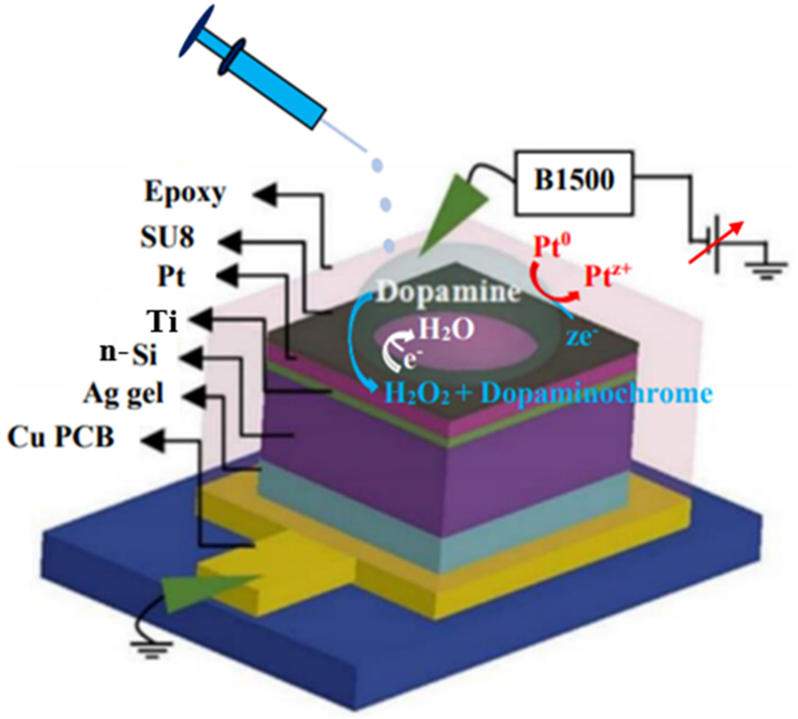
2.3. Sensor Structure and Measurement Procedure

The schematic view of the Pt/Ti/n-Si sensors and measurement set up is shown in Figure 1. The structure is similar to the as-deposited Pt/n-Si sensor [26]. The split table of all the sensors is given in Table 1. I-V characteristics and current–time (I-t) responses were performed using an Agilent B1500 semiconductor analyzer. For the measurements, a solution drop with a volume of 10 µL was poured on the sensing area by using a micropipette. Sweeping bias was applied by connecting the tungsten probe into it, whereas the other side of the sensor or n-Si was grounded. The concentration of dopamine preparation is discussed below. Current with different dopamine concentrations were measured by using a N2/O2 annealed Pt/Ti/n-Si MEMS sensor, as shown in Figure 1.

2.4. Details of Materials and Preparation of the Solutions

The n-Si wafers were obtained from Sadhu Design Corporation Limited, Taiwan. For the RCA cleaning process, the H2SO4, H2O2, NH4OH, and HF solutions were purchased from J. T. Baker, United States of America (USA). The metallic Pt and Ti targets were taken from Sadhu Design Corporation Limited, Taiwan. The photoresist SU8 and SU8 developer solutions were bought from MICROCHEM, USA. The colloidal silver gel was purchased from TED PELLA, INC., California (CA). Epoxy solutions were bought from Uni Region Bio-Tech, Taiwan. The dopamine hydrochloride solution was obtained from Sigma Aldrich, Germany. The molecular weight of dopamine is 153.18 g/mol. By using molarity calculation, firstly 1 mM, 100 mL dopamine solution was prepared as the main stock. By diluting this main stock in buffer solution pH7, sub-stock solution up to 10 pM dopamine was prepared. The 0.5 M, 1 L pH7 buffer solution was grabbed from Honeywell, Germany. The 0.5 M, 1 L pH6, pH8, and pH10 commercial buffer solutions were purchased from Alfa Aesar, USA. The micropipettes were taken from Nichipet Premium, Japan.

3. Results and Discussion

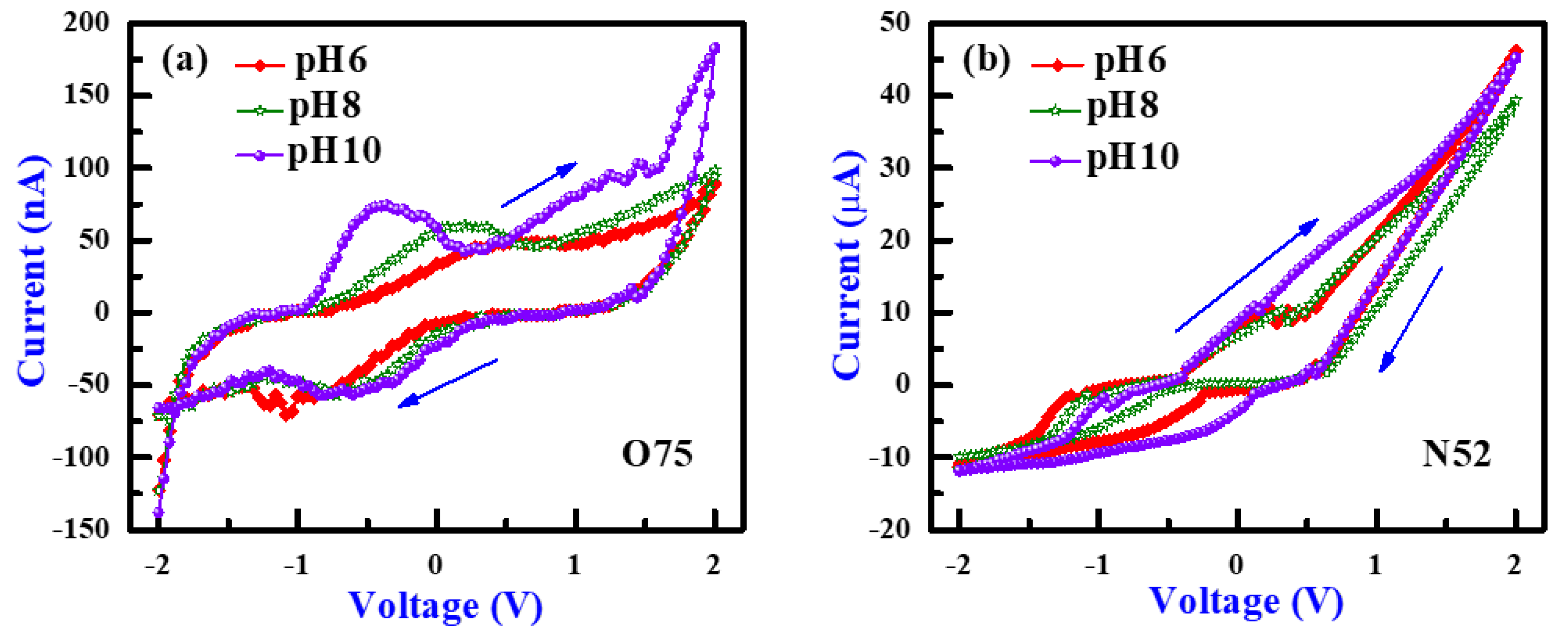
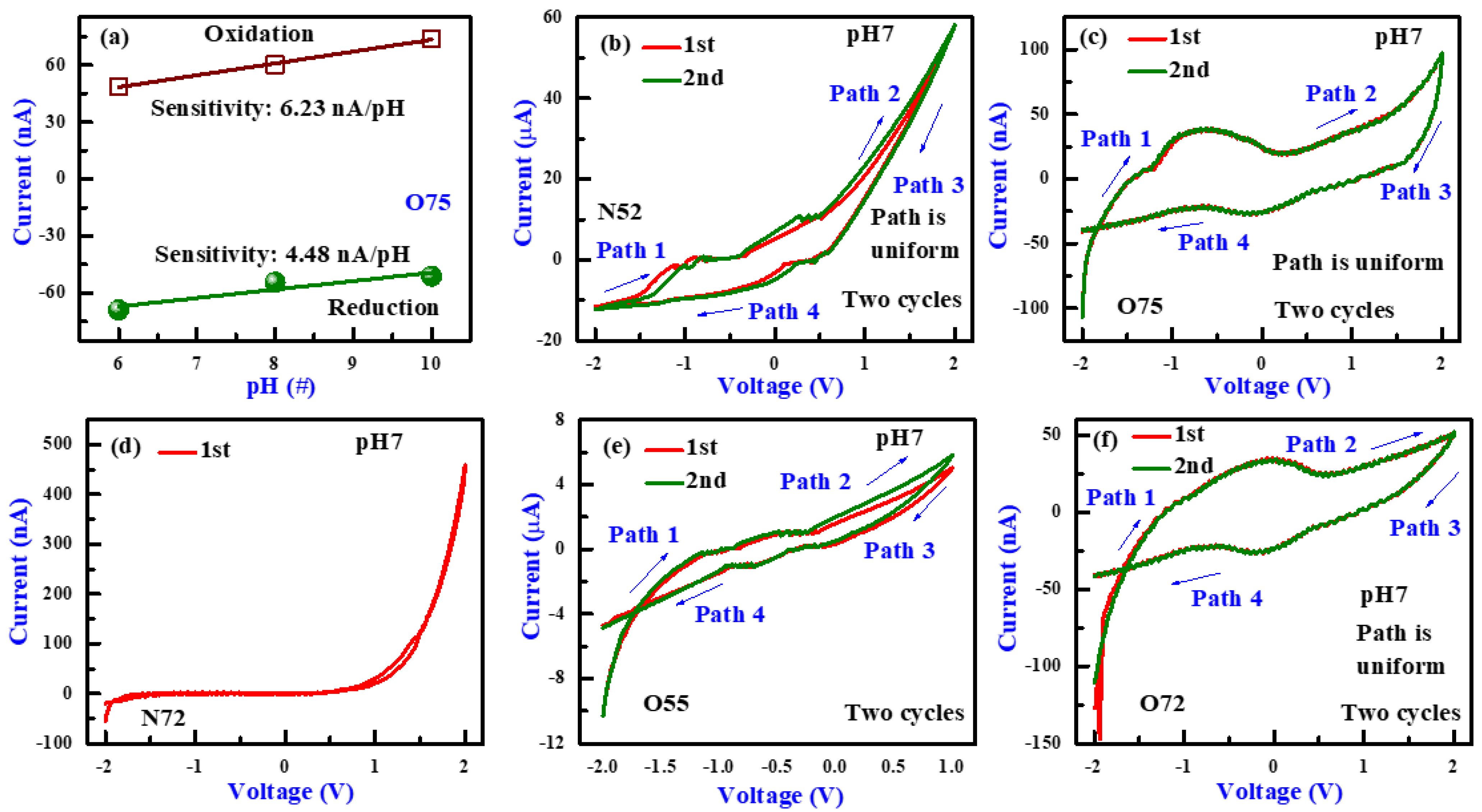
3.1. PH Sensing and I-V Sweeping Characteristics

I-V characteristics of the Pt/Ti/n-Si optimized O2 (O75) and N2 (N52) annealed sensors in different pH solutions are shown in Figure 2a,b, respectively. The split table of all the thicknesses, annealing temperature, and annealing ambient-dependent Pt-based sensors is shown in Table 1. The pH-sensing characteristics of the Pt/Ti/n-Si based O75 sensors in the MEMS structure are obtained with pH6, pH8, and pH10 solutions, as shown in Figure 2a. The 5 nm-thick Pt sensors were annealed at 700 °C for a certain time, 10 min in constant 2.5 slm O2 (O75 sensors) flow, as shown in Figure 2a. I-V characteristics of the 2 nm-thick Pt sensors annealed at 500 °C in N2 ambient (N52 sensors) at pH6, pH8, and pH10 solutions are shown in Figure 2b. In contrast, N2 annealing was done in constant 2.5 slm N2 flow for a certain time of 10 min. I-V responses of both the sensors show cyclic voltammetry (CV) nature. It is noticed that the oxidation peak currents are shifting toward more negative potential, and the reduction peak currents are shifting toward more positive potential with increasing in pH value of the solutions for O75 sensors (Figure 2a). The oxidation peak current increases with increasing the pH value from 6 to 10, which is due to more OH- ions on the Pt membrane surface. The oxidation peaks are obtained comparatively in positive potential than reduction peaks for the O75 sensors (Figure 2a). The plot of pH value vs. current for the O75 sensors is shown in Figure 3a. In the oxidation region, the pH sensitivity value obtained is 6.23 nA/pH and the pH sensitivity value in reduction portion obtained is 4.48 nA/pH. For optimized N2 annealed Pt (N52) sensors, the current peaks are shifting toward positive potential as the pH value of the solution is increased from pH 6 to pH 10, as shown in Figure 2b. I-V characteristics of the Pt/Ti/n-Si-based optimized N2 and O2 annealed sensors in pH7 solution are shown in Figure 3b,c. The I-V responses of the other Pt-based N2 and O2 annealed sensors in neutral pH7 solution are also shown in Figure 3d–f. Figure 3b shows the I-V characteristics of the N52 sensors and Figure 3c shows the I-V responses of the O75 sensors in pH7 solution. For I-V measurements, sweeping voltage is kept at ±2 V with an optimized step voltage of 40 mV, and the delay time is 0 s. The sweeping path is given as path 1→path 2→path 3→path 4. For all sensors, path 1 and path 4 denote the reduction portions, and path 2 and path 3 denote the oxidation portion of the CV curves. For both optimized N2 and O2 annealed sensors, I-V characteristics are measured for two cycles, and O75 is recorded to be more uniform, which symbolizes that the sensor surface of the N52 sensors is rougher, and this is helpful for bio-analyte sensing.
However, the current value is much higher for the N52 sensors in a high µA range, whereas the current value is in a few nA range for the O75 sensors. This indicates that N52 has more surface area than O75, which will be helpful for low concentration bio-analyte detection. Figure 3d shows the I-V responses of the 2 nm-thick Pt sensors annealed in N2 ambient at 700 °C (N72 sensors). One cycle of the I-V curve was measured, and no oxidation–reduction peak current is observed. A proper I-V curve is also not obtained, and the current value is in the nA (≈550 nA) range i.e., too low compared to other N2 annealed sensors. The I-V curves of the 5 nm-thick Pt sensors annealed in O2 ambient at 50 °C for 10 min are shown in Figure 3e (O55 sensors). Two cycles of the I-V curves for the sensors are recorded, and the window area is moderate. The current value is higher in the 5 µA range, but the path is not uniform. The prominent oxidation–reduction peak currents are also not obtained.
Figure 3f shows the I-V responses of the 2 nm-thick Pt sensors annealed in 2.5 SLM O2 ambient at 700 °C (O72 sensors). Two cycles were measured, and the path is repeatable. The window is also broad. The current value is ≈100 nA. In case of a thicker Pt membrane with 5 nm-thickness annealed at 700 °C (O75 sensors), I-V responses have shown a more repeatable broader window with prominent oxidation–reduction peak currents. So, it can be concluded that O2 annealing at 700 °C (O75 and O72 sensors) provides repeatable, stable, and uniform I-V curves with a broader window. The O75 sensors are considered as optimized O2 annealed sensors. It shows a comparatively broader window with more prominent current peaks and a higher current value. Among the N2 annealed sensors, 2 nm-thick Pt-based sensors annealing at 500 °C (N52) in N2 ambient show higher current value in the µA range, and almost repeatable I-V curves are obtained. The thicker Pt membrane in O2 annealed at higher temperatures (700 °C) has shown good effect. That is why sensors optimization is also further checked with a thicker (10 nm) Pt membrane at elevated temperatures (800 °C) annealing (not shown here). However, the O75 sensors turn out to be more useful than other O2 annealed sensors.
It is found that the current is much lower for the O2 annealed sensors than the N2 annealed sensors because the Pt membrane is oxidized and the resistivity of the membrane increases as well. On the other hand, the Si surface is also oxidized at elevated temperature owing to the reduced forward current at positive bias. Therefore, a lower N2 annealing temperature at 500 °C (N52 sensors) is more useful (Figure 3b). For O2 annealing, higher annealing temperature at 700 °C (O75 sensors) is useful as it shows prominent oxidation–reduction peaks (Figure 3c). Among the N2 annealed sensors, the N52 sensor is considered an optimized sensor. It shows the highest ≈50 µA current value, which symbolizes higher sensor surface area and an almost repeatable path (Figure 3b).
It is also found that both O75 and N52 sensors show good hysteresis. This indicates that the thinner film (2 nm) in N2 annealing at lower temperature (500 °C) and thicker film (5 nm) in O2 annealing at higher temperature show better hysteresis. On the other hand, the N2 annealing sensors show a higher Schottky diode current at positive voltage than the current at negative voltage. It is also observed that the reduction portion of N52 sensors is more stable, and the reduction current peaks are prominent as the sensor surface is reduced due to N2 annealing. For the O75 sensors (Figure 3c), the oxidation portion is more stable, and the oxidation current shows prominent peaks owing to O2 annealing. The Pt membrane is more oxygen enriched. Therefore, we have used both sensors for dopamine sensing below.

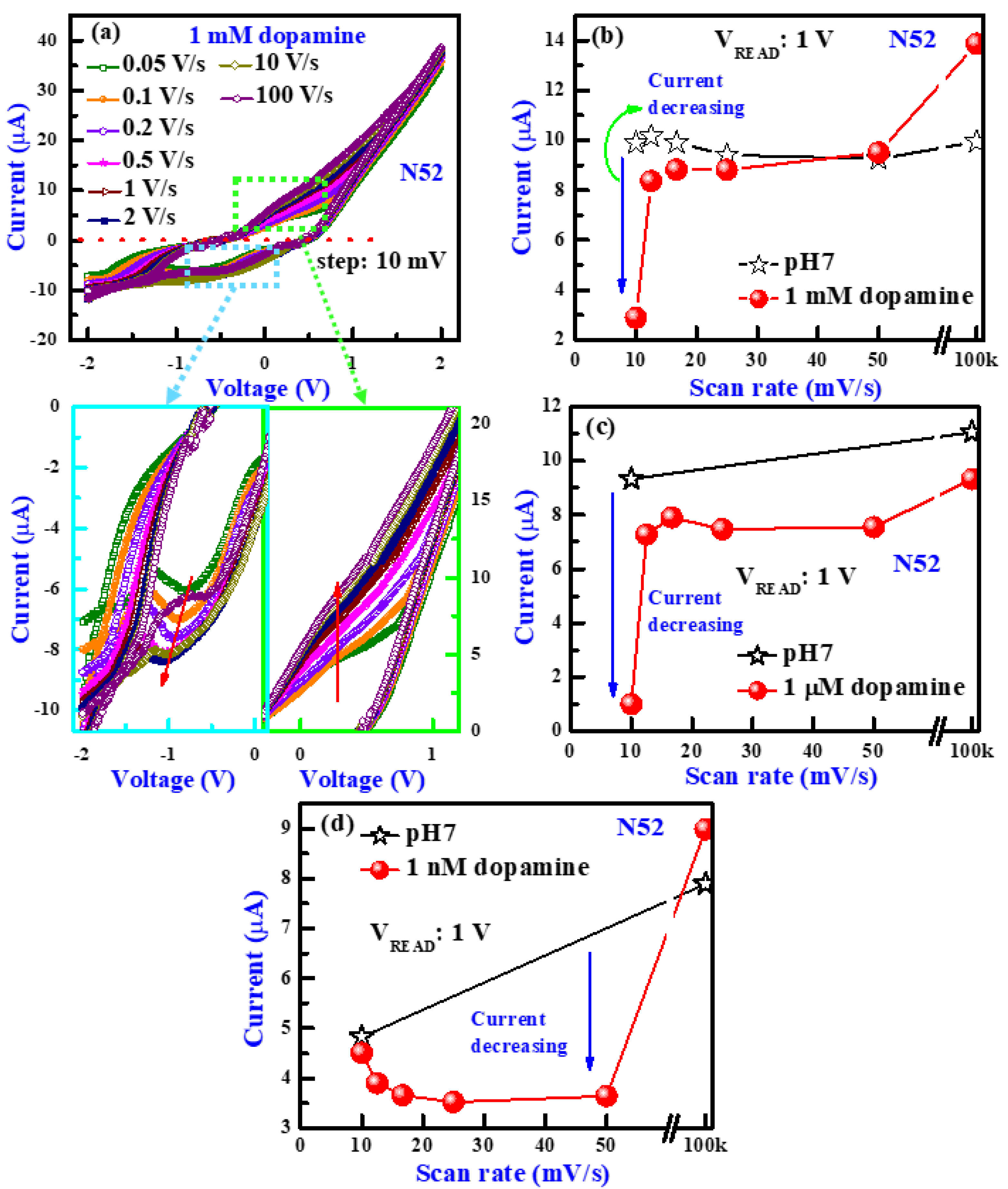
3.2. Scan Rate Dependence

The effects of change in scan rates of the N52 (Figure 4a,c) and O75 (Figure 4b,d) sensors are checked with pH7 solutions and respective dopamine solutions. Figure 4a,b shows the effect of change in the scan rate of N52 and O75 sensors in pH7 solutions. The scan rate is changed by keeping the step voltage constant at 10 mV and increasing the delay time from 0 s to 200 ms. A prominent I-V curve is recorded by the N52 sensors at a much lower scan rate of 0.05 V/s i.e., 50 mV/s. For the O75 sensors, below a scan rate of 500 mV/s, prominent oxidation and reduction current peaks are not found. In case of all the figures shown in Figure 4, it is also observed that the oxidation–reduction peak currents with increasing in the scan rate are increasing for both sensors in pH7 and dopamine solutions. Owing to better dopamine sensing of the N52 sensors, a lower concentration of 1 µM is measured than that of the O75 sensors. The N52 sensors (Figure 4a,c) show a stable and distinct peak response for changing in the scan rate, and it follows a definite pattern. For O75 sensors (Figure 4b,d), the response is comparatively less stable, and the peaks are less prominent at lower scan rates. With an increase in the scan rate, oxidation and reduction peaks are increasing for both cases. It is observed that oxidation and reduction peaks in the dopamine solution are less prominent with respect to pH 7 at lower scan rates. With an increase in scan rate, oxidation and reduction peaks are increasing for both cases. It is noted that the oxidation and reduction peaks in dopamine solution are shifting to more positive potential with respect to pH7 for both of the sensors. The detailed performance of the dopamine sensing is explained below.

3.3. I-V Characteristics of Dopamine Sensing

Figure 5a,b show the dopamine sensing characteristics of the N52 and O75 sensors for a step voltage 40 mV and delay time fixed at default value. As the current change of N52 sensors is greater in the case of the 1 mM dopamine concentration shown in Figure 6a so, it is further used to sense dopamine with lower (1 µM) concentration (Figure 5a). The dopamine solution of concentration 1 µM (Figure 5a) is sensed by the N52 sensors, and 1 mM dopamine concentration is sensed by the O75 sensors (Figure 5b). As the reduction portion of the N52 sensors is very good owing to the N2 annealing Pt membrane, it is reduced. A rapid and stable current change for 1 µM dopamine solution with respect to the pH 7 solution is observed in the reduction portion of the I-V curves (Figure 5a). In the case of both of the sensors, dopamine sensing characteristics are shown to be opposite. For the O75 sensors, dopamine sensing characteristics are observed in the oxidation portion owing to the O2 annealing Pt membrane, which is more oxygen rich. For both N52 (Figure 5a) and O75 (Figure 5b) sensors, the current value is decreased in the case of dopamine with respect to pH 7 solutions. As dopamine auto oxidizes to H2O2, in contact with H2O2, Pt shifts to higher oxidation states, resulting in an increment in the Schottky barrier height (ϕSB) of the Pt membrane. The current value decreases as well. The current value of the N52 sensors shows a clearer change between dopamine and pH7 solutions than the O75 sensors.
It is found that the oxidation–reduction peak positions in case of the O75 sensors are shifted for dopamine with respect to pH 7 solution. For the O75 sensors, the oxidation peak position is −0.276 V for pH7 and 0.012 V for 1 mM dopamine solutions. The reduction peak position of the O75 sensors is −0.1 V for pH7 and −0.267 V for 1 mM dopamine. For the N52 sensors, the current value at read voltage (VREAD) of −0.2 V changes from 0.88 µA to 0.84 µA equivalent to 4.5% changed. This can be useful to identify other analytes in the future. Similarly, the peak positions of other analytes will be different, and the interference effect will be avoided. However, further study is needed.

3.4. Scan Rate-Dependent Dopamine Sensing

The scan rate-dependent I-V characteristics of the N52 sensors in 1 mM dopamine solution is shown in Figure 6a. The scan rate values are varied from 50 mV/s to 100 V/s, and it shows a definite pattern for changing in oxidation–reduction current peaks. The optimized nitrogenated (N52) sensors are stable, repeatable, and robust. To check these phenomena, the scan rate-dependent I-V characteristics are measured by using these sensors after a few months in pH7 and dopamine solutions. I-V curves have shown similar nature to the previous curves (not shown here). This result indicates that the nitrogenated Pt membrane shows good stability and robustness even after being exposed in air. The scan rate vs. current change is plotted by taking the pH7 solutions as a reference for different dopamine concentrations of 1 mM, 1 µM, and 1 nM (Figure 6b–d). There is a drastic current change with respect to the current of pH 7 at 10 mV/s scan rate for both 1 mM and 1 µM dopamine solutions at a read voltage of 1 V, as shown in Figure 6b and Figure 6c, respectively. This suggests that slow I-V sweeping is needed to eliminate higher ionic current through a higher concentration of dopamine and measured dopamine as well. However, further study is needed to explore this scan rate behavior. On the other hand, the current of 1 nM dopamine in pH 7 is decreased at 50 mV/s, as shown in Figure 6d. In fact, the optimized scan rate has played an important role for different concentrations of dopamine sensing.
To obtain proper oxidation–reduction current peaks, further scan rates are lowered to 50 mV/s and 10 mV/s. The measurement was done with a lower concentration of dopamine solutions. It is well known that the normal concentration of dopamine in human blood is 26–40 nM. The dopamine concentration both higher and lower than this value is associated with some abnormalities. The O75 sensors can sense dopamine with higher concentration value (1 mM) owing to more oxygen-rich thicker Pt membrane (5 nm) due to O2 annealing at elevated temperatures (700 °C). So, the O75 sensors will be helpful to sense higher abnormal dopamine concentrations with very prominent oxidation–reduction current peaks. As the sensor surface of the O75 sensors is already oxidized, it cannot show a good response at low concentration dopamine. On the other hand, the N52 sensors will be used to sense dopamine solutions with lower concentrations. This has been discussed below.

3.5. Low Concentration Dopamine Detection

The N52 sensors are robust and show more distinct peaks at low scan rates. The change in scan rate vs. change in current value for the pH7 solution and low 1 pM concentration dopamine solution are plotted in Figure 7. In reference to the pH7 solution, it is observed that the current value is decreased at a lower scan rate 50 mV/s for the 1 pM dopamine solution in pH7. To obtain a more distinguished larger shift for 1 pM dopamine solution with respect to pH7 solution, the scan rate is optimized for a long range from 10 mV/s to 100 V/s. At a lower 50 mV/s scan rate, N2 annealed optimized Pt sensors (N52) have shown the largest decrement in current value owing to the higher oxidation states of Pt and higher Schottky barrier height.
In this work, low concentration dopamine of 1 pM is sensed by using thickness optimized and N2 annealed Pt N52 sensors in a metal-electrolyte-sensing membrane. This work is a novel and useful research of early-stage 1 pM low concentration dopamine detection. It is using novel redox properties of the Pt membrane in a two-terminal measurement setup, whereas the current–voltage curves show a cyclic voltammetry nature. As dopamine is an important research topic, many groups are working on it. A comparison of Pt-sensing electrode-based dopamine-sensing performances among the recent published data is shown in Table 2. However, the sensing mechanism is important, which is explained below.

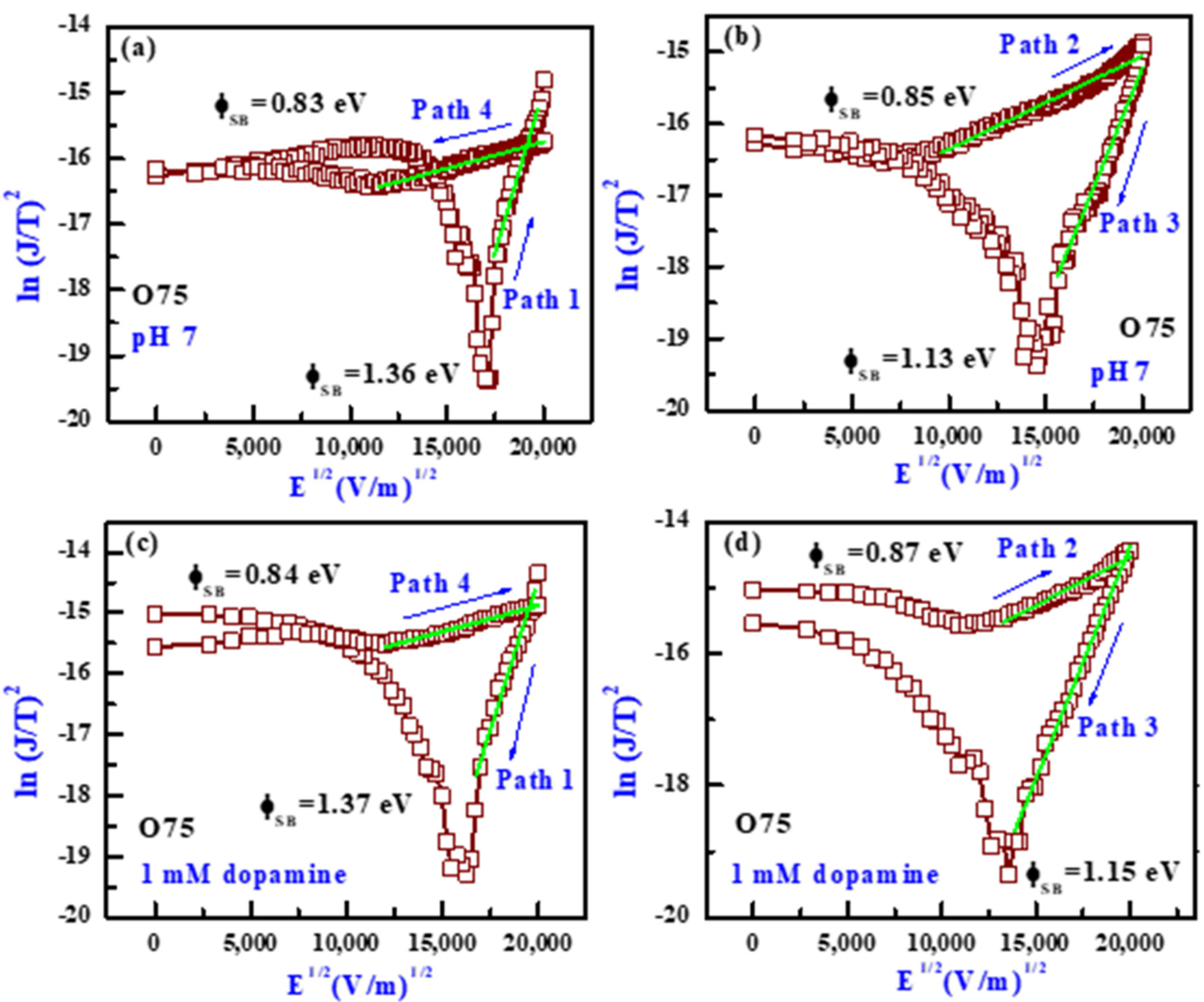
3.6. Transport and Dopamine-Sensing Mechanism

The fitting curves (ln J/T2 vs. E1/2) in the negative region for the O75 sensors are plotted for path 1 and path 4 (reduction region) for both pH7 and 1 mM dopamine solutions, as shown in Figure 8a,c. The ln (J/T2) vs. E1/2 plots in the positive region for path 2 and path 3 (oxidation region) for both pH7 and 1 mM dopamine solution are shown in Figure 8b,d. The sensor shows Schottky phenomena, and the Schottky barrier heights (ɸSB) are calculated from the Schottky equation [26,27]. A comparison of ϕSB values for both pH 7 and 1 mM dopamine solutions for four sweeping paths is shown in Figure 9. The ϕSB values in the pH7 solution for path 1, path 2, path 3, and path 4 are found to be 1.36, 0.85, 1.13, and 0.83 eV, respectively (Figure 8a,b). The ϕSB values in the 1 mM dopamine solutions for path 1, path 2, path 3, and path 4 are found to be 1.37, 0.87, 1.15, and 0.84 eV, respectively (Figure 8c,d). The ɸSB values are found to be less for the pH7 solution, and the values are 0.85 eV (path 2) and 1.13 eV (path 3). For 1 mM dopamine solution, ɸSB is increased, and the values are 0.87 eV (path 2) and 1.15 eV (path 3). The ɸSB value is higher for dopamine than pH7. That is why the current value decreases in case of dopamine solutions owing to the increase in Schottky barrier height. The ϕSB values are comparable with the reported results by Schmitz et al. [28].
It is observed that the optimized Pt/Ti/n-Si (N52) structure over other types of Pt based sensors has detected a low 1 pM dopamine concentration. The nitrogenated thin Pt membrane annealed at comparatively lower temperature (N52 sensors) has shown the lowest concentration dopamine detection, which is useful for early-stage dopamine sensing. In contrast, the comparatively thicker Pt membrane oxygen annealed at comparatively elevated temperatures (O75 sensors) has shown high-concentration dopamine sensing with prominent current peaks. This is also very useful for the detection of higher abnormal dopamine concentration in human blood. Both of the optimized N52 and O75 sensors are first checked in pH 7 solutions. Then measurement is performed in dopamine solutions by varying the scan rates. It is found that a higher concentration of dopamine is detected at a lower scan rate.
The effect of scan rates on the sensing performances of the sensors has been investigated. From a study done with the N52 sensors, it is observed that the scan rate plays an important role for the detection of different concentrations of dopamine. It is expected that higher concentrations of dopamine solutions have more ionic current. Therefore, a longer time or lower scan rate is needed to oxidize the surface of the Pt membrane. At a lower scan rate, the oxidation current dominates over the ionic current. On the other hand, a lower concentration (1 pM) of dopamine solutions is detected at a moderate scan rate (50 mV/s) or shorter delay time. For a lower concentration of dopamine solutions, few dopamine molecules are present in the solutions, or this has a lower ionic current. Therefore, it needs a shorter time to oxidize the Pt surface or the oxidation current is higher than the ionic current at a shorter time. In fact, there is a contradiction that a lower concentration of dopamine cannot be measured at longer delay time or lower scan rates owing to the presence of a few dopamine molecules in the solutions, which can oxidize the surface of the Pt membrane. It is also found that the current of pH7 is slightly decreased (approximately 10 to 7.5 µA) with repeated measurement, and it is remains oxidized at the Pt membrane surface. It needs some time to return to its original surface condition. However, this nitrogenated Pt membrane shows robust and longer time measurement. Therefore, the stability is very good. It can show ten repeatable cycles for bio-analyte sensing. This study indicates that the optimized Pt/Ti/n-Si structure is also useful for biosensing; especially, the Pt membrane is very effective. In the near future, by using this structure, other bio-analytes can be also detected.

4. Conclusions

A low dopamine concentration of 1 pM with a small volume of 10 µL is sensed by using a thickness optimized and annealed Pt membrane in MEMS structure for the first time. Post deposition annealing temperatures ranging from 500 to 700 °C in N2 or O2 ambient have been used. By using optimized nitrogenated thin Pt membrane in N2 ambient at a lower scan rate of 50 mV/s, 1 pM dopamine is sensed. A high concentration dopamine with prominent oxidation–reduction current peaks is sensed by using a high-temperature oxygen annealed optimized thicker Pt membrane. The pH and dopamine sensing mechanisms are owing to Schottky barrier height modulation in contact of pH/dopamine on the Pt membrane. The sensing performance of the sensors is optimized, and a stable time response is also obtained by the said sensor. This novel approach of dopamine sensing in a simple method by using an optimized Pt/Ti/n-Si sensor can achieve a milestone in the bio-medical field of abnormality behavior, especially Parkinson’s disease diagnosis at early stage in the near future.

Author Contributions

Both Y.-P.C. and A.R. wrote the first draft under the instruction of S.M.; A.R. measured the sensors; Both P.-H.W. and S.-Y.H. analyzed and modified the manuscript.; Y.-P.C. suggested and discussed modifying the manuscript.; S.M. finalized the manuscript. All authors have read and agreed to the published version of the manuscript.

Funding

Ministry of Science and Technology (MOST) Taiwan, under contract numbers: MOST-105-2221-E-182-002 and MOST-110-2221-E-182-061.

Acknowledgments

The authors are grateful to the Ministry of Science and Technology (MOST) Taiwan under contract numbers: MOST-105-2221-E-182-002 and MOST-110-2221-E-182-061. The authors are grateful to Pei-Jer Tzeng, Industrial Technology Research Institute (ITRI), Hsinchu, Taiwan for discussion on sensing characteristics.

Conflicts of Interest

The authors declare no conflict of interest.

References

  1. Roy, A.; Chen, Y.P.; Qiu, J.T.; Maikap, S. Sarcosine prostate cancer biomarker detection by controlling oxygen in NiOx membrane on vertical silicon nanowires in electrolyte–insulator–nanowire structure. Anal. Chem. 2020, 92, 8064–8071. [Google Scholar] [CrossRef]
  2. Mehrjardi, H.R.Z. Electrochemical sensing of dopamine in the presence of ascorbic acid using carbon paste electrode modified with molybdenum Schiff base complex/1-butyl-3-methylimidazolium tetrafluoroborate. Iran. Chem. Commun. 2018, 6, 300–311. [Google Scholar]
  3. Ramachandran, A.; Panda, S.; Yesodha, S.K. Physiological level and selective electrochemical sensing of dopamine by a solution processable graphene and its enhanced sensing property in general. Sens. Actuators B 2018, 256, 488–497. [Google Scholar] [CrossRef]
  4. Wang, H.H.; Chen, X.J.; Li, W.T.; Zhou, W.H.; Guo, X.C.; Kang, W.Y.; Kou, D.X.; Zhou, Z.J.; Meng, Y.N.; Tian, Q.W.; et al. ZnO nanotubes supported molecularly imprinted polymers arrays as sensing materials for electrochemical detection of dopamine. Talanta 2018, 176, 573–581. [Google Scholar] [CrossRef] [PubMed]
  5. Shukla, S.K.; Lavon, A.; Shmulevich, O.; Yoav, H.B. The effect of loading carbon nanotubes onto chitosan films on electrochemical dopamine sensing in the presence of biological interference. Talanta 2018, 181, 57–64. [Google Scholar] [CrossRef]
  6. Dai, H.; Chen, D.; Li, Y.; Cao, P.; Wang, N.; Lin, M. Voltammetric sensing of dopamine based on a nanoneedle array consisting of NiCo2S4 hollow core-shells on a nickel foam. Microchim. Acta 2018, 185, 157. [Google Scholar] [CrossRef]
  7. Zhuang, X.; Chen, D.; Zhang, S.; Luan, F.; Chen, L. Reduced graphene oxide functionalized with a CoS2/ionic liquid composite and decorated with gold nanoparticles for voltammetric sensing of dopamine. Microchim. Acta 2018, 185, 166. [Google Scholar] [CrossRef] [PubMed]
  8. Yang, H.; Zhao, J.; Qiu, M.; Sun, P.; Han, D.; Niu, L.; Cui, G. Hierarchical bi-continuous Pt decorated nanoporous Au-Sn alloy on carbon fiber paper for ascorbic acid, dopamine and uric acid simultaneous sensing. Biosens. Bioelectron. 2019, 124, 191–198. [Google Scholar] [CrossRef] [PubMed]
  9. Lee, J.S.; Oh, J.; Kim, S.G.; Jang, J. Highly sensitive and selective field-effect transistor nonenzyme dopamine sensors based on Pt/conducting polymer hybrid nanoparticles. Small 2015, 11, 2399. [Google Scholar] [CrossRef] [PubMed]
  10. Ma, L.; Zhang QWu, C.; Zhang, Y.; Zeng, L. PtNi bimetallic nanoparticles loaded MoS2 nanosheets: Preparation and electrochemical sensing application for the detection of dopamine and uric acid. Anal. Chim. Acta 2019, 1055, 17–25. [Google Scholar] [CrossRef]
  11. Wang, D.; Xian, H.; Wang, R.; Li, Y. Permselectivity and dopamine detection with Pt electrodes covered with PAA-modified porous silicate film. J. Electrochem. Soc. 2019, 166, H19–H24. [Google Scholar] [CrossRef]
  12. Deepika, J.; Sha, R.; Badhulika, S. A ruthenium (IV) disulfide based non-enzymatic sensor for selective and sensitive amperometric determination of dopamine. Microchim. Acta 2019, 186, 480. [Google Scholar] [CrossRef] [PubMed]
  13. Toledo, K.C.F.; Bonacin, J.A. Preferential coordination of ruthenium complex as an electroactive self-assembled monolayer on gold substrate and its application in sensing of dopamine. Inorg. Chem. Commun. 2019, 99, 52–59. [Google Scholar] [CrossRef]
  14. Shakkthivel, P.; Chen, S.M. Simultaneous determination of ascorbic acid and dopamine in the presence of uric acid on ruthenium oxide modified electrode. Biosens. Bioelectron. 2007, 22, 1680–1687. [Google Scholar] [CrossRef]
  15. Quast, T.; Mariani, F.; Scavetta, E.; Schuhmann, W.; Andronescu, C. Reduced-graphene-oxide-based needle-type field-effect transistor for dopamine sensing. ChemElectroChem 2020, 7, 1922–1927. [Google Scholar] [CrossRef]
  16. Lin, C.H.; Hsiao, C.Y.; Hung, C.H.; Lo, Y.R.; Lee, C.C.; Su, C.J.; Lin, H.C.; Ko, F.H.; Huang, T.Y.; Yang, Y.S. Ultrasensitive detection of dopamine using a polysilicon nanowire field-effect transistor. Chem. Commun. 2008, 5749–5751. [Google Scholar] [CrossRef]
  17. Freeman, R.; Elbaz, J.; Gill, R.; Zayats, M.; Willner, I. Analysis of dopamine and tyrosinase activity on ion-sensitive field-effect transistor (ISFET) devices. Chem. Eur. J. 2007, 13, 7288–7293. [Google Scholar] [CrossRef]
  18. Casalini, S.; Leonardi, F.; Cramer, T.; Biscarini, F. Organic field-effect transistor for label-free dopamine sensing. Org. Electron. 2013, 14, 156–163. [Google Scholar] [CrossRef]
  19. Rithesh, R.D.; Sudarsanakumar, C. Surface plasmon resonance-based fiber optic dopamine sensor using BSA-gold cluster/polymer composites. Adv. Sci. Lett. 2018, 24, 5598–5602. [Google Scholar]
  20. Kumar, M.A.; Lakshminarayanan, V.; Ramamurthy, S.S. Platinum nanoparticles decorated graphene-modified glassy carbon electrode toward the electrochemical determination of ascorbic acid, dopamine, and paracetamol. C. R. Chim. 2019, 22, 58–72. [Google Scholar] [CrossRef]
  21. Yang, Z.; Liu, X.; Zheng, X.; Zheng, J. Synthesis of Au@Pt nanoflowers supported on graphene oxide for enhanced electrochemical sensing of dopamine. J. Electroanal. Chem. 2018, 817, 48–54. [Google Scholar] [CrossRef] [Green Version]
  22. Jin, H.; Zhao, C.; Gui, R.; Gao, X.; Wang, Z. Reduced graphene oxide/nile blue/gold nanoparticles complex modified glassy carbon electrode used as a sensitive and label-free aptasensor for ratiometric electrochemical sensing of dopamine. Anal. Chim. Acta 2018, 1025, 154–162. [Google Scholar] [CrossRef]
  23. Ahmed, J.; Faisal, M.; Harraz, F.A.; Jalalah, M.; Alsareii, S.A. Development of an amperometric biosensor for dopamine using novel mesoporous silicon nanoparticles fabricated via a facile stain etching approach. Phys. E Low-Dimens. Syst. Nanostruct. 2022, 135, 114952. [Google Scholar] [CrossRef]
  24. Herlinger, E.; Jameson, R.F.; Linert, W. Spontaneous autoxidation of dopamine. J. Chem. Soc. Perkin Trans. 1995, 2, 259–263. [Google Scholar] [CrossRef]
  25. Zoccarato, F.; Toscano, P.; Alexandre, A. Dopamine-derived dopaminochrome promotes H2O2 release at mitochondrial complex I. J. Biol. Chem. 2005, 280, 15587–15594. [Google Scholar] [CrossRef] [PubMed] [Green Version]
  26. Roy, A.; Maikap, S.; Tzeng, P.J.; Qiu, J.T. Sensing characteristics of dopamine using Pt/n-Si structure. Vacuum 2020, 172, 109050. [Google Scholar] [CrossRef]
  27. Chiu, F.C. A review on conduction mechanisms in dielectric films. Ann. Mater. Sci. Eng. 2014, 2014, 578168. [Google Scholar] [CrossRef] [Green Version]
  28. Schmitz, A.C.; Ping, A.T.; Khan, M.A.; Chen, Q.; Yang, J.W.; Adesida, I. Schottky barrier properties of various metals on n-type GaN. Semicond. Sci. Technol. 1996, 11, 1464–1467. [Google Scholar] [CrossRef]
Figure 1. Schematic diagram of the Pt/Ti/n-Si sensors along with the dopamine sensing mechanism, which has a similar structure to the as-deposited Pt/n-Si sensor [26]. This is a two-terminal sensor and CV characteristics are observed. Current passes through the W probe/electrolyte/Pt membrane/Ti/n-Si (MEMS) structure under external bias.
Electronics 10 03146 g001
Figure 2. (a) The pH sensing characteristics by using the O75 and (b) N52 sensors.
Electronics 10 03146 g002
Figure 3. (a) The pH sensitivity plot of the O75 sensors. I-V characteristics of the (b) N52 sensors, (c) O75 sensors, (d) 2 nm-thick Pt sensors annealed at 700 °C in N2 ambient (N72 sensors), (e) 5 nm-thick Pt sensors annealed at 500 °C in O2 ambient (O55 sensors), and (f) 2 nm-thick Pt sensors at 700 °C (O72 sensors) in pH7 solutions.
Electronics 10 03146 g003
Figure 4. Effect of change in scan rate of the (a) N52 and (b) O75 sensors in pH7 solutions. Effect of increase in scan rate is shown for the (c) N52 and (d) O75 sensors in dopamine solutions. A zoom-in view of the oxidation–reduction peaks is also shown for the N52 sensors.
Electronics 10 03146 g004
Figure 5. Dopamine sensing characteristics of the (a) N52 and (b) O75 sensors.
Electronics 10 03146 g005
Figure 6. (a) Effect of increase in scan rate for N52 sensors in 1 mM dopamine solution. Zoom-in view at the oxidation–reduction peaks is also shown. This figure shows that the current shifts are more intense for N52 sensors. The current versus scan rates at different concentrations of dopamine (b) 1 mM, (c) 1 µM, and (d) 1 nM in comparison with the current of pH7 solutions is shown.
Electronics 10 03146 g006
Figure 7. Plot of scan rate vs. current at 1 pM concentration of dopamine solutions with reference to current at pH 7 solution for the N52 sensors. For low concentration dopamine solutions, 50 mV/s is the optimized scan rate.
Electronics 10 03146 g007
Figure 8. Schottky fitting of the O75 sensors in pH7 solution in (a) reduction portion and (b) oxidation portion of the I-V curves is shown. Schottky fitting of the O75 sensors in 1 mM dopamine solutions in the (c) reduction portion and (d) oxidation portion of the I-V curves is shown.
Electronics 10 03146 g008
Figure 9. Comparison of Schottky barrier height (ϕSB) for all sweeping paths in pH7 and 1 mM dopamine for the O75 sensors.
Electronics 10 03146 g009
Table 1. Split table of the Pt/Ti/n-Si based sensors.
SamplesThickness in nmPost-Deposition Annealing (PDA) for 10 Min
PtN2 (2.5 slm)O2 (2.5 slm)
25Temperatures (°C)Temperatures (°C)
500700500700
N52
N72
O72
O55
O75
Table 2. Comparison of our dopamine sensing performance with other Pt sensing electrode-based published data in the literature [8,10,11,20,21]. (The short forms are defined here, PAA-SNFs/Pt: polyacrylic acid silicate nanopore films on Pt, GPtNPs-GCE: platinum nanoparticles-decorated graphene-modified glassy carbon electrode, Pt@NP-AuSn/Ni/CFP: Pt nanoparticle- modified nanoporous AuSn(Pt@NP-AuSn) alloy on Ni-buffered flexible carbon fiber paper (CFP), Au@Pt/GO/GCE: Au@Pt nanoflowers supported on graphene oxide, PtNi@MoS2: PtNi bimetallic nanoparticles loaded MoS2 nanosheets).
ElectrodeMinimum Detection (nm)MethodSubstance (pH)References
Pt@NP-AuSn/Ni/CFP 130DPV7Yang et al. [8]
PtNi@MoS2 100CV, DPV7.4Ma et al. [10]
PAA-SNFs/Pt 1.7DPV7.6Wang et al. [11]
GPtNPs-GCE 5DPV7.4Kumar et al. [20]
Au@Pt/GO/GCE0.11CV7.0Yang et al. [21]
Pt/Ti/n-Si0.001I-V7This work
Publisher’s Note: MDPI stays neutral with regard to jurisdictional claims in published maps and institutional affiliations.

