Article

Thermodynamic Stability of Psychrophilic and Mesophilic Pheromones of the Protozoan Ciliate Euplotes

1
Department of Molecular Biology, The Scripps Research Institute, La Jolla, CA 92037, USA
2
Department of Environmental and Natural Sciences, University of Camerino, Camerino 62032, Italy
3
Skaggs Institute for Chemical Biology, The Scripps Research Institute, La Jolla, CA 92037, USA
*
Authors to whom correspondence should be addressed.
Biology 2013, 2(1), 142-150; https://doi.org/10.3390/biology2010142
Submission received: 6 December 2012 / Revised: 27 December 2012 / Accepted: 31 December 2012 / Published: 14 January 2013
(This article belongs to the Special Issue Polar Microbiology: Recent Advances and Future Perspectives)

Abstract

:
Three psychrophilic protein pheromones (En-1, En-2 and En-6) from the polar ciliate, Euplotes nobilii, and six mesophilic pheromones (Er-1, Er-2, Er-10, Er-11, Er-22 and Er-23) from the temperate-water sister species, Euplotes raikovi,were studied in aqueous solution for their thermal unfolding and refolding based on the temperature dependence of their circular dichroism (CD) spectra. The three psychrophilic proteins showed thermal unfolding with mid points in the temperature range 55–70 °C. In contrast, no unfolding was observed for any of the six mesophilic proteins and their regular secondary structures were maintained up to 95 °C. Possible causes of these differences are discussed based on comparisons of the NMR structures of the nine proteins.

1. Introduction

Protozoan ciliates represent a major micro-eukaryotic component of the polar ecosystem [1,2], which can readily be collected from every aquatic habitat for use in stable laboratory cultures [3]. Strains of Euplotes species such as E. patella, E. raikovi, E. octocarinatus and E. crassus inhabiting non-polar temperate waters, and of E. nobilii inhabiting Arctic and Antarctic waters are capable of secreting cell type-specific signaling proteins genetically specified at a single multi-allelic locus (designated as mating-type, or mat locus) [4,5]. These water-borne “pheromones” are functionally associated with the genetic mechanism of the mating types and act as prototypic autocrine (autologous) growth factors and as paracrine (heterologous) inducers of mating pair formation [6,7]. In addition to the full-length coding gene sequences [8,9,10], the three-dimensional molecular structures of a significant number of pheromones were determined by NMR spectroscopy in solution, firstly, from the temperate-water species, E. raikovi [11,12,13,14,15,16,17], and subsequently from the polar-water species, E. nobilii [18,19,20]. These mesophilic (E. raikovi) and psychrophilic (E. nobilii) pheromone families, both characterized by small, helical and disulfide-rich proteins of 37 to 63 amino acids, thus represent an interesting source of material for structure based comparative studies of protein adaptation to cold.
Here we present data on the thermal denaturation of the three pheromones En-1, En-2 and En-6 from the psychrophilic pheromone family of E. nobilii, and the six pheromones Er-1, Er-2, Er-10, Er-11, Er-22 and Er-23 from the mesophilic pheromone family of E. raikovi (Figure 1). Considering that NMR solution structures are available for all the nine proteins, and for one (i.e., Er-1) is available also the crystallographic structure [21], we expect that these data will be of interest for in-depth studies of correlations between molecular protein structure, thermodynamic stability, and cold adaptation.
Figure 1. Amino acid sequences of E. nobilii and E. raikovi pheromones. The sequence alignment was maximized by insertion of gaps. Cysteines are marked in red and by progressive Roman numerals, and their pairing into disulfide bonds is indicated by brackets. The sequence regions involved in the formation of helical structures are shadowed. The PDB codes of the pheromone NMR and crystal (Er-1) structures are the following: Er-1, 1ERC, 1ERl; Er-2, 1ERD; Er-10, 1ERP; Er-11, 1ERY; Er-22, 1HD6; Er-23, 1HA8; En-1, 2NSV; En-2, 2NSW; En-6, 2JMS.
Biology 02 00142 g001

2. Materials and Methods
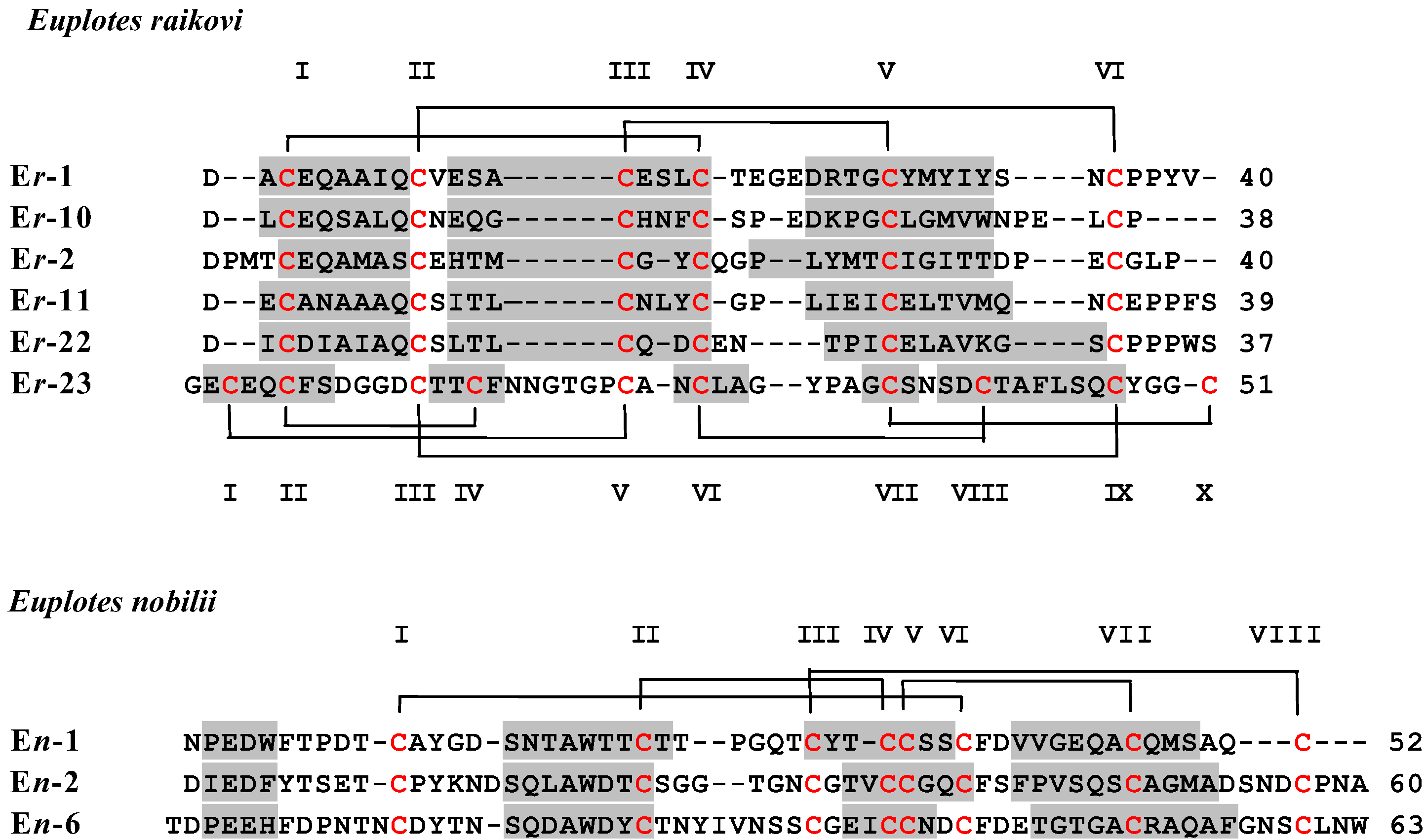
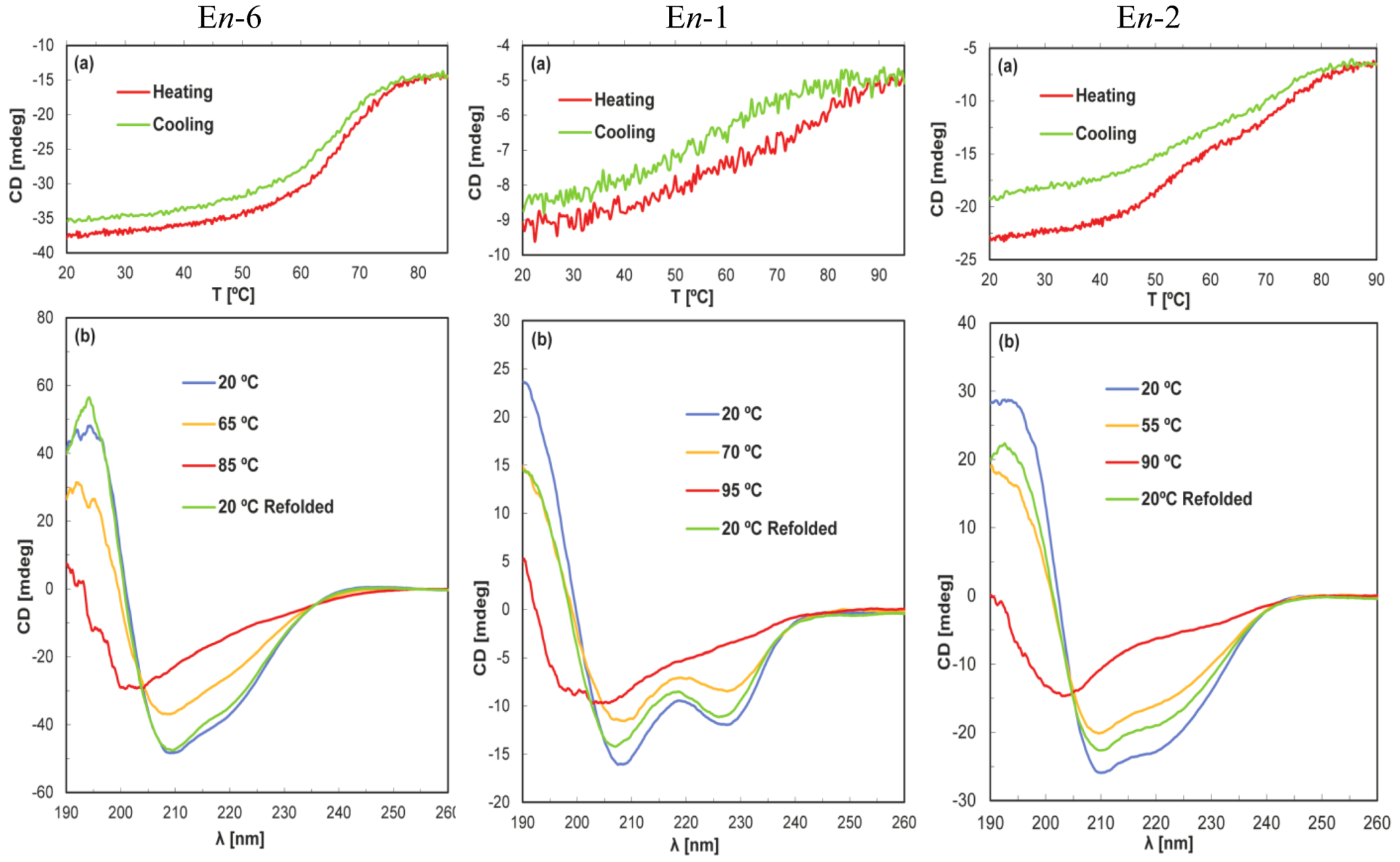
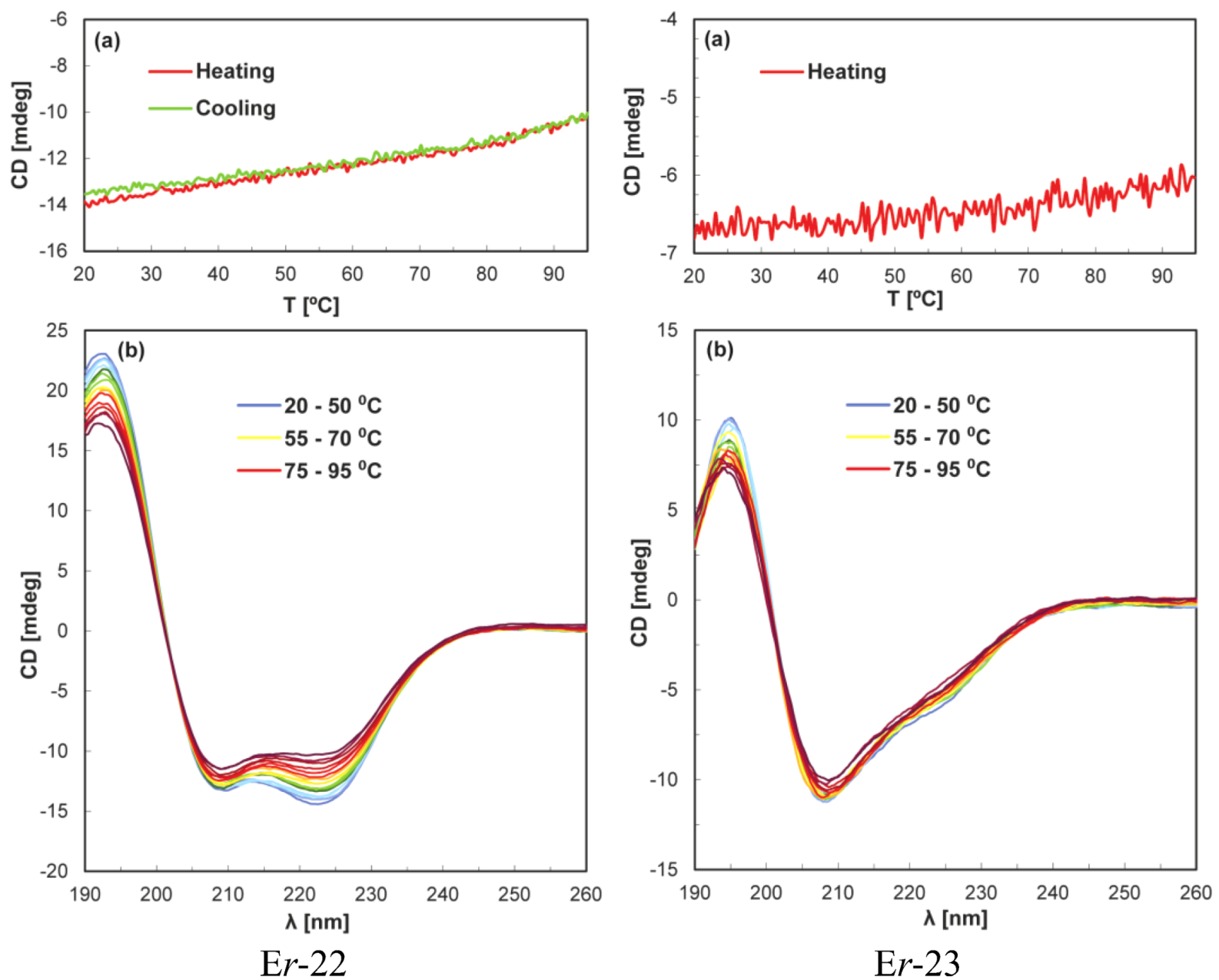
The isolation and purification of the nine proteins investigated in this paper has previously been described [22,23,24,25]. Lyophilized samples of each pheromone were dissolved in 20 mM sodium phosphate buffer at pH 6.0 and diluted to a protein concentration of 20 µM before aliquoting into a 0.1 cm path length quartz cuvette. CD experiments were recorded using the Temperature/Wavelength Scan software supplied with the Jasco 815 CD spectrophotometer. Melting curves over the range 20–95 °C were measured at a constant wavelength of 220 nm by increasing the temperature at a rate of 1.0 or 0.5 °C/min. Wavelength scans from 260–190 nm were measured at 5 °C intervals.
In additional exploratory studies, the chemical denaturants guanidine-HCl and urea were added to solutions of the mesophilic pheromones Er-1, Er-10, Er-22 and Er-23 to further test their stabilities. In particular melting curves were recorded for Er-1 in 7.8 M urea and 20 mM sodium phosphate at pH 6.0, 6 M guanidine-HCl and 20 mM sodium phosphate at pH 6.0, 4 M guanidine-HCl and 20 mM sodium phosphate at pH 6.0, 4.5 and 3.0, and 6 M urea at pH 3.0. Similar analyses on Er-10, Er-22 and Er-23 were performed using 4 M guanidine-HCl in 20 mM formic acid at pH 2.0.

3. Results

For all the three psychrophilic pheromones En-1, En-2, and En-6 of E. nobilii, the CD spectra (Figure 2) show that the regular secondary structures are unfolded at 95 °C, and that this unfolding is reversible upon cooling of the solutions to the starting temperature at 20 °C. Nevertheless, among the individual proteins there are appreciable variations with regard to the shape of the thermal unfolding curves. For En-6 a nearly symmetrical sigmoidal denaturation curve was observed with a midpoint near 65 °C, and a sigmoidal curve was also obtained for the refolding upon cooling of the solution; in addition, only a very small loss of protein was recorded during the unfolding/refolding procedure. On the other hand, En-1 and En-2 showed more sluggish unfolding transitions, and for En-2 there was an indication that the unfolding and refolding processes involve equilibria between more than two states. For the present qualitative survey of the stability of these three proteins we retain that, similar to En-6, the regular secondary structures in En-1 and En-2 are unfolded at 95 °C and refolded upon cooling of the solutions to 20 °C.
In the family of the six mesophilic pheromones Er-1, Er-2, Er-10, Er-11, Er-22, and Er-23 from E. raikovi, the regular secondary structures manifested in the CD spectra at 20 °C were found to be maintained up to the highest temperature studied (Figure 2). The temperature dependence of the signal intensity at 220 nm did not provide evidence for unfolding, and for Er-22 the small reduction of the signal intensity at the higher temperatures appeared to be fully reversible upon solution cooling. The data recorded for this pheromone are representative of the observations made with the other pheromones Er-1, Er-2, Er-10, and Er-11. Also the pheromone Er-23 showed qualitatively similar behavior, but it was clearly the most stable member of the E. raikovi mesophilic protein family since it did not undergo denaturation (Figure 3).
Figure 2. Temperature-induced denaturation of the E. nobilii psychrophilic pheromones En-6, En-1 and En-2 monitored by CD spectroscopy. (a) Temperature variation of the signal intensity at 220 nm during heating and cooling over the range from 20 °C to 95 °C. (b) CD spectra at different temperatures, as indicated by the color code in the figure. The protein concentration was 20 µM in 20 mM sodium phosphate at pH 6.0, and the cell length 0.1 cm. The temperature variation of the signal intensity at 220 nm in panels (a) was recorded at a rate of 1.0 °C/min. Scans were recorded with a speed of 100 nm/min, in 5 °C intervals over the temperature range of panels (a); for improved clarity, only four traces are shown in panels (b).
Biology 02 00142 g002
In view of the high thermal stability of the E. raikovi pheromones in neutral aqueous solution, we also performed exploratory experiments with the addition of chemical denaturants (see Materials and Methods). These experiments provided further indications of the remarkably high stability of these proteins. We did not observe their full unfolding in the temperature range 20–95 °C with the solution conditions listed in the Materials and Methods section, although partial melting within this temperature range was observed for some of them.

4. Discussion

The psychrophilic and mesophilic pheromone families of the protozoan ciliate Euplotes studied here are both represented by single-domain small disulfide-rich proteins, which have usually been shown to be outstandingly stable with regard to thermal denaturation in aqueous solution [26,27,28]. However, despite their extensive homology on the level of the amino acid sequences and three-dimensional structures, the psychrophilic E. nobilii pheromones showed a significantly lower thermal stability than their mesophilic E. raikovi counterparts. This finding coincides with observations derived from comparisons between other psychrophilic and mesophilic homologous proteins [29,30,31,32,33]. However, while these comparisons are essentially based on individual proteins from distantly related organisms, our finding involves families of proteins with known NMR solution structures [11,12,13,14,15,16,17,18,19,20,21] from two closely related species [34,35]. It therefore provides data which, at least in principle, are more reliable (being unaffected by the evolutionary noise which is intrinsic to comparisons between distantly related systems) for further detailed analyses by other groups of researchers who are interested in studying the correlations between protein structure, thermodynamic stability, and cold-adaptation.
Figure 3. Temperature-induced denaturation of the E. raikovi mesophilic pheromones Er-22 and Er-23 monitored by CD spectroscopy. Er-22 has been taken as representative of the other E. raikovi pheromones Er-1, Er-2, Er-10 and Er-11. Same experimental conditions and presentations as in Figure 2, except that all the spectra recorded in 5 °C temperature intervals are shown in panels (b).
Biology 02 00142 g003
As previously discussed [3,10,21], the two homologous protein families of E. nobilii psychrophilic pheromones and E. raikovi mesophilic pheromones differ significantly in the composition of polar, hydrophobic and aromatic amino acids, in global hydrophilicity and hydrophobicity, as well as in various aspects of their three-dimensional structures. It will now be interesting to explore the extent to which these physico-chemical and structural variations can be related to variations of the stability of the folded proteins. Two structural features distinctive of the two pheromone families appear to be particularly promising starting points for further investigations into the structural basis of the observed differences in the thermal stability of these proteins. One is the N-terminal elongation of 10 to 12 residues, which is common to all the E. nobilii pheromones and has no counterparts in any of the E. raikovi pheromones. It includes only a 310-helical turn as regular secondary structure and is devoid of connections through disulfide bonds to other parts of the proteins. Secondly, there is a higher density of disulfide bonds in the mesophilic E. raikovi pheromones than in the psychrophilic ones of E. nobilii. On average, the E. raikovi pheromones contain one disulfide bond per 13 amino acid residues, while in the E. nobilii pheromones there is one disulfide bond per 15 residues. In particular, the high density of one disulfide bond per 10 residues appears to be the dominant feature that makes Er-23 most stable among the E. raikovi pheromones, notwithstanding that Er-23 mimics the E. nobilii psychrophilic pheromones with regard to the contents of polar and hydrophobic residues as well as the aliphatic index.

5. Conclusions

The key observation reported in this paper is that the E. nobilii and E. raikovi pheromone families show a thermal denaturation behavior that is uniform among their members and significantly divergent between them. The mesophilic E. raikovi pheromones have higher stability by at least 30 °C when compared to the psychrophilic E. nobilii pheromones. Given the small size of these water-borne signaling proteins and the availability of their three-dimensional structures, the data presented in this communication should be of interest for systematic investigations of protein adaptation to cold environments.

Acknowledgments

The authors are grateful to R. Glockshuber (ETH, Zürich) for helpful discussions. This research was financially supported by the National Program for Antarctic Research (PNRA).

References

  1. Petz, W. Ciliates. In Antarctic Marine Protists; Scott, F.J., Marchant, H.J., Eds.; Australian Biological Resources Study: Canberra and Australian Antarctic Division, Hobart, Australia, 2005; pp. 347–448. [Google Scholar]
  2. Petz, W.; Valbonesi, A.; Schiftner, U.; Quesada, A.; Cynan Ellis-Evans, J. Ciliate biogeography in Antarctic and Arctic freshwater ecosystems: endemism or global distribution of species? FEMS Microbiol. Ecol. 2007, 59, 396–408. [Google Scholar] [CrossRef]
  3. Di Giuseppe, G.; Erra, F.; Dini, F.; Alimenti, C.; Vallesi, A.; Pedrini, B.; Wüthrich, K.; Luporini, P. Antarctic and Arctic populations of the ciliate Euplotes nobilii show common pheromone-mediated cell-cell signaling and cross-mating. Proc. Natl. Acad. Sci. USA 2011, 108, 3181–3186. [Google Scholar]
  4. Luporini, P.; Alimenti, C.; Ortenzi, C.; Vallesi, A. Ciliate mating type and their specific protein pheromones. Acta Protozool. 2005, 44, 89–101. [Google Scholar]
  5. Alimenti, C.; Vallesi, A.; Federici, S.; di Giuseppe, G.; Dini, F.; Carratore, V.; Luporini, P. Isolation and structural characterization of two water-borne pheromones from Euplotes crassus, a ciliate commonly known to carry membrane-bound pheromones. J. Eukaryot. Microbiol. 2011, 58, 234–241. [Google Scholar] [CrossRef]
  6. Vallesi, A.; Giuli, G.; Bradshaw, R.A.; Luporini, P. Autocrine mitogenic activity of pheromone produced by the protozoan ciliate Euplotes raikovi. Nature 1995, 376, 522–524. [Google Scholar] [CrossRef]
  7. Vallesi, A.; Ballarini, P.; di Pretoro, B.; Alimenti, C.; Miceli, C.; Luporini, P. Autocrine, mitogenic pheromone receptor loop of the ciliate Euplotes raikovi: Pheromone-induced receptor internalization. Eukaryot. Cell 2005, 4, 1221–1227. [Google Scholar] [CrossRef]
  8. La Terza, A.; Dobri, N.; Alimenti, C.; Vallesi, A.; Luporini, P. The water-borne protein signals (pheromones) of the Antarctic ciliated protozoan Euplotes nobilii: Structure of the gene coding for the En-6 pheromone. Can. J. Microbiol. 2009, 5, 57–62. [Google Scholar]
  9. Vallesi, A.; Alimenti, C.; La Terza, A.; di Giuseppe, G.; Dini, F.; Luporini, P. Characterization of the pheromone gene family of an Antarctic and Arctic protozoan ciliate, Euplotes nobilii. Mar. Genomics 2009, 2, 27–32. [Google Scholar] [CrossRef]
  10. Vallesi, A.; Alimenti, C.; Pedrini, B.; di Giuseppe, G.; Dini, F.; Wüthrich, K.; Luporini, P. Coding genes and molecular structures of the diffusible signalling proteins (pheromones) of the polar ciliate, Euplotes nobilii. Mar. Genomics 2012, 8, 9–13. [Google Scholar] [CrossRef]
  11. Brown, L.R.; Mronga, S.; Bradshaw, R.A.; Ortenzi, C.; Luporini, P.; Wüthrich, K. Nuclear magnetic resonance solution structure of the pheromone Er-10 from the ciliated protozoan Euplotes raikovi. J. Mol. Biol. 1993, 231, 800–816. [Google Scholar] [CrossRef]
  12. Mronga, S.; Luginbühl, P.; Brown, L.R.; Ortenzi, C.; Luporini, P.; Bradshaw, R.A.; Wüthrich, K. The NMR solution structure of the pheromone Er-1 from the ciliated protozoan Euplotes raikovi. Protein Sci. 1994, 3, 1527–1536. [Google Scholar] [CrossRef]
  13. Ottiger, M.; Szyperski, T.; Luginbühl, P.; Ortenzi, C.; Luporini, P.; Bradshaw, R.A.; Wüthrich, K. The NMR solution structure of the pheromone Er-2 from the ciliated protozoan Euplotes raikovi. Protein Sci. 1994, 3, 1517–1526. [Google Scholar]
  14. Luginbühl, P.; Ottiger, M.; Mronga, S.; Wüthrich, K. Structure comparison of the NMR structures of the pheromones Er-1, Er-10, and Er-2 from Euplotes raikovi. Protein Sci. 1994, 3, 1537–1546. [Google Scholar] [CrossRef]
  15. Luginbühl, P.; Wu, J.; Zerbe, O.; Ortenzi, C.; Luporini, P.; Wüthrich, K. The NMR solution structure of the pheromone Er-11 from the ciliated protozoan Euplotes raikovi. Protein Sci. 1996, 5, 1512–1522. [Google Scholar] [CrossRef]
  16. Liu, A.; Luginbühl, P.; Zerbe, O.; Ortenzi, C.; Luporini, P.; Wüthrich, K. NMR structure of the pheromone Er-22 from Euplotes raikovi. J. Biomol. NMR 2001, 19, 75–78. [Google Scholar] [CrossRef]
  17. Zahn, R.; Damberger, F.; Ortenzi, C.; Luporini, P.; Wüthrich, K. NMR structure of the Euplotes raikovi pheromone Er-23 and identification of its five disulfide bonds. J. Mol. Biol. 2001, 313, 923–931. [Google Scholar] [CrossRef]
  18. Placzek, W.J.; Etezady-Esfarjani, T.; Herrmann, T.; Pedrini, B.; Peti, W.; Alimenti, C.; Luporini, P.; Wüthrich, K. Cold-adapted signal proteins: NMR structures of pheromones from the Antarctic ciliate Euplotes nobilii. IUBMB Life 2007, 59, 578–585. [Google Scholar] [CrossRef]
  19. Pedrini, B.; Placzek, W.J.; Koculi, E.; Alimenti, C.; La Terza, A.; Luporini, P.; Wüthrich, K. Cold-adaptation in sea-waterborne signal proteins: Sequence and NMR structure of the pheromone En-6 from the Antarctic ciliate Euplotes nobilii. J. Mol. Biol. 2007, 372, 277–286. [Google Scholar] [CrossRef]
  20. Alimenti, C.; Vallesi, A.; Pedrini, B.; Wüthrich, K.; Luporini, P. Molecular cold-adaptation: Comparative analysis of two homologous families of psychrophilic and mesophilic signal proteins of the protozoan ciliate Euplotes. IUBMB Life 2009, 61, 838–845. [Google Scholar] [CrossRef]
  21. Weiss, M.S.; Anderson, D.H.; Raffioni, S.; Bradshaw, R.A.; Ortenzi, C.; Luporini, P.; Eisenberg, D.A. Cooperative model for ligand recognition and cell adhesion: Evidence from the molecular packing in the 1.6 Å crystal structure of the pheromone Er-1 from the ciliate protozoan Euplotes raikovi. Proc. Natl. Acad. Sci. USA 1995, 92, 10172–10176. [Google Scholar]
  22. Concetti, A.; Raffioni, S.; Miceli, C.; Barra, D.; Luporini, P. Purification to apparent homogeneity of the mating pheromone of mat-1 Euplotes raikovi. J. Biol. Chem. 1986, 61, 10582–10587. [Google Scholar]
  23. Raffioni, S.; Miceli, C.; Vallesi, A.; Chowdhury, S.K.; Chait, B.T.; Luporini, P.; Bradshaw, R.A. Primary structure od the Euplotes raikovi pheromones: Comparison of five sequences of pheromones from cell with variable mating interactions. Proc. Natl. Acad. Sci. USA 1992, 89, 2071–2075. [Google Scholar]
  24. Alimenti, C.; Ortenzi, C.; Carratore, V.; Luporini, P. Structural characterization of protein pheromone from a cold-adapted (Antarctic) single-cell eukaryote, the ciliate Euplotes nobilii. FEBS Lett. 2002, 514, 329–332. [Google Scholar] [CrossRef]
  25. Alimenti, C.; Ortenzi, C.; Carratore, V.; Luporini, P. Structural characterization of En-1, a cold-adapted protein pheromone isolated from the Antarctic ciliate Euplotes nobilii. Biochem. Biophys. Acta 2003, 1621, 17–21. [Google Scholar] [CrossRef]
  26. Devi, V.S.; Sprecher, C.B.; Hunziker, P.; Mittl, P.R.; Bosshard, H.R.; Jelesarov, I. Disulfide formation and stability of a cysteine-rich repeat protein from Helicobacter pylori. Biochemistry 2006, 45, 1599–1607. [Google Scholar]
  27. Trivedi, M.V.; Laurence, J.S.; Siahaan, T.J. The Role of thiols and disulfides on protein stability. Curr. Protein Pept. Sci. 2009, 10, 614–625. [Google Scholar] [CrossRef]
  28. Fass, D. Disulfide bonding in protein biophysics. In Annual Review of Biophysics; Rees, D.C., Ed.; Annual Reviews: Palo Alto, CA, USA, 2012; Volume 41, pp. 63–79. [Google Scholar]
  29. Aghajari, N.; Feller, G.; Gerday, C.; Haser, R. Structures of the psychrophilic Alteromonas haloplanctis α-amylase give insights into cold adaptation at a molecular level. Structure 1998, 6, 1503–1516. [Google Scholar] [CrossRef]
  30. Bae, E.; Phillips, G.N., Jr. Structure and analysis of highly homologous psychrophilic, mesophilic, and thermophilic adenylate kinases. J. Biol. Chem. 2004, 279, 28202–28208. [Google Scholar] [CrossRef]
  31. Fedoy, A.E.; Yang, N.; Martinez, A.; Leiros, H.K.; Steen, I.H. Structural and functional properties of isocitrate dehydrogenase from the psychrophilic bacterium Desulfotalea psychrophila reveal a cold-active enzyme with an unusual high thermal stability. J. Mol. Biol. 2007, 372, 130–149. [Google Scholar] [CrossRef]
  32. Garcia-Arribas, O.; Mateo, R.; Melanie, M.; Tomczak, M.M.; Davies, P.L.; Mateu, M.G. Thermodynamic stability of a cold-adapted protein, type III antifreeze protein, and energetic contribution of salt bridges. Protein Sci. 2007, 16, 227–238. [Google Scholar]
  33. Lockwood, B.L.; Somero, G.N. Functional determinants of temperature adaptation in enzymes of cold- versus warm-adapted mussels (genus Mytilus). Mol. Biol. Evol. 2012, 29, 3061–3070. [Google Scholar] [CrossRef]
  34. Vallesi, A.; di Giuseppe, G.; Dini, F.; Luporini, P. Pheromone evolution in the protozoan ciliate, Euplotes: The ability to synthesize diffusibile forms is ancestral and secondarily lost. Mol. Phylogenet. Evol. 2008, 47, 439–442. [Google Scholar] [CrossRef]
  35. Jiang, J.; Zhang, Q.; Warren, A.; Al-Rasheid, K.A.; Song, W. Morphology and SSUrRNA gene-based phylogeny of two marine Euplotes species, E. orientalis spec. nov. and E. raikovi (Ciliophora, Euplotida). Eur. J. Protisotol. 2010, 46, 121–132. [Google Scholar]

