Article

Unveiling Genomic Islands Hosting Antibiotic Resistance Genes and Virulence Genes in Foodborne Multidrug-Resistant Patho-Genic Proteus vulgaris

Tianjin Key Laboratory of Food Biotechnology, School of Biotechnology and Food Science, Tianjin University of Commerce, Tianjin 300134, China
*
Author to whom correspondence should be addressed.
Biology 2025, 14(7), 858; https://doi.org/10.3390/biology14070858
Submission received: 29 May 2025 / Revised: 5 July 2025 / Accepted: 14 July 2025 / Published: 15 July 2025

Simple Summary

Proteus vulgaris is a foodborne pathogen commonly found in seafood, particularly shrimp, that can cause serious infections in humans. Of particular concern are multidrug-resistant strains like P3M, which was isolated from farmed shrimp and poses significant treatment challenges. Our study examined the genetic mechanisms behind this bacterium’s antimicrobial resistance and disease-causing potential. Through genomic analysis, we identified specialized DNA segments called genomic islands that harbor both antibiotic resistance genes and virulence factors. These mobile genetic elements can transfer between bacteria, rapidly spreading dangerous traits. Additionally, we detected clusters of resistance genes associated with highly mobile DNA sequences that may accelerate their dissemination. Understanding these genetic transmission pathways is essential for developing effective strategies to control infections and safeguard food supplies. Our findings emphasize the urgent need for improved antibiotic stewardship in aquaculture systems to curb the spread of resistant bacteria. This work offers important guidance for researchers and public health officials striving to enhance food safety and protect community health.

Abstract

Proteus vulgaris is an emerging multidrug-resistant (MDR) foodborne pathogen that poses a significant threat to food safety and public health, particularly in aquaculture systems where antibiotic use may drive resistance development. Despite its increasing clinical importance, the genomic mechanisms underlying antimicrobial resistance (AMR) and virulence transmission in foodborne Proteus vulgaris remain poorly understood, representing a critical knowledge gap in One Health frameworks. To investigate its AMR and virulence transmission mechanisms, we analyzed strain P3M from Penaeus vannamei intestines through genomic island (GI) prediction and comparative genomics. Our study provides the first comprehensive characterization of mobile genetic elements in aquaculture-derived Proteus vulgaris, identifying two virulence-associated GIs (GI12/GI15 containing 25/6 virulence genes) and three AMR-linked GIs (GI7/GI13/GI16 carrying 1/1/5 antibiotic resistance genes (ARGs)), along with a potentially mobile ARG cluster flanked by IS elements (tnpA-tnpB), suggesting horizontal gene transfer capability. These findings elucidate previously undocumented genomic mechanisms of AMR and virulence dissemination in Proteus vulgaris, establishing critical insights for developing One Health strategies to combat antimicrobial resistance and virulence in foodborne pathogens.

1. Introduction

Foodborne pathogens, which include notable representatives such as Salmonella, Escherichia coli, Proteus spp., Staphylococcus aureus [1,2,3,4], are ubiquitous in human food systems and pose significant public health risks through their transmission via contaminated food, causing illnesses ranging from diarrhea to severe food poisoning [5]. Recent advances in molecular biology have led to the development of innovative detection techniques and analytical tools, which have substantially improved our ability to track transmission pathways, identify infection sources, and elucidate pathogenic mechanisms. These technological developments have consequently enhanced the diagnostic accuracy, detection sensitivity, and overall understanding of foodborne diseases [6,7,8].
The escalating use of antibiotics has driven a concerning rise in antimicrobial resistance (AMR) among foodborne pathogens [9,10]. This resistance mechanism functions as a biological defense system, effectively neutralizing antibiotic activity and promoting pathogen survival and proliferation [11]. Notably, the convergence of virulence factors and AMR mechanisms equips pathogens with enhanced capacity to overcome host defenses and complicates food safety interventions, thereby posing unprecedented challenges to public health regulation [12]. Consequently, comprehensive investigations into the molecular basis of pathogenicity and resistance are critical for developing effective food safety protocols, optimizing antibiotic stewardship, and designing targeted control measures.
The genomic repertoire of foodborne pathogens contains antibiotic resistance genes (ARGs) and virulence determinants, which collectively drive their pathogenic potential and drug resistance [13,14]. Horizontal gene transfer (HGT) serves as the primary vehicle for disseminating these traits, enabling rapid genetic adaptation through the acquisition of exogenous DNA [15]. Genomic islands (GIs), which are mobile DNA segments frequently harboring ARGs and virulence genes, have been extensively documented as major HGT substrates [16,17]. These modular genetic elements can integrate into microbial chromosomes and facilitate interspecies gene exchange, thereby expanding functional diversity and evolutionary potential [18,19,20]. Complementing GIs, insertion sequences (ISs) constitute another class of mobile genetic elements (MGEs) that orchestrate DNA rearrangements and accelerate trait propagation [21,22]. Collectively, these HGT mechanisms underpin bacterial evolution, including the emergence of enhanced virulence and multidrug resistance profiles.
Proteus vulgaris is a ubiquitous environmental bacterium that readily disseminates through food chains, with increasing prevalence as an emerging multidrug-resistant pathogen in aquaculture systems [23,24,25]. Clinically, this organism is a well-documented etiological agent of gastroenteritis, urinary tract infections, and other opportunistic diseases, establishing its role as a priority foodborne pathogen [26,27]. Notably, Proteus vulgaris exhibits concerning resistance phenotypes, including multidrug resistance (MDR) and intrinsic resistance to polymyxins (last-resort antibiotics), which severely limits therapeutic options for associated infections [28,29]. This resistance profile is especially alarming in aquaculture settings, where frequent gene exchange between environmental and clinical strains may accelerate the spread of resistance determinants [30,31,32,33].
While GIs have been well characterized as key vectors for AMR and virulence gene dissemination in other Enterobacterales like E. coli and Salmonella [17,34,35,36], their specific roles in mediating ARGs and virulence genes transfer in Proteus vulgaris remain unexplored, particularly within foodborne transmission chains. This study aims to characterize the GI repertoire of the multidrug-resistant Proteus vulgaris strain P3M from aquaculture sources, with a particular focus on assessing evolutionary conservation of resistance/virulence-associated GIs across 13 sequenced Proteus vulgaris strains through comparative genomics. Our focus on P3M’s GI architecture is motivated by its clinical resistance profile (resistant to seven antibiotic classes) [28], and the absence of prior systematic GI analyses in foodborne Proteus vulgaris, addressing critical knowledge gaps in understanding how this pathogen acquires and disseminates resistance and virulence traits under the One Health framework.

2. Materials and Methods

2.1. Proteus Vulgaris Genomes and Data Acquisition

The complete genome sequence of Proteus vulgaris strain P3M (accession: CP060211), along with 12 additional sequenced Proteus vulgaris genomes were retrieved from NCBI Nucleotide database (https://www.ncbi.nlm.nih.gov/nuccore, accessed on 11 April 2025) in GenBank (GBK) format. The analyzed strains included the following: Proteus vulgaris strain CCU063 (CP032663), FADDRGOS_366 (CP150645), FADDR-GOS_566 (CP033736), FADDRGOS_1507 (CP083628), HH17 (CP054157), LC-693 (CP063314), PvSC3 (CP034668), TAF3 (CP126335), USDA-ARS-USMARC-49741 (CP104121), ZN3 (CP047344), Ld01 (CP090064), and 2023JQ-00005 (CP137920). All genomes represented complete, annotated assemblies, with sequencing platforms, library protocols, and assembly methods detailed in their original NCBI submissions (accessible via the provided accession numbers). Detailed information of all these 13 Proteus vulgaris strains is listed in Table S1.

2.2. Prediction and Comparison of Genomic Islands (GIs)

The online tool IslandViewer 4 (v4.0.1) was employed to predict the potential genomic islands (https://www.pathogenomics.sfu.ca/islandviewer/browse/, accessed on 15 April 2025) [37]. IslandViewer 4 integrates three distinct algorithms (SIGI-HMM, IslandPick, and IslandPath-DIMOB) to compensate for limitations of individual methods, achieving >90% consensus accuracy for GI boundaries. Among these, SIGI-HMM analyzes codon usage bias to identify potential genomic islands, while Island-Path-DIMOB assesses dinucleotide deviation for the same purpose. The IslandPick method employs genome-wide sequencing to detect large DNA fragments that exist independently within a genome but are absent in related genomes. Additionally, a comparative analysis of genome islands was conducted using the IslandCompare online tool (v1.1.0) [38] (https://islandcompare.ca/analysis?id=ff959500-555c-11ef-b9c1-059f1c7f7fe4, accessed on 15 April 2025). IslandCompare was employed because it uniquely visualizes GI conservation patterns across all 13 sequenced Proteus vulgaris strains, enabling evolutionary inferences about ARG dissemination.

2.3. Antibiotic Resistance Genes (ARGs) and Virulence Genes Prediction

ARGs and virulence genes were predicted from the P3M genome using CARD (https://card.mcmaster.ca/, accessed on 16 April 2025) (v6.0.0) [39] and VFDB (v2021) (https://www.mgc.ac.cn/VFs/main.htm, accessed on 16 April 2025) databases, respectively. For ARG prediction we applied stringent thresholds of ≥80% sequence identity and ≥80% coverage as recommended by CARD to ensure reliable detection of resistance determinants, while virulence gene identification employed slightly relaxed thresholds (≥70% identity, ≥50% coverage) following VFDB guidelines to capture functionally conserved yet evolutionarily divergent elements. These parameter selections were based on database recommendations, empirical validation studies demonstrating optimal sensitivity-specificity balance, with all analyses performed using default parameters unless otherwise specified, and full command-line documentation is available upon request to ensure complete reproducibility.

2.4. Phylogenetic Tree Construction

Phylogenetic analysis was performed using MEGA (v7.0.26) [40,41] to construct unrooted neighbor-joining trees of target genes and genomic islands. Coding sequences were first aligned using MUSCLE (v3.8.31) with default parameters. The Maximum Composite Likelihood (MCL) evolutionary model was selected after model testing in MEGA7. To assess node reliability, bootstrap analysis with 1000 replicates was conducted. For genomic island comparisons, phylogenetic analysis was based on core genome sequences. The resulting tree is drawn to scale, with branch lengths representing the evolutionary distances used for phylogenetic inference.

2.5. Sequence Alignments

Linear comparison and map generation of the genomic islands were performed and visualized using Easyfig software (v2.2.3) [42] with BLASTn (v 2.2.31+) under a 50% minimum identity threshold, where input sequences in GenBank format were annotated by Prokka and visualized as linear maps highlighting conserved syntenic blocks, boundary regions, and ARG/virulence gene clusters, followed by manual verification of domain architecture using NCBI’s CD-Search to ensure alignment accuracy.
Detailed tool specifications are provided in Table S2.

3. Results

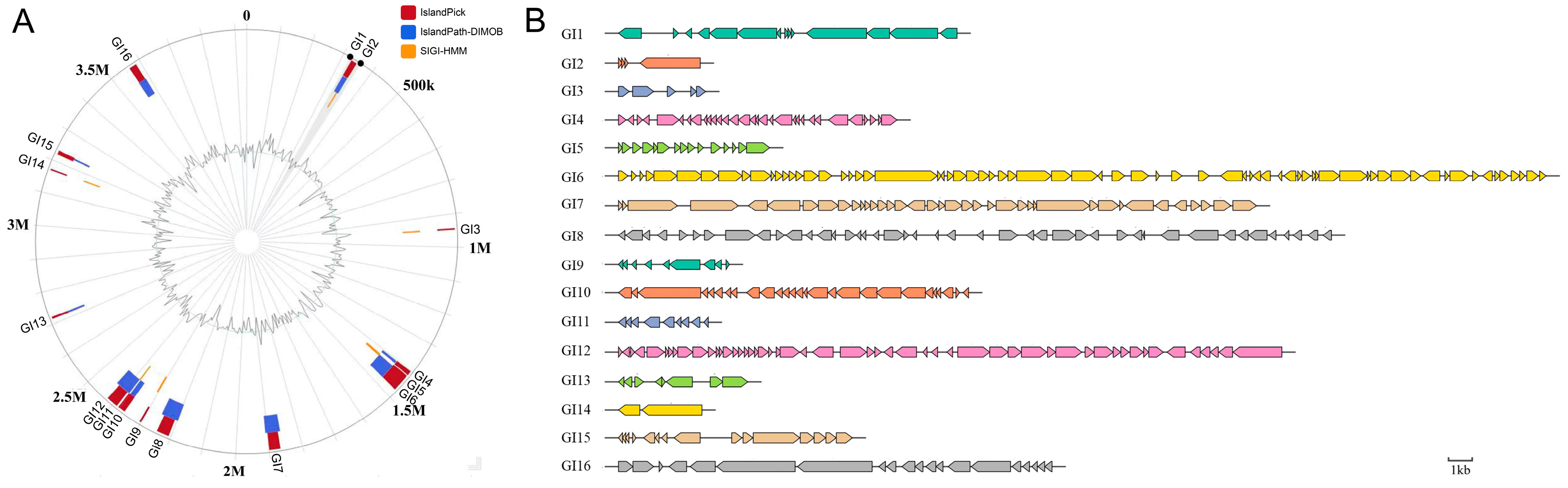
3.1. P3M Genomic Island Analysis

Comprehensive genomic characterization identified 16 GIs in P3M (Table 1, Figure 1), ranging from 4.4 kb (GI2) to 49.9 kb (GI6) in size and distributed throughout the chromosome, exhibiting significant structural diversity with coding gene content varying from 2 (GI14) to 69 (GI6) genes per island [43]. These GIs demonstrated notable variations in genomic location, size distribution, and gene composition, confirming their established roles in horizontal gene transfer and genome plasticity while highlighting their substantial capacity for adaptive gene acquisition and diverse functional potential.

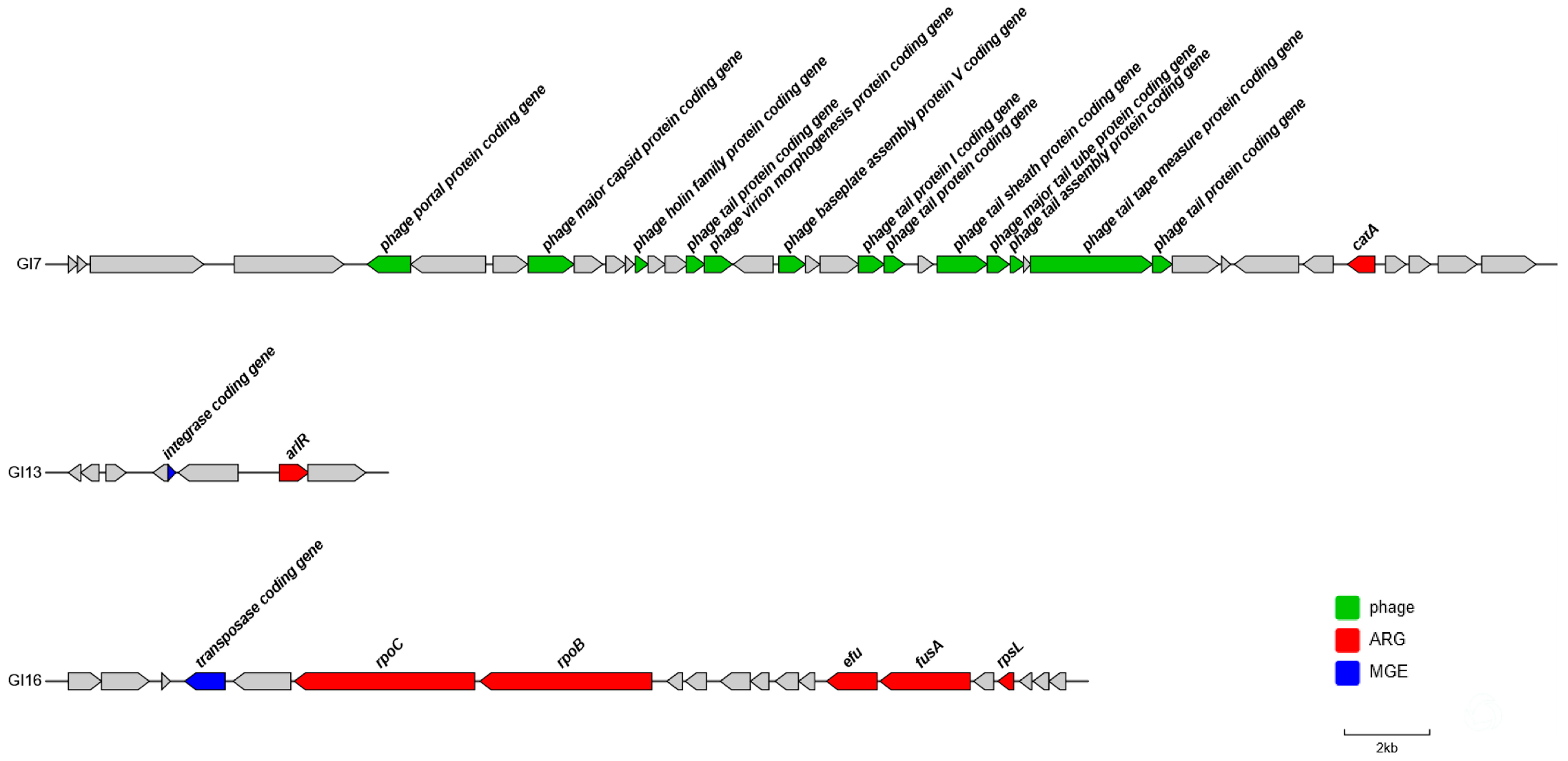
3.2. Identification of Antibiotic Resistance-Associated Genomic Islands in P3M

Genomic analysis identified 218 potential ARGs in strain P3M (Table S3: Potential ARGs on the P3M genome predicted by CARD database), with three GIs (GI7, GI13, GI16) harboring clinically significant resistance determinants (Figure 2). GI7 contained catA encoding chloramphenicol O-acetyltransferase [44,45] (highlighted in blue in Table S3), while GI13 carried hprR associated with fluoroquinolone resistance [46] (highlighted in pink in Table S3). GI16 possessed five ARGs (rpoC, rpoB, tuf, fusA, and rpsL) conferring resistance to daptomycin, rifampicin, kirromycin, fusidic acid, and aminoglycosides, respectively (highlighted in green in Table S3), with tuf currently displaying resistance-conferring mutations. Notably, GI13 and GI16 contained integrases/transposases facilitating horizontal transfer, whereas GI7’s mobilization potential may involve novel mechanisms through uncharacterized ORFs. The localization of these ARGs within MGEs underscores their dissemination risk, particularly under selective pressures in aquatic environments [39].
Comparative genomic analyses of GI7, GI13, and GI16 revealed distinct evolutionary origins, with phylogenetic reconstruction (Figure 3A–C) demonstrating that GI7 showed closest affinity to Hafnia alvei strain 2023JQ-00054, while GI16 clustered with Proteus penneri strain S178-2, suggesting intergeneric horizontal transfer events. Sequence alignments (Figure 3D–F) confirmed high conservation of all ARGs across related strains, though the GI13 integrase gene appeared unique to P3M, indicating potential strain-specific mobilization mechanisms. These findings collectively demonstrate the mosaic evolutionary history of resistance GIs in P3M, with both conserved resistance determinants and strain-specific elements contributing to the dissemination.
Whole genome analysis identified 42 IS elements in P3M, predominantly from the IS200/IS605 family encoding TnpA transposase and its regulatory partner TnpB (Table S4: IS on the P3M genome) [21,22,47]. Notably, we characterized a 30 kb genomic segment (1,609,703–1,639,229 bp) flanked by identical tnpA-tnpB genes, containing three putative ARGs (fabG, pgsA, and satB) with potential resistance to triclosan, daptomycin, and chloramphenicol, respectively (highlighted in yellow in Table S3). Although fabG and pgsA currently lack resistance-conferring mutations, their unique genomic organization between functional IS elements suggests an evolutionarily active hotspot for ARG acquisition and dissemination through transposition-mediated mechanisms, representing an alternative pathway to GI-mediated ARG transfer.
Comparative genomic analysis under stringent alignment parameters (100% query coverage, >85% identity) revealed distinct conservation patterns: while pgsA and satB were universally conserved across all 13 Proteus vulgaris strains, fabG was uniquely present in P3M and 2023JQ-00005, and the flanking tnpA-tnpB genes were retained in only four strains (P3M, CCU063, 2023JQ-00005, and HH17) (Figure 4). Notably, 2023JQ-00005 maintained complete conservation of all cluster components, suggesting this strain may represent an important reservoir for IS-mediated resistance dissemination. The strain-specific distribution patterns of these MGEs highlight substantial genomic plasticity within Proteus vulgaris populations, likely driven by differential transposition activity across lineages. And comprehensive BLAST analysis (sequence coverage > 70%, identity > 90%) and subsequent phylogenetic reconstruction (Figure 5) revealed that the tnpA-tnpB-flanked ARG cluster in P3M exhibited closer genetic affinity to Proteus faecis strain 19MO01SH08 than to its conspecific Proteus vulgaris strain 2023JQ-00005, suggesting either horizontal acquisition from divergent Proteus lineages or convergent evolution under comparable selection pressures in aquatic environments.

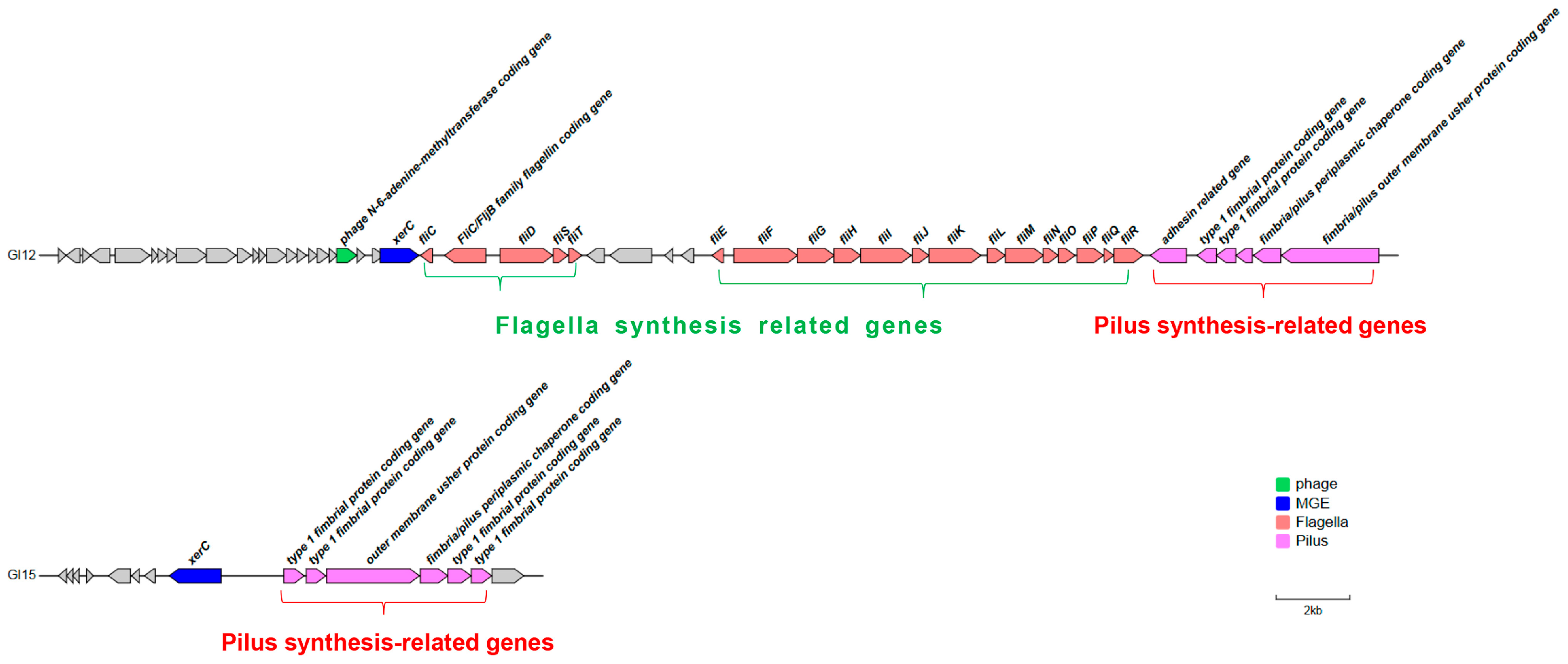
3.3. Identification Virulence-Associated Genomic Islands in P3M

Genomic analysis identified over 150 virulence-associated genes in the foodborne strain P3M (Table S5: Virulence genes on the P3M genome), with two GIs (GI12 and GI15) exhibiting significant enrichment of virulence determinants (Figure 6). GI12 contained 25 virulence genes (48.1% of its 52 total genes), including clusters for flagellar assembly and type I fimbriae synthesis, while GI15 harbored six virulence genes (40.0% of 15 genes) specifically encoding P-type fimbriae components. Both GIs carried the xerC integrase gene (Figure 6), a marker of horizontal transfer potential, consistent with their proposed role in disseminating motility and adhesion traits that are critical for host colonization [48,49]. The concentration of these virulence factors within MGEs suggests an efficient mechanism for pathogenicity acquisition in Proteus vulgaris, particularly concerning its transmission through food chains.
Phylogenetic reconstruction revealed distinct evolutionary origins for the virulence GIs, with GI12 showing closest affinity to Proteus sp. CD3 while GI15 clustered with Proteus penneri strain S178-2 (Figure 7A,B). Functional analysis demonstrated differential conservation patterns: GI12’s flagellar genes were widely conserved across species, whereas its fimbrial genes were P3M-specific; conversely, GI15’s P-type fimbrial genes showed high conservation among related species (Figure 7C,D). The unique presence of xerC integrase in P3M’s virulence GIs suggests strain-specific mobilization of these pathogenicity determinants, potentially contributing to its distinctive virulence profile as a foodborne pathogen.

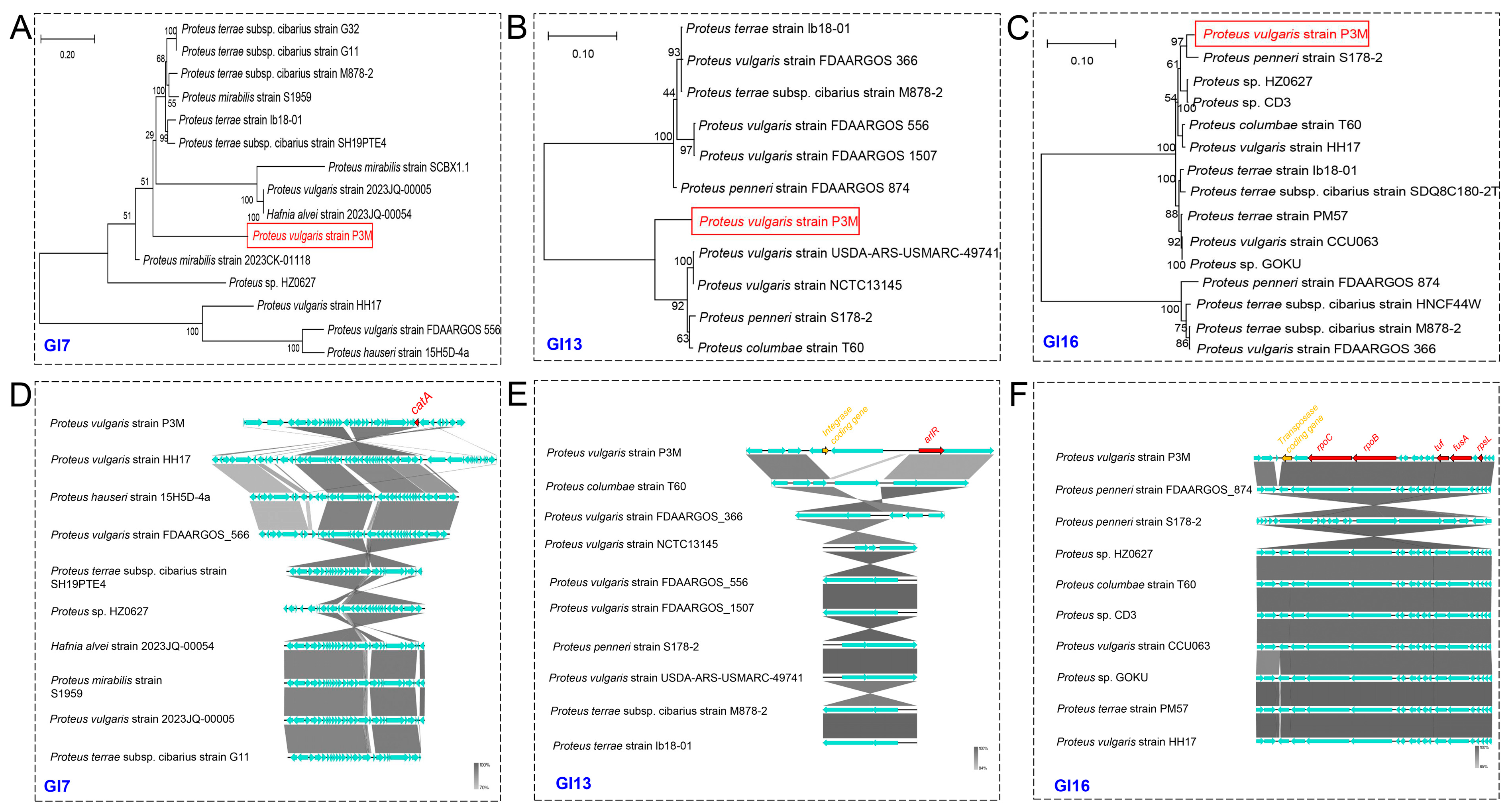
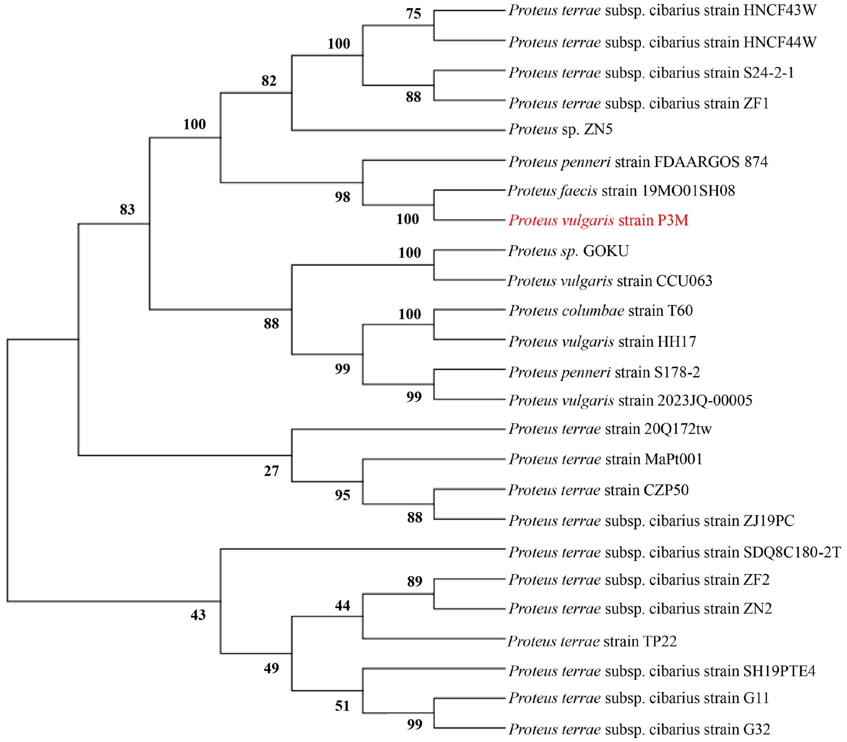
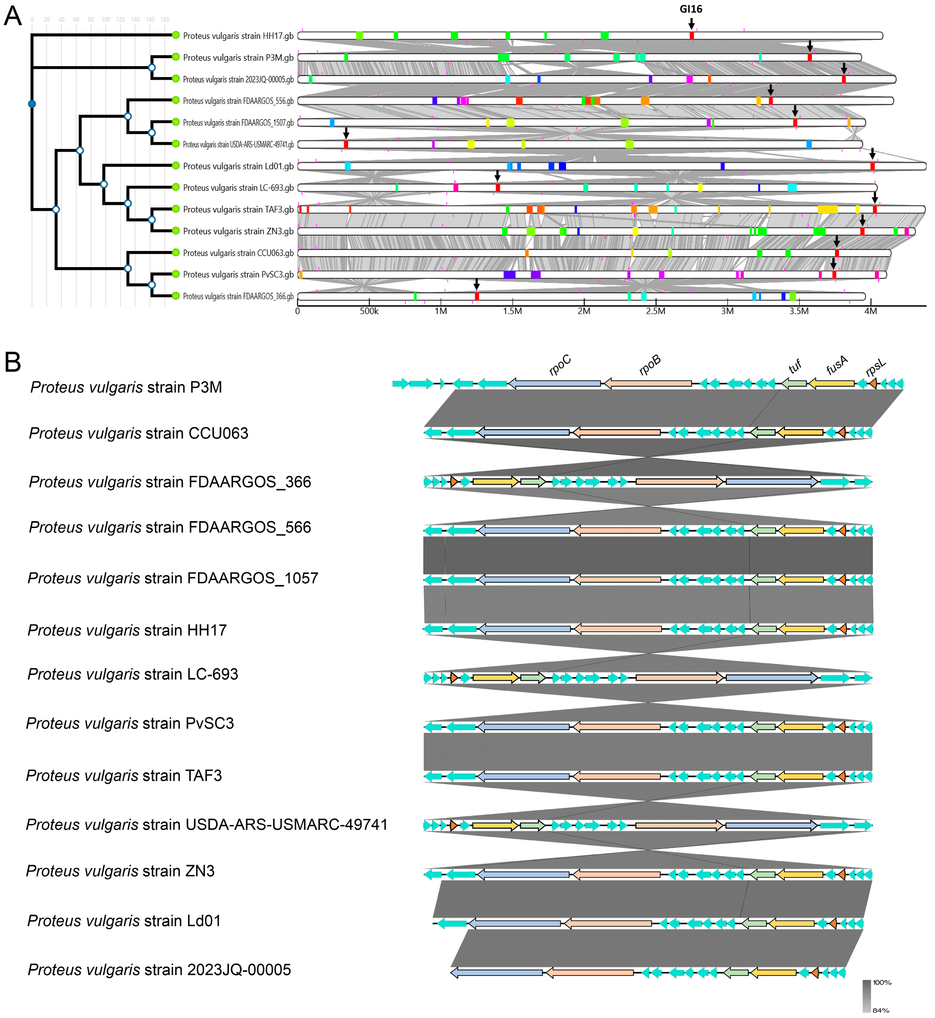
3.4. Evolutionary Conservation Analysis of Genomic Islands in P3M

Comparative genomic analysis of 13 Proteus vulgaris strains revealed substantial variation in GI content, with GI16 emerging as the sole universally conserved GI across all strains (Figure 8A). Detailed characterization of GI16 identified 15 highly conserved genes (75% of its 20-gene content) among the 12 comparative strains, exhibiting minimal sequence variation (Figure 8B). Notably, this conserved core included all five previously identified ARGs (rpoC, rpoB, tuf, fusA, and rpsL), suggesting GI16 may harbor essential genetic elements maintained through purifying selection in Proteus vulgaris populations. The exceptional conservation of this resistance-associated island underscores its potential evolutionary significance in this bacterial species, warranting further investigation into its functional roles.

4. Discussion

MGEs, particularly GIs, serve as key drivers of microbial adaptation through their ability to rapidly disseminate functional genes across bacterial populations [50,51,52]. Our characterization of 16 GIs in the aquaculture-derived Proteus vulgaris strain P3M revealed distinct functional specialization, with two islands (GI12/GI15) enriched for virulence determinants and three (GI7/GI13/GI16) harboring ARGs. These islands exhibited remarkable structural diversity (4.4–49.9 kb) and varied genomic distributions, reflecting their dynamic evolutionary trajectories and potential for niche adaptation. Of particular significance was the identification of an IS tnpA-tnpB-associated ARG cluster exhibiting strain-specific conservation patterns, suggesting an alternative mobilization pathway to classical genomic island transfer. The chromosomal localization of all resistance and virulence determinants, consistent with previous reports of limited plasmid carriage in P3M [53], underscores the importance of integrative genetic elements in this strain’s adaptive arsenal. These findings collectively highlight the dual role of GIs in both enhancing Proteus vulgaris pathogenicity and facilitating the dissemination of resistance traits through food production chains.
Three potential mechanisms may account for these genomic arrangements: (i) Integron-mediated gene cassette capture, as observed in Serratia’s intI1-associated gene arrays; (ii) Tn3-like transposition via tnpA-tnpB; and (iii) Phage-derived recombination at inverted repeats flanking GI12. Notably, the unique presence of specific integrases in resistance-associated GI13 and virulence-associated GI12/GI15 implies P3M has evolved distinctive genetic transfer strategies compared to other Proteus strains, providing novel insights into the evolutionary dynamics of genomic islands in environmental pathogens. These findings significantly expand our understanding of how genomic plasticity contributes to bacterial adaptation in aquaculture systems.
From a One Health standpoint, the persistence of these GIs in foodborne Proteus vulgaris underscores important concerns about potential resistance gene dissemination to human-associated pathogens [54]. While our bioinformatic analyses reveal compelling patterns of genomic island conservation and diversification, the study has inherent limitations that warrant acknowledgment. Future investigations should prioritize experimental validation through functional genomics approaches, including targeted gene knockout studies and comprehensive characterization of resistance and virulence gene expression profiles under relevant environmental conditions. Such efforts will be crucial for elucidating the precise mechanisms governing antibiotic resistance development and pathogenicity in this emerging foodborne pathogen, ultimately informing more effective surveillance and control strategies at the human–animal-environment interface.

5. Conclusions

This study provides important genomic insights into the multidrug-resistant foodborne pathogen Proteus vulgaris strain P3M, characterizing 16 GIs with specialized functional profiles, including virulence-enriched GI12/GI15 and resistance-associated GI7/GI13/GI16. The universal conservation of GI16 across Proteus vulgaris strains contrasts with strain-specific features like GI13’s unique integrase, suggesting complex adaptation strategies in aquaculture environments. Although our bioinformatic analyses reveal compelling evidence for horizontal gene transfer potential, the study’s limitations—particularly the need for functional validation of predicted resistance/virulence genes and experimental confirmation of genomic island mobility—highlight important directions for future research. Moving forward, integrating transcriptomic profiling, conjugation assays, and expanded environmental surveillance will be essential to fully elucidate the transmission dynamics of these genetic elements and develop effective control measures at the aquaculture–human interface, ultimately contributing to more robust antimicrobial resistance containment strategies within the One Health framework.

Supplementary Materials

The following supporting information can be downloaded at https://www.mdpi.com/article/10.3390/biology14070858/s1, Table S1: Detailed information of the 13 sequenced Proteus vulgaris strains used in this study; Table S2: Summary of bioinformatics tools and databases used in this study; Table S3: Potential antibiotic resistance genes (ARGs) on the P3M genome predicted by CARD database; Table S4: IS on the P3M genome; Table S5: Virulence genes on the P3M genome.

Author Contributions

Conceptualization, H.Z.; methodology, H.Z. and T.W.; validation, H.R.; data curation, H.Z., T.W. and H.R.; writing—original draft preparation, H.Z.; writing—review and editing, T.W. and H.R.; Supervision: H.R. All authors have read and agreed to the published version of the manuscript.

Funding

This research was funded by Tianjin Education Commission Scientific Research Project (grant number 2022KJ004).

Institutional Review Board Statement

Not applicable.

Informed Consent Statement

Not applicable.

Data Availability Statement

Data supporting the findings of the present study are included in the article and Supplementary Materials. The complete genomic information of Proteus vulgaris strains can be accessed through the database (https://www.ncbi.nlm.nih.gov/nucleotide/, accessed on 11 April 2025).

Conflicts of Interest

The authors declare no conflicts of interest.

References

  1. Ferrari, R.G.; Rosario, D.K.A.; Cunha-Neto, A.; Mano, S.B.; Figueiredo, E.E.S.; Conte-Junior, C.A. Worldwide Epidemiology of Salmonella Serovars in Animal-Based Foods: A Meta-analysis. Appl. Environ. Microbiol. 2019, 85, e00591-19. [Google Scholar] [CrossRef]
  2. Babines-Orozco, L.; Balbuena-Alonso, M.G.; Barrios-Villa, E.; Lozano-Zarain, P.; Martínez-Laguna, Y.; Del Carmen Rocha-Gracia, R.; Cortés-Cortés, G. Antimicrobial resistance in food-associated Escherichia coli in Mexico and Latin America. Biosci. Microbiota Food Health 2024, 43, 4–12. [Google Scholar] [CrossRef]
  3. Li, R.; Zhou, M.; Lu, J.; Wei, J. Antibiofilm Effects of Epigallocatechin Gallate Against Proteus mirabilis Wild-Type and Ampicillin-Induced Strains. Foodborne Pathog. Dis. 2022, 19, 136–142. [Google Scholar] [CrossRef]
  4. Sergelidis, D.; Angelidis, A.S. Methicillin-resistant Staphylococcus aureus: A controversial food-borne pathogen. Lett. Appl. Microbiol. 2017, 64, 409–418. [Google Scholar] [CrossRef]
  5. Cangemi, J.R. Food poisoning and diarrhea: Small intestine effects. Curr. Gastroenterol. Rep. 2011, 13, 442–448. [Google Scholar] [CrossRef] [PubMed]
  6. Kocot, A.M.; Briers, Y.; Plotka, M. Phages and engineered lysins as an effective tool to combat Gram-negative foodborne pathogens. Compr. Rev. Food Sci. Food Saf. 2023, 22, 2235–2266. [Google Scholar] [CrossRef]
  7. Shakeran, Z.; Javadi-Zarnaghi, F.; Emamzadeh, R. Novel luminescent affiprobes for molecular detection of Staphylococcus aureus using flow cytometry. J. Appl. Microbiol. 2021, 130, 493–503. [Google Scholar] [CrossRef] [PubMed]
  8. Bakhshandeh, B.; Sorboni, S.G.; Haghighi, D.M.; Ahmadi, F.; Dehghani, Z.; Badiei, A. New analytical methods using carbon-based nanomaterials for detection of Salmonella species as a major food poisoning organism in water and soil resources. Chemosphere 2022, 287, 132243. [Google Scholar] [CrossRef] [PubMed]
  9. Ge, H.; Wang, Y.; Zhao, X. Research on the drug resistance mechanism of foodborne pathogens. Microb. Pathog. 2022, 162, 105306. [Google Scholar] [CrossRef]
  10. Asfaw, T.; Genetu, D.; Shenkute, D.; Shenkutie, T.T.; Amare, Y.E.; Yitayew, B. Foodborne Pathogens and Antimicrobial Resistance in Ethiopia: An Urgent Call for Action on “One Health”. Infect. Drug Resist. 2022, 15, 5265–5274. [Google Scholar] [CrossRef]
  11. Patil, A.; Banerji, R.; Kanojiya, P.; Saroj, S.D. Foodborne ESKAPE Biofilms and Antimicrobial Resistance: Lessons Learned from Clinical Isolates. Pathog. Glob. Health 2021, 115, 339–356. [Google Scholar] [CrossRef]
  12. Wu-Wu, J.W.F.; Guadamuz-Mayorga, C.; Oviedo-Cerdas, D.; Zamora, W.J. Antibiotic Resistance and Food Safety: Perspectives on New Technologies and Molecules for Microbial Control in the Food Industry. Antibiotics 2023, 12, 550. [Google Scholar] [CrossRef]
  13. Parra-Flores, J.; Holý, O.; Bustamante, F.; Lepuschitz, S.; Pietzka, A.; Contreras-Fernández, A.; Castillo, C.; Ovalle, C.; Alarcón-Lavín, M.P.; Cruz-Córdova, A.; et al. Virulence and Antibiotic Resistance Genes in Listeria monocytogenes Strains Isolated From Ready-to-Eat Foods in Chile. Front. Microbiol. 2022, 12, 796040. [Google Scholar] [CrossRef]
  14. Hartantyo, S.H.P.; Chau, M.L.; Koh, T.H.; Yap, M.; Yi, T.; Cao, D.Y.H.; GutiÉrrez, R.A.; Ng, L.C. Foodborne Klebsiella pneumoniae: Virulence Potential, Antibiotic Resistance, and Risks to Food Safety. J. Food Prot. 2020, 83, 1096–1103. [Google Scholar] [CrossRef] [PubMed]
  15. Tonkin-Hill, G.; Corander, J.; Parkhill, J. Challenges in prokaryote pangenomics. Microb. Genom. 2023, 9, mgen001021. [Google Scholar] [CrossRef] [PubMed]
  16. Li, W.; Wang, A. Genomic islands mediate environmental adaptation and the spread of antibiotic resistance in multiresistant Enterococci-evidence from genomic sequences. BMC Microbiol. 2021, 21, 55. [Google Scholar] [CrossRef] [PubMed]
  17. Lloyd, A.L.; Henderson, T.A.; Vigil, P.D.; Mobley, H.L. Genomic islands of uropathogenic Escherichia coli contribute to virulence. J. Bacteriol. 2009, 191, 3469–3481. [Google Scholar] [CrossRef]
  18. Li, B.; Chen, D.; Lin, F.; Wu, C.; Cao, L.; Chen, H.; Hu, Y.; Yin, Y. Genomic Island-Mediated Horizontal Transfer of the Erythromycin Resistance Gene erm(X) among Bifidobacteria. Appl. Environ. Microbiol. 2022, 88, e0041022. [Google Scholar] [CrossRef]
  19. Juhas, M.; van der Meer, J.R.; Gaillard, M.; Harding, R.M.; Hood, D.W.; Crook, D.W. Genomic islands: Tools of bacterial horizontal gene transfer and evolution. FEMS Microbiol. Rev. 2009, 33, 376–393. [Google Scholar] [CrossRef]
  20. Molina-Pardines, C.; Haro-Moreno, J.M.; López-Pérez, M. Phosphate-related genomic islands as drivers of environmental adaptation in the streamlined marine alphaproteobacterial HIMB59. mSystems 2023, 8, e0089823. [Google Scholar] [CrossRef]
  21. Chen, S.; Liu, H.; Liang, W.; Hong, L.; Zhang, B.; Huang, L.; Guo, X.; Duan, G. Insertion sequences in the CRISPR-Cas system regulate horizontal antimicrobial resistance gene transfer in Shigella strains. Int. J. Antimicrob. Agents 2019, 53, 109–115. [Google Scholar] [CrossRef] [PubMed]
  22. Che, Y.; Yang, Y.; Xu, X.; Břinda, K.; Polz, M.F.; Hanage, W.P.; Zhang, T. Conjugative plasmids interact with insertion sequences to shape the horizontal transfer of antimicrobial resistance genes. Proc. Natl. Acad. Sci. USA 2021, 118, e2008731118. [Google Scholar] [CrossRef] [PubMed]
  23. Das, B.K.; Kumar, V.; Baitha, R.; Ramteke, M.H.; Adhikari, A.; Bisai, K.; Jana, A.K.; Das, A.K. The emergence of multidrug-resistant Proteus vulgaris infection in cage reared Pangasianodon hypophthalmus: Molecular characterization and host-pathogen response. Microb. Pathog. 2024, 197, 107029. [Google Scholar] [CrossRef] [PubMed]
  24. Kateete, D.P.; Kabugo, U.; Baluku, H.; Nyakarahuka, L.; Kyobe, S.; Okee, M.; Najjuka, C.F.; Joloba, M.L. Prevalence and antimicrobial susceptibility patterns of bacteria from milkmen and cows with clinical mastitis in and around Kampala, Uganda. PLoS ONE 2013, 8, e63413. [Google Scholar] [CrossRef]
  25. Sridhar, A.; Krishnasamy, S.R.; Manikandan, D.B.; Arumugam, M.; Veeran, S.; Ramasamy, T. Activity profile of innate immune-related enzymes and bactericidal of freshwater fish epidermal mucus extract at different pH. Environ. Sci. Pollut. Res. Int. 2021, 28, 33914–33926. [Google Scholar] [CrossRef]
  26. Hamilton, A.L.; Kamm, M.A.; Ng, S.C.; Morrison, M. Proteus spp. as Putative Gastrointestinal Pathogens. Clin. Microbiol. Rev. 2018, 31, e00085-17. [Google Scholar] [CrossRef]
  27. Yu, X.; Torzewska, A.; Zhang, X.; Yin, Z.; Drzewiecka, D.; Cao, H.; Liu, B.; Knirel, Y.A.; Rozalski, A.; Wang, L. Genetic diversity of the O antigens of Proteus species and the development of a suspension array for molecular serotyping. PLoS ONE 2017, 12, e0183267. [Google Scholar] [CrossRef]
  28. Zhang, H.; Wang, H.; Ma, Z.; Liu, Y.; Wu, Z.; Xu, H.; Qiao, M. Characterization of Proteus vulgaris Strain P3M, a Foodborne Multidrug-Resistant Bacterium Isolated from Penaeus vannamei in China. Microb. Drug Resist. 2021, 27, 1360–1370. [Google Scholar] [CrossRef]
  29. Baron, S.; Leulmi, Z.; Villard, C.; Olaitan, A.O.; Telke, A.A.; Rolain, J.M. Inactivation of the arn operon and loss of aminoarabinose on lipopolysaccharide as the cause of susceptibility to colistin in an atypical clinical isolate of Proteus vulgaris. Int. J. Antimicrob. Agents 2018, 51, 50–457. [Google Scholar] [CrossRef]
  30. Sheng, H.; Zhao, L.; Suo, J.; Yang, Q.; Cao, C.; Chen, J.; Cui, G.; Fan, Y.; Ma, Y.; Huo, S.; et al. Niche-specific evolution and gene exchange of Salmonella in retail pork and chicken. Food Res. Int. 2024, 197, 115299. [Google Scholar] [CrossRef]
  31. Su, H.; Xia, T.; Xu, W.; Hu, X.; Xu, Y.; Wen, G.; Cao, Y. Temporal variations, distribution, and dissemination of antibiotic resistance genes and changes of bacterial communities in a biofloc-based zero-water-exchange mariculture system. Ecotoxicol. Environ. Saf. 2023, 256, 114904. [Google Scholar] [CrossRef] [PubMed]
  32. Zhang, Y.; Lei, C.W.; Wang, H.N. Identification of a novel conjugative plasmid carrying the multiresistance gene cfr in Proteus vulgaris isolated from swine origin in China. Plasmid 2019, 105, 102440. [Google Scholar] [CrossRef] [PubMed]
  33. Drzewiecka, D. Significance and roles of Proteus spp. bacteria in natural environments. Microb. Ecol. 2016, 72, 741–758. [Google Scholar] [CrossRef] [PubMed]
  34. Beutlich, J.; Jahn, S.; Malorny, B.; Hauser, E.; Hühn, S.; Schroeter, A.; Rodicio, M.R.; Appel, B.; Threlfall, J.; Mevius, D.; et al. Med-Vet-Net WP21 Project Group. Antimicrobial resistance and virulence determinants in European Salmonella genomic island 1-positive Salmonella enterica isolates from different origins. Appl. Environ. Microbiol. 2011, 77, 5655–5664. [Google Scholar] [CrossRef]
  35. Hall, R.M. Salmonella genomic islands and antibiotic resistance in Salmonella enterica. Future Microbiol. 2010, 5, 1525–1538. [Google Scholar] [CrossRef]
  36. Yang, D.; Yang, Y.; Qiao, P.; Jiang, F.; Zhang, X.; Zhao, Z.; Cai, T.; Li, G.; Cai, W. Genomic Island-Encoded Histidine Kinase and Response Regulator Coordinate Mannose Utilization with Virulence in Enterohemorrhagic Escherichia coli. mBio 2023, 14, e0315222. [Google Scholar] [CrossRef]
  37. Zhou, Y.; Li, J.; Huang, F.; Ai, H.; Gao, J.; Chen, C.; Huang, L. Characterization of the pig lower respiratory tract antibiotic resistome. Nat. Commun. 2023, 14, 4868. [Google Scholar] [CrossRef]
  38. Turner, L.; Zhang, R.; Darnton, N.C.; Berg, H.C. Visualization of Flagella during bacterial Swarming. J. Bacteriol. 2010, 192, 3259–3267. [Google Scholar] [CrossRef]
  39. Chahales, P.; Thanassi, D.G. Structure, Function, and Assembly of Adhesive Organelles by Uropathogenic Bacteria. Microbiol. Spectr. 2015, 3, 39. [Google Scholar] [CrossRef]
  40. Lou, L.; Zhang, P.; Piao, R.; Wang, Y. Salmonella Pathogenicity Island 1 (SPI-1) and Its Complex Regulatory Network. Front. Cell. Infect. Microbiol. 2019, 9, 270. [Google Scholar] [CrossRef]
  41. Desvaux, M.; Dalmasso, G.; Beyrouthy, R.; Barnich, N.; Delmas, J.; Bonnet, R. Pathogenicity Factors of Genomic Islands in Intestinal and Extraintestinal Escherichia coli. Front. Microbiol. 2020, 11, 2065. [Google Scholar] [CrossRef]
  42. Alibayov, B.; Baba-Moussa, L.; Sina, H.; Zdeňková, K.; Demnerová, K. Staphylococcus aureus mobile genetic elements. Mol. Biol. Rep. 2014, 41, 5005–5018. [Google Scholar] [CrossRef]
  43. Bertelli, C.; Laird, M.R.; Williams, K.P.; Simon Fraser University Research Computing Group; Lau, B.Y.; Hoad, G.; Winsor, G.L.; Brinkman, F.S.L. IslandViewer 4: Expanded prediction of genomic islands for larger-scale datasets. Nucleic Acids Res. 2017, 45, W30–W35. [Google Scholar] [CrossRef]
  44. Bertelli, C.; Gray, K.L.; Woods, N.; Lim, A.C.; Tilley, K.E.; Winsor, G.L.; Hoad, G.R.; Roudgar, A.; Spencer, A.; Peltier, J.; et al. Enabling genomic island prediction and comparison in multiple genomes to investigate bacterial evolution and outbreaks. Microb. Genom. 2022, 8, mgen000818. [Google Scholar] [CrossRef] [PubMed]
  45. Alcock, B.P.; Huynh, W.; Chalil, R.; Smith, K.W.; Raphenya, A.R.; Wlodarski, M.A.; Edalatmand, A.; Petkau, A.; Syed, S.A.; Tsang, K.K.; et al. CARD 2023: Expanded curation, support for machine learning, and resistome prediction at the Comprehensive Antibiotic Resistance Database. Nucleic Acids Res. 2023, 51, D690–D699. [Google Scholar] [CrossRef]
  46. Kumar, S.; Stecher, G.; Tamura, K. MEGA7: Molecular Evolutionary Genetics Analysis Version 7.0 for Bigger Datasets. Mol. Biol. Evol. 2016, 33, 1870–1874. [Google Scholar] [CrossRef] [PubMed]
  47. Tamura, K.; Nei, M.; Kumar, S. Prospects for inferring very large phylogenies by using the neighbor-joining method. Proc. Natl. Acad. Sci. USA 2004, 101, 11030–11035. [Google Scholar] [CrossRef]
  48. Sullivan, M.J.; Petty, N.K.; Beatson, S.A. Easyfig: A genome comparison visualizer. Bioinformatics 2011, 27, 1009–1010. [Google Scholar] [CrossRef] [PubMed]
  49. Tumapa, S.; Holden, M.T.; Vesaratchavest, M.; Wuthiekanun, V.; Limmathurotsakul, D.; Chierakul, W.; Feil, E.J.; Currie, B.J.; Day, N.P.; Nierman, W.C.; et al. Burkholderia pseudomallei genome plasticity associated with genomic island variation. BMC Genom. 2008, 9, 190. [Google Scholar] [CrossRef]
  50. Jorquera, M.A.; Acuña, J.J.; Huerta, N.; Bai, J.; Zhang, L.; Xiao, R.; Sadowsky, M.J. Multiple antibiotic resistance and herbicide catabolic profiles of bacteria isolated from Lake Villarrica surface sediments (Chile). Environ. Pollut. 2024, 358, 124538. [Google Scholar] [CrossRef]
  51. Letchumanan, V.; Yin, W.F.; Lee, L.H.; Chan, K.G. Prevalence and antimicrobial susceptibility of Vibrio parahaemolyticus isolated from retail shrimps in Malaysia. Front. Microbiol. 2015, 6, 33. [Google Scholar] [CrossRef]
  52. Gu, W.; Li, W.; Jia, S.; Zhou, Y.; Yin, J.; Wu, Y.; Fu, X. Antibiotic resistance and genomic features of Clostridioides difficile in southwest China. PeerJ 2022, 10, e14016. [Google Scholar] [CrossRef] [PubMed]
  53. Zhang, H.; Chang, M.; Zhang, X.; Cai, P.; Dai, Y.; Song, T.; Wu, Z.; Xu, H.; Qiao, M. Functional Identification and Evolutionary Analysis of Two Novel Plasmids Mediating Quinolone Resistance in Proteus vulgaris. Microorganisms 2020, 8, 1074. [Google Scholar] [CrossRef] [PubMed]
  54. Marques, P.H.; Prado, L.C.D.S.; Felice, A.G.; Rodrigues, T.C.V.; Pereira, U.P.; Jaiswal, A.K.; Azevedo, V.; Oliveira, C.J.F.; Soares, S. Insights into the Vibrio Genus: A One Health Perspective from Host Adaptability and Antibiotic Resistance to In Silico Identification of Drug Targets. Antibiotics 2022, 11, 1399. [Google Scholar] [CrossRef] [PubMed]
Figure 1. Genomic island distribution and gene content in Proteus vulgaris strain P3M. (A) Circular genome map showing locations of 16 predicted GIs (GI1–GI16) with IslandViewer4. Colors represent different prediction methods. (B) Bar plot of coding gene counts per GI (range: 2–69 genes). Scale bar: 1 kb.
Biology 14 00858 g001
Figure 2. Distribution of ARGs on GI7, GI13, and GI16.
Biology 14 00858 g002
Figure 3. Phylogenetic analysis of resistance-associated GIs. (AC) Neighbor-joining trees of GI7/GI13/GI16 core sequences. Bootstrap values > 70% shown. Scale bar: 0.05 substitutions per site. (DF) BLASTn alignments (Easyfig) with closely related species.
Biology 14 00858 g003
Figure 4. Conservation analysis of tnpA-tnpB-linked ARG cluster.
Biology 14 00858 g004
Figure 5. Phylogenetic analysis of the tnpA-tnpB-linked ARG cluster. Maximum-likelihood tree based on concatenated tnpA-tnpB sequences (1000 bootstrap replicates).
Biology 14 00858 g005
Figure 6. Distribution of virulence genes on GI12 and GI15.
Biology 14 00858 g006
Figure 7. Phylogenetic analysis and linear alignment of GI12 and GI15. Phylogenetic analysis of virulence-associated GIs. (A,B) Neighbor-joining trees of GI12/GI15 core sequences. (C,D) BLASTn alignments (Easyfig) with closely related species.
Biology 14 00858 g007
Figure 8. (A) Alignment of genomic islands in whole genome-sequenced Proteus vulgaris strains. (B) Linear alignment of GI16 among whole-genome sequenced Proteus vulgaris strains.
Biology 14 00858 g008
Table 1. Predicted genomic islands from the P3M genome (obtained using IslandViewer 4).
Genomic Island (GI)StartEndSize
GI1330,090348,33618,246
GI2339,053343,4604407
GI3943,652948,3534701
GI41,401,5311,415,71314,182
GI51,418,8451,426,9928147
GI61,423,8361,473,83049,994
GI71,865,0711,899,23734,166
GI82,206,1962,244,62438,428
GI92,299,8492,305,5985749
GI102,358,9932,377,86118,868
GI112,378,7982,383,2414443
GI122,388,8002,424,55435,754
GI132,714,8802,721,5646684
GI143,170,9603,175,4564496
GI153,221,9603,234,56112,601
GI163,561,5103,584,87723,367
Disclaimer/Publisher’s Note: The statements, opinions and data contained in all publications are solely those of the individual author(s) and contributor(s) and not of MDPI and/or the editor(s). MDPI and/or the editor(s) disclaim responsibility for any injury to people or property resulting from any ideas, methods, instructions or products referred to in the content.

