Article

Comparative Analysis of Microbial Communities in Each Developmental Stage of Dermacentor nuttalli in Two Regions in Inner Mongolia, China

1
College of Veterinary Medicine, Inner Mongolia Agricultural University, Hohhot 010018, China
2
Key Laboratory of Clinical Diagnosis and Treatment Technology in Animal Disease, Ministry of Agriculture and Rural Affairs, Hohhot 010011, China
3
College of Life Science and Technology, Inner Mongolia Normal University, Hohhot 010022, China
4
Alxa Left Banner Animal Disease Prevention and Control Center, Alxa League 750300, China
*
Authors to whom correspondence should be addressed.
These authors contributed equally to this work.
Biology 2025, 14(6), 613; https://doi.org/10.3390/biology14060613
Submission received: 16 April 2025 / Revised: 20 May 2025 / Accepted: 22 May 2025 / Published: 27 May 2025
(This article belongs to the Special Issue Tickborne Diseases and Their Vectors)

Simple Summary

In this study, nucleic acids were extracted from Dermacentor nuttalli collected from Ordos and Hinggan League in the Inner Mongolia Autonomous Region. Subsequently, different developmental stages of D. nuttalli were artificially fed under controlled laboratory conditions. Then, microbial community structure analysis was conducted using high-throughput sequencing. We first compared the microbial compositions of different developmental stages of D. nuttalli from the tow regions of Inner Mongolia under identical artificial feeding conditions, and annotated Rickettsia japonica, Tacheng tick virus 2, and bovine viral diarrhea virus in D. nuttalli for the first time.

Abstract

Dermacentor is the most widely distributed tick genus in China. Dermacentor nuttalli, a predominant tick species in Inner Mongolia, can carry and transmit pathogenic microorganisms. Here, D. nuttalli were collected from Ordos (O-D) and Hinggan League (H-D) in the Inner Mongolia. D. nuttalli specimens at different developmental stages were subsequently reared under identical laboratory conditions. Sample processing, nucleic acid extraction, high-throughput sequencing, and microbial community analyses were conducted. Bacterial communities in O-D and H-D were annotated to 8 phyla, 145 genera and 16 phyla, 141 genera, respectively, with Proteobacteria showing the highest relative abundance. Differences in dominant bacterial genera were observed across developmental stages between the two regions. The most abundant bacterial species were Arsenophonus_uncultured_bacterium in O-D and Rickettsia japonica in H-D. Viral communities were annotated to 4 orders, 25 families, 61 genera, and 126 species in O-D and 6 orders, 28 families, 49 genera, 135 species in H-D. Notable difference in the viral genera with >1% abundance were identified at different developmental stages in the two regions. To our knowledge, this is the first study to compare microbial community compositions of D. nuttalli across developmental stages in two Inner Mongolian regions under under identical rearing conditions and to report the presence of R. japonica, Tacheng Tick Virus-2, and bovine viral diarrhea virus in D. nuttalli.

1. Introduction

Ticks are obligatory blood-feeding arthropods that parasitize various vertebrates, including wild animals, livestock, and humans [1]. There are 949 tick species worldwide, belonging to four families—Ixodidae, Argasidae, Nuttalliellidae, and Deinocrotonidae—of which 731 species belong to Ixodidae [2]. In total, 124 species of ticks (113 hard ticks and 11 soft ticks) are found in China [3]. Dermacentor nuttalli, a hard tick, has been recorded in at least 11 provinces in China [4]. Its distribution range in mainland China is second only to that of Haemaphysalis longicornis, and it is the dominant tick species in Inner Mongolia [3,5].
Ticks transmit a remarkable diversity of pathogens, and the range of infection sources they carry exceeds that of other blood-feeding arthropods [6]. In China, D. nuttalli has been identified as a vector of various pathogenic microorganisms in the provinces of Inner Mongolia, Xinjiang, Yunnan, Gansu, Shaanxi, Shanxi, Heilongjiang, and Jilin, as well as in neighboring countries such as Mongolia and Russia [3]. Previous studies have reported the detection of adult D. nuttalli ticks [7,8,9,10,11,12,13,14,15,16,17,18,19,20,21,22,23,24,25,26,27,28] and D. nuttalli at different developmental stages carrying specific pathogens [29,30,31,32,33,34]. Moreover, pathogens have been annotated in adult D. nuttalli ticks [35,36,37,38,39,40,41,42,43] and in ticks at different developmental stages using next-generation sequencing [44]. These findings adequately demonstrate the strong ability of D. nuttalli to harbor and transmit pathogenic microorganisms.
High-throughput sequencing enables rapid and efficient analysis of microbial communities without the need for bacterial culture, allowing simultaneous identification of nonculturable bacteria and unknown pathogens [44,45,46]. Moreover, different tick species have preferences for specific biotopes or environments, which in turn determine their geographical distribution. Ticks with different geographical distributions may carry and transmit different pathogenic species [44]. In this study, D. nuttalli collected from southwestern and northeastern Inner Mongolia, regions separated by >1500 km, were artificially fed in the laboratory. High-throughput sequencing was performed using 16S rDNA amplicons and viral metagenomics techniques. We compared the microbial community structures of D. nuttalli at different developmental stages in the two regions, aiming to provide a scientific basis for risk assessment of tick-borne diseases.

2. Materials and Methods

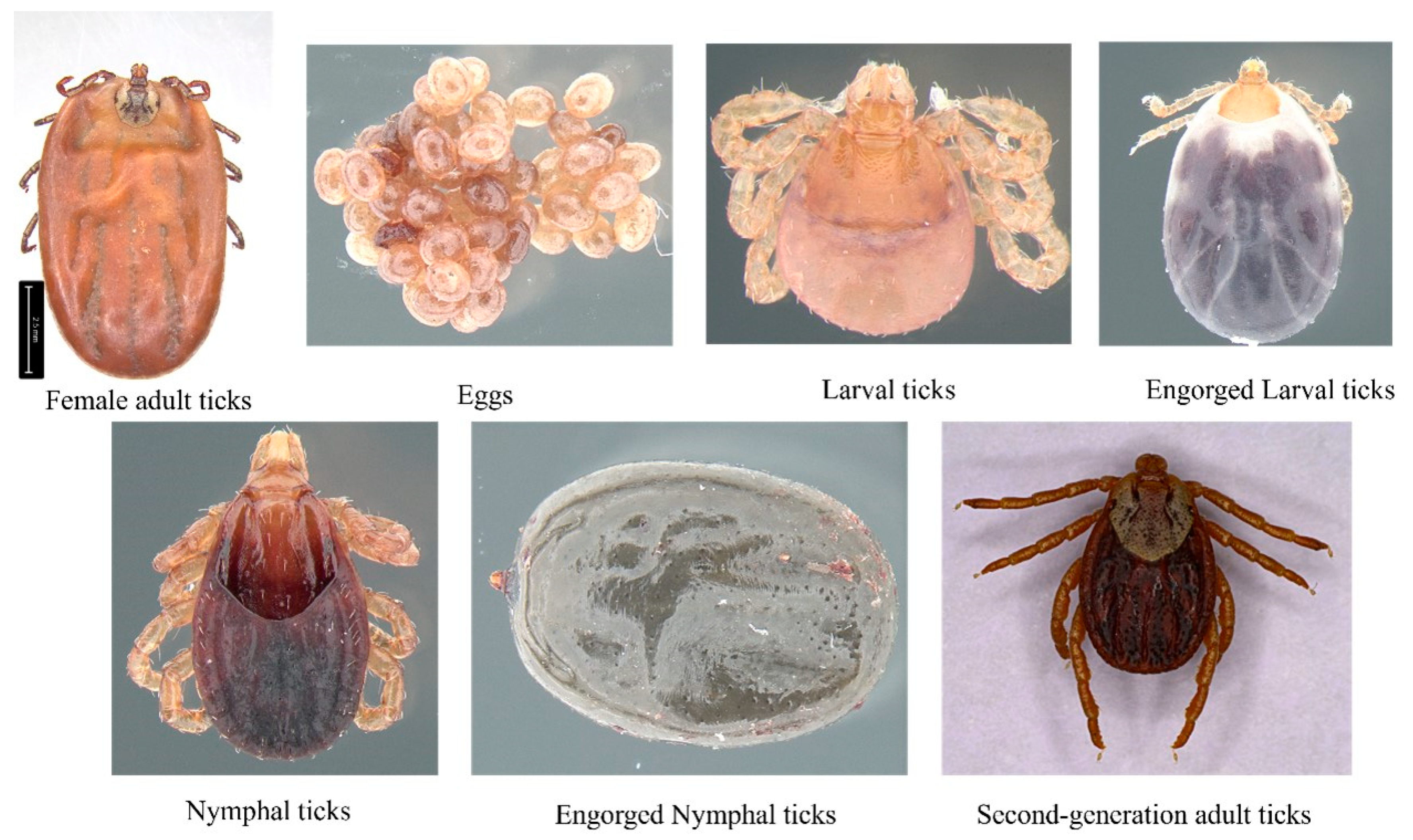
2.1. Study Area and Sample Collection

A large number of adult D. nuttalli ticks were collected from sheep in Ordos (950 m above sea level; 109.27°′ E, 40.25° N) and cattle in the Hinggan League (950 m above sea level; 120.57°′ E, 46.57° N) in Inner Mongolia, China. Adult ticks were identified as D. nuttalli (designated as O-D and H-D, respectively) using morphological and molecular biological identification methods [44]. The remaining six developmental stages of D. nuttalli (O-D-E and H-D-E: eggs; O-D-L and H-D-L: larvae; O-D-EL and H-D-EL: engorged larvae; O-D-N and H-D-N: nymphs; O-D-EN and H-D-EN: engorged nymphs; and O-D-SA and H-D-SA: second-generation adults) were obtained by artificially rearing the first-generation adult female ticks (O-D-FA and H-D-FA) collected from the field (Figure 1). Male Kunming mice (age: 6–8 weeks; body weight: 28–32 g), purchased from Sibefu (Beijing) Biotechnology Co., Ltd. (license number : SYXK [Meng] 2020-0003), were used as the sole blood source for tick feeding. All samples were stored at −80 °C until further analysis.

2.2. DNA and RNA Extraction

The 14 samples represented ticks at seven developmental stages from the two regions. For each region, a well-developed tick breeding series was selected, in which six developmental stages were the offspring of an adult female tick. Each developmental stage was a pool comprising varying numbers of individual ticks. The samples were washed with sterile water followed by 75% ethanol, and DNA was extracted using the TaKaRa Mini BEST Universal Genomic DNA Extraction Kit Ver.5.0 (Takara, Beijing, China). For RNA and viral nucleic acid extraction, six mixed sample pools were prepared from the O-D and H-D groups. The pools included O-D-FA-MIX and H-D-FA-MIX (pooled first-generation adult female ticks); O-D-EL-MIX and H-D-EL-MIX (mixed samples of eggs and larvae); and O-D-SA-MIX and H-D-SA-MIX (second-generation adult ticks). All sample pools were washed with sterile water and 75% ethanol prior to nucleic acid extraction using the RaPure Viral RNA/DNA Kit (MAGEN, Shanghai, China). The extracted nucleic acids were then stored at −80 °C until further use.

2.3. Library Construction and High-Throughput Sequencing

The 16S rDNA V3−V4 region was amplified by using TransStart R© FastPfu DNA polymerase (TransStart, Beijing, China) from the DNA of each sample. The primers 338F (5′-ACTCCTRCGGGAGGCAGCAG-3′) and 806R (5′-GGACTACCVGGGTATCTAAT-3′) were used under the following conditions: 95 °C for 2 min, followed by 25 cycles at 95 °C for 30 s, 55 °C for 30 s, and 72 °C for 30 s with a final extension at 72 °C for 5 min [47]. The PCR products were identified via electrophoresis and recovered using the AxyPrep DNA gel extraction kit (AXYGEN, Suzhou, China). Subsequently, the VAHTS ® ssDNA Library Prep Kit (Illumina, San Diego, CA, USA) was used to construct the Illumina PE250 library. High-throughput sequencing of 16S rRNA was performed on the Illumina Novaseq 6000 platform (San Diego, CA, USA).
After determining the RNA concentration, the RNA samples were fragmented, followed by the addition of first strand buffer and first strand enzyme for cDNA synthesis. End repair was performed using SEA Enzyme Mix and SEA Buffer. The Fast RNA-Seq Library Prep Kit (Illumina, San Diago, CA, USA) was used to construct the Illumina PE150 library. Viral metagenomic sequencing was conducted on the Illumina NovaSeq 6000 platform (San Diego, CA, USA).

2.4. Data Analysis

The Illumina PE250 sequencing data were first demultiplexed based on barcode information to obtain valid sequences., Paired-end reads of the original DNA fragments were merged using FLASH (v.1.2.11), and sequence analysis was conducted using USEARCH (v.7.0.1090, http://drive5.com/uparse/) (accessed on 20 April 2024). Sequences with ≥97% similarity were clustered into operational taxonomic units (OTUs), and taxonomic classification was performed using the RDP Classifier (v.2.2, http://sourceforge.net/projects/rdp-classifier/) (accessed on 20 April 2024), based on a Bayesian algorithm. Alpha diversity analysis was conducted to evaluate community richness and diversity. Richness was assessed using the number of unique OTUs per sample, the Chao index (http://www.mothur.org/wiki/Chao) (accessed on 20 April 2024), and the abundance-based coverage estimator (ACE). Diversity was accessed using the Shannon index (http://www.mothur.org/wiki/Shannon) (accessed on 20 April 2024), and Simpson index (http://www.mothur.org/wiki/Simpson) (accessed on 20 April 2024), whereas sequencing depth was evaluated using Good’s coverage (http://www.mothur.org/wiki/Coverage) (accessed on 20 April 2024). Species accumulation curves were used to assess sample community diversity and estimate species richness.
The Illumina PE150 sequencing data were first optimized and quality-controlled using Cutadapt (v.1.16, https://cutadapt.readthedocs.io/en/stable/) (accessed on 20 April 2024) and FastQC (v.0.11.4, https://www.bioinformatics.babraham.ac.uk/projects/fastqc/) (accessed on 20 April 2024). High-quality sequences were then assembled using MEGAHIT (v.1.2.9, https://github.com/voutcn/megahit) (accessed on 20 April 2024) and gene prediction was performed with MetaGeneMark (v.3.38, http://exon.gatech.edu/GeneMark/) (accessed on 20 April 2024). Gene annotation was conducted using DIAMOND (https://github.com/bbuchfink/diamond) (accessed on 20 April 2024).
Sequencing and sequence analysis were conducted in collaboration with Origin-gene Biology Co., Ltd. (Shanghai, China).

3. Results

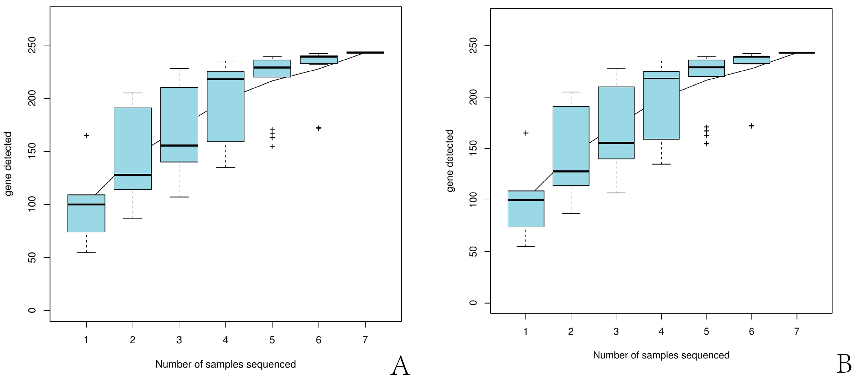
3.1. Sequencing Results of Illumina PE250

3.1.1. General Statistics

Fourteen samples were sequenced using the Illumina PE250 platform. The O-D and H-D groups yielded 1,191,556 and 954,312 raw reads, respectively. After optimization, 562,680 and 435,060 high-quality sequences were obtained. The total number of bases in the optimized data was 237,005,186 bp for O-D and 183,324,532 bp for H-D. The average sequences lengths were 421.20 bp and 421.38 bp, respectively (Table 1).

3.1.2. Alpha Diversity Analysis

Based on the 97% similarity threshold, the Shannon, Chao, and ACE indices are presented in Table 2. The Good’s coverage of all O-D and H-D samples exceeded 99.9% (Table 2). The species accumulation curves for O-D and H-D (Figure 2) showed a tendency to plateau as sample size increased.

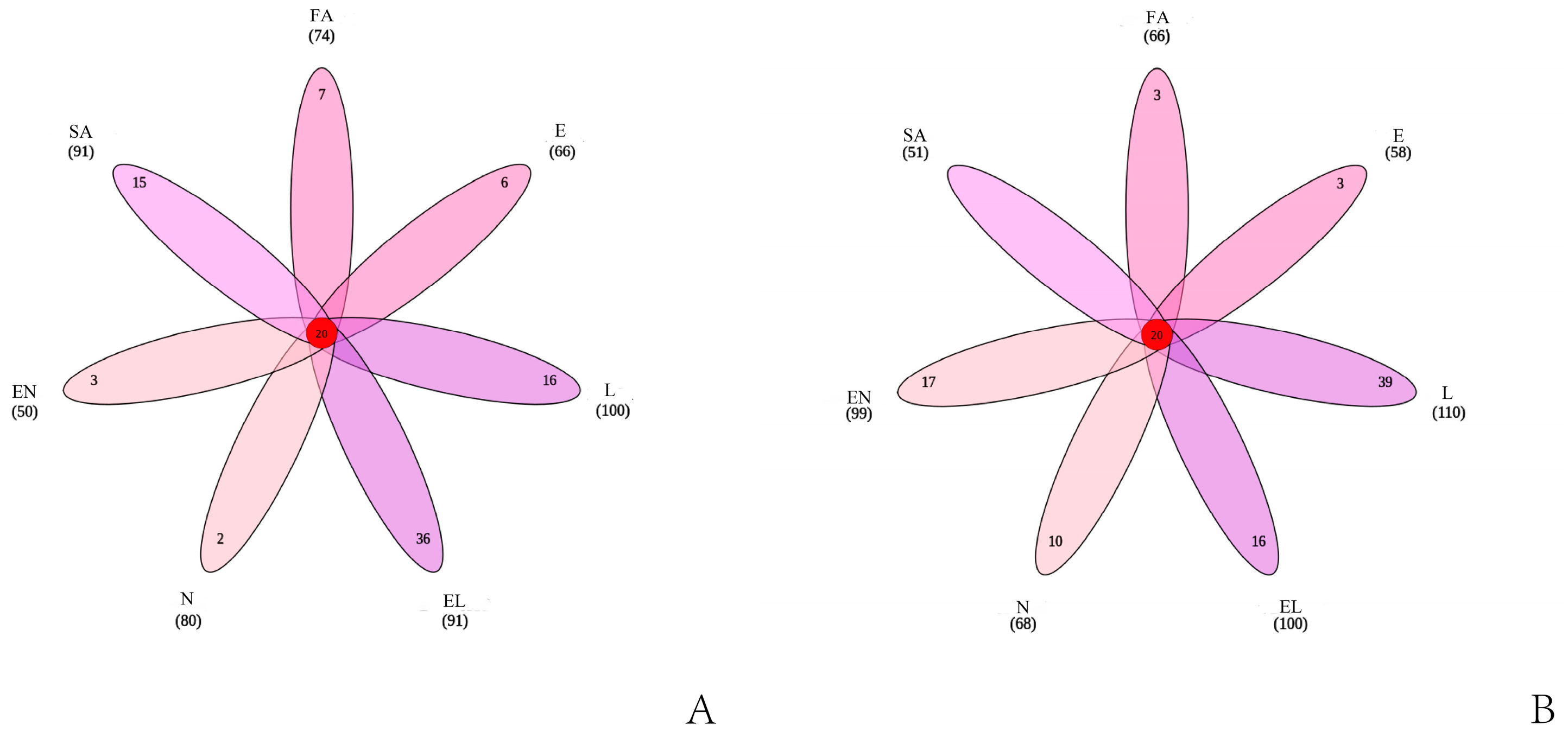
3.1.3. OTU Cluster Analysis

A total of 660 and 620 OTUs were obtained from O-D and H-D, respectively, with 20 unique OTUs shared across all developmental stages. Each developmental stage of O-D contained unique OTUs, whereas H-D-SA did not have any unique OTUs (Figure 3). In total, 99.13% and 99.95% of tags from O-D and H-D, respectively, were assigned to the genus level. Among these, 49.88% of O-D tags and 73.38% of H-D tags were further classified at the species level.

3.2. Microbial Population

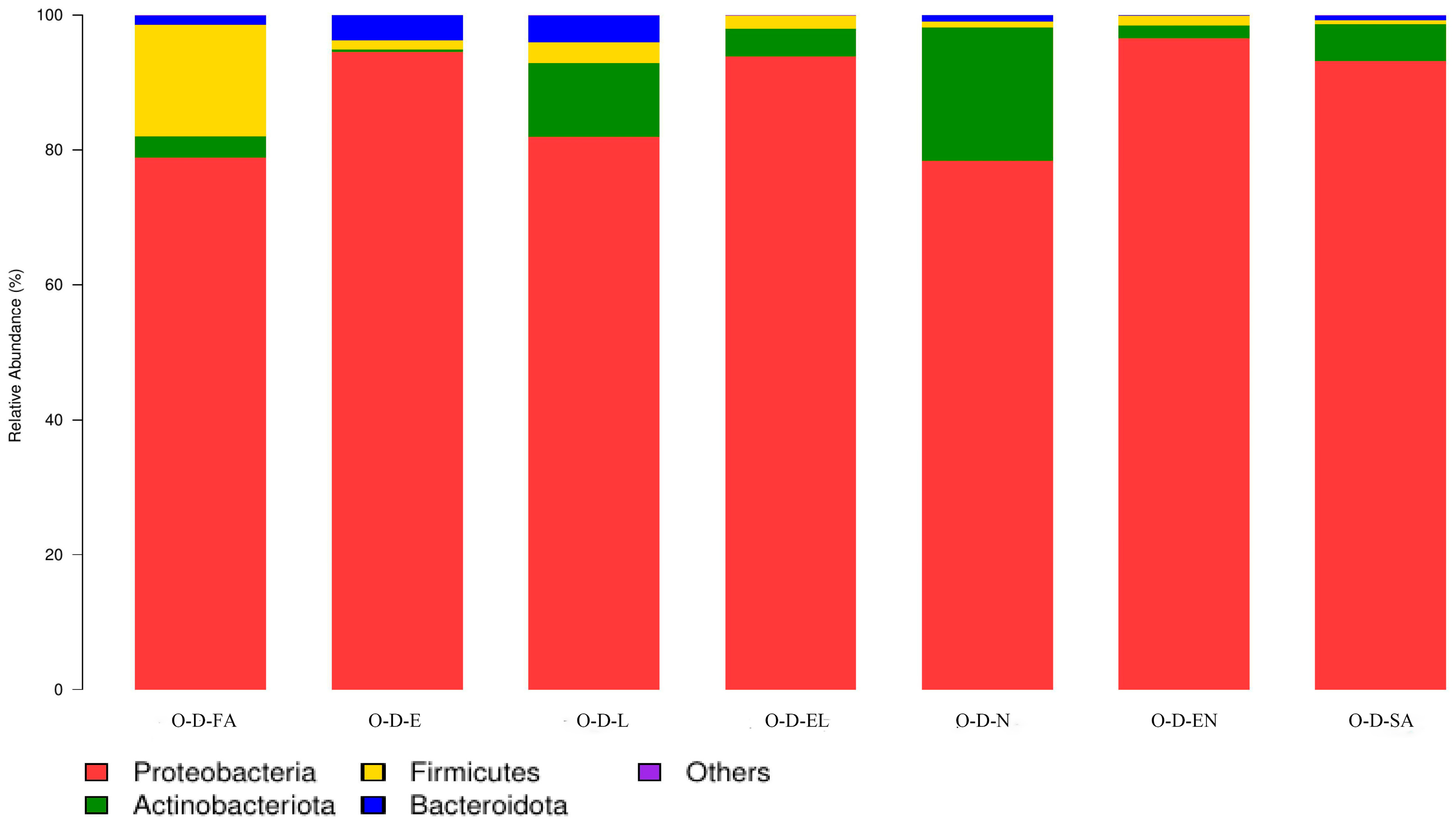
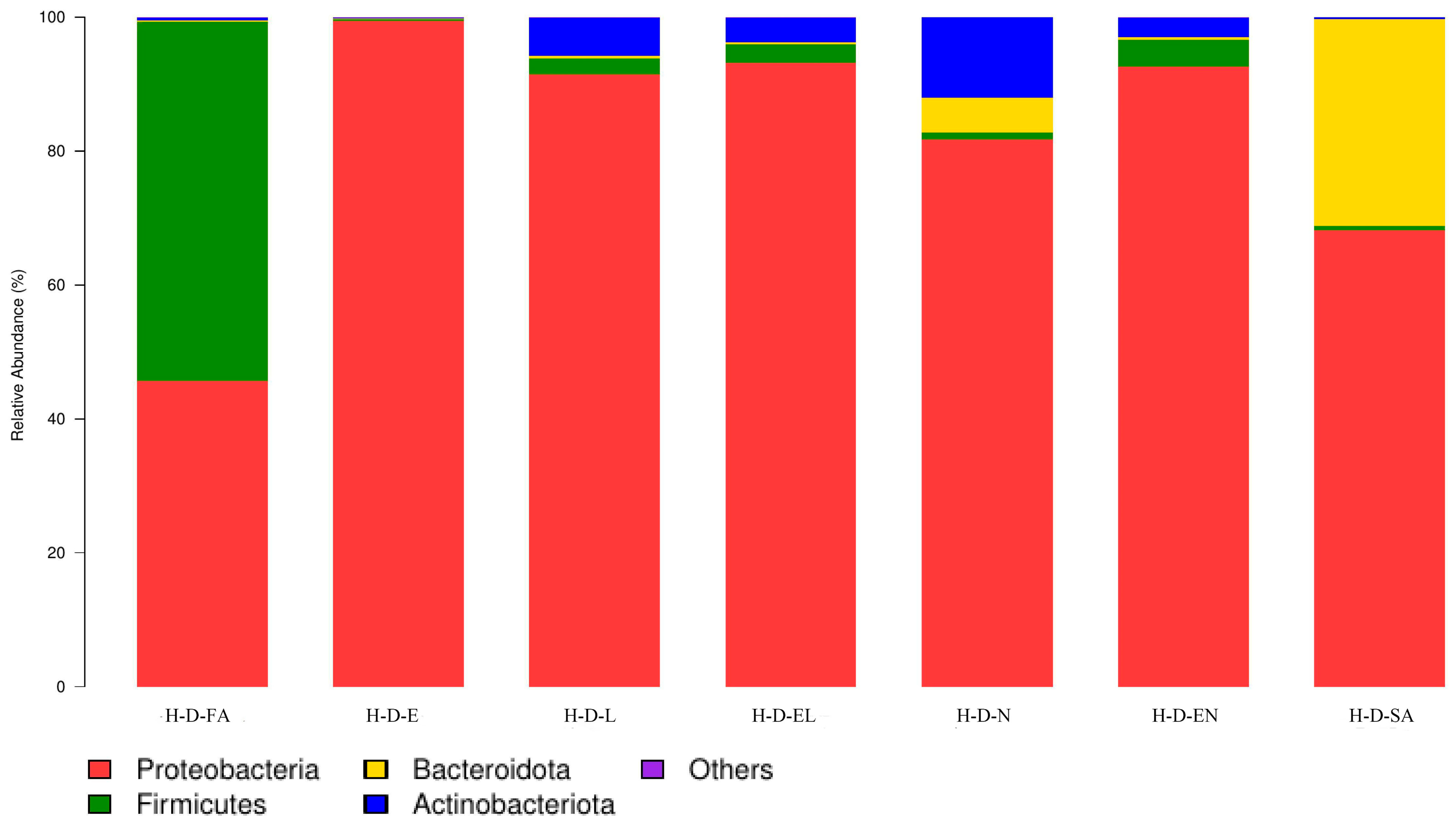
3.2.1. Microbial Community Composition at the Phylum Level

O-D and H-D were co-annotated to eight and sixteen bacterial phyla, respectively. Across all developmental stages in both regions, four common phyla were consistently identified (Figure 4 and Figure 5). Proteobacteria was the dominant phylum in all developmental stages of O-D and also predominated in H-D, except in H-D-FA, where Firmicutes was more abundant. Proteobacteria had the highest total relative abundance among bacterial phyla in both regions—88.1% in O-D and 81.7% in H-D (Table 3).

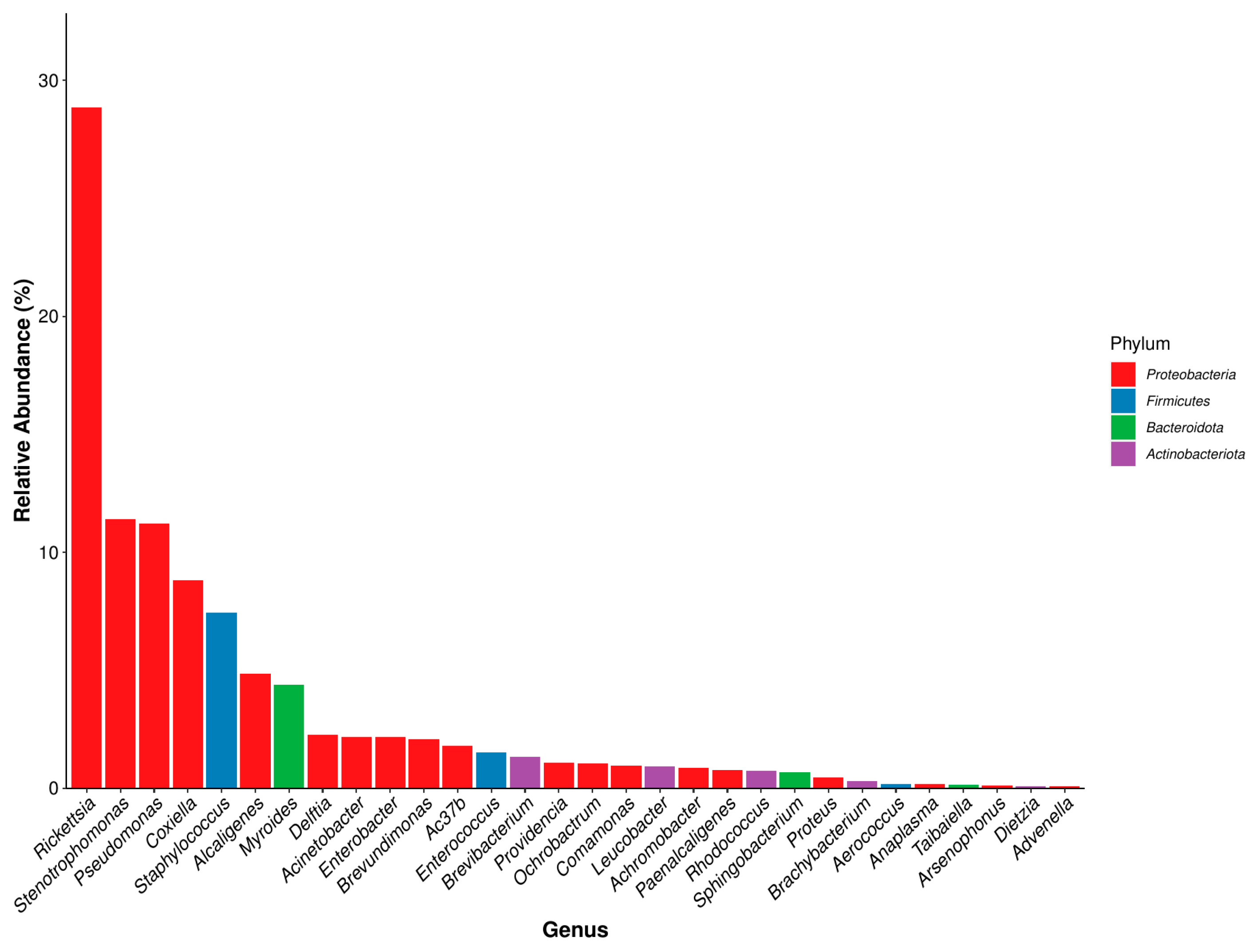
3.2.2. Microbial Community Composition at the Genus Level

O-D and H-D samples were co-annotated to 145 and 141 bacterial genera, respectively. The most abundant genera overall were Arsenophonus (18.31%) and Rickettsia (28.83%). In O-D, 18 of the 30 most abundant genera belonged to Proteobacteria, whereas in H-D, 19 were classified under Proteobacteria. In total, 15 and 16 genera had relative abundances greater than 1% in O-D and H-D, respectively. Across the seven developmental stages, 19 genera were annotated in O-D and 16 in H-D. The top five most abundant genera in O-D and the top four in H-D all belonged to Proteobacteria (Figure 6 and Figure 7). The dominant genera varied across developmental stages and between the two regions (Table S1).

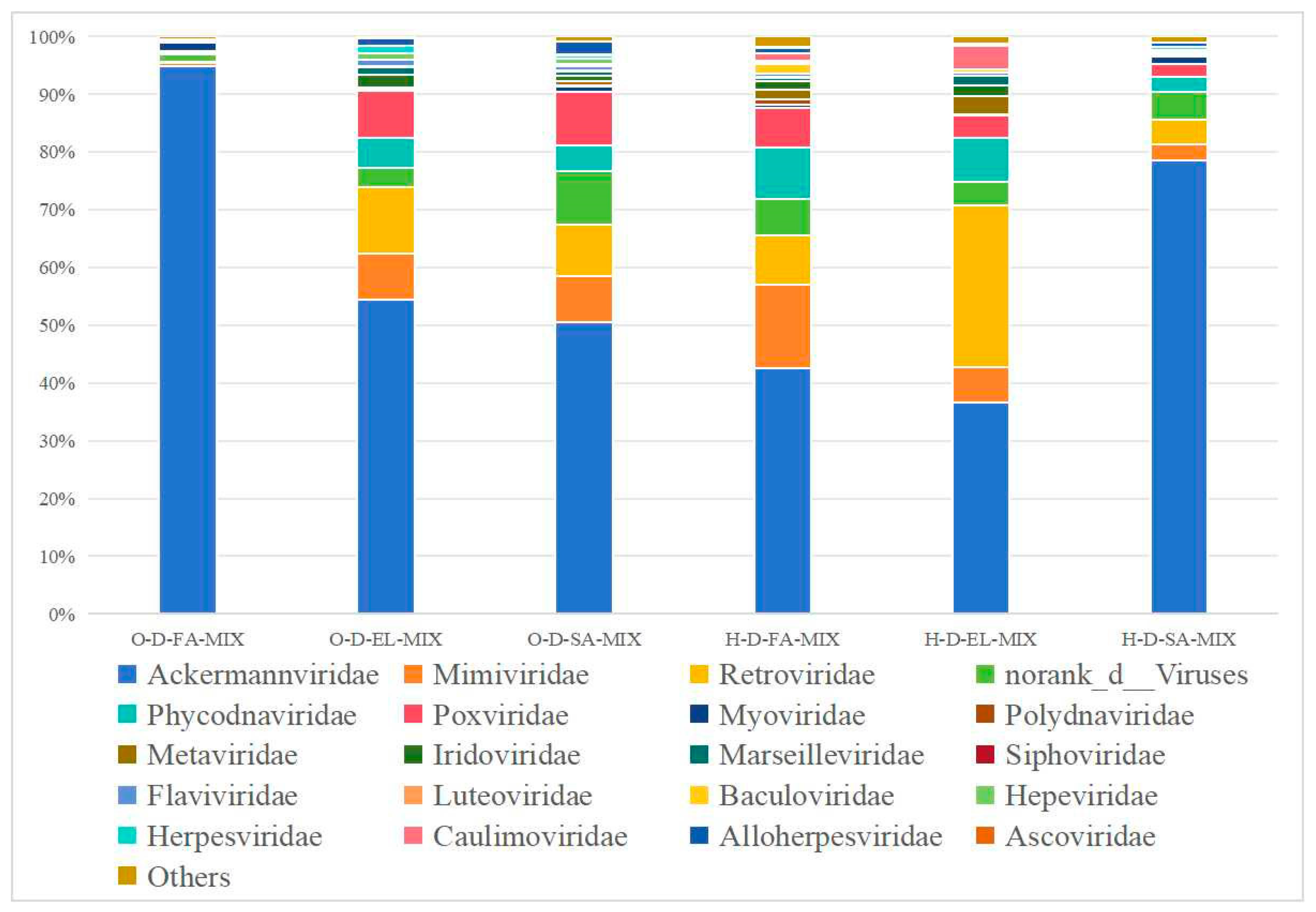
3.2.3. Microbial Community Composition at the Species Level

O-D and H-D were co-annotated to 28 and 36 bacterial species with an abundance of >1%, 16 and 19 bacterial species were present at seven developmental stages, respectively. The dominant species of O-D-FA, H-D-FA, O-D-E, H-D-E, and H-D-EL was Rickettsia japonica. The dominant species of the remaining nine samples were different (Table S2).

3.3. Illumina PE150 Sequencing Results

3.3.1. Species Classification Annotation Data

In the three O-D samples, 74,766,240; 63,544,512; and 59,774,580 clean reads were obtained, with CG contents of 57.31%, 54.13%, and 53.69%, respectively. Following open reading frame (ORF) prediction, 342,780; 253,560; and 640,677 reads were annotated as viral sequences. For the three H-D samples, 31,468,902; 55,601,958; and 52,845,094 clean reads were obtained, with CG contents of 55.25%, 53.86%, and 54.54%, respectively. Following ORF prediction, 123,987; 23,898; and 2,851,584 reads were annotated as viral sequences.

3.3.2. Species Composition at the Order Level

O-D and H-D were annotated to four and six viral orders, respectively. Among them, four viral orders, including Caudovirales, Herpesvirales, Ortervirales, and norank_d_Viruses, co-existed in all six samples (Table 4).

3.3.3. Species Composition at the Family Level

O-D and H-D samples were annotated to 25 and 28 viral families, respectively (Figure 8). Among them, three pooled samples from each region shared 16 and 13 viral families, respectively. Additionally, O-D-FA-MIX and O-D-EL-MIX were co-annotated with five viral families, namely, Ascoviridae, Baculoviridae, Caulimoviridae, Metaviridae, and Polydnaviridae; H-D-EL-MIX and H-D-SA-MIX were also co-annotated with seven viral families, namely, Asfarviridae, Caulimoviridae, Geminiviridae, Iflaviridae, Marseilleviridae, Metaviridae, and Polydnaviridae; H-D-FA-MIX and H-D-SA-MIX were co-annotated with five viral families, namely, Ascoviridae, Hepeviridae, Phenuiviridae, Siphoviridae, and norank_o_Caudovirales; and O-D-FA-MIX and O-D-SA-MIX were co-annotated with norank_o_Caudovirales.

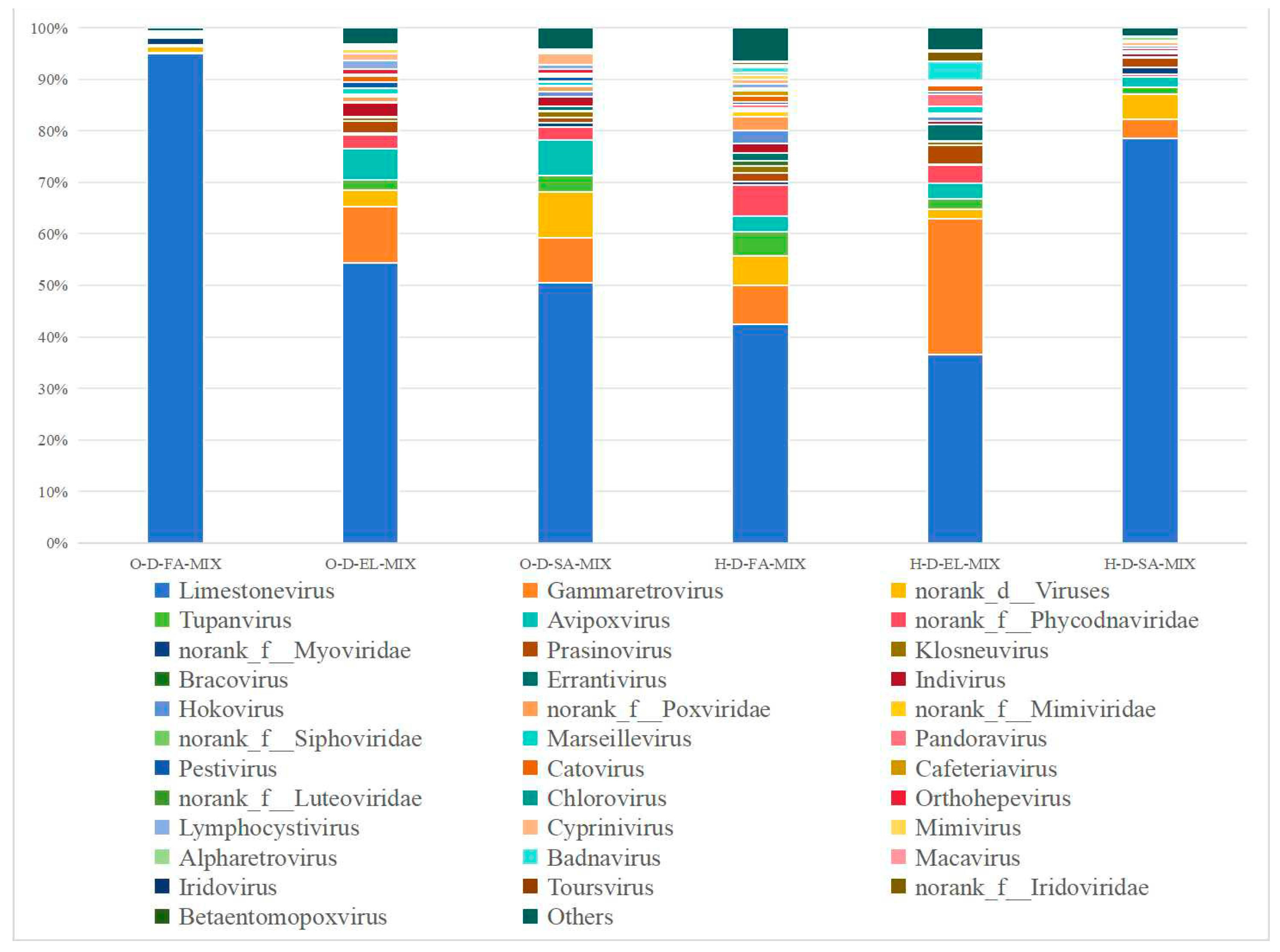
3.3.4. Species Composition at the Genus Level

O-D and H-D samples were annotated to 61 and 49 viral genera, respectively (Figure 9, Table S3). Among them, three pooled samples from each region shared 35 genera in O-D and 23 genera in H-D. Additionally, O-D-FA-MIX and O-D-EL-MIX were co-annotated with 7 genera; O-D-FA-MIX and O-D-SA-MIX were co-annotated with Phaeovirus and norank _o_Caudovirales; O-D-EL-MIX and O-D-SA-MIX were co-annotated with 4 genera; H-D-FA-MIX and H-D-SA-MIX were co-annotated with 12 genera; and H-D-EL-MIX and H-D-SA-MIX were co-annotated with 19 genera.

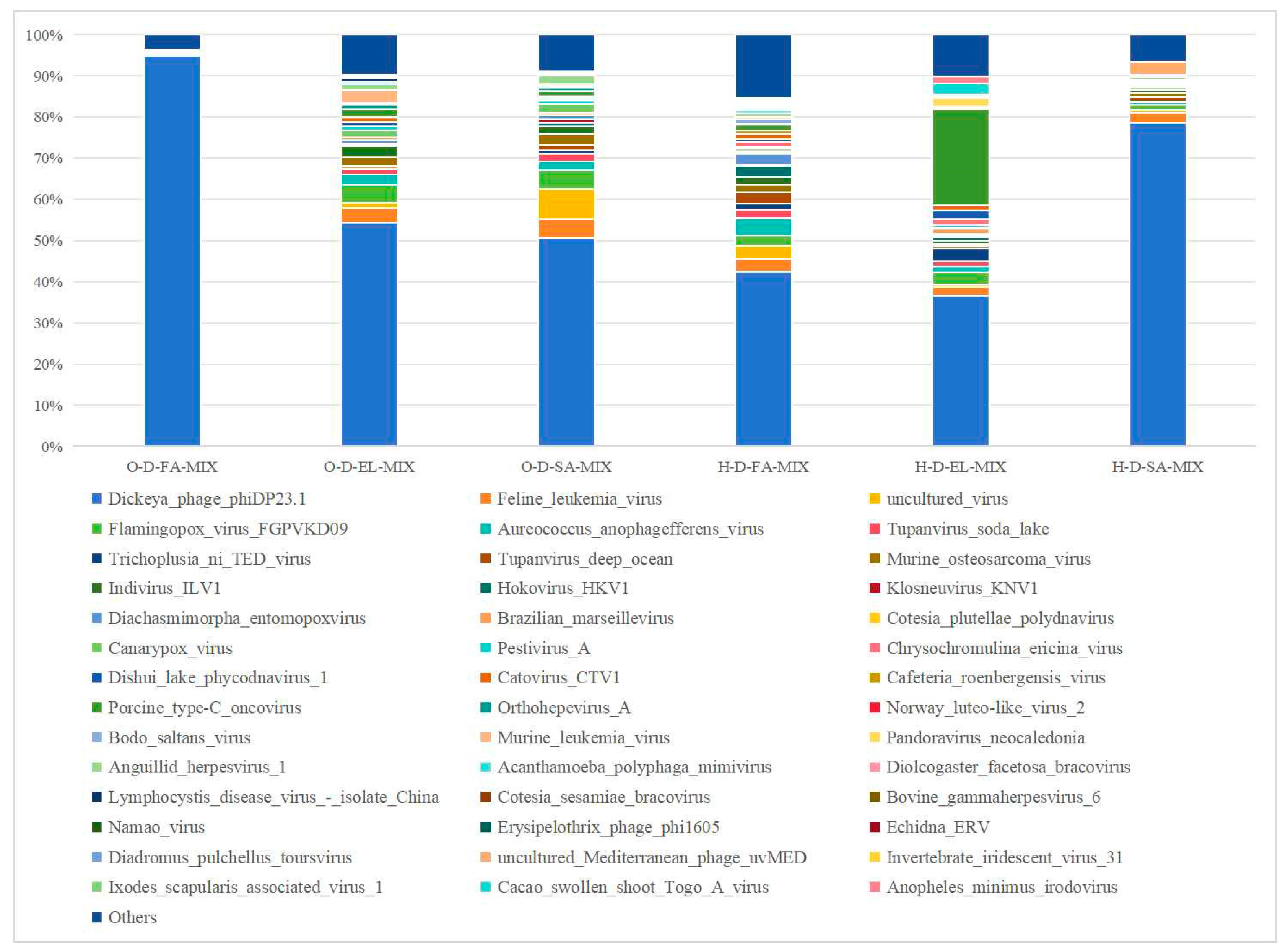
3.3.5. Species Composition at the Species Level

O-D and H-D samples were annotated to 126 and 135 viral species, respectively (Figure 10, Table S4). Among them, three pooled samples from each region shared 48 species in O-D and 37 species in H-D. Additionally, O-D-FA-MIX and O-D-EL-MIX were co-annotated with 10 species; O-D-FA-MIX and O-D-SA-MIX were co-annotated with 4 species; and O-D-EL-MIX and O-D-SA-MIX were co-annotated with 7 species.
H-D-FA-MIX and H-D-SA-MIX were co-annotated with 18 species; H-D-EL-MIX and H-D-SA-MIX were co-annotated with 29 species. Notably, 29 species were co-annotated across all six samples. Among them, Dickeya phage phiDP23.1 accounted for the highest relative abundance, with an average proportion of 59.60%. Additionally, African swine fever virus (ASFV) was annotated in both H-D-EL-MIXs and H-D-SA-MIX.

4. Discussion

In the 16S rRNA high-throughput sequencing results, a higher Shannon index and lower Simpson index indicated higher microbial community diversity, whereas a higher ACE value reflected a larger estimated total number of species [44,45]. In this study, these indices varied across samples, indicating inconsistent bacterial diversity and species richness among them. Based on these estimators, the sequencing data of this study showed that at the phylum level, except for the dominant phylum of H-D-FA being Firmicutes, Proteobacteria was the dominant phylum in 13 of the 14 samples, with the exception of H-D-FA, wherein Firmicutes was predominant. This finding differs from previous reports that consistently identified Proteobacteria as the dominant phylum in ticks [38,44,48,49,50,51,52,53,54]. Moreover, although the total number of annotated phyla varied between O-D and H-D, the co-annotated phyla across all samples were consistent. This observation is consistent with those of prior studies that identified four dominant phyla in D. nuttalli [38,44,48].
At the genus level, although the dominant genera varied between O-D and H-D at different growth stages, there were some similarities. The total relative abundance of Rickettsia was the highest in H-D and second highest in O-D. The relative abundance of Rickettsia in O-D and H-D exhibited an alternating trend of increase and decrease from first-generation adult female ticks to second-generation adult ticks, with the highest abundance observed in O-D-E and H-D-E. Notably, the abundance of Rickettsia increased significantly during the two blood-feeding stages, namely from larvae to engorged larvae and from nymphs to engorged nymphs. This trend is consistent with previous results reported in D. nuttalli [44] and supports the general observation that blood-feeding behavior affects the composition and abundance of the microbiome in ticks [55,56]. However, it has also been reported that blood feeding reduces microbial richness and internal tick microbiota diversity [44], suggesting that the process of blood ingestion affects the tick microbiome. Rickettsia, as one of the symbionts of ticks, can provide ticks with B vitamins such as folic acid [57]. Pathogenic Rickettsia can modulate the immune response of ticks [58]. In a previous study, multiple Rickettsia species were detected in D. nuttalli; therefore, it is a serious threat to human and animal health worldwide [44] and requires increased attention.
Staphylococcus was one of the dominant genera in O-D-FA and H-D-FA. However, its abundance was relatively low in the other six developmental stages. Staphylococcus can cause skin infections, food poisoning, and sepsis in both humans and animals [59]. Moreover, Staphylococcus is the dominant genus on the skin near the site of tick bites [60]. Staphylococcus is resistant to various antibiotics, and the treatment of related infections is complex and difficult [61]. In this study, the high abundance of Staphylococcus may be attributed to the host.
Furthermore, it is worth noting that Coxiella showed high abundance in O-D-E and H-D-L. Coxiella is regarded as an endosymbiont genus that can be propagated through tansovarian and transstadial transmission [62,63]. Coxiella is primarily enriched in the midgut, ovaries, and Malpighian tubules of ticks and plays a significant role in tick reproduction and development [64]. The removal of Coxiella-like endosymbionts can reduce the satiation weight of Amblyomma americanum and H. longicornis and the egg-laying ability of adult ticks [64,65]. This may explain the high abundance of Coxiella in O-D-E and H-D-L in this study.
Further, we annotated various Pseudomonas speices across all developmental stages of O-D and H-D. P. aeruginosa has also been detected in H. flava [66], Rhipicehalus microplus [67], and Dermacentor variabilis [68]. P. aeruginosa was first isolated from purulent wound secretions [69]. P. aeruginosa poses a serious threat to animal health and can lead to significant economic losses in the livestock industry. Therefore, more attention should be focused on the control of D. nuttalli infestations.
Viruses are an important component of tick-borne pathogens. According to previous reports, 19 tick-borne viruses have been identified in China [3], most of which are transmitted by hard ticks [70]. In this study, four common viral orders were detected in O-D and H-D samples, whereas Bunyavirales and Picornavirale were uniquely identified in H-D. Almost all studies on tick viruses have detected viruses associated with existing members of Bunyavirales [71]. Most of them belong to the Nairoviridae and Phenuiviridae families, which usually infect vertebrates and pose a threat to human and animal health [46]. At the family level, 25 and 28 viral families were annotated in O-D and H-D, respectively, with 11 viral families common to both. The family Mimiviridae belongs to a class of nucleocytoplasmic large DNA viruses, which are giant viruses that infect amoebae [46]. At the species level, Pestivirus A (bovine viral diarrhea virus 1, BVDV-1) was identified at notable abundance in all six samples. BVDV-1 primarily affects cattle and can lead to high mortality in severe cases [72]. Moreover, it is associated with persistent infection and immunosuppression in affected cattle [73]. In this study, Tacheng Tick Virus 2 (TcTV-2), a member of the genus Phlebovirus, was annotated in H-D-FA-MIX and H-D-SA-MIX. TcTV-2 was first discovered through next-generation sequencing of ticks in China [74]. To date, the pathogenic potential of TcTV-2 infection remains unclear, with only one human case reported in China in 2019 [75]. Interestingly, ASFV was detected in H-D-EL-MIX and H-D-SA-MIX but not in H-D-FA-MIX. ASFV was first reported in Asia in 2018 in Liaoning Province, China [76], and rapidly spread throughout mainland China [77]. Although ticks of the genus Ornithodoros are the biological vectors for ASFV, DNA fragments of this virus have also been detected in hard ticks, including D. nuttalli [78,79]. However, there is currently no conclusive evidence to suggest that hard ticks can transmit ASFV [80].
The microbial community composition and diversity of ticks are affected by several factors, including tick species and hosts [81], sex [82], developmental stage [83], blood meal [84], engorged state [85,86], and geographical location [87]. In this study, the two D. nuttalli sampling locations were >1500 km apart. These regions differ significantly in average temperature, precipitation, and sunshine duration, which may partly explain the observed differences in the diversity and abundance of microorganisms carried by D. nuttalli. Overall, this study compared the microorganisms of D. nuttalli from different regions in Inner Mongolia and analyzed the differences in the spectrum of pathogens in D. nuttalli. Our findings may help predict emerging pathogens and evaluate the potential risks to both animal and public health.

5. Conclusions

In both O-D and H-D samples, Proteobacteria was the phylum with the highest total abundance. Notable differences in dominant bacterial genera were observed across different developmental stages in O-D and H-D. To our knowledge, this study is the first to compare the composition of the microbial community across the developmental stages of D. nuttalli from two locations in Inner Mongolia under identical artificial feeding conditions. Moreover, for the first time, microorganisms such as R. japonica, TcTV-2, and BVDV were annotated in D. nuttalli with certain abundance.

Supplementary Materials

The following supporting information can be downloaded at https://www.mdpi.com/article/10.3390/biology14060613/s1. Table S1. Genus-level microbial community composition of Dermacentor nuttalli at different developmental stages in Ordos and Hinggan League. Table S2. Species-level microbial community composition of Dermacentor nuttalli at different developmental stages in Ordos and Hinggan League. Table S3. Viral genus data of Dermacentor nuttalli. Table S4. Viral species data of Dermacentor nuttalli.

Author Contributions

Conceptualization, L.Z., X.-N.D., H.C. and Y.-H.L.; investigation, X.-N.D., H.C., L.-Y.S., R.M., M.N., J.-M.K., N.B., Y.-S.Z., Z.-H.Q., Z.-X.L., Z.-L.Z., X.-Y.Z., Y.-L.D., R.W. and Y.W.; data curation, L.-Y.S., R.M., M.N., J.-M.K., N.B., Y.-S.Z., Z.-H.Q., Z.-X.L., Z.-L.Z. and X.-Y.Z.; resources, H.C., Y.-L.D., R.W., Y.W. and Y.-H.L.; writing—original draft, L.Z., X.-N.D., H.C. and Y.-H.L.; writing—review and editing, L.Z., X.-N.D., H.C. and Y.-H.L.; methodology, L.Z., X.-N.D., H.C. and Y.-H.L.; formal analysis, L.Z.; funding acquisition, Y.-H.L. All authors have read and agreed to the published version of the manuscript.

Funding

This study was funded by the National Natural Science Foundation of China (32260887), Higher Education Science and Technology Research Project of Inner Mongolia (NJZZ22560), and Fundamental Research Funds for the Inner Mongolia Normal University (2022JBQN090).

Institutional Review Board Statement

This study was carried out in strict accordance with international standards as published in the Guide to the feeding, management and use of experimental animals (8th edition) and follows the “Regulations on the management of experimental animals” and other relevant laws and regulations. The biomedical research ethics committee of Inner Mongolia Agricultural University specifically approved this study (No. 2020[081]). In addition, permission was obtained from the farm owners before the specimens were collected, and all efforts were made to minimize suffering.

Informed Consent Statement

Not applicable.

Data Availability Statement

The raw tags were deposited in Sequence Read Archive (SRA) from the NCBI under BioProject accession number PRJNA1222544 and PRJNA1222561. The individual run files received the accession numbers SAMN46783900–SAMN46783913 and SAMN46784101–SAMN46784106.

Conflicts of Interest

The authors declare that they have no competing interests.

Abbreviations

The following abbreviations are used in this manuscript:
PCRpolymerase chain reaction
OTUsoperational taxonomic units.
ACEabundance-based coverage estimator

References

  1. Zhong, Z.; Wang, K.; Wang, J. Tick symbiosis. Curr. Opin. Insect Sci. 2024, 62, 101163. [Google Scholar] [CrossRef] [PubMed]
  2. Filipe, D.T. Species concepts: What about ticks? Trends Parasitol. 2018, 34, 1017–1026. [Google Scholar]
  3. Zhao, G.P.; Wang, Y.X.; Fan, Z.W.; Ji, Y.; Liu, M.J.; Zhang, W.H.; Li, X.L.; Zhou, S.X.; Li, H.; Liang, S.; et al. Mapping ticks and tick-borne pathogens in China. Nat. Commun. 2021, 12, 1075. [Google Scholar] [CrossRef]
  4. Zhang, Y.K.; Zhang, X.Y.; Liu, J.Z. Ticks (Acari: Ixodoidea) in China: Geographical distribution, host diversity, and specificity. Arch. Insect Biochem. Physiol. 2019, 102, e21544. [Google Scholar] [CrossRef]
  5. Gui, Z.; Cai, H.; Qi, D.; Zhang, S.; Fu, S.; Yu, J.; Si, X.; Cai, T.; Mao, R. Identification and genetic diversity analysis of Rickettsia in Dermacentor nuttalli within inner Mongolia, China. Parasites Vectors 2022, 15, 286. [Google Scholar] [CrossRef]
  6. Gulia-Nuss, M.; Nuss, A.B.; Meyer, J.M.; Sonenshine, D.E.; Roe, R.M.; Waterhouse, R.M.; Sattelle, D.B.; de la Fuente, J.; Ribeiro, J.M.; Megy, K.; et al. Genomic insights into the Ixodes scapularis tick vector of Lyme disease. Nat. Commun. 2016, 7, 10507. [Google Scholar] [CrossRef]
  7. Voorhees, M.A.; Padilla, S.L.; Jamsransuren, D.; Koehler, J.W.; Delp, K.L.; Adiyadorj, D.; Baasandagwa, U.; Jigjav, B.; Olschner, S.P.; Minogue, T.D.; et al. Crimean-Congo hemorrhagic fever virus, Mongolia, 2013–2014. Emerg. Infect Dis. 2018, 24, 2202–2209. [Google Scholar] [CrossRef]
  8. Wang, Q.; Pan, Y.S.; Jiang, B.G.; Ye, R.Z.; Chang, Q.C.; Shao, H.Z.; Cui, X.M.; Xu, D.L.; Li, L.F.; Wei, W.; et al. Prevalence of multiple tick-borne pathogens in various tick vectors in Northeastern China. Vector Borne Zoonotic Dis. 2021, 21, 162–171. [Google Scholar] [CrossRef] [PubMed]
  9. Enkhtaivan, B.; Narantsatsral, S.; Davaasuren, B.; Otgonsuren, D.; Amgalanbaatar, T.; Uuganbayar, E.; Zoljargal, M.; Myagmarsuren, P.; Suganuma, K.; Molefe, N.I.; et al. Molecular detection of Anaplasma ovis in small ruminants and ixodid ticks from Mongolia. Parasitol. Int. 2019, 69, 4753. [Google Scholar] [CrossRef]
  10. Yin, X.; Guo, S.; Ding, C.; Cao, M.; Kawabata, H.; Sato, K.; Ando, S.; Fujita, H.; Kawamori, F.; Su, H.; et al. Spotted fever group rickettsiae in inner Mongolia, China, 2015–2016. Emerg. Infect Dis. 2018, 24, 2105–2107. [Google Scholar] [CrossRef]
  11. von Fricken, M.E.; Qurollo, B.A.; Boldbaatar, B.; Wang, Y.W.; Jiang, R.R.; Lkhagvatseren, S.; Koehler, J.W.; Moore, T.C.; Nymadawa, P.; Anderson, B.D.; et al. Genetic diversity of anaplasma and ehrlichia bacteria found in dermacentor and ixodes ticks in Mongolia. Ticks Tick Borne Dis. 2020, 11, 101316. [Google Scholar] [CrossRef] [PubMed]
  12. von Fricken, M.E.; Voorhees, M.A.; Koehler, J.W.; Asbun, C.; Lam, B.; Qurollo, B.; Hogan, K.M.; Baasandagva, U.; Jigjav, B.; Schoepp, R.J. Molecular characteristics of rickettsia in ticks collected along the Southern Border of Mongolia. Pathogens 2020, 9, 943. [Google Scholar] [CrossRef]
  13. Gao, Y.; Lv, X.L.; Han, S.Z.; Wang, W.; Liu, Q.; Song, M. First detection of Borrelia miyamotoi infections in ticks and humans from the northeast of Inner Mongolia, China. Acta Trop. 2021, 217, 105857. [Google Scholar] [CrossRef] [PubMed]
  14. Li, Y.; Wen, X.; Li, M.; Moumouni, P.F.A.; Galon, E.M.; Guo, Q.; Rizk, M.A.; Liu, M.; Li, J.; Ji, S.; et al. Molecular detection of tick-borne pathogens harbored by ticks collected from livestock in the Xinjiang Uygur Autonomous Region, China. Ticks Tick Borne Dis. 2020, 11, 101478. [Google Scholar] [CrossRef]
  15. Song, S.N.; Chen, C.F.; Yang, M.H.; Zhao, S.; Wang, B.; Hornok, S.; Makhatov, B.; Rizabek, K.; Wang, Y. Diversity of Rickettsia species in border regions of northwestern China. Parasites Vectors 2018, 11, 634. [Google Scholar] [CrossRef] [PubMed]
  16. Liu, H.; Liang, X.; Wang, H.; Sun, X.; Bai, X.; Hu, B.; Shi, N.; Wang, N.; Zhang, X.; Huang, L.; et al. Molecular evidence of the spotted fever group Rickettsiae in ticks from Yunnan Province, Southwest China. Exp. Appl. Acarol. 2020, 80, 339–348. [Google Scholar] [CrossRef]
  17. Shao, J.W.; Zhang, X.L.; Li, W.J.; Huang, H.L.; Yan, J. Distribution and molecular characterization of rickettsiae in ticks in Harbin area of Northeastern China. PLoS Negl. Trop. Dis. 2020, 14, e0008342. [Google Scholar] [CrossRef]
  18. Ni, J.; Lin, H.L.; Xu, X.F.; Ren, Q.Y.; Aizezi, M.; Luo, J.; Luo, Y.; Ma, Z.; Chen, Z.; Tan, Y.C.; et al. Coxiella burnetii is widespread in ticks (Ixodidae) in the Xinjiang areas of China. BMC Vet. Res. 2020, 16, 317. [Google Scholar]
  19. Han, R.; Yang, J.F.; Niu, Q.L.; Liu, Z.J.; Chen, Z.; Kan, W.; Hu, G.W.; Liu, G.Y.; Luo, J.X.; Yin, H. Molecular prevalence of spotted fever group rickettsiae in ticks from Qinghai Province, northwestern China. Infect. Genet. Evol. 2018, 57, 1–7. [Google Scholar] [CrossRef]
  20. Wang, Y.C.; Li, S.; Wang, Z.D.; Zhang, L.; Cai, Y.N.; Liu, Q. Prevalence and Identification of Borrelia burgdorferi Sensu Lato Genospecies in Ticks from Northeastern China. Vector Borne Zoonotic Dis. 2019, 19, 309–315. [Google Scholar] [CrossRef]
  21. Zhang, L.; Li, S.; Huang, S.J.; Wang, Z.D.; Wei, F.; Feng, X.M.; Jiang, D.X.; Liu, Q. Isolation and genomic characterization of lymphocytic choriomeningitis virus in ticks from northeastern China. Transbound. Emerg. Dis. 2018, 65, 17339. [Google Scholar] [CrossRef] [PubMed]
  22. Song, R.; Wang, Q.; Guo, F.; Liu, X.; Song, S.; Chen, C.; Tu, C.; Wureli, H.; Wang, Y. Detection of Babesia spp. Theileria spp. and Anaplasma ovis in Border Regions, northwestern China. Transbound. Emerg. Dis. 2018, 65, 1537–1544. [Google Scholar] [PubMed]
  23. Liu, X.F.; Zhang, X.; Wang, Z.D.; Dong, Z.H.; Xie, S.S.; Jiang, M.M.; Song, R.X.; Ma, J.; Chen, S.Y.; Chen, K.C.; et al. A tentative tamdy orthonairovirus related to febrile illness in Northwestern China. Clin. Infect. Dis. 2020, 70, 2155–2160. [Google Scholar] [CrossRef]
  24. Narankhajid, M.; Yeruult, C.; Gurbadam, A.; Battsetseg, J.; Aberle, S.W.; Bayartogtokh, B.; Joachim, A.; Duscher, G.G. Some aspects on tick species in Mongolia and their potential role in the transmission of equine piroplasms, Anaplasma phagocytophilum and Borrelia burgdorferi L. Parasitol. Res. 2018, 117, 355766. [Google Scholar] [CrossRef]
  25. Shen, S.; Duan, X.M.; Wang, B.; Zhu, L.; Zhang, Y.; Zhang, J.; Wang, J.; Luo, T.; Kou, C.; Liu, D.; et al. A novel tickborne phlebovirus, closely related to severe fever with thrombocytopenia syndrome virus and Heartland virus, is a potential pathogen. Emerg. Microbes Infect. 2018, 7, 95. [Google Scholar] [CrossRef]
  26. Su, S.; Hong, M.; Cui, M.Y.; Gui, Z.; Ma, S.; Wu, L.; Xing, L.; Mu, L.; Yu, J.; Fu, S.; et al. Microbial diversity of ticks and a novel typhus group Rickettsia species (Rickettsiales bacterium Ac37b) in Inner Mongolia, China. Parasite 2023, 30, 58. [Google Scholar] [CrossRef]
  27. Shi, Q.; Song, F.; Yang, Y.; Gao, Y.; Ci, Y.; Cheng, X.; Nie, C.; Liu, L.; Zhang, X.; Wang, J. Epidemiological and Molecular Study on Tick-Borne Pathogens in Argun Port Area Near the Chinese–Russian Border. Vector-Borne Zoonotic Dis. 2023, 23, 447–457. [Google Scholar] [CrossRef] [PubMed]
  28. Lee, Y.J.; Kim, B.; Kwak, D.; Nyamsuren, O.; Guugandaa, N.; Seo, M.G. Tick-borne pathogens in Mongolian ticks: The high prevalence of Rickettsia raoultii and its public health implications. Acta Trop. 2024, 260, 107412. [Google Scholar] [CrossRef]
  29. Huang, T.; Zhang, J.; Sun, C.; Liu, Z.; He, H.; Wu, J.; Geriletu. A novel arthropod host of brucellosis in the arid steppe ecosystem. Front. Vet. Sci. 2020, 7, 566253. [Google Scholar] [CrossRef]
  30. Kholodilov, I.; Belova, O.; Burenkova, L.; Korotkov, Y.; Romanova, L.; Morozova, L.; Kudriavtsev, V.; Gmyl, L.; Belyaletdinova, I.; Chumakov, A.; et al. Ixodid ticks and tick-borne encephalitis virus prevalence in the South Asian part of Russia (Republic of Tuva). Ticks Tick Borne Dis. 2019, 10, 959–969. [Google Scholar] [CrossRef]
  31. Kholodilov, I.S.; Belova, O.A.; Morozkin, E.S.; Litov, A.G.; Ivannikova, A.Y.; Makenov, M.T.; Shchetinin, A.M.; Aibulatov, S.V.; Bazarova, G.K.; Bell-Sakyi, L.; et al. Geographical and tick-dependent distribution of flavi-like Alongshan and Yanggou Tick viruses in Russia. Viruses 2021, 13, 458. [Google Scholar] [CrossRef] [PubMed]
  32. Fischer, T.; Myalkhaa, M.; Krücken, J.; Battsetseg, G.; Batsukh, Z.; Baumann, M.P.O.; Clausen, P.; Nijhof, A. Molecular detection of tick-borne pathogens in bovine blood and ticks from Khentii, Mongolia. Transbound. Emerg. Dis. 2020, 67 (Suppl. S2), 111–118. [Google Scholar] [CrossRef] [PubMed]
  33. Guo, W.P.; Wang, Y.H.; Lu, Q.; Xu, G.; Luo, Y.; Ni, X.; Zhou, E.M. Molecular detection of spotted fever group rickettsiae in hard ticks, northern China. Transbound. Emerg. Dis. 2019, 66, 1587–1596. [Google Scholar] [CrossRef]
  34. Zhao, S.; Yang, M.H.; Jiang, M.M.; Yan, B.; Zhao, S.; Yuan, W.; Wang, B.; Hornok, S.; Wang, Y. Rickettsia raoultii and Rickettsia sibirica in ticks from the long-tailed ground squirrel near the China-Kazakhstan border. Exp. Appl. Acarol. 2019, 77, 425–433. [Google Scholar]
  35. Moore, T.C.; Pulscher, L.A.; Caddell, L.; von Fricken, M.E.; Anderson, B.D.; Gonchigoo, B.; Gray, G.C.; Gonchigoo, B. Evidence for transovarial transmission of tick-borne rickettsiae circulating in Northern Mongolia. PLoS Negl. Trop. Dis. 2018, 12, e0006696. [Google Scholar] [CrossRef]
  36. Jiao, J.; Lu, Z.Y.; Yu, Y.H.; Ou, Y.; Fu, M.; Zhao, Y.; Wu, N.; Zhao, M.; Liu, Y.; Sun, Y.; et al. Identification of tickborne pathogens by metagenomic next-generation sequencing in Dermacentor nuttalli and Ixodes persulcatus in Inner Mongolia, China. Parasites Vectors 2021, 14, 287. [Google Scholar] [CrossRef] [PubMed]
  37. Dedkov, V.G.; Dolgova, A.S.; Safonova, M.V.; Samoilov, A.E.; Belova, O.A.; Kholodilov, I.S.; Matsvay, A.D.; Speranskaya, A.S.; Khafizov, K.; Karganova, G.G. Isolation and characterization of Wad Medani virus obtained in the tuva Republic of Russia. Ticks Tick Borne Dis. 2021, 12, 101612. [Google Scholar] [CrossRef]
  38. Meng, F.; Ding, M.; Tan, Z.; Zhao, Z.; Xu, L.; Wu, J.; He, B.; Tu, C. Virome analysis of tick-borne viruses in Heilongjiang Province, China. Ticks Tick Borne Dis. 2019, 10, 412–420. [Google Scholar] [CrossRef]
  39. Song, K.; Ji, Y.X.; Sun, S.R.; Yue, X.H.; Wang, C.; Luo, T.; Moming, A.; Song, Y.J.; Zhang, Y.J.; Yang, R.F.; et al. Bacterial microbiota in unfed ticks (Dermacentor nuttalli) from Xinjiang detected through 16S rDNA amplicon sequencing and culturomics. Zoonoses 2021, 12, 118. [Google Scholar]
  40. Matulis, G.A.; Sakolvaree, J.; Boldbaatar, B.; Cleary, N.; Takhampunya, R.; Poole-Smith, B.K.; Lilak, A.A.; Altantogtokh, D.; Tsogbadrakh, N.; Chanarat, N.; et al. Applying next generation sequencing to detect tick-pathogens in Dermacentor nuttalli, Ixodes persulcatus, and Hyalomma asiaticum collected from Mongolia. Ticks Tick-Borne Dis. 2023, 14, 102203. [Google Scholar] [CrossRef]
  41. Li, C.; Ma, R.; Gao, A.; Jiang, N.; Sang, C.; Zhang, Y.; Tian, H.; Li, J.; Hu, W.; Feng, X. Deciphering the microbial communities in ticks of Inner Mongolia: Ecological determinants and pathogen profiles. Parasites Vectors 2024, 17, 448. [Google Scholar] [CrossRef] [PubMed]
  42. Su, S.; Cui, M.Y.; Xing, L.L.; Gao, R.J.; Mu, L.; Hong, M.; Guo, Q.Q.; Ren, H.; Yu, J.F.; Si, X.Y.; et al. Metatranscriptomic analysis reveals the diversity of RNA viruses in ticks in Inner Mongolia, China. PLoS Negl. Trop. Dis. 2024, 18, e0012706. [Google Scholar] [CrossRef] [PubMed]
  43. Su, S.; Cui, M.Y.; Gui, Z.; Guo, Q.Q.; Ren, H.; Ma, S.F.; Mu, L.; Yu, J.F.; Fu, S.Y.; Qi, D.D. First detection of Candidatus Rickettsia tarasevichiae in Hyalomma marginatum ticks. PLoS ONE 2024, 19, e0296757. [Google Scholar] [CrossRef]
  44. Zhao, L.; Ma, Y.M.; Yang, B.; Han, W.X.; Zhao, W.H.; Chai, H.L.; Zhang, Z.S.; Zhan, Y.J.; Wang, L.F.; Xing, Y.; et al. Comparative analysis of microbial communities in different growth stages of Dermacentor nuttalli. Front. Vet. Sci. 2022, 9, 1021426. [Google Scholar] [CrossRef] [PubMed]
  45. Duan, D.Y.; Cheng, T.Y. Determination of the microbial community features of Haemaphysalis flava in different developmental stages by high-throughput sequencing. J. Basic Microbiol. 2017, 57, 302–308. [Google Scholar] [CrossRef]
  46. Liu, Y.H.; Ma, Y.M.; Tian, H.O.; Yang, B.; Han, W.X.; Zhao, W.H.; Chai, H.L.; Zhang, Z.S.; Wang, L.F.; Chen, L.; et al. First determination of DNA virus and some additional bacteria from Melophagus ovinus (sheep ked) in Tibet, China. Front. Microbiol. 2022, 13, 988136. [Google Scholar] [CrossRef]
  47. Liu, J.H.; Zhang, M.L.; Zhang, R.Y.; Zhu, W.Y.; Mao, S.Y. Comparative studies of the composition of bacterial microbiota associated with the ruminal content, ruminal epithelium and in the faeces of lactating dairy cows. Microb Biotechnol. 2016, 9, 257–268. [Google Scholar] [CrossRef]
  48. Perveen, N.; Muzaffar, S.B.; Vijayan, R.; Al-Deeb, M.A. Microbial communities associated with the camel tick, Hyalomma dromedarii: 16S rRNA gene-based analysis. Sci. Rep. 2020, 10, 17035. [Google Scholar] [CrossRef]
  49. Tokarz, R.; Tagliafierro, T.; Sameroff, S.; Cucura, D.M.; Oleynik, A.; Che, X.; Jain, K.; Lipkin, W.I. Microbiome analysis of Ixodes scapularis ticks from New York and Connecticut. Ticks Tick Borne Dis. 2019, 10, 894–900. [Google Scholar] [CrossRef]
  50. Gurfield, N.; Grewal, S.; Cua, L.S.; Torres, P.J.; Kelley, S.T. Endosymbiont interference and microbial diversity of the Pacific coast tick, Dermacentor occidentalis, in San Diego County, California. PeerJ 2017, 5, e3202. [Google Scholar] [CrossRef]
  51. Kim, M.; Oh, S.; Yi, M.; Kim, M.; Yun, S.; Choi, J.H.; Yoon, M.; Yong, T.-S.; Lee, B.; Noh, K.T.; et al. Comparative microbiome analysis of Haemaphysalis longicornis ticks at the Korea Combat Training Center in 2022. Entomol. Res. 2023, 53, 627–635. [Google Scholar] [CrossRef]
  52. Benyedem, H.; Lekired, A.; Mhadhbi, M.; Dhibi, M.; Romdhane, R.; Chaari, S.; Rekik, M.; Ouzari, H.I.; Hajji, T.; Darghouth, M.A.; et al. First insights into the microbiome of Tunisian Hyalomma ticks gained through next-generation sequencing with a special focus on H. scupense. PLoS ONE 2022, 17, e0268172. [Google Scholar] [CrossRef] [PubMed]
  53. Budachetri, K.; Browning, R.E.; Adamson, S.W.; Dowd, S.E.; Chao, C.C.; Ching, W.M.; Karim, S. An insight into the microbiome of the Amblyomma maculatum (Acari: Ixodidae). J. Med. Entomol. 2014, 51, 119–129. [Google Scholar] [CrossRef]
  54. Budachetri, K.; Gaillard, D.; Williams, J.; Mukherjee, N.; Karim, S. A snapshot of the microbiome of Amblyomma tuberculatum ticks infesting the gopher tortoise, an endangered species. Ticks Tick Borne Dis. 2016, 7, 1225–1229. [Google Scholar] [CrossRef]
  55. Ma, H.; Lao, Y.; Liu, S.; Ai, J.; Sun, X.; Zhang, W.; Kang, M.; Li, j.; Sun, Y. The diurnal salivary glands transcriptome of Dermacentor nuttalli from the first four days of blood feeding. Ticks Tick Borne Dis. 2023, 14, 102178. [Google Scholar] [CrossRef] [PubMed]
  56. Brinkerhoff, R.J.; Clark, C.; Ocasio, K.; Gauthier, D.T.; Hynes, W.L. Factors affecting the microbiome of Ixodes scapularis and Amblyomma americanum. PLoS ONE 2020, 15, e0232398. [Google Scholar] [CrossRef]
  57. Duron, O.; Morel, O.; Noël, V.; Buysse, M.; Binetruy, F.; Lancelot, R.; Loire, E.; Ménard, C.; Bouchez, O.; Vavre, F.; et al. Tick-Bacteria Mutualism Depends on B Vitamin Synthesis Pathways. Curr. Biol. 2018, 28, 1896–1902.e5. [Google Scholar] [CrossRef]
  58. Wu-Chuang, A.; Hodžić, A.; Mateos-Hernández, L.; Estrada-Peña, A.; Obregon, D.; Cabezas-Cruz, A. Current debates and advances in tick microbiome research. Curr. Res. Parasitol. Vector Borne Dis. 2021, 1, 100036. [Google Scholar] [CrossRef]
  59. Nemeghaire, S.; Argudín, M.A.; Feßler, A.T.; Hauschild, T.; Schwarz, S.; Butaye, P. The ecological importance of the Staphylococcus sciuri species group as a reservoir for resistance and virulence genes. Vet. Microbiol. 2014, 171, 342–356. [Google Scholar] [CrossRef]
  60. Zhang, J.; Zheng, Y.C.; Chu, Y.L.; Buysse, M.; Binetruy, F.; Lancelot, R.; Loire, E.; Ménard, C.; Bouchez, O.; Vavre, F.; et al. Skin infectome of patients with a tick bite history. Front. Cell. Infect. Microbiol. 2023, 13, 1113992. [Google Scholar] [CrossRef]
  61. Guo, G.; Wang, J.; You, Y.; Tan, J.; Shen, H. Distribution characteristics of Staphylococcus spp. in different phases of periprosthetic joint infection: A review. Exp. Ther. Med. 2017, 13, 2599–2608. [Google Scholar] [CrossRef] [PubMed]
  62. Bonnet, S.I.; Binetruy, F.; Hernández-Jarguín, A.M.; Duron, O. The Tick Microbiome: Why Non-pathogenic Microorganisms Matter in Tick Biology and Pathogen Transmission. Front. Cell. Infect. Microbiol. 2017, 7, 236. [Google Scholar] [CrossRef] [PubMed]
  63. Machado-Ferreira, E.; Vizzoni, V.F.; Balsemão-Pires, E.; Moerbeck, L.; Gazeta, G.S.; Piesman, J.; Voloch, C.M.; Soares, C.A.G. Coxiella symbionts are widespread into hard ticks. Parasitol. Res. 2016, 115, 4691–4699. [Google Scholar] [PubMed]
  64. Zhang, C.M.; Li, N.X.; Zhang, T.T.; Qiu, Z.X.; Li, Y.; Li, L.W.; Liu, J.Z. Endosymbiont CLS-HI plays a role in reproduction and development of Haemaphysalis longicornis. Exp. Appl. Acarol. 2017, 73, 429–438. [Google Scholar] [CrossRef]
  65. Zhong, J.; Jasinskas, A.; Barbour, A.G. Antibiotic treatment of the tick vector Amblyomma americanum reduced reproductive fitness. PLoS ONE 2007, 2, e405. [Google Scholar] [CrossRef]
  66. Cheng, T.Y.; Liu, G.H. PCR denaturing gradient gel electrophoresis as a useful method to identify of intestinal bacteria flora in Haemaphysalis flava ticks. Acta Parasitol. 2017, 62, 269–272. [Google Scholar] [CrossRef]
  67. Zimmer, K.R.; Macedo, A.J.; Nicastro, G.G.; Baldini, R.L.; Termignoni, C. Egg wax from the cattle tick Rhipicephalus (Boophilus) microplus inhibits Pseudomonas aeruginosa biofilm. Ticks Tick Borne Dis. 2013, 4, 366–376. [Google Scholar] [CrossRef]
  68. Johns, R.; Sonenshine, D.E.; Hynes, W.L. Control of bacterial infections in the hard tick Dermacentor variabilis (Acari: Ixodidae): Evidence for the existence of antimicrobial proteins in tick hemolymph. J. Med. Entomol. 1998, 35, 458–464. [Google Scholar] [CrossRef]
  69. Gessard, C. Classics in infectious diseases. On the blue and green coloration that appears on bandages. By Carle Gessard (1850–1925). Rev. Infect. Dis. 1984, 6, S775–S776. [Google Scholar] [CrossRef]
  70. Bartíková, P.; Holíková, V.; Kazimírová, M.; Štibrániová, I. Tick-borne viruses. Acta Virol. 2017, 61, 413–427. [Google Scholar] [CrossRef]
  71. Narasimhan, S.; Fikrig, E. Tick microbiome: The force within. Trends Parasitol. 2015, 31, 315–323. [Google Scholar] [CrossRef] [PubMed]
  72. Ricci, S.; Bartolini, S.; Morandi, F.; Cuteri, V.; Preziuso, S. Genotyping of Pestivirus A (Bovine Viral Diarrhea Virus 1) detected in faeces and in other specimens of domestic and wild ruminants at the wildlife-livestock interface. Vet. Microbiol. 2019, 235, 180–187. [Google Scholar] [CrossRef]
  73. Peterhans, E.; Jungi, T.W.; Schweizer, M. BVDV and innate immunity. Biologicals 2003, 31, 107–112. [Google Scholar] [CrossRef]
  74. Li, C.X.; Shi, M.; Tian, J.H.; Lin, X.D.; Kang, Y.J.; Chen, L.J.; Qin, X.C.; Xu, J.; Holmes, E.C.; Zhang, Y.Z. Unprecedented genomic diversity of RNA viruses in arthropods reveals the ancestry of negative-sense RNA viruses. Elife 2015, 4, e05378. [Google Scholar] [CrossRef] [PubMed]
  75. Dong, Z.; Yang, M.; Wang, Z.; Zhao, S.; Xie, S.; Yang, Y.; Liu, G.; Zhao, S.; Xie, J.; Liu, Q.; et al. Human Tacheng Tick Virus 2 Infection, China, 2019. Emerg. Infect. Dis. 2021, 27, 594–598. [Google Scholar] [CrossRef]
  76. Wang, T.; Sun, Y.; Qiu, H.J. African swine fever: An unprecedented disaster and challenge to China. Infect. Dis. Poverty 2018, 7, 111. [Google Scholar] [CrossRef] [PubMed]
  77. Lu, G.; Pan, J.; Zhang, G. African swine fever virus in Asia: Its rapid spread and potential threat to unaffected countries. J. Infect. 2020, 80, 350–371. [Google Scholar] [CrossRef]
  78. Golnar, A.J.; Martin, E.; Wormington, J.D.; Kading, R.C.; Teel, P.D.; Hamer, S.A.; Hamer, G.L. Reviewing the Potential Vectors and Hosts of African Swine Fever Virus Transmission in the United States. Vector Borne Zoonotic Dis. 2019, 19, 512–524. [Google Scholar] [CrossRef]
  79. Chen, Z.; Xu, X.F.; Wang, Y.F.; Bei, J.L.; Jin, X.F.; Dou, W.H.; Ji, H.S.; Duan, Y.J.; Yang, X.J.; Gao, S.; et al. DNA segments of African Swine Fever Virus detected for the first time in hard ticks from sheep and bovines. Syst. Appl. Acarol. 2019, 24, 180–184. [Google Scholar] [CrossRef]
  80. Bonnet, S.I.; Bouhsira, E.; De Regge, N.; Fite, J.; Etoré, F.; Garigliany, M.; Jori, F.; Lempereur, L.; Le Potier, M.; Quillery, E.; et al. Putative Role of Arthropod Vectors in African Swine Fever Virus Transmission in Relation to Their Bio-Ecological Properties. Viruses 2020, 12, 778. [Google Scholar] [CrossRef]
  81. Xu, X.L.; Cheng, T.Y.; Yang, H.; Yan, F. Identification of intestinal bacterial flora in Rhipicephalus microplus ticks by conventional methods and PCR-DGGE analysis. Exp. Appl. Acarol. 2015, 66, 257–268. [Google Scholar] [CrossRef] [PubMed]
  82. Williams-Newkirk, A.J.; Rowe, L.A.; Mixson-Hayden, T.R.; Dasch, G.A. Characterization of the bacterial communities of life stages of free living lone star ticks (Amblyomma americanum). PLoS ONE 2014, 9, e102130. [Google Scholar] [CrossRef] [PubMed]
  83. Clay, K.; Klyachko, O.; Grindle, N.; Civitello, D.; Oleske, D.; Fuqua, C. Microbial communities and interactions in the lone star tick, Amblyomma americanum. Mol. Ecol. 2008, 17, 4371–4381. [Google Scholar] [CrossRef] [PubMed]
  84. Swei, A.; Kwan, J.Y. Tick microbiome and pathogen acquisition altered by host blood meal. ISME J. 2017, 11, 813–816. [Google Scholar] [CrossRef] [PubMed]
  85. Heise, S.R.; Elshahed, M.S.; Little, S.E. Bacterial diversity in Amblyomma americanum (Acari: Ixodidae) with a focus on members of the genus Rickettsia. J. Med. Entomol. 2010, 47, 258–268. [Google Scholar] [CrossRef]
  86. Zhang, X.C.; Yang, Z.N.; Lu, B.; Ma, X.F.; Zhang, C.X.; Xu, H.J. The composition and transmission of microbiome in hard tick, Ixodes persulcatus, during blood meal. Ticks Tick Borne Dis. 2014, 5, 864–870. [Google Scholar] [CrossRef]
  87. Carpi, G.; Cagnacci, F.; Wittekindt, N.E.; Zhao, F.; Qi, J.; Tomsho, L.P.; Drautz, D.I.; Rizzoli, A.; Schuster, S.C. Metagenomic profile of the bacterial communities associated with Ixodes ricinus ticks. PLoS ONE 2011, 6, e25604. [Google Scholar] [CrossRef]
Figure 1. Different developmental stages of Dermacentor nuttalli.
Biology 14 00613 g001
Figure 2. Species accumulation curves of Dermacentor nuttalli from the two regions: (A) shows the species accumulation curve for D. nuttalli from Ordos, and (B) shows the curve for D. nuttalli from Hinggan League. + represents outlier.
Biology 14 00613 g002
Figure 3. Venn diagrams of microbial OTUs identified in Dermacentor nuttalli from the two regions: (A) shows the Venn diagram of D. nuttalli from Ordos, and (B) shows that of D. nuttalli from Hinggan League.
Biology 14 00613 g003
Figure 4. Microbial community composition at the phylum level in each developmental stage of Dermacentor nuttalli from Ordos. (Note: To enhance visualization, taxa with relative abundance <1% are grouped as “Others” in the graph).
Biology 14 00613 g004
Figure 5. Microbial community composition at the phylum level in each developmental stage of Dermacentor nuttalli from Hinggan League. (Note: To enhance visualization, taxa with relative abundance <1% are grouped as “Others” in the graph).
Biology 14 00613 g005
Figure 6. Dominant bacterial genera in Dermacentor nuttalli from Ordos.
Biology 14 00613 g006
Figure 7. Dominant bacterial genera in Dermacentor nuttalli from Hinggan League.
Biology 14 00613 g007
Figure 8. Species composition distribution at the family level. (Note: To enhance visualization, taxa with relative abundance <1% are grouped as “Others” in the graph).
Biology 14 00613 g008
Figure 9. Species composition distribution at the genus level. (Note: To enhance visualization, taxa with relative abundance <1% are grouped as “Others” in the graph).
Biology 14 00613 g009
Figure 10. Species composition distribution at the species level. (Note: To enhance visualization, taxa with relative abundance <1% are grouped as “Others” in the graph).
Biology 14 00613 g010
Table 1. Sequencing data of Dermacentor nuttalli samples from Ordos and Hinggan League.
RegionSampleSequencesBases (bp)Average Length (bp)
OrdosO-D-EN41,33417,437,036421.86
O-D-EL255,821107,654,007420.82
O-D-SA62,40526,458,024423.97
O-D-E33,56514,034,524418.13
O-D-N59,70225,172,642421.64
O-D-FA36,78915,430,339419.43
O-D-L73,06930,818,614421.77
Hinggan LeagueH-D-EN30,49512,936,993424.23
H-D-EL28,43511,635,803409.21
H-D-SA131,10155,929,480426.61
H-D-E60,09024,652,783410.26
H-D-N80,15333,865,155422.51
H-D-FA68,05028,708,458421.87
H-D-L36,73615,595,860424.54
Note: O-D-FA: first-generation adult female ticks from Ordos, O-D-E: eggs from Ordos, O-D-L: larvae from Ordos, O-D-EL: engorged larvae from Ordos, O-D-N: nymphs from Ordos, O-D-EN: engorged nymphs from Ordos, O-D-SA: second-generation adult ticks from Ordos. H-D-FA: first-generation adult female ticks from Hinggan League, H-D-E: eggs from Hinggan League, H-D-L: larvae from Hinggan League, H-D-EL: engorged larvae from Hinggan League, H-D-N: nymphs from Hinggan League, H-D-EN: engorged nymphs from Hinggan League, H-D-SA: second-generation adult ticks from Hinggan League.
Table 2. Alpha diversity indices of Dermacentor nuttalli from two regions.
RegionSample IDReads0.97
OTUAceChaoCoverageShannonSimpson
OrdosO-D-FA27,3467494810.9995252.460.1424
O-D-E30,0806884830.9995011.280.3973
O-D-L57,4071091131130.9998432.580.1732
O-D-EL245,8451651711690.9999431.170.467
O-D-N52,228891021010.9997131.960.2667
O-D-EN37,5955561590.9997871.610.3281
O-D-SA53,5411001071030.9998132.190.1792
Hinggan LeagueH-D-FA60,3357581810.9998341.770.2695
H-D-E55,1826877750.9997830.880.6072
H-D-L32,8731231391320.9993311.810.2977
H-D-EL25,5391001091070.9994521.520.4618
H-D-N66,0818494950.9998182.810.0814
H-D-EN21,434991081060.9993472.380.1461
H-D-SA113,3467179760.9999121.990.1888
Note: O-D-FA: first-generation adult female ticks from Ordos, O-D-E: eggs from Ordos, O-D-L: larvae from Ordos, O-D-EL: engorged larvae from Ordos, O-D-N: nymphs from Ordos, O-D-EN: engorged nymphs from Ordos, O-D-SA: second-generation adult ticks from Ordos. H-D-FA: first-generation adult female ticks from Hinggan League, H-D-E: eggs from Hinggan League, H-D-L: larvae from Hinggan League, H-D-EL: engorged larvae from Hinggan League, H-D-N: nymphs from Hinggan League, H-D-EN: engorged nymphs from Hinggan League, H-D-SA: second-generation adult ticks from Hinggan League.
Table 3. Relative abundance of bacterial phyla in different developmental stages of Dermacentor nuttalli from Ordos and Hinggan League.
RegionSampleProteobacteria (%)Actinobacteria (%)Firmicutes (%)Bacteroidetes (%)Other (%)
OrdosO-D-FA78.813.1916.551.420.03
O-D-E94.560.351.323.76
O-D-L81.9111.003.044.04
O-D-EL93.864.111.980.030.02
O-D-N78.3219.850.900.94
O-D-EN96.491.991.470.05
O-D-SA93.135.490.610.760.01
Total proportion88.166.573.691.570.01
Hinggan LeagueH-D-FA45.6453.700.210.44
H-D-E99.430.300.080.18
H-D-L91.422.390.375.710.11
H-D-EL93.172.780.333.620.11
H-D-N81.741.075.1312.06
H-D-EN92.574.100.312.980.04
H-D-SA68.170.6830.820.33
Total proportion81.739.295.323.620.04
Note: O-D-FA: first-generation adult female ticks from Ordos, O-D-E: eggs from Ordos, O-D-L: larvae from Ordos, O-D-EL: engorged larvae from Ordos, O-D-N: nymphs from Ordos, O-D-EN: engorged nymphs from Ordos, O-D-SA: second-generation adult ticks from Ordos. H-D-FA: first-generation adult female ticks from Hinggan League, H-D-E: eggs from Hinggan League, H-D-L: larvae from Hinggan League, H-D-EL: engorged larvae from Hinggan League, H-D-N: nymphs from Hinggan League, H-D-EN: engorged nymphs from Hinggan League, H-D-SA: second-generation adult ticks from Hinggan League. — represents no annotation.
Table 4. Statistical data of viral taxa in Dermacentor nuttalli samples.
RegionSampleKingdomPhylumClassOrderFamilyGenusSpecies
OrdosO-D-FA-MIXnoranknoranknorank4244877
O-D-EL-MIXnoranknoranknorank5224975
O-D-SA-MIXnoranknoranknorank4184791
Total---42661126
Hinggan LeagueH-D-FA-MIXnoranknoranknorank5183561
H-D-EL-MIXnoranknoranknorank5204266
H-D-SA-MIXnoranknoranknorank62849129
Total---62849135
Note: O-D-FA-MIX: first-generation adult female ticks from Ordos; O-D-EL-MIX: mixed samples of eggs and larvae from Ordos; O-D-SA-MIX: second-generation adult ticks from Ordos; H-D-FA-MIX: first-generation adult female ticks from Hinggan League; H-D-EL-MIX: mixed samples of eggs and larvae from Hinggan League; H-D-SA: second-generation adult ticks from Hinggan League.
Disclaimer/Publisher’s Note: The statements, opinions and data contained in all publications are solely those of the individual author(s) and contributor(s) and not of MDPI and/or the editor(s). MDPI and/or the editor(s) disclaim responsibility for any injury to people or property resulting from any ideas, methods, instructions or products referred to in the content.

