Article

PIWIs Regulate Spermatogonia Self-Renewal and Differentiation by Wnt/β-Catenin Signaling Pathway in Eriocheir sinensis

1
College of Marine Sciences, South China Agricultural University, Guangzhou 510642, China
2
The Sperm Laboratory, College of Life Sciences, Zhejiang University, Hangzhou 310058, China
*
Authors to whom correspondence should be addressed.
Biology 2025, 14(10), 1440; https://doi.org/10.3390/biology14101440 (registering DOI)
Submission received: 27 August 2025 / Revised: 7 October 2025 / Accepted: 15 October 2025 / Published: 18 October 2025
(This article belongs to the Section Developmental and Reproductive Biology)

Abstract

Simple Summary

PIWI (P-element-induced wimpy testis) belongs to the Argonaute/Piwi family, with versatile regulatory functions such as gene expression, stem cell maintenance, spermatogenesis, etc. Our study reveals novel functions for PIWI proteins in regulating spermatogenesis in Eriocheir sinensis. Knocking down Piwis enhanced the proliferation and differentiation of spermatogonia, potentially via regulation of the Wnt-signaling pathway. Based on these findings, we propose a model wherein PIWIs maintain the normal spermatogenic cycle by suppressing excessive spermatogonia proliferation and differentiation. These results provide a foundation for exploring PIWI functions in E. sinensis and other crustaceans. In a parallel context, our findings in E. sinensis offer new insights into the mechanism of Piwis in regulating spermatogenesis.

Abstract

The roles of PIWI in mammalian spermatogenesis have been well-studied but are largely unknown in invertebrates such as the Chinese mitten crab (Eriocheir sinensis), which produces non-flagellar sperm. Here, we demonstrate that knockdown of PIWIs significantly promotes the proliferation of spermatogonia and the transformation into spermatocytes. Expression of PIWIs in HEK 293T significantly inhibits cell proliferation through the Wnt-signaling pathway. PIWIs suppress transcriptional activity of the Wnt pathway to down-regulate Cyclin D and Cyclin E by inhibiting β-catenin and the phosphorylation of β-catenin at Ser552. The intracellular structure of the adherens junction is destroyed by PIWIs due to downregulated α-catenin, β-catenin, and ZO1. Overall, our results suggest that PIWIs regulate spermatogonia self-renewal and differentiation through inhibiting the Wnt-signaling pathway and stabilize the structure of the adherens junction by regulating the expression and location of α-catenin, β-catenin, and ZO1 in E. sinensis, which are different from the functions in mammals. Our findings revealed novel functions and molecular mechanisms of PIWIs in regulating spermatogonia self-renewal and differentiation during the Crustacea spermatogenesis.

1. Introduction

Germ cells play a crucial role in the continuation of species, as they are responsible for transmitting genetic information to offspring. For males, the formation of mature germ cells is composed of several processes and can be divided into the following three main steps: mitotic, meiotic, and spermiogenesis phases [1,2]. Mitotic proliferation of spermatogonia provides a stem cell pool for spermatogenesis. Meiotic proliferation produces haploid spermatids. And spermiogenesis is the process of maturation of haploid spermatids. These three processes lay the foundation for the production of functional spermatozoa; any mistake in these processes can impair male fertility. As the complex of these processes, many factors are involved in the regulation of these biological processes [3,4]. Among them, PIWI proteins were found to play significant roles in the regulation of germ cell development [5,6].
The Piwi (P-element-induced wimpy testis) gene was initially identified in Drosophila melanogaster, playing key roles in Germline Stem Cells (GSCs) asymmetric division, and its mutation resulted in the cessation of GSC division [7]. As a member of the Argonaute/Piwi family, PIWI proteins contain two important domains, named the PAZ and PIWI domains, which are highly conserved during evolution [8,9]. In recent years, evidence has accumulated that PIWI has multitudinous roles in regulating the germline cycle [10]. PIWI is required for the formation of primordial germ cells (PGCs) in Drosophila [11]. The absence of maternal PIWI significantly reduces the number of PGCs; the number of PGCs was positively associated with the dose of maternal PIWI [11]. In medaka (Oryzias latipes), PIWI was also found to regulate PGC migration. Reduced PGCs were observed when Piwi was knocked down, and the depletion of Piwi significantly changed the distribution of PGCs [12]. The function of PIWI proteins in germline stem cell maintenance is the most clearly illustrated function. Since it was identified in Drosophila, PIWI was found to act in the maintenance of germline stem cells in both males and females [8]. In mice, MILI was required for germline stem cell self-renewal. Depletion of MILI resulted in sterility since no post-meiotic germ cells were found in adult Mili-null mice; the expression pattern of spermatogenic stage markers indicated that spermatogenesis was arrested at early stages of meiosis [13]. However, further study found that few spermatogonia were developing at this stage, and the meiotic rate was significantly reduced in Mili-null mice at 8 days postpartum, which indicated that MILI was required for spermatogonial stem cell division [14]. PIWI proteins also participated in the regulation of spermatogenic meiosis. Similarly to MILI, the mutation of MIWI and MIWI2 in mice abolished male fertility. No elongated spermatid was found in Miwinull mice, and spermatogenesis was arrested at the round spermatid stage [15]. Prior to the round spermatid stage, drastic apoptosis was found in spermatocytes in Miwi mutants [15]. Depletion of MIWI2 showed no haploid germ cells; the spermatogenesis in Miwi2-deficient mice was arrested at the prophase of meiosis I, to be more exact, at the leptotene stage, as only a few mutant spermatocytes reached zygotene, and no pachytene spermatocytes were observed [16]. Two PIWI proteins were found in zebrafish: Ziwi and Zili [17,18]. Zili was also found to function during meiosis. The number of germ cells was reduced in Zili mutant fish at the age of 6 weeks without the development of differentiated germ cells [18]. The maturation of spermatids undergoes several biological processes, such as nuclear condensation, which are driven by the transition of histone to protamine [19]. Human PIWI (HIWI) participated in the transition regulation of histone to protamine. HIWI elimination during spermiogenesis was shown to be responsible for the ubiquitination of histones [20], and MIWI was also found to regulate spermiogenic mRNAs [21]. On one hand, MIWI was involved in the elimination of massive mRNA during late spermiogenesis [22]. On the other hand, MIWI also stabilized and activated the translation of the mRNA essential for functional sperm formation [23,24]. The above studies indicate the complex and diverse roles of PIWI proteins in regulating spermatogenesis. During the spermatogenesis process of different species, the functions performed by PIWIs also vary.
Similar to mammalian spermatogenesis, the germ cell development of the Chinese mitten crab (Eriocheir sinensis) also undergoes mitotic, meiotic, and spermiogenesis phases. However, several specific characteristics during germ cell development are found in this species. Compared to mammalian round seminiferous tubules, the morphology of seminiferous tubules in crabs seems to be irregular. The seminiferous tubules in the crab can be divided into three parts: germinal, transformation, and evacuation zone [25]. The germinal zone represents one side of the seminiferous tubules, the evacuation zone is the lumen of the tubules, and the transformation zone fills the region between the germinal and the evacuation zone [25]. As the development of germ cells progresses, the germ cells will translocate from the germinal to the evacuation zone. During mammalian spermiogenesis, the most remarkable change is the condensed nucleus with histone to protamine exchange, while uncompacted chromatin is found in crustacean sperm [26]. The differences in the reproductive systems and germ cell development between mammals and crustaceans may indicate different roles of PIWI in mammals and crustaceans.
In our previous study, we found that a lack of PIWIs increased the ratio of abnormal mature sperm in the testis and reduced the apoptotic gene transcriptional level in E. sinensis [27]. In the present study, we interfered with Piwis in the crabs using double-stranded RNA (dsRNA). The knockdown significantly promoted the proliferation of spermatogonia and their development into spermatocytes. The expression of PIWIs from E. sinensis significantly suppressed cell proliferation in HEK 293T cells. PIWIs also downregulated the expression of Cyclin D1, Cyclin D2, Cyclin E1, and Cyclin E2, leading to a cell cycle arrest at the G0/G1 phase. In addition, we found that the regulatory effect of PIWIs on cell proliferation was mediated through the β-catenin-dependent Wnt-signaling pathway. Furthermore, PIWIs markedly reduced the expression of cell junction proteins. The findings of the present study substantially expand our understanding of PIWI functions in crustaceans, highlighting distinct differences compared to those observed in mammals.

2. Materials and Methods

2.1. Experimental Animals

Healthy male crabs (E. sinensis) with an average weight of 105 ± 13 g were purchased from Chongming Research Base of Shanghai Ocean University. Then, all crabs with intact appendages and good vitality were randomly divided into two parts: one part for the cycle of the seminiferous epithelium analysis (30 crabs) and the other part for the experiment of RNA interference (60 crabs). In the first part, 30 crabs were randomly divided into six groups with 5 crabs in each group. In the second part, 60 crabs were divided into four groups with 15 crabs in each group. All the crabs in 1 group were cultured in a plastic box with flowing oxygen at 25 °C. They were fed commercial food once per day, and water was changed daily to maintain the conditions of water quality. Before treatment, the crabs were fed in the feeding room for about a week, and we made sure they adapted to the new breeding environment.

2.2. RNA Interference

RNA interference was performed using dsRNA. The dsRNA against three Piwis was prepared according to previous reports [27]. dsRNA against Gfp was used as a negative control. dsRNA against piwi1, piwi2, and piwi3 were used as treatment groups. dsRNA for all four groups was injected into crabs through the first walking leg at a ratio of 2 μg/g body weight (200 μg dsRNA in 200 μL PBS for every crab). All dsRNA was injected every third day, reaching a total of 5 injections. Two days after the fifth injection, testes from the crabs were collected for further analysis.

2.3. RNA Extraction and Real-Time PCR

After dsRNA injection, the testes of the crabs were collected. Testes were homogenized in RNAiso Plus reagent (Takara, Beijing, China), and total RNA was extracted according to the manufacturer’s protocol. For RNA from cells, the cells were trypsinized with 0.25% trypsin. After centrifugation, the cells were then resuspended in RNAiso Plus and followed by the same steps applied for the testes RNA extraction. The quality and quantity were measured using a Nano-1000 Micro-spectrophotometer (Allsheng, Hangzhou, China); only the RNA with a ratio of A260/A280 around 1.8–2.0 was used for further analysis. First-Strand cDNA was synthesized using PrimeScript™ RT Master Mix (Takara, Beijing, China). For a 10 μL reaction, 500 ng RNA was used. The cDNA was diluted five times before the Real-Time PCR reaction. The Real-Time PCR reaction was performed in a BioRad CFX Connect Real-Time System (BioRad, Berkeley, CA, USA) using TB Green® Premix Ex Taq™ (Takara, Beijing, China). The reaction contained 12.5 μL of TB Green Premix Ex Taq, 0.5 μL of forward and reverse primer, 2 μL of diluted cDNA, and 9.5 μL of sterile water. A melting-curve analysis was added according to the manufacturer’s protocol to check the specificity of the primer. The expression of the genes was normalized to esβ-actin. The primers used in this study are shown in Table 1. The quantifications of gene expression were measured using the 2−ΔΔCt method. Semi-quantitative PCR was used to check the expression of Piwis in HEK 293T cells; the expression of the genes was normalized to hβ-actin. PCR was conducted using 2xFlash Hot Start MasterMix (Dye) (CWBIO, Guangzhou, China). The PCR reaction was performed as follows: denaturation at 98 °C for 10 s, annealing at 55 °C for 5 s, and elongation at 72 °C for 15 s, for 25 cycles. PCR products were separated on 1% agarose gels and stained using 4S Red Plus Nucleic Acid Stain (BBI, Sherbrooke, QC, Canada). Images were recorded using Tanon 4200 (Tanon, Shanghai, China).

2.4. Western Blotting

For protein from crab testis, the testes were lysed using RIPA Lysis Buffer (Strong) (Beyotime, Shanghai, China) supplemented with PMSF. The testes were homogenized in RIPA buffer, and the DNA complex in the homogenized solution was destroyed by ultrasonication. As for protein from cells, the media were removed, and the cells were washed with PBS. Then, the cells were lysed using Cell Lysis Buffer for Western or IP (Beyotime, Shanghai, China) supplemented with PMSF. For the phosphorylation analysis, a phosphatase inhibitor cocktail was also added. The supernatant was obtained by centrifugation at 12,000× g for 10 min. The concentrations were measured using a BCA Protein Assay Kit (Beyotime, Shanghai, China). Lysates from the testis or cells were separated on SDS-PAGE gels. Western blots were performed according to the methods described in a previous study [27]. After the transfer, the PVDF membrane was blocked using 5% non-fat milk in PBST (PBS with 0.05% Tween 20, v/v) for 2–4 h. Then, the membrane was incubated with primary antibodies overnight at 4 °C. The antibodies against PIWIs were prepared by our lab; the detailed information can be found in our previous report [27]. The next day, the membranes were washed with PBST thrice and then incubated with respective HRP-conjugated secondary antibodies for 60 min at room temperature. Image development was performed using a Tanon 5200 chemiluminescent imaging system by applying BeyoECL Moon (Beyotime, Shanghai, China). All the primary antibodies used for the cell samples were brought from the Beyotime Biotechnology, Shanghai, China. The dilution ratio of the primary antibodies was 1:2000. The blot values were measured by ImageJ v1.8.0.

2.5. EdU Incorporation Assay

EdU was used both in vivo and in vitro. For crabs, EdU was injected into the crab from the base of the fourth walking leg at a ratio of 20 mg/kg body weight according to the instructions. After injection of EdU for 2 h, 3, 6, 9, 12, and 15 days, the crab testes were collected for the analysis of the crab spermatogenic cycle. For the RNA interference assay, crabs were injected with EdU two hours before their sacrifice. The collected testes were fixed in 4% PFA overnight at 4 °C and were embedded in paraffin and sectioned at 8 μm. The click reaction was used to label the EdU in the tissues according to the protocol of BeyoClick™ EdU Cell Proliferation Kit with Alexa Fluor 555 (Beyotime, China). The section was immersed in xylene to remove the paraffin. Then, the section was incubated with the click reaction solution for 30 min at room temperature. After washing with 3% BSA in PBS thrice, the section was analyzed under a laser scanning confocal microscope FV3000 (Olympus, Tokyo, Japan). The EdU incorporation assay was also used in vitro for cell proliferation analysis. EdU was added to the cell culture medium at a final concentration of 10 μM. After 1.5 h of culture, cells were trypsinized with 0.25% trypsin and fixed in 4% PFA, then permeabilized using 0.3% Triton X-100 in PBS. EdU immunostaining was conducted according to the protocol of the BeyoClick™ EdU Cell Proliferation Kit with Alexa Fluor 488 (Beyotime, China). EdU-positive cells were counted using Cytoflex (Beckman Coulter, Brea, CA, USA).

2.6. Cell Culture and Expression of PIWIs

The full length of PIWI1 was cloned using the primer containing BamH I and Xba I. The full lengths of PIWI2 and PIWI3 were synthesized in Generay (Shanghai, China). Then, the expression fragments were constructed into pCMV-N-flag and pEGFP-c1, respectively. The expressed PIWI 1, 2, and 3 were referred to as OE1, OE2, and OE3 in this study, respectively. Two main domains of PIWI were also cloned using the primers in Table 1, and they were constructed into pEGFP-c1 using ClonExpress II One Step Cloning Kit (Vazyme, Nanjing, China), which we marked as 1PAZ, 2PAZ, 3PAZ (for the PAZ domain), 1PI, 2PI, and 3PI (for the PIWI domain). Recombinant pEGFP-c1 plasmids were used for the immunofluorescence analysis, while the other analyses used the recombinant pCMV-N-flag plasmids. Recombinant plasmids were transformed into TOP10 competent cells (Weidi, Shanghai, China). An EndoFree Plasmid Midi Kit (CWBIO, Guangzhou, China) was used to prepare the endotoxin-free plasmid for cell transfection. HEK 293T cells were cultured using the DMEM media supplemented with 10% FBS and antibiotics in the desired plates. After the cells grew to confluency of 70–80%, endo-free recombinant plasmids were transfected into the cells using Lipo8000 (Beyotime, Shanghai, China), according to the manufacturer’s protocol. The cells were collected for further analysis after being transfected with recombinant plasmids for 48 h.

2.7. Immunofluorescence

HEK 293T cells were sub-cultured into 24-well plates, which contained a sterile coverslip inside. Cells were transfected with the recombinant pEGFP-c1 plasmids when the confluency reached 50%. After transfection for 48 h, the medium was removed, and the cells were washed with PBS twice. Then, the cells were fixed with 4% PFA for 30 min and permeabilized using 0.3% Triton X-100 in PBS. After washing with PBS thrice, the cells were blocked using 1% BSA in PBS for 60 min at room temperature. Following blocking, the cells were incubated with primary antibodies at 4 °C overnight. The next day, the cells were incubated with the respective secondary antibodies after washing with PBS thrice using Alexa Fluor 555-labeled goat anti-mouse IgG or Alexa Fluor 555-labeled goat anti-rabbit IgG (Beyotime, Shanghai, China) for 60 min at room temperature in the dark. Tubulin was incubated using Tubulin-Tracker Red (Beyotime, Shanghai, China) with a dilution ratio of 1:100. The nucleus was stained with DAPI. Then, coverslips were mounted on glass slides using an Antifade Mounting Medium (Beyotime, Shanghai, China). Images were observed under a laser scanning confocal microscope FV3000 (Olympus, Tokyo, Japan). All the primary antibodies used for the cell samples were bought from Beyotime Biotechnology, China. The dilution ratio of the primary antibodies was 1:50.

2.8. Cell Cycle Analysis

The cells were grown in 12-well plates and transfected with the recombinant pCMV-N-flag plasmids when the confluency reached 70%. After 48 h, the cells were trypsinized with 0.25% trypsin and collected by centrifuging at 1000 rpm for 5 min. Then, the cells were washed with cold PBS, fixed with 70% cold ethanol overnight after another round of centrifugation. The next day, ethanol was removed after centrifugation. The ethanol residue was washed with cold PBS. Then, the cells were treated with RNase and stained with Propidium at 37 °C for 30 min using the Cell Cycle and Apoptosis Analysis Kit (Beyotime, Shanghai, China). Cytoflex (Beckman Coulter, California, USA) was used to detect any fluorescence. Ratios of cells in different cell cycle phases were analyzed using FlowJo V10.

2.9. TCF Reporter Assay

Transcriptional factor TCF is the downstream factor of the canonical Wnt-signaling pathway. The transcriptional activity of TCF represents the activated level of the canonical Wnt-signaling pathway. We used the TOP Flash plasmid (Millipore, Burlington, MA, USA) to analyze the transcriptional activity of TCF, as it contains two sets of three copies of the TCF binding site upstream of the Firefly Luciferase open reading frame. The pRL-SV40 is used as an internal control reporter, which contains a cDNA encoding Renilla luciferase. HEK 293T was seeded in the 12-well plates, and PIWI recombinant plasmids were co-transfected with TOP-flash and pRL-SV40 plasmids after 24 h at a ratio of 3:2:1. The media were changed after 4 h. After 48 h of transfection, the media were removed, and the cells were washed with PBS. Then, 100 μL of the Firefly Luciferase Reporter Gene Assay Cell Lysis Buffer (Beyotime, Shanghai, China) was added to the cells. The cell lysate was centrifuged at 12,000× g for 10 min, and the supernatant was collected for further experiments. Luciferase activity was detected by Dual Luciferase Reporter Gene Assay Kit (Beyotime, Shanghai, China). The cell lysate, the substrate luciferin, and coelenterazine were dissolved to room temperature. The cell lysate (20 μL) was added to the black cell cluster plates, and 100 μL of the luciferin buffer was added to the cell lysate and incubated at room temperature for 5 min. The relative light unit was measured with VICTOR Nivo (Perkin Elmer, Shanghai, China). After luciferin, 100 μL of the coelenterazine buffer was added. The relative light unit was measured again. The relative luciferase activity was calculated as Firefly luciferase activity/Renilla luciferase activity.

2.10. Statistical Analysis

All data in this study were presented as means ± SD (n = 3). The homogeneity of data variance was examined using Levene’s test. The statistical differences were calculated via an unpaired two-tailed Student’s t-test using SPSS Statistics 22 (IBM, New York, NY, USA). A p-value < 0.05 was considered statistically significant, and a p-value < 0.01 indicated an extremely significant change. Graphs in this study were made using GraphPad Prism 9 (GraphPad, Boston, MA, USA). Adobe Illustrator CC 2018 was used to construct the figures.

3. Results

3.1. Effects of PIWI Deficiency on Spermatogonia Proliferation and Differentiation in E. sinensis

To investigate the functions of PIWIs during spermatogenesis in E. sinensis, we constructed specific dsRNA to knock down Piwis. As Piwis were mainly expressed in the testes, we just checked the expression of Piwi genes and PIWI proteins in the testes. After injection of dsRNA, the transcriptional levels of Piwis were significantly reduced compared to the control group, where dsRNA specific to gfp gene from Aequorea Victoria was injected (Figure 1A). The dsRNA against Piwi1 did not have any effects on Piwi2 or Piwi3. The same results were observed in the knockdown of Piwi2 and Piwi3 (Figure 1A), which indicated that specific dsRNA against Piwi significantly reduced the transcriptional level of the corresponding Piwi without affecting others. As the downregulation of Piwi genes occurred, the dsRNA injection also significantly reduced the protein level of PIWIs (Figure 1B,C). Therefore, we successfully inhibited the expression of Piwis and PIWIs in E. sinensis testes by using dsRNA injection.
To investigate the role of PIWIs during spermatogenesis in E. sinensis, we should be familiar with the spermatogenic cycle. So, we first investigated the spermatogenic cycle using an EdU incorporation assay. EdU can be incorporated into germ cells during DNA replication, which happens at the beginning of mitosis and meiosis. The time point at which the EdU signal can be first detected in spermatocytes, spermatids, or spermatozoa represents the duration required for spermatogonia to develop into germ cells at the corresponding stage. We checked the EdU signaling every 3 days after we injected the EdU, as shown in Supplemental Figure S1. The EdU signaling could be detected in the spermatocytes after being injected with EdU for 2 h, 3, 6, 9, 12, and 15 d. We could even detect EdU in spermatocytes 2 h after EdU injection and not find the EdU in spermatozoa even after 15 d (Supplemental Figure S1). On day 12, we observed the existence of EdU in spermatids (Supplemental Figure S1). Therefore, spermatogonia of E. sinensis could develop into spermatocytes within 2 h, into spermatids in about 12 d, and into spermatozoa in more than 15 d. In this study, we continuously injected dsRNA for 15 days, which covered the entire period required for spermatogonia to develop into spermatids.
PIWIs have been reported to have roles in germline stem cell maintenance and proliferation [9,28]. We speculated that PIWIs in E. sinensis participated in the regulation of spermatogonia proliferation. We used EdU incorporation to investigate the effects of Piwi knockdown on spermatogonia proliferation. EdU signaling was detected both in Piwi-knockdown testes and the control testes. However, the knockdown groups showed much more EdU signaling in both spermatogonia and spermatocytes compared to the control group (Figure 1D).

3.2. Localization of PIWIs in HEK 293T Cells

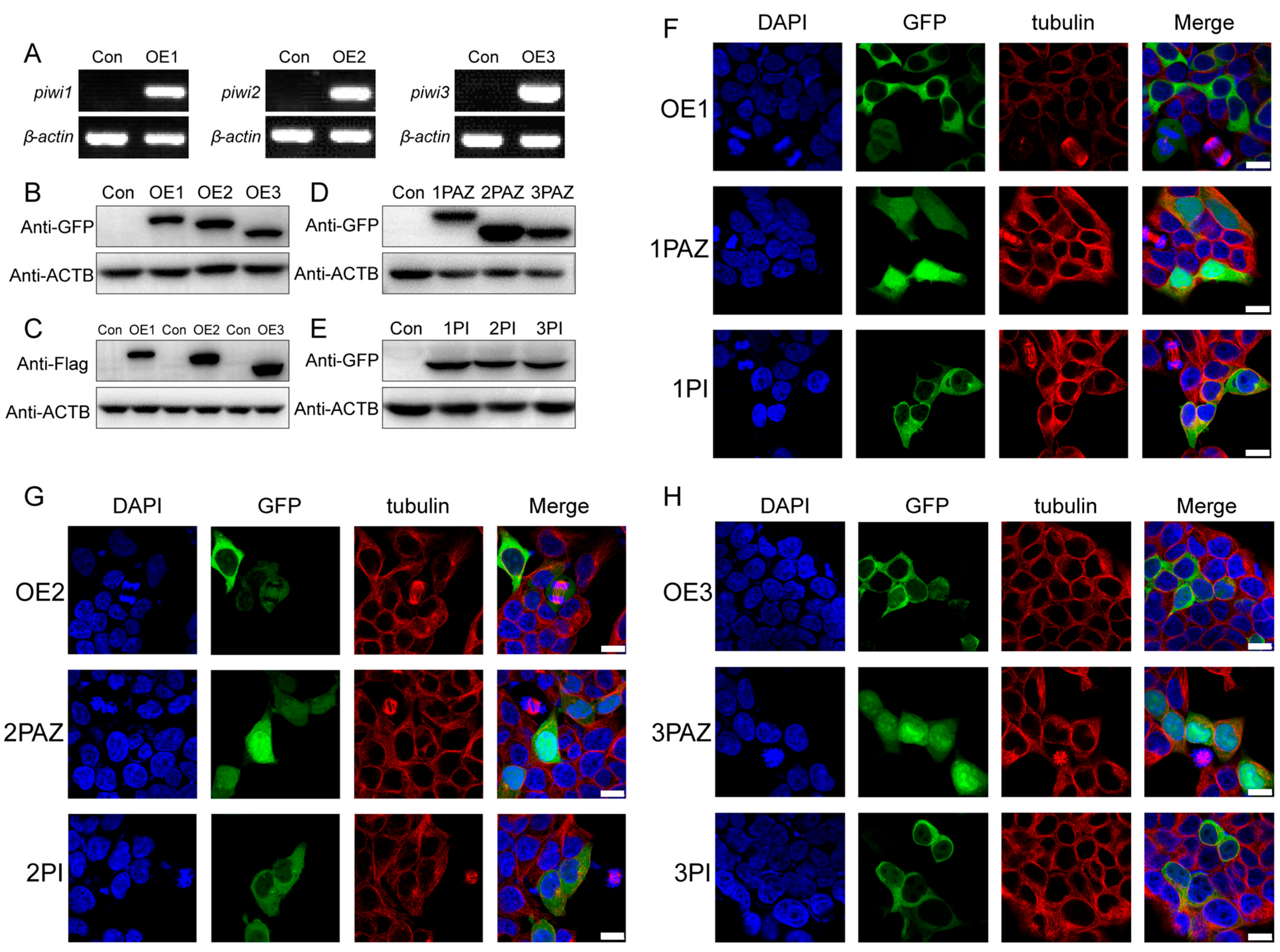
As a limitation of cell culture technology in crustaceans, we used HEK 293T cells to investigate the mechanism of the regulation of PIWIs on the proliferation and differentiation of spermatogonia in E. sinensis. We constructed PIWI-expressed plasmids, after transfection to HEK 293T cells, and all three Piwis could be successfully expressed, with no transcript being found in the control group (Figure 2A). The results of the Western blot showed that the expressed proteins in HEK 293T cells had the same molecular weight as we predicted (Figure 2B,C). We also investigated the potential contribution of two main domains of PIWI, the PAZ and PIWI domains. We expressed these two domains in HEK 293T cells and confirmed their expression via the Western blot (Figure 2D,E).
The localization of PIWIs in E. sinensis testes was reported in our previous study, which showed that three PIWIs were expressed in the cytoplasm of spermatogonia, spermatocytes, and spermatids, and PIWI 1 and PIWI 3 were also localized in the cup-like nucleus of spermatozoa [27]. In this paper, we analyzed the localization of PIWIs as well as their PAZ and PIWI domains in HEK 293T cells (Figure 2F–H). Similar to the results in E. sinensis, PIWIs were mainly expressed in the cytoplasm, while the PAZ domains were extensively expressed in the cells, including cytoplasm and nuclei, with many more signals being found in nuclei. Compared with the PAZ domain, the PIWI domain was strictly expressed in the cytoplasm. Therefore, PIWIs are mainly expressed in the cytoplasm of HEK 293T cells, and the PIWI domain seemed to contribute more to the localization of PIWIs.

3.3. Cell Proliferation Inhibited by Down-Regulating Cyclin After PIWI Expression

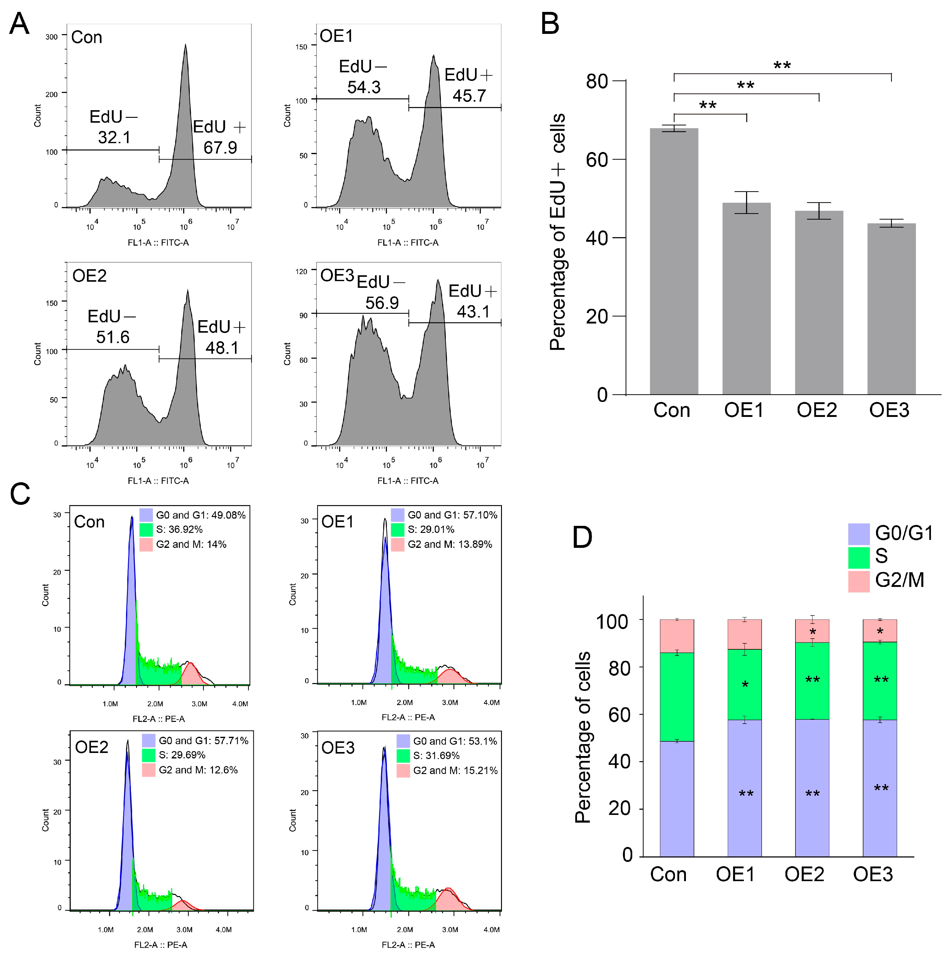
As interference of Piwis promoted the proliferation of spermatogonia in E. sinensis, we checked the cell proliferation in HEK 293T cells after PIWI expression. The ratio of EdU-positive cells was counted by flow cytometry after EdU addition for 1.5 h. The ratio of EdU-positive cells was 67.9% in the control group (Figure 3A), while it was 45.7%, 48.1%, and 43.1% in the piwi1-, piwi2-, and piwi3-expressed groups, respectively (Figure 3A,B), which indicated that PIWIs significantly decreased the proliferation of HEK 293T cells.
PIWIs regulated cell proliferation by reducing DNA replication. We speculated that the cell cycle should also be changed by PIWIs. Fluorescence-activated cell sorting (FACS) was used to analyze the cell cycle in cells transfected with PIWIs. As we speculated, the cell cycle was significantly altered by PIWI expression (Figure 3C,D). All three PIWIs significantly increased the ratio of cells in the G1 phase and decreased the cells in the S phase. The cell cycle arrest at the G1 phase suggested that fewer cells went through the DNA replication process, and PIWIs reduced the cell proliferation.
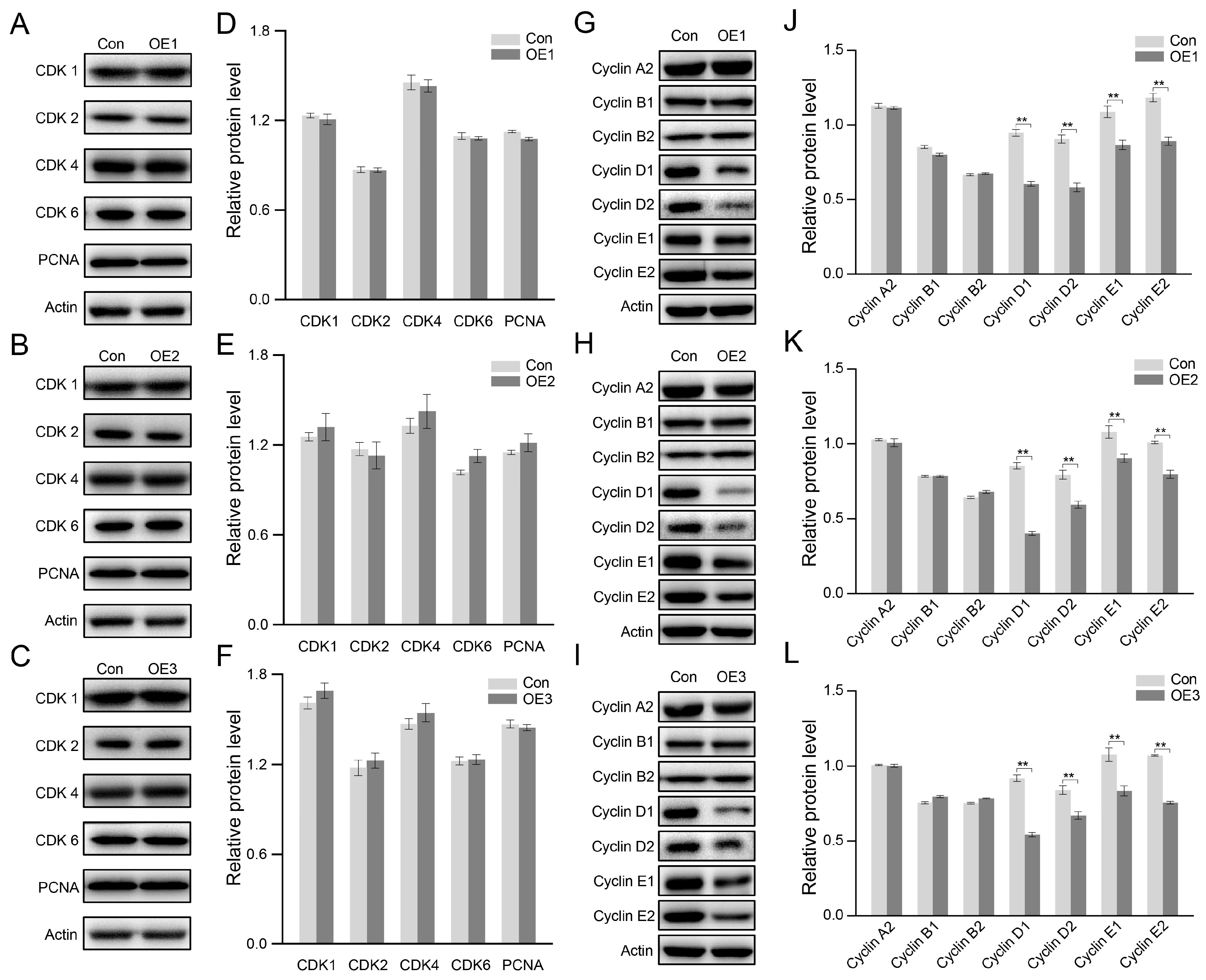
Then, the cell cycle-related proteins were checked using the Western blot. PIWI expression did not change the expression of any cyclin-dependent kinase (CDK), including CDK1, CDK2, CDK4, and CDK6 (Figure 4A–F). Same with CDKs, no charge was detected in the expression of the DNA replication marker PCNA. However, the expression of cyclins was significantly changed as the expression of PIWIs (Figure 4G–L). Three PIWIs significantly decreased Cyclin D1, D2, E1, and E2, without altering Cyclin A2, B1, and B2, suggesting that PIWIs inhibited cell proliferation at the G1 phase by down-regulating Cyclin D and Cyclin E.

3.4. Canonical Wnt-Signaling Pathway Inhibited After PIWI Expression

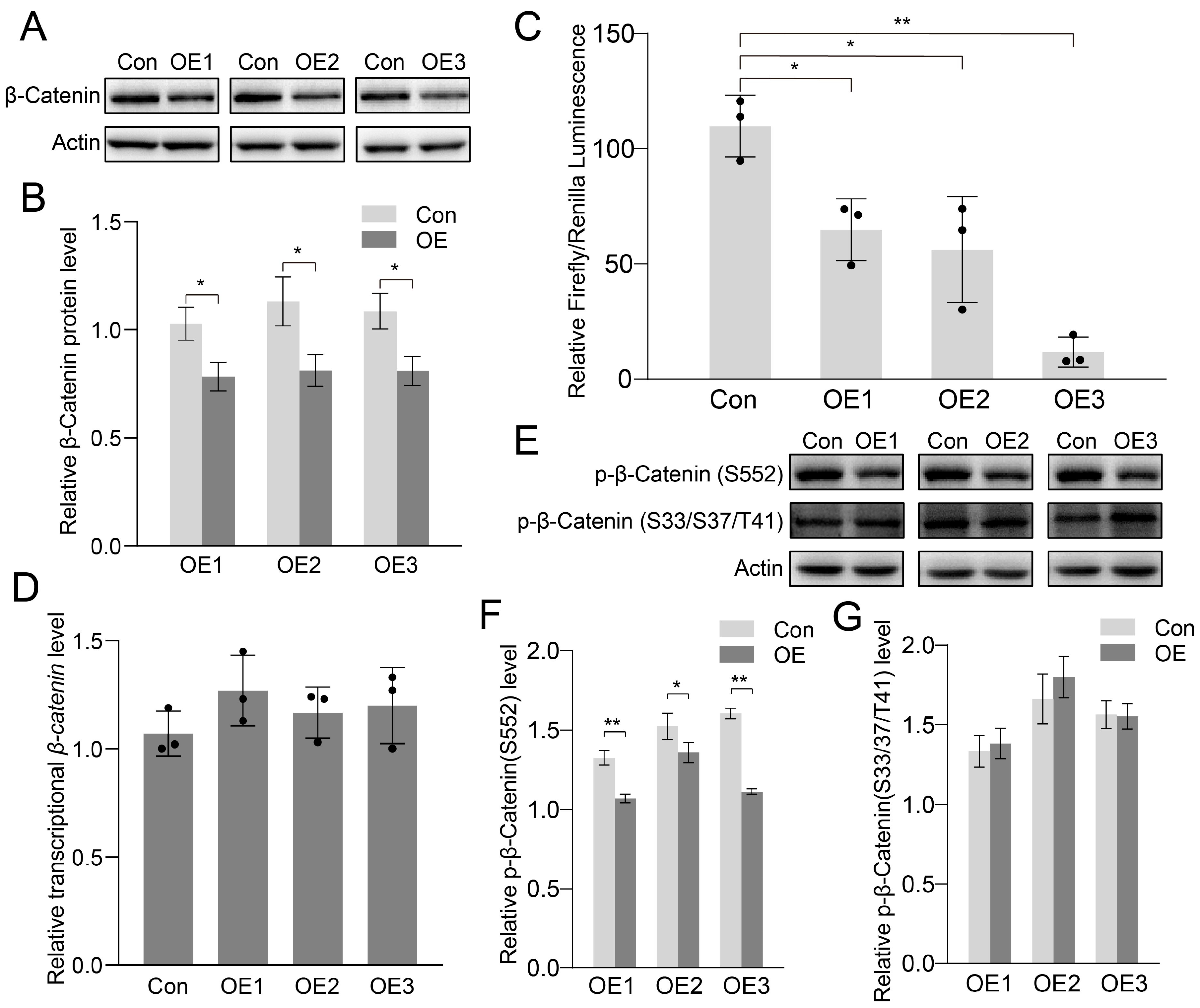
It is reported that Cyclin D is a target gene of the Wnt-signaling pathway [29,30]. We then checked the effects of PIWIs on the Wnt-signaling pathway. The Wnt-signaling pathway is divided into two different types: the canonical pathway (or β-catenin pathway) and the noncanonical pathway [31,32,33]. The most important protein in the canonical pathway is β-catenin. We found that PIWI expression significantly reduced β-catenin (Figure 5A,B). The downregulated β-catenin might have significant effects on the activation of the canonical Wnt pathway. The TOP flash plasmid, which contains the TCF binding site upstream of the Firefly luciferase open reading frame, was used to confirm the activation of the canonical Wnt pathway. As shown in Figure 5C, the luciferase activity in PIWIs was significantly lower than in the control group, suggesting that the canonical Wnt pathway transcriptional activity was inhibited after PIWI expression. Taken together, PIWIs inhibited the canonical Wnt pathway by downregulating β-catenin.
PIWI interacts with piRNA to regulate the expression of genes and transposons at both transcriptional and post-transcriptional levels [34]. We hypothesized that PIWIs regulate β-catenin at the transcriptional level. However, it turned out that the transcriptional level of β-catenin was not changed by PIWI expression (Figure 5D). Thus, the downregulated β-catenin might be due to the post-translational modification. As the most important phosphorylation sites, Ser33, Ser37, and Thr41 were responsible for the stability of β-catenin in cells. The phosphorylation of these sites destabilized β-catenin and increased the ubiquitination and degradation of β-catenin [35]. The mutation of these sites increased the stability of β-catenin and resulted in the accumulation in the cytoplasm, which would find entry into nuclei and further activate Wnt signaling [36]. In our study, downregulated β-catenin and Wnt-signaling activation were found in PIWI-expressing cells. This suggests the possibility of higher phosphorylation at Ser33, Ser37, and Thr41. However, Western blotting showed that the phosphorylation at Ser33, Ser37, and Thr41 was similar between control and the PIWI expression groups (Figure 5E,G). However, the phosphorylated β-catenin at Ser552, which could promote Wnt signaling transcriptional activity [37,38], was decreased in the PIWI expression groups than in the control group (Figure 5E,F).

3.5. Effects on Regulation of Cell Junction Proteins After PIWI Expression

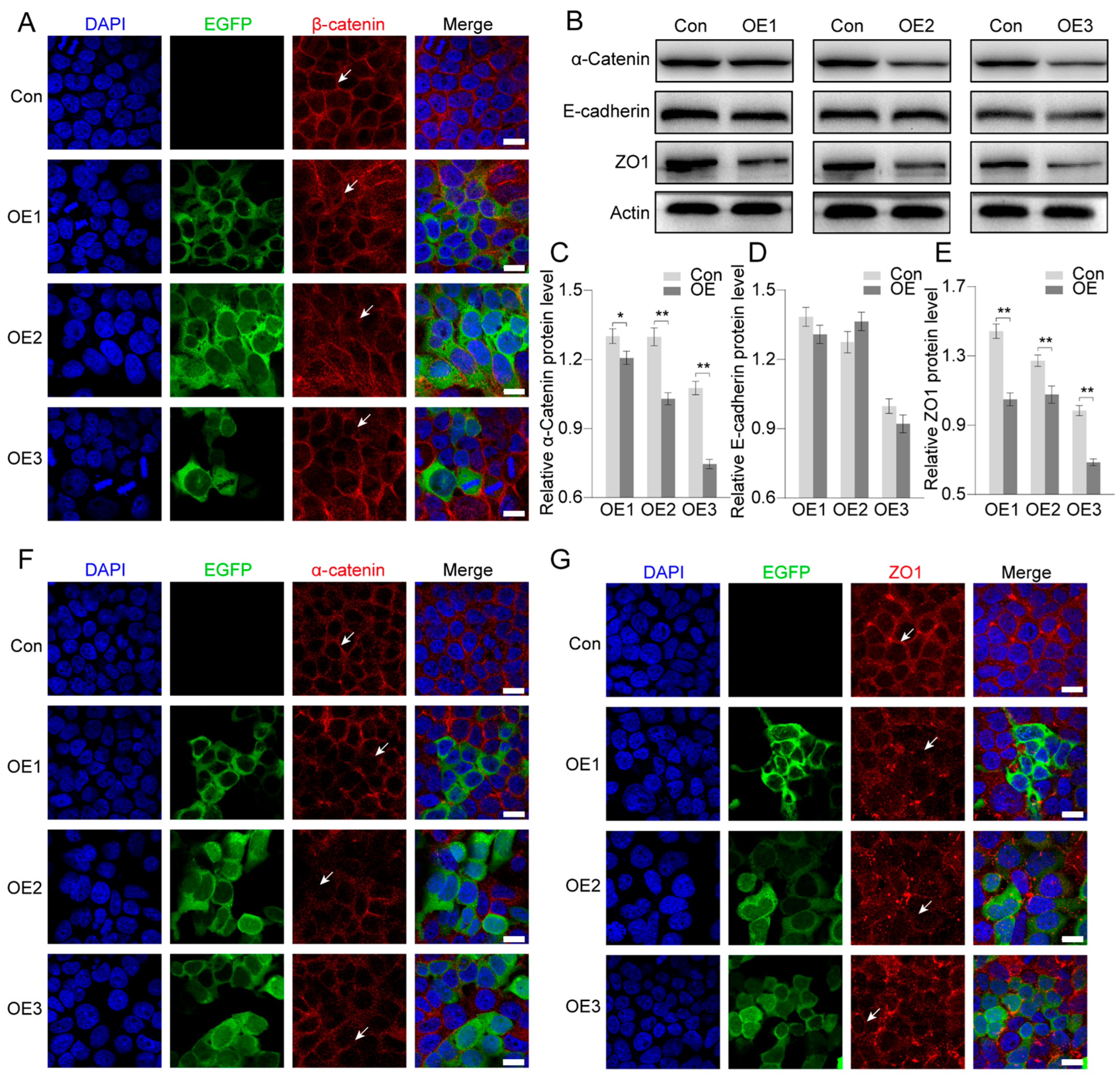
Except for the important factors in the Wnt-signaling pathway, β-catenin also functions in cell–cell interactions, as it links cadherin to the cytoskeleton [39]. In the present study, β-catenin was significantly inhibited by PIWIs. We speculated that the adherens junction might also be affected by PIWIs. The localization of cell junction proteins indicated significant damage in cell–cell interactions. In the control group, β-catenin was strictly and compactly distributed on the cell membrane, while in PIWI expression groups, it seemed that they were translocated from the membrane to the cytoplasm, which would decrease cell–cell interactions (Figure 6A).
The results of Western blots indicated that the adherens junction protein α-catenin was also significantly changed by PIWI expression (Figure 6B,C). However, E-cadherin was unchanged (Figure 6B,D). As β-catenin and α-catenin were significantly reduced, ZO-1 (zonula occludens-1), a tight junction protein, was also significantly reduced by PIWIs (Figure 6B,E). Immunofluorescence indicated that cells after PIWI expression showed less signaling of α-catenin in the cell membrane compared to control cells (Figure 5F). In the control group, cells had an intact ZO1 signal, but in PIWI-expressed groups, the structure of ZO1 in the membrane was destroyed completely (Figure 6G). These results indicated that PIWIs inhibited cell proliferation and destroyed the cell–cell interaction.

4. Discussion and Perspectives

4.1. Novel Functions of PIWIs in Regulating Spermatogonia Self-Renewal and Differentiation During E. sinensis Spermatogenesis

Spermatogonia are a type of Germline Stem Cells (GSCs) and are characterized by their capacity for self-renewal and differentiation into spermatocytes. They play a fundamental role in spermatogenic cell development and maintenance throughout life. The Piwi genes were initially identified as key regulators of GSC asymmetric division in D. melanogaster [7]. PIWI proteins, which exhibit RNA endonucleolytic activity guided by piRNAs, are highly expressed in animal gonads [40]. Early studies using Piwi mutants demonstrated that Piwi regulates GSC self-renewal and differentiation in different species. Decreased Piwi expression in Caenorhabditis elegans reduces the proliferation of GSC-equivalent cells [9]. In the Drosophila ovary, Piwi is required in somatic signaling cells to maintain germline stem cells, such as self-renewal and differentiation [28,41]. However, Piwi abnormalities in mammals result in different outcomes compared with those of invertebrates, and their loss leads to male sterility [42,43]. A recent study showed that a deficiency of Piwi resulted in severe developmental disorders in spermatogenesis of golden hamsters, such as developmental arrest or cell death beginning after meiosis [44]. Therefore, all these studies have revealed the diverse functions of PIWIs in various biological and developmental processes.
In the present study, much more EdU signaling was detected in both spermatogonia and spermatocytes in each piwi knockdown group. That is, more spermatogonia finished the proliferation and differentiation into spermatocytes in the Piwi knockdown groups. Therefore, PIWIs suppressed the proliferation of spermatogonia and the transformation of spermatogonia into spermatocytes to maintain the dynamic balance between spermatogonia and spermatocytes during spermatogenesis in E. sinensis. Similar studies were conducted in Penaeus monodon and Scylla paramamosain [45,46]. Both studies found that piwi2 knockdown led to a significant decrease in the number of sperm cells. Furthermore, the authors held that Sp-Piwi2 may play an important role in spermatogonia proliferation and differentiation during spermatogenesis in S. paramamosain. However, they predicted that Sp-Piwi2 silencing presumably inhibited spermatogonia self-replication and the transition from spermatogonia to spermatocytes through transcriptome sequencing and differential gene analysis (DEGs), which is exactly in contrast with our results [46]. Therefore, inhibiting spermatogonia self-replication and the transition from spermatogonia to spermatocytes in E. sinensis may be Piwi’s new function, and further studies are required on the roles of PIWIs in crustacean spermatogenesis.

4.2. PIWIs Suppressed Spermatogonia Proliferation and Differentiation Through Wnt Signaling Pathway

PIWI was initially identified in the reproductive system and is predominantly expressed in the gonads of Drosophila. Furthermore, it has also been detected in tumor tissues of mammals [47]. In tumor tissues, PIWI interacts with piRNA to regulate cell proliferation, differentiation, and survival [48]. PIWIL2 bound with piRNA-54265 to activate the STAT3 pathway and then promoted cell proliferation in colorectal adenocarcinoma [49]. In non-small cell lung carcinoma, piR-651 increased the expression of Cyclin D1 and CDK4 to promote tumor progression [50], while piR-55490 inhibited cell proliferation in lung cancer cells by targeting the 3′ UTR (untranslated region) of mTOR mRNA [51]. Similarly, piR-39980 targeted RRM2 (ribonucleoside–diphosphate reductase subunit M2) mRNA 3′ UTR to inhibit cell proliferation of HT1080 fibrosarcoma cells [52]. These results suggest that the effects of PIWIs on cell proliferation vary between different piRNAs and different cell types, which raises our concern about the role of PIWI on germ cell proliferation in E. sinensis.
In the present study, we found that knockdown of Piwis promoted spermatogonia proliferation. To investigate the potential mechanism of the roles of PIWIs in this process, we expressed PIWIs of E. sinensis in HEK 293T. Consistent with our conjecture, PIWIs significantly inhibited the cell proliferation of HEK 293T. The Wnt-signaling pathway is a well-known pathway in the regulation of cell proliferation [53]. During spermatogenesis, Wnt5a activates JNK to promote spermatogonial stem cell self-renewal [54]. Wnt3a also positively regulated spermatogonial stem cell proliferation [55]. Wnt/β-catenin signaling is induced by Wnt6 to promote the proliferation of undifferentiated spermatogonia [56]. We checked the effects of PIWIs on Wnt signaling. The analysis of the TCF Reporter Gene showed that the luciferase activity in PIWIs was significantly lower than in the control group, suggesting that the canonical Wnt pathway transcriptional activity was inhibited after PIWI expression. In addition, Cyclin D and Cyclin E are target genes of the Wnt-signaling pathway, so their expression levels are regulated by the Wnt-signaling pathway. It exactly matches our result that Cyclin D and Cyclin E decreased significantly after the expression of PIWIs from E. sinensis in HEK 293T cells.
In this study, we found that PIWIs significantly inhibited the Wnt-signaling pathway by regulating β-catenin, which is consistent with that of HILI in HeLa and HepG2 cells. However, knockdown of HILI in HeLa and HepG2 significantly inhibited β-catenin by regulating the phosphorylation and ubiquitylation of β-catenin, while the overexpression of HILI activated the Wnt-signaling pathway due to the accumulation of β-catenin in the nucleus [57]. In Drosophila, PIWI was reduced in the aged niche of germline stem cells. The reduced PIWI derepressed transposons/transposable elements and resulted in the activation of Toll-GSK3 signaling. The activated Toll-GSK3 signaling would further induce degradation of β-catenin [58].
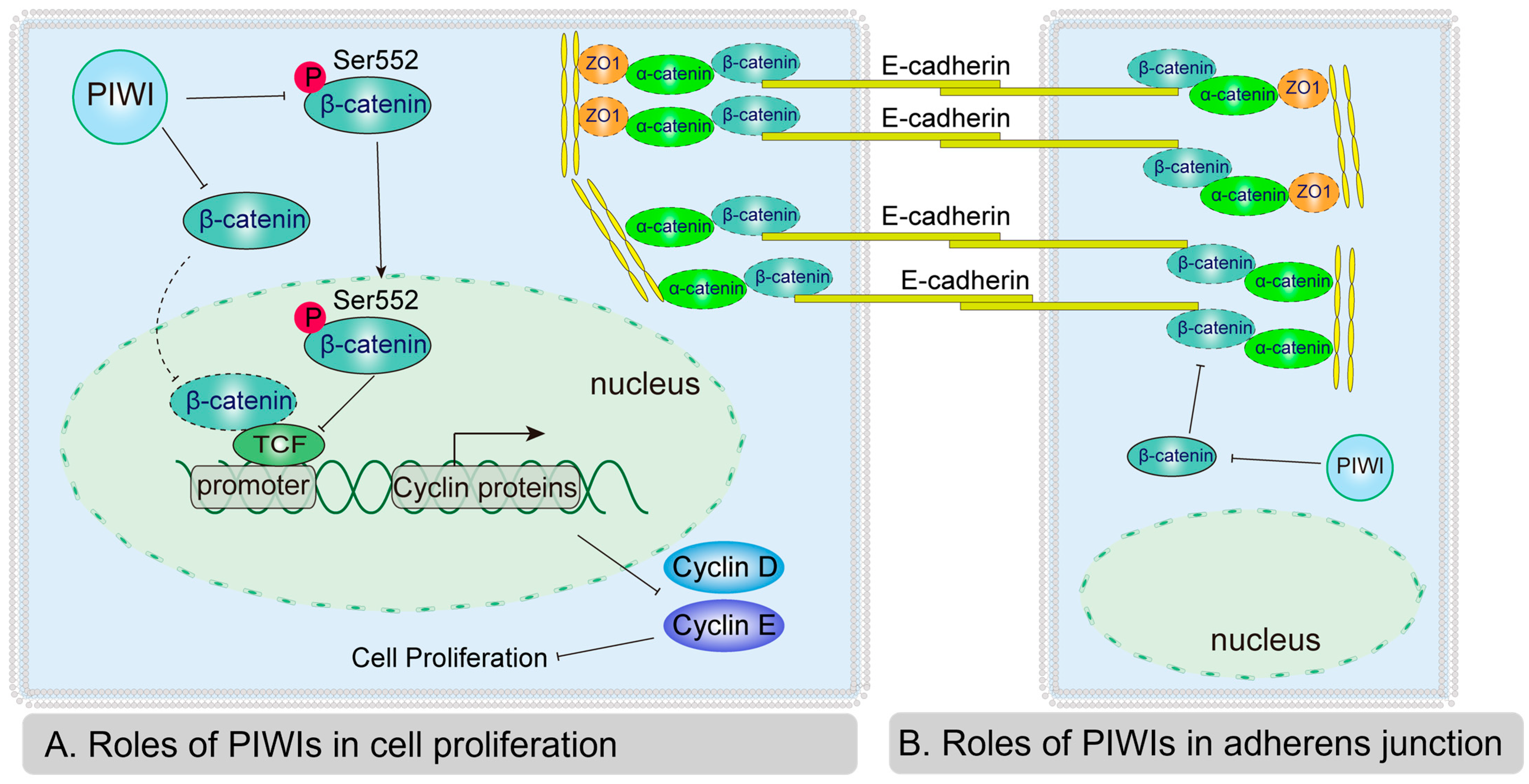
Previous studies found that PIWI regulated β-catenin by changing the phosphorylation and ubiquitylation of β-catenin. We investigated the potential mechanism of PIWIs from E. sinensis in β-catenin regulation. A well-known function of PIWI is transposons or transposable element repression, as well as mRNA degradation at the guide of piRNA [59,60]. We analyzed the effects of PIWIs on β-catenin mRNA. However, the mRNA level of β-catenin was not affected by the expression of PIWIs from E. sinensis, which showed similar results obtained from HeLa and Hep2G [57]. In the canonical Wnt-signaling pathway, the phosphorylation of β-catenin induced by GSK3β and CK1 contributes to the degradation of β-catenin [61]. The level of phosphorylated β-catenin at Ser33/Ser37/Thr41, measured by Western blotting, was unchanged. This indicates that downregulated β-catenin due to the expression of PIWIs does not rely on GSK3. PIWI also had roles in the regulation of translation processes. PIWI interacts with eIF3a, eIF4E, and eIF4G to regulate protein expression by translational activation and repression [16,62,63]. Thus, we speculate that PIWIs from E. sinensis may regulate β-catenin at the translational level. Although the phosphorylation of β-catenin at Ser33/Ser37/Thr41 was unchanged by PIWIs, the phospho-β-catenin (Ser552) was significantly downregulated. The phosphorylation of β-catenin at Ser552 induced by PKA (protein kinase A) and AKT is responsible for the activation of the Wnt-signaling pathway [37,38]. Collectively, our results indicated that PIWIs inhibited the Wnt-signaling pathway by directly downregulating β-catenin, as well as the phosphorylation of β-catenin at Ser552. In conclusion, a possible mechanism of PIWI-inhibited spermatogonia proliferation and differentiation was proposed in our study: on one hand, PIWIs directly downregulate β-catenin and decrease the accumulation of β-catenin in nuclei to inhibit Wnt signaling; on the other hand, PIWIs inhibit the phosphorylation of β-catenin at Ser552 and further inhibit the expression of Cyclin D and Cyclin E (Figure 7A).

4.3. PIWIs Affect Adherens Junctions by Regulating α-Catenin and ZO1

In addition to functioning in the Wnt-signaling pathway, β-catenin also contributes to the formation of adherens junctions [39,64]. The adherens junction contains E-cadherin, α-catenin, β-catenin, and p120-catenin [65]. E-cadherins in different cells bind with each other through an extracellular domain [66]. α-catenin binds with the cytoplasmic domain of E-cadherin through β-catenin [67,68]. α-catenin can directly link the complex to the cytoskeleton or interact with ZO1 [69]. As the downregulated β-catenin was due to the expression of PIWIs, we also checked the adherens junction changes. When PIWIs were expressed, E-cadherin was unchanged, but the protein levels of α-catenin and ZO1 were significantly decreased. At the same time, the subcellular localization of α-catenin and ZO1 was altered, transitioning from a predominantly membrane-associated distribution to a more diffuse cytoplasmic localization. Therefore, we concluded that PIWIs E. sinensis affected the adherens junction through regulation of the expression and localization of α-catenin and ZO1 (Figure 7B). Unfortunately, we were unable to obtain more relevant data on E. sinensis; there is still a lot of room for research in the future.

5. Conclusions

In conclusion, new functions of PIWIs were found in regulating spermatogenesis in E. sinensis. The knockdown of Piwis may promote the proliferation and differentiation of spermatogonia in E. sinensis by regulating the Wnt-signaling pathway. Abnormal expression of PIWIs destroys the adherens junction by changing the localization or expression of α-catenin and ZO1. Furthermore, we proposed a possible mechanism of PIWIs in suppressing the excessive proliferation and differentiation of spermatogonia to maintain the normal spermatogenesis cycle in E. sinensis. These results of the present study could provide important information for further exploration of PIWI’s function in E. sinensis and other crustacean species.

Supplementary Materials

The following supporting information can be downloaded at: https://www.mdpi.com/article/10.3390/biology14101440/s1, Supplemental Figure S1. Cycle of seminiferous epithelium analysis in E sinensis by EdU incorporation assay. EdU was injected to crabs, and the signaling of EdU (red) was detected at 2 h, 3, 6, 9, 12 and 15 d using Confocal Laser Scanning Microscope. Scale bar shows 50 μm. DNA replication happens in spermatogonia, which means that EdU can only be incorporated in spermatogonia. The timing when we can detect EdU signaling in spermatocyte, spermatid or spermatozoa can represent the time required in the development of spermatogonia to the specific stage germ cells. The supplementary Figures S2–S6 are the original full Western blot figures in this study.

Author Contributions

Conceptualization, B.-H.W., S.-L.H. and W.-X.Y.; methodology, data curation, writing—original draft preparation, and preparing all the figures, B.-H.W.; crab culturing and RNA interference experiments, Z.Z., H.-Y.Q. and Z.-F.L.; manuscript frame constructing, revision, and funding provision, S.-L.H. and W.-X.Y. All authors have read and agreed to the published version of the manuscript.

Funding

This work was supported by the National Natural Science Foundation of China [Nos. 32102786 and 32270555].

Institutional Review Board Statement

Not applicable.

Informed Consent Statement

Not applicable.

Data Availability Statement

All data are included in the main text or Supplementary Material. Further inquiries can be directed to the corresponding author.

Conflicts of Interest

The authors declare no conflicts of interest.

References

  1. Stukenborg, J.B.; Kjartansdóttir, K.R.; Reda, A.; Colon, E.; Albersmeier, J.P.; Söder, O. Male germ cell development in humans. Horm. Res. Paediatr. 2014, 81, 2–12. [Google Scholar] [CrossRef]
  2. Matson, C.K.; Murphy, M.W.; Griswold, M.D.; Yoshida, S.; Bardwell, V.J.; Zarkower, D. The Mammalian Doublesex Homolog DMRT1 Is a Transcriptional Gatekeeper that Controls the Mitosis versus Meiosis Decision in Male Germ Cells. Dev. Cell 2010, 19, 612–624. [Google Scholar] [CrossRef]
  3. Mei, X.X.; Wang, J.; Wu, J. Extrinsic and intrinsic factors controlling spermatogonial stem cell self-renewal and differentiation. Asian J. Androl. 2015, 17, 347–354. [Google Scholar] [CrossRef] [PubMed]
  4. Tseng, Y.T.; Liao, H.F.; Yu, C.Y.; Mo, C.F.; Lin, S.P. Epigenetic factors in the regulation of prospermatogonia and spermatogonial stem cells. Reproduction 2015, 150, R77–R91. [Google Scholar] [CrossRef] [PubMed]
  5. Juliano, C.; Wang, J.; Lin, H. Uniting germline and stem cells: The function of Piwi proteins and the piRNA pathway in diverse organisms. Annu. Rev. Genet. 2011, 45, 447–469. [Google Scholar] [CrossRef] [PubMed]
  6. Ramat, A.; Simonelig, M. Functions of PIWI Proteins in Gene Regulation: New Arrows Added to the piRNA Quiver. Trends Genet. 2021, 37, 188–200. [Google Scholar] [CrossRef]
  7. Lin, H.; Spradling, A.C. A novel group of pumilio mutations affects the asymmetric division of germline stem cells in the Drosophila ovary. Development 1997, 124, 2463–2476. [Google Scholar] [CrossRef]
  8. Meister, G. Argonaute proteins: Functional insights and emerging roles. Nat. Rev. Genet. 2013, 14, 447–459. [Google Scholar] [CrossRef]
  9. Cox, D.N.; Chao, A.; Baker, J.; Chang, L.; Qiao, D.; Lin, H. A novel class of evolutionarily conserved genes defined by piwi are essential for stem cell self-renewal. Genes Dev. 1998, 12, 3715–3727. [Google Scholar] [CrossRef]
  10. Thomson, T.; Lin, H. The biogenesis and function of PIWI proteins and piRNAs: Progress and prospect. Annu. Rev. Cell Dev. Biol. 2009, 25, 355–376. [Google Scholar] [CrossRef]
  11. Megosh, H.B.; Cox, D.N.; Campbell, C.; Lin, H. The role of PIWI and the miRNA machinery in Drosophila germline determination. Curr. Biol. 2006, 16, 1884–1894. [Google Scholar] [CrossRef]
  12. Li, M.; Hong, N.; Gui, J.; Hong, Y. Medaka piwi is essential for primordial germ cell migration. Curr. Mol. Med. 2012, 12, 1040–1049. [Google Scholar] [CrossRef]
  13. Kuramochi-Miyagawa, S.; Kimura, T.; Ijiri, T.W.; Isobe, T.; Asada, N.; Fujita, Y.; Ikawa, M.; Iwai, N.; Okabe, M.; Deng, W.; et al. Mili, a mammalian member of piwi family gene, is essential for spermatogenesis. Development 2004, 131, 839–849. [Google Scholar] [CrossRef]
  14. Unhavaithaya, Y.; Hao, Y.; Beyret, E.; Yin, H.; Kuramochi-Miyagawa, S.; Nakano, T.; Lin, H. MILI, a PIWI-interacting RNA-binding protein, is required for germ line stem cell self-renewal and appears to positively regulate translation. J. Biol. Chem. 2009, 284, 6507–6519. [Google Scholar] [CrossRef]
  15. Deng, W.; Lin, H. miwi, a murine homolog of piwi, encodes a cytoplasmic protein essential for spermatogenesis. Dev. Cell 2002, 2, 819–830. [Google Scholar] [CrossRef]
  16. Carmell, M.A.; Girard, A.; van de Kant, H.J.; Bourc’his, D.; Bestor, T.H.; de Rooij, D.G.; Hannon, G.J. MIWI2 is essential for spermatogenesis and repression of transposons in the mouse male germline. Dev. Cell 2007, 12, 503–514. [Google Scholar] [CrossRef]
  17. Houwing, S.; Kamminga, L.M.; Berezikov, E.; Cronembold, D.; Girard, A.; van den Elst, H.; Filippov, D.V.; Blaser, H.; Raz, E.; Moens, C.B.; et al. A role for Piwi and piRNAs in germ cell maintenance and transposon silencing in Zebrafish. Cell 2007, 129, 69–82. [Google Scholar] [CrossRef]
  18. Houwing, S.; Berezikov, E.; Ketting, R.F. Zili is required for germ cell differentiation and meiosis in zebrafish. EMBO J. 2008, 27, 2702–2711. [Google Scholar] [CrossRef] [PubMed]
  19. Meistrich, M.L.; Mohapatra, B.; Shirley, C.R.; Zhao, M. Roles of transition nuclear proteins in spermiogenesis. Chromosoma 2003, 111, 483–488. [Google Scholar] [CrossRef] [PubMed]
  20. Gou, L.T.; Kang, J.Y.; Dai, P.; Wang, X.; Li, F.; Zhao, S.; Zhang, M.; Hua, M.M.; Lu, Y.; Zhu, Y.; et al. Ubiquitination-deficient Mutations in human Piwi cause male infertility by impairing histone-to-protamine exchange during spermiogenesis. Cell 2017, 169, 1090–1104.e13. [Google Scholar] [CrossRef] [PubMed]
  21. Dai, P.; Wang, X.; Liu, M.F. A dual role of the PIWI/piRNA machinery in regulating mRNAs during mouse spermiogenesis. Sci. China Life Sci. 2020, 63, 447–449. [Google Scholar] [CrossRef]
  22. Gou, L.T.; Dai, P.; Yang, J.H.; Xue, Y.; Hu, Y.P.; Zhou, Y.; Kang, J.Y.; Wang, X.; Li, H.; Hua, M.M.; et al. Pachytene piRNAs instruct massive mRNA elimination during late spermiogenesis. Cell Res. 2014, 24, 680–700. [Google Scholar] [CrossRef]
  23. Vourekas, A.; Zheng, Q.; Alexiou, P.; Maragkakis, M.; Kirino, Y.; Gregory, B.D.; Mourelatos, Z. Mili and Miwi target RNA repertoire reveals piRNA biogenesis and function of Miwi in spermiogenesis. Nat. Struct. Mol. Biol. 2012, 19, 773–781. [Google Scholar] [CrossRef]
  24. Dai, P.; Wang, X.; Gou, L.T.; Li, Z.T.; Wen, Z.; Chen, Z.G.; Hua, M.M.; Zhong, A.; Wang, L.; Su, H.; et al. A Translation-activating function of MIWI/piRNA during mouse spermiogenesis. Cell 2019, 179, 1566–1581.e16. [Google Scholar] [CrossRef] [PubMed]
  25. Wang, Y.L.; Sun, W.J.; He, L.; Li, Q.; Wang, Q. Morphological alterations of all stages of spermatogenesis and acrosome reaction in Chinese mitten crab Eriocheir sinensis. Cell Tissue Res. 2015, 360, 401–412. [Google Scholar] [CrossRef] [PubMed]
  26. Kurtz, K.; Martínez-Soler, F.; Ausió, J.; Chiva, M. Histones and nucleosomes in cancer sperm (Decapod: Crustacea) previously described as lacking basic DNA-associated proteins: A new model of sperm chromatin. J. Cell. Biochem. 2008, 105, 574–584. [Google Scholar] [CrossRef]
  27. Wei, B.H.; Ni, J.H.; Yang, T.; Hao, S.L.; Yang, W.X. PIWIs maintain testis apoptosis to remove abnormal germ cells in Eriocheir sinensis. Reproduction 2021, 162, 193–207. [Google Scholar] [CrossRef] [PubMed]
  28. Szakmary, A.; Cox, D.N.; Wang, Z.; Lin, H. Regulatory relationship among piwi, pumilio, and bag-of-marbles in Drosophila germline stem cell self-renewal and differentiation. Curr. Biol. 2005, 15, 171–178. [Google Scholar] [CrossRef]
  29. Tetsu, O.; McCormick, F. Beta-catenin regulates expression of cyclin D1 in colon carcinoma cells. Nature 1999, 398, 422–426. [Google Scholar] [CrossRef]
  30. Shtutman, M.; Zhurinsky, J.; Simcha, I.; Albanese, C.; D’Amico, M.; Pestell, R.; Ben-Ze’ev, A. The cyclin D1 gene is a target of the beta-catenin/LEF-1 pathway. Proc. Natl. Acad. Sci. USA 1999, 96, 5522–5527. [Google Scholar] [CrossRef]
  31. Logan, C.Y.; Nusse, R. The Wnt signaling pathway in development and disease. Cell Dev. Biol. 2004, 20, 781–810. [Google Scholar] [CrossRef]
  32. Seifert, J.R.; Mlodzik, M. Frizzled/PCP signalling: A conserved mechanism regulating cell polarity and directed motility. Nat. Rev. Genet. 2007, 8, 126–138. [Google Scholar] [CrossRef]
  33. Semenov, M.V.; Habas, R.; Macdonald, B.T.; He, X. SnapShot: Noncanonical Wnt Signaling Pathways. Cell 2007, 131, 1378. [Google Scholar] [CrossRef]
  34. Ku, H.Y.; Lin, H. PIWI proteins and their interactors in piRNA biogenesis, germline development and gene expression. Natl. Sci. Rev. 2014, 1, 205–218. [Google Scholar] [CrossRef] [PubMed]
  35. Yost, C.; Torres, M.; Miller, J.R.; Huang, E.; Kimelman, D.; Moon, R.T. The axis-inducing activity, stability, and subcellular distribution of beta-catenin is regulated in Xenopus embryos by glycogen synthase kinase 3. Genes Dev. 1996, 10, 1443–1454. [Google Scholar] [CrossRef] [PubMed]
  36. Morin, P.J.; Sparks, A.B.; Korinek, V.; Barker, N.; Clevers, H.; Vogelstein, B.; Kinzler, K.W. Activation of beta-catenin-Tcf signaling in colon cancer by mutations in beta-catenin or APC. Science 1997, 275, 1787–1790. [Google Scholar] [CrossRef] [PubMed]
  37. Taurin, S.; Sandbo, N.; Qin, Y.; Browning, D.; Dulin, N.O. Phosphorylation of beta-catenin by cyclic AMP-dependent protein kinase. J. Biol. Chem. 2006, 281, 9971–9976. [Google Scholar] [CrossRef]
  38. Fang, D.; Hawke, D.; Zheng, Y.; Xia, Y.; Meisenhelder, J.; Nika, H.; Mills, G.B.; Kobayashi, R.; Hunter, T.; Lu, Z. Phosphorylation of beta-catenin by AKT promotes beta-catenin transcriptional activity. J. Biol. Chem. 2007, 282, 11221–11229. [Google Scholar] [CrossRef]
  39. Wheelock, M.J.; Johnson, K.R. Cadherins as modulators of cellular phenotype. Annu. Rev. Cell Dev. Biol. 2003, 19, 207–235. [Google Scholar] [CrossRef] [PubMed]
  40. Özata, D.M.; Yu, T.; Mou, H.; Gainetdinov, I.; Colpan, C.; Cecchini, K.; Kaymaz, Y.; Wu, P.H.; Fan, K.; Kucukural, A.; et al. Evolutionarily conserved pachytene piRNA loci are highly divergent among modern humans. Nat. Ecol. Evol. 2020, 4, 156–168. [Google Scholar] [CrossRef]
  41. Cox, D.N.; Chao, A.; Lin, H. piwi encodes a nucleoplasmic factor whose activity modulates the number and division rate of germline stem cells. Development 2000, 127, 503–514. [Google Scholar] [CrossRef]
  42. Dowling, M.; Homolka, D.; Raad, N.; Gos, P.; Pandey, R.R.; Pillai, R.S. In Vivo PIWI slicing in mouse testes deviates from rules established in vitro. RNA Soc. 2023, 29, 308–316. [Google Scholar] [CrossRef]
  43. Reuter, M.; Berninger, P.; Chuma, S.; Shah, H.; Hosokawa, M.; Funaya, C.; Antony, C.; Sachidanandam, R.; Pillai, R.S. Miwi catalysis is required for piRNA amplification-independent LINE1 transposon silencing. Nature 2011, 480, 264–267. [Google Scholar] [CrossRef]
  44. Lv, X.; Xiao, W.; Lai, Y.; Zhang, Z.; Zhang, H.; Qiu, C.; Hou, L.; Chen, Q.; Wang, D.; Gao, Y.; et al. The non-redundant functions of PIWI family proteins in gametogenesis in golden hamsters. Nat. Commun. 2023, 14, 5267. [Google Scholar] [CrossRef]
  45. Sukthaworn, S.; Panyim, S.; Udomkit, A. Homologues of Piwi control transposable elements and development of male germline in Penaeus monodon. Comp. Biochem. Physiol. Part A Mol. Integr. Physiol. 2020, 250, 110807. [Google Scholar] [CrossRef]
  46. Wang, T.; Lu, L.; Liu, F.; Liu, A.; Ye, H. Transcriptome analysis of testis in response to silencing of piwi2 sheds light on spermatogenesis in the mud crab scylla paramamosain. Aquac. Rep. 2025, 40, 102550. [Google Scholar] [CrossRef]
  47. Kwon, C.; Tak, H.; Rho, M.; Chang, H.R.; Kim, Y.H.; Kim, K.T.; Balch, C.; Lee, E.K.; Nam, S. Detection of PIWI and piRNAs in the mitochondria of mammalian cancer cells. Biochem. Biophys. Res. Commun. 2014, 446, 218–223. [Google Scholar] [CrossRef] [PubMed]
  48. Zeng, Q.; Wan, H.; Zhao, S.; Xu, H.; Tang, T.; Oware, K.A.; Qu, S. Role of PIWI-interacting RNAs on cell survival: Proliferation, apoptosis, and cycle. IUBMB Life 2020, 72, 1870–1878. [Google Scholar] [CrossRef]
  49. Mai, D.; Ding, P.; Tan, L.; Zhang, J.; Pan, Z.; Bai, R.; Li, C.; Li, M.; Zhou, Y.; Tan, W.; et al. PIWI-interacting RNA-54265 is oncogenic and a potential therapeutic target in colorectal adenocarcinoma. Theranostics 2018, 8, 5213–5230. [Google Scholar] [CrossRef] [PubMed]
  50. Li, D.; Luo, Y.; Gao, Y.; Yang, Y.; Wang, Y.; Xu, Y.; Tan, S.; Zhang, Y.; Duan, J.; Yang, Y. piR-651 promotes tumor formation in non-small cell lung carcinoma through the upregulation of cyclin D1 and CDK4. Int. J. Mol. Med. 2016, 38, 927–936. [Google Scholar] [CrossRef]
  51. Peng, L.; Song, L.; Liu, C.; Lv, X.; Li, X.; Jie, J.; Zhao, D.; Li, D. piR-55490 inhibits the growth of lung carcinoma by suppressing mTOR signaling. Tumour Biol. 2016, 37, 2749–2756. [Google Scholar] [CrossRef]
  52. Das, B.; Roy, J.; Jain, N.; Mallick, B. Tumor suppressive activity of PIWI-interacting RNA in human fibrosarcoma mediated through repression of RRM2. Mol. Carcinog. 2019, 58, 344–357. [Google Scholar] [CrossRef]
  53. Clevers, H.; Loh, K.M.; Nusse, R. Stem cell signaling. An integral program for tissue renewal and regeneration: Wnt signaling and stem cell control. Science 2014, 346, 1248012. [Google Scholar] [CrossRef]
  54. Yeh, J.R.; Zhang, X.; Nagano, M.C. Wnt5a is a cell-extrinsic factor that supports self-renewal of mouse spermatogonial stem cells. J. Cell Sci. 2011, 124 Pt 14, 2357–2366. [Google Scholar] [CrossRef] [PubMed]
  55. Yeh, J.R.; Zhang, X.; Nagano, M.C. Indirect effects of Wnt3a/β-catenin signalling support mouse spermatogonial stem cells in vitro. PLoS ONE 2012, 7, e40002. [Google Scholar] [CrossRef] [PubMed]
  56. Takase, H.M.; Nusse, R. Paracrine Wnt/β-catenin signaling mediates proliferation of undifferentiated spermatogonia in the adult mouse testis. Proc. Natl. Acad. Sci. USA 2016, 113, E1489–E1497. [Google Scholar] [CrossRef] [PubMed]
  57. Qiu, B.; Zeng, J.; Zhao, X.; Huang, L.; Ma, T.; Zhu, Y.; Liu, M.; Tao, D.; Liu, Y.; Lu, Y.; et al. PIWIL2 stabilizes β-catenin to promote cell cycle and proliferation in tumor cells. Biochem. Biophys. Res. Commun. 2019, 516, 819–824. [Google Scholar] [CrossRef]
  58. Lin, K.Y.; Wang, W.D.; Lin, C.H.; Rastegari, E.; Su, Y.H.; Chang, Y.T.; Liao, Y.F.; Chang, Y.C.; Pi, H.; Yu, B.Y.; et al. Piwi reduction in the aged niche eliminates germline stem cells via Toll-GSK3 signaling. Nat. Commun. 2020, 11, 3147. [Google Scholar] [CrossRef]
  59. Robine, N.; Lau, N.C.; Balla, S.; Jin, Z.; Okamura, K.; Kuramochi-Miyagawa, S.; Blower, M.D.; Lai, E.C. A broadly conserved pathway generates 3′UTR-directed primary piRNAs. Curr. Biol. 2009, 19, 2066–2076. [Google Scholar] [CrossRef]
  60. Rojas-Ríos, P.; Simonelig, M. piRNAs and PIWI proteins: Regulators of gene expression in development and stem cells. Development 2018, 145, dev161786. [Google Scholar] [CrossRef]
  61. Stamos, J.L.; Weis, W.I. The β-catenin destruction complex. Cold Spring Harb. Perspect. Biol. 2013, 5, a007898. [Google Scholar] [CrossRef]
  62. Grivna, S.T.; Pyhtila, B.; Lin, H. MIWI associates with translational machinery and PIWI-interacting RNAs (piRNAs) in regulating spermatogenesis. Proc. Natl. Acad. Sci. USA 2006, 103, 13415–13420. [Google Scholar] [CrossRef] [PubMed]
  63. Ramat, A.; Garcia-Silva, M.R.; Jahan, C.; Naït-Saïdi, R.; Dufourt, J.; Garret, C.; Chartier, A.; Cremaschi, J.; Patel, V.; Decourcelle, M.; et al. The PIWI protein Aubergine recruits eIF3 to activate translation in the germ plasm. Cell Res. 2020, 30, 421–435. [Google Scholar] [CrossRef]
  64. Gumbiner, B.M. Regulation of cadherin-mediated adhesion in morphogenesis. Nat. Rev. Mol. Cell Biol. 2005, 6, 622–634. [Google Scholar] [CrossRef]
  65. Maker, A.; Gumbiner, B.M. Reconstitution of the full transmembrane cadherin-catenin complex. Protein Expr. Purif. 2022, 193, 106056. [Google Scholar] [CrossRef]
  66. Harrison, O.J.; Jin, X.; Hong, S.; Bahna, F.; Ahlsen, G.; Brasch, J.; Wu, Y.; Vendome, J.; Felsovalyi, K.; Hampton, C.M.; et al. The extracellular architecture of adherens junctions revealed by crystal structures of type I cadherins. Structure 2011, 19, 244–256. [Google Scholar] [CrossRef] [PubMed]
  67. Pokutta, S.; Weis, W.I. Structure of the dimerization and beta-catenin-binding region of alpha-catenin. Mol. Cell 2000, 5, 533–543. [Google Scholar] [CrossRef] [PubMed]
  68. Huber, A.H.; Weis, W.I. The structure of the beta-catenin/E-cadherin complex and the molecular basis of diverse ligand recognition by beta-catenin. Cell 2001, 105, 391–402. [Google Scholar] [CrossRef] [PubMed]
  69. Itoh, M.; Nagafuchi, A.; Moroi, S.; Tsukita, S. Involvement of ZO-1 in cadherin-based cell adhesion through its direct binding to alpha catenin and actin filaments. J. Cell Biol. 1997, 138, 181–192. [Google Scholar] [CrossRef]
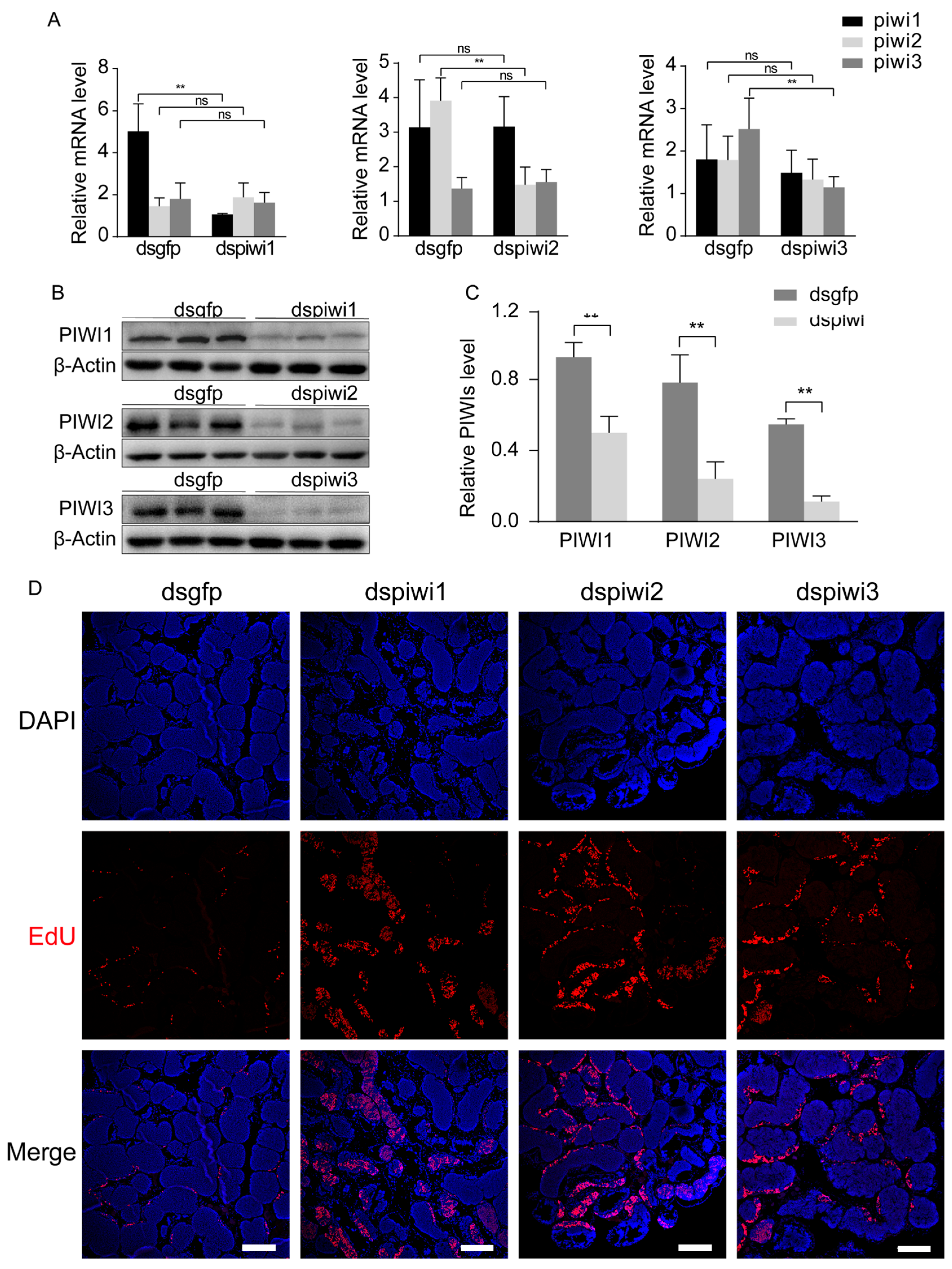
Figure 1. PIWIs regulate the transformation of spermatogonia to spermatocytes in E. sinensis. (A) Transcriptional level of Piwis in the testis of E. sinensis injected with dsRNA specific to Piwi1-3, respectively. (B,C) Western blot analysis and quantification of PIWIs in E. sinensis testes injected with dsRNA specific to Piwi1-3, respectively. The integrated density was measured using ImageJ. Crabs in the control group were injected with dsRNA against Gfp. The actin mRNA and actin protein from crabs were set as an internal control. Three crabs (n = 3) were used in each group. Significant differences were analyzed with an unpaired two-tailed Student’s t-test. “**” indicates p < 0.01. “ns” indicates no significant change. (D) Immunostaining of EdU in the testes of E. sinensis injected with dsRNA. Testis samples were collected 2 h after the injection of EdU. Scale bar = 200 μm.
Biology 14 01440 g001
Figure 2. The expression model of PIWIs and PIWI domains from E. sinensis in HEK 293T. (A) Detection of Piwis mRNA in HEK 293T using RT-PCR. β-Actin from humans was set as an internal control. (B,C) Western blot analysis of PIWIs (constructed into pEGFP-c1 and pCMV-N-flag) from E. sinensis in HEK 293T. The actin protein was set as an internal control. (D,E) Western blot analysis of PIWIs domains (PAZ and PIWI domains constructed into pEGFP-c1) from E. sinensis in HEK 293T. The actin protein was set as an internal control. OE1, OE2, and OE3 represented piwi1, piwi2, and piwi3, respectively. (FH) Immunostaining analysis on the localization of PIWIs in HEK 293T cells transfected with PIWI expression vectors, which was shown by EGFP (green). The nuclei were stained with DAPI (blue). Tubulin was marked by Tubulin-Tracker Red (red). Scale bar = 15 μm.
Biology 14 01440 g002
Figure 3. PIWIs from E. sinensis inhibited cell proliferation of HEK 293T. (A,B) EdU incorporation assay and quantification analysis of PIWIs on cell proliferation of HEK 293T by flow cytometry. (C,D) Cell cycle analysis and quantification analysis in HEK 293T cells transfected with PIWI expression vectors. Triplicate were used in each group (n = 3). Significant differences were analyzed by an unpaired two-tailed Student’s t-test. “*” above the columns indicates p < 0.05, while “**” indicates p < 0.01.
Biology 14 01440 g003
Figure 4. PIWIs from E. sinensis inhibit the expression of Cyclin in HEK 293T. (AF) Western blot analysis and quantification of cyclin-dependent kinase in HEK 293T cells transfected with PIWI expression vectors. (GL) Western blot analysis and quantification of Cyclin in HEK 293T cells transfected with PIWI expression vectors. The actin protein was set as an internal control. The integrated density was measured using ImageJ. Triplicates were used in each group (n = 3). Significant differences were analyzed with an unpaired two-tailed Student’s t-test. “**” above the columns indicates p <0.01.
Biology 14 01440 g004
Figure 5. PIWIs from E. sinensis inhibit the Wnt-signaling pathway. (A,B) Western blot analysis and quantification of β-catenin in HEK 293T cells transfected with PIWI expression vectors. (C) TCF reporter gene assay in HEK 293T cells transfected with PIWI expression vectors. (D) Transcriptional level of β-catenin in HEK 293T cells transfected with PIWI expression vectors. (EG) Western blot analysis and quantification of phosphorylation of β-catenin in HEK 293T cells transfected with PIWI expression vectors. The actin mRNA and actin protein were set as an internal control. The integrated density was measured by ImageJ. Triplicate were used in each group (n = 3). Significant differences were analyzed using an unpaired two-tailed Student’s t-test. “*” above the columns indicates p < 0.05, while “**” indicates p < 0.01.
Biology 14 01440 g005
Figure 6. PIWIs regulate adherens junctions in HEK 293T. (A) Immunostaining analysis of β-catenin (red) in HEK 293T cells transfected with PIWI expression vectors, which was shown by EGFP (green). The nuclei were stained with DAPI (blue). Scale bar = 15 μm. (BE) Western blot analysis and quantification of proteins relative to adherens junctions in HEK 293T cells transfected with PIWI expression vectors. The actin protein was set as an internal control. The integrated density was measured by Image J v1.8.0. Triplicates were used in each group (n = 3). Significant differences were analyzed by an unpaired two-tailed Student’s t-test. “*” above the columns indicates p < 0.05, while “**” indicates p < 0.01. (F,G) Immunostaining analysis of α-catenin and ZO1 (red) in HEK 293T cells transfected with PIWI expression vectors, which was shown by EGFP (green). The distribution of β-catenin, α-catenin and ZO1 is denoted by white arrows. The nuclei were stained with DAPI (blue). Scale bar = 15 μm.
Biology 14 01440 g006
Figure 7. Schematic diagram illustrates the roles of PIWIs in spermatogenesis. Roles of PIWIs in cell proliferation (A) and adherens junctions (B). PIWIs down-regulate both β-catenin and the phosphorylation of β-catenin at Ser552 to inhibit the transcriptional activity of the Wnt-signaling pathway. Repressed Wnt signaling reduces the expression of Cyclin D and Cyclin E to inhibit cell proliferation, which may inhibit spermatogonia proliferation and transformation of spermatogonia to spermatocytes in E. sinensis. Down-regulated β-catenin due to PIWIs also significantly destroys adherens junctions by inhibiting and impairing the localization of α-catenin and ZO1.
Biology 14 01440 g007
Table 1. The primer sequences used in the present study.
Gene Primer Sequence (5′–3′)Purpose
Piwi1FACACCCCAAAATCCGAGACCqPCR
RCTTGTCCATGCTCCCCTTGT
Piwi2FTTGATGCGCCGATGTATGGAqPCR
RGGATGGTGCTGGTGTATCCC
Piwi3FAAGGCATACCAAGTAGCCGTGqPCR
RTAGCAATGACTCCATGGTCGG
esβ-actinFCGAGGCTACACCTTCACGACqPCR
RACGCGGCAGTGGTCATTT
β-cateninFCATCTACACAGTTTGATGCTGCTqPCR
RGCAGTTTTGTCAGTTCAGGGA
hβ-actinFCTCCATCCTGGCCTCGCTGTqPCR
RGCTGTCACCTTCACCGTTCC
1PAZFTCGAGCTCAAGCTTCGAATTCCAGCACTTGCCTCAATGTAATGGexpression
RTTATCTAGATCCGGTGGATCCAGTGGGTGTGACACGGG
1PIFTCGAGCTCAAGCTTCGAATTCGTTGGCAGTCATAATCTTTCCAACexpression
RTTATCTAGATCCGGTGGATCCCTTCACATTCTGACCAATGAGATA
2PAZFTCGAGCTCAAGCTTCGAATTCGGAGACAGCCTACACCGTCGexpression
RTTATCTAGATCCGGTGGATCCGTCTGGGGCGGTGCG
2PIFTCGAGCTCAAGCTTCGAATTCGCTGGTGCTGGTGGTGCTCexpression
RTTATCTAGATCCGGTGGATCCTGCCTTGTTGACGATCTGGC
3PAZFTCGAGCTCAAGCTTCGAATTCCGACACTGTGTACAACCTCCTTCGexpression
RTTATCTAGATCCGGTGGATCCGTCTGGGCTGAGGCGTGT
3PIFTCGAGCTCAAGCTTCGAATTCCCTGGTGGTGTGCGTCATCCCexpression
RTTATCTAGATCCGGTGGATCCATGGATTGCTTGGCCTGTGATGG
Disclaimer/Publisher’s Note: The statements, opinions and data contained in all publications are solely those of the individual author(s) and contributor(s) and not of MDPI and/or the editor(s). MDPI and/or the editor(s) disclaim responsibility for any injury to people or property resulting from any ideas, methods, instructions or products referred to in the content.