The Spatial Precision of Contextual Feedback Signals in Human V1

1
Centre for Cognitive Neuroimaging, School of Psychology and Neuroscience, College of Medical, Veterinary and Life Sciences, University of Glasgow, Glasgow G12 8QB, UK
2
Imaging Centre for Excellence (ICE), College of Medical, Veterinary and Life Sciences, University of Glasgow, Glasgow G51 4LB, UK
3
School of Psychology, University of East Anglia, Norwich Research Park, Norwich NR4 7TJ, UK
*
Authors to whom correspondence should be addressed.
Biology 2023, 12(7), 1022; https://doi.org/10.3390/biology12071022
Submission received: 5 April 2023
/
Revised: 23 June 2023
/
Accepted: 5 July 2023
/
Published: 20 July 2023
(This article belongs to the Section Neuroscience)
Simple Summary
Higher levels of our visual systems process information from broad regions of our visual space, recognising scenes, objects, and faces, often regardless of their location. In contrast, earlier levels of our visual system respond to small detailed visual features, such as lines oriented in a certain direction and at precise spatial locations. High- and low-level visual areas are reciprocally connected, and visual perception emerges from recurrent neuronal computations between these areas. When tuning our brain imaging analysis to the spatial precision of the top-down connections from higher- to lower-level visual areas, do higher areas project their coarse spatial tuning or is this processing translated to the high-precision coding of the lower-level target region they are projecting to? For example, while neurons in early visual areas respond to the precise location of a cup on your desk, and higher-level areas continue to respond to the cup even when the cup is slightly moved, how is communication between higher-level visual areas and lower-level visual areas resolved? We hypothesised that the more generalised contextual information of ‘the cup is somewhere here’ is broadcast to cells in lower visual areas that would otherwise not respond, and those that are responding with the information ‘an object boundary is precisely detected here’ can now confirm whether it is consistent with the explanation that this contour belongs to a cup. In line with this hypothesis, we found that early visual areas receive feedback information that generalises across images in which the features have been spatially displaced. The data inform our understanding of how early visual areas are modulated by higher areas during visual perception.
Abstract
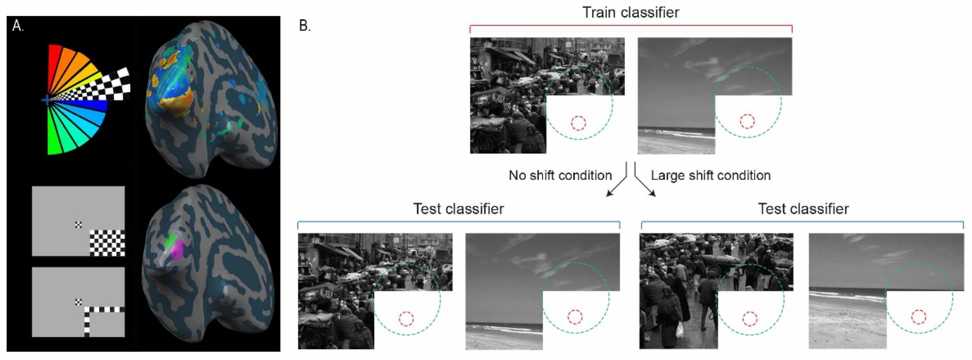
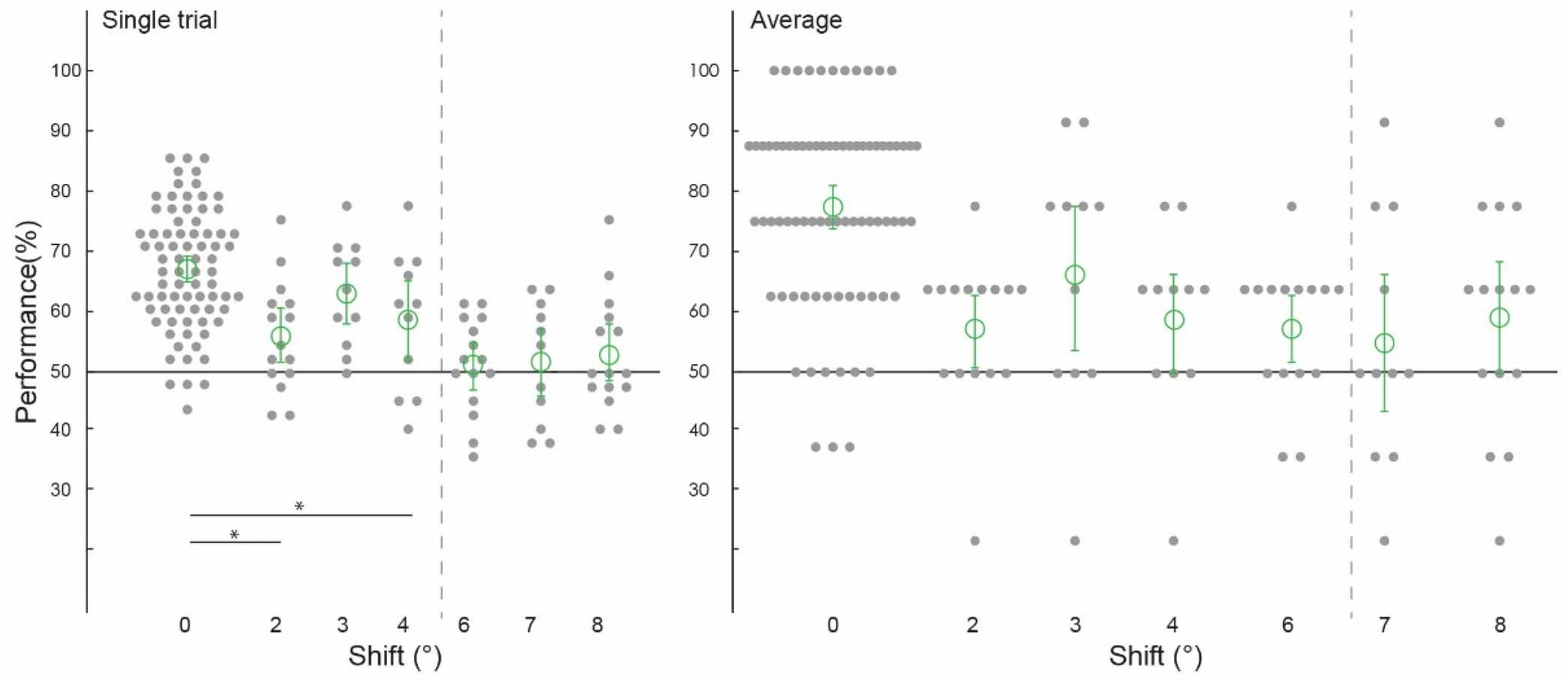
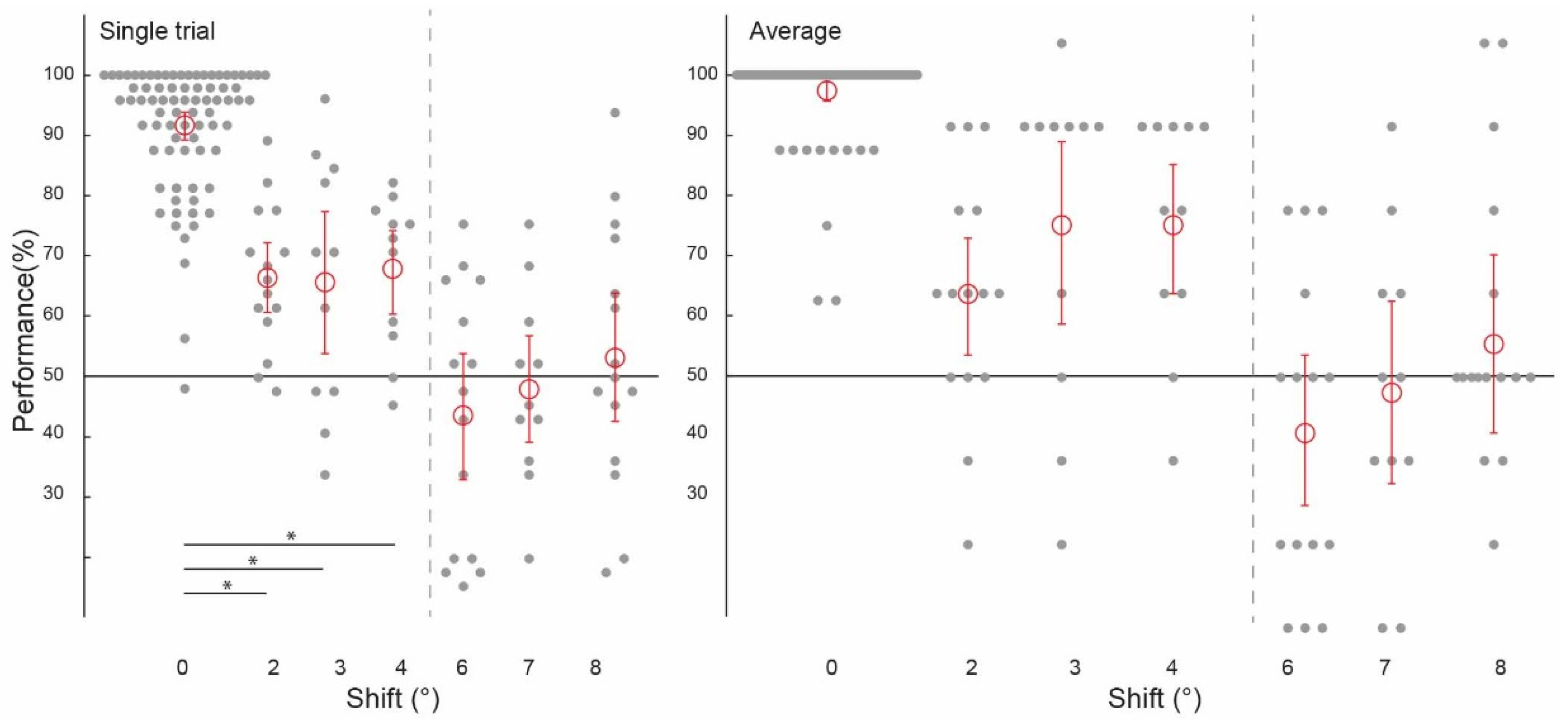
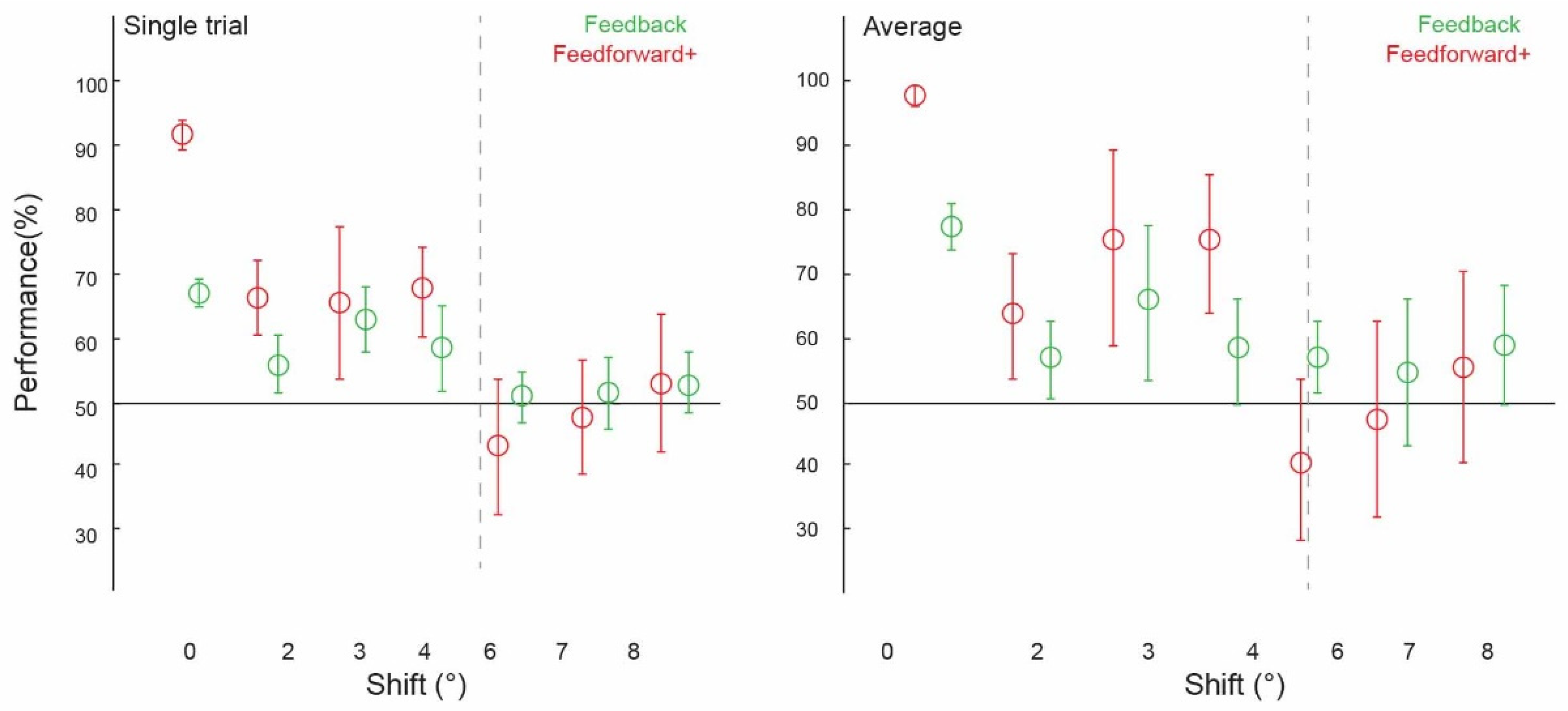
Neurons in the primary visual cortex (V1) receive sensory inputs that describe small, local regions of the visual scene and cortical feedback inputs from higher visual areas processing the global scene context. Investigating the spatial precision of this visual contextual modulation will contribute to our understanding of the functional role of cortical feedback inputs in perceptual computations. We used human functional magnetic resonance imaging (fMRI) to test the spatial precision of contextual feedback inputs to V1 during natural scene processing. We measured brain activity patterns in the stimulated regions of V1 and in regions that we blocked from direct feedforward input, receiving information only from non-feedforward (i.e., feedback and lateral) inputs. We measured the spatial precision of contextual feedback signals by generalising brain activity patterns across parametrically spatially displaced versions of identical images using an MVPA cross-classification approach. We found that fMRI activity patterns in cortical feedback signals predicted our scene-specific features in V1 with a precision of approximately 4 degrees. The stimulated regions of V1 carried more precise scene information than non-stimulated regions; however, these regions also contained information patterns that generalised up to 4 degrees. This result shows that contextual signals relating to the global scene are similarly fed back to V1 when feedforward inputs are either present or absent. Our results are in line with contextual feedback signals from extrastriate areas to V1, describing global scene information and contributing to perceptual computations such as the hierarchical representation of feature boundaries within natural scenes.