Article

Dusky-like Is Critical for Morphogenesis of the Cellular Protuberances and Formation of the Cuticle in Henosepilachna vigintioctopunctata

Education Ministry Key Laboratory of Integrated Management of Crop Diseases and Pests, State & Local Joint Engineering Research Center of Green Pesticide Invention and Application, Department of Entomology, College of Plant Protection, Nanjing Agricultural University, Nanjing 210095, China
*
Author to whom correspondence should be addressed.
Biology 2023, 12(6), 866; https://doi.org/10.3390/biology12060866
Submission received: 30 April 2023 / Revised: 12 June 2023 / Accepted: 13 June 2023 / Published: 15 June 2023
(This article belongs to the Section Developmental Biology)

Abstract

:

Simple Summary

In Drosophila melanogaster and Tribolium castaneum, dusky-like (Dyl) is related to linking the insect cuticle to the plasma membrane of epidermal cells, regulating the cytoskeleton and deposition of the cuticle. A deficiency in Dyl impairs the formation of embryonic denticles and adult sensory bristles and wing hairs. In a polyphagous beetle, Henosepilachna vigintioctopunctata, we characterized Hvdyl using RNAi. The results showed that the knockdown of Hvdyl inhibited the growth of cellular protuberances, inhibited foliage consumption, and affected the survival of the beetles. Our findings indicated that two functions of Dyl in Drosophila are conserved in H. vigintioctopunctata.

Abstract

Dusky-like (Dyl) is a transmembrane protein containing a zona pellucida domain. Its physiological roles during metamorphosis have been well explored in Drosophila melanogaster and have also been documented in Tribolium castaneum. However, Dyl has undergone a functional shift between Diptera and Coleoptera insects. Further investigation of Dyl in other insects will be helpful to further clarify its function in insect growth and development. Henosepilachna vigintioctopunctata is an important Coleoptera that causes enormous economic losses in agriculture in China. In this study, we found that the expression of Hvdyl was detectable in embryos, larvae, prepupae, pupae, and adults. We knocked down Hvdyl in third- and fourth-instar larvae and pupae with RNA interference (RNAi). RNAi of Hvdyl mainly caused two phenotypic defects. Firstly, the growth of epidermal cellular protuberances was suppressed. Injection of dsdyl (double-stranded dusky-like RNA) at the third-instar larval stage truncated the scoli throughout the thorax and abdomen and shortened the setae on the head capsules and mouthparts of the fourth-instar larvae. Introduction of dsdyl at the third- and fourth-instar stages led to misshapen pupal setae. The setae were shortened or became black nodules. Treatment with dsdyl at the larval and pupal stages resulted in deformed adults with completely suppressed wing hairs. Moreover, the knockdown of Hvdyl at the third-instar stage caused deformed larval mouthparts at the fourth-instar period. As a result, foliage consumption was inhibited, and larval growth was slowed. The results indicate that Dyl is associated with the growth of cellular protuberances throughout development and with the formation of the cuticle in H. vigintioctopunctata.

1. Introduction

Dusky-like (Dyl) is a member of the zona pellucida (ZP) domain family, a group of proteins related to linking the insect cuticle to the plasma membrane of epidermal cells, regulating the cytoskeleton and deposition of cuticle [1,2,3,4,5,6]. In Drosophila melanogaster, Dyl has been documented to be a transmembrane and secreted protein [3,7]. It is involved in the organization of the apical extracellular domain in the epidermis and the deposition of chitin. As a result, a deficiency in Dyl impairs the formation of embryonic denticles, adult sensory bristles, and wing hairs [3,5,8].
Dyl also plays a crucial role in the development of the Coleoptera Tribolium castaneum [3,6,9]. Dyl in T. castaneum reaches peak expression in the late embryonic stage, and RNAi of Tcdyl in the parents leads to significant embryonic lethality. Depletion of Dyl results in defects in larval epidermal pigmentation and completely blocks the transitions from larval-to-pupal and pupal-to-adult stages [9].
It appears that Dyl has undergone a functional shift between Diptera and Coleoptera insects. Further investigation of Dyl in other insects will be helpful to further clarify its function and help us understand its role in insect growth and development. Henosepilachna vigintioctopunctata (Coleoptera: Coccinellidae), a destructive pest that attacks potato, tomato, eggplant, and Chinese boxthorn, possesses several kinds of cellular protuberances. The larvae have two types of extensions. Firstly, an H. vigintioctopunctata larva has 11 rows of dark-branched spines (scoli) located on brown epidermal mastoids (strumae) [10], similar to its sibling species [11]. Secondly, sensory setae are located on the larval mouthparts and legs, the pupal thorax, and the adult mouthparts and elytra. Furthermore, H. vigintioctopunctata is sensitive to RNA interference (RNAi) [10,12,13,14,15]. This offers an ideal insect model to investigate the roles of Dyl throughout the entire lifecycle.
In this study, we characterized Hvdyl in H. vigintioctopunctata using RNAi. We observed two characteristic RNAi phenotypes seen in D. melanogaster and T. castaneum. Firstly, the knockdown of Hvdyl inhibited the growth of cellular protuberances, a defect observed in the D. melanogaster [3,5,8]. Secondly, the knockdown of Hvdyl inhibited foliage consumption and affected the survival of the beetles, a major phenotype described in Dyl RNAi experiments in T. castaneum [9]. We argued that the link between the cuticle and the plasma membrane of epidermal cells is disturbed in the Dyl-deficient H. vigintioctopunctata larvae. This represses the development of cellular protuberances and blocks the formation of the cuticle. As a result, food ingestion is reduced, and survival is affected.

2. Methods and Materials

2.1. Insect

H. vigintioctopunctata was routinely maintained in an insectary at 25 ± 1 °C under a 16 h:8 h light–dark photoperiod and 50–60% relative humidity with potato foliage at vegetative growth or tuber initiation stages provided as food.

2.2. Molecular Cloning

The Hvdyl sequence was obtained from H. vigintioctopunctata transcriptome data. The total RNA was extracted using TRIzol reagent (Invitrogen, New York, NY, USA) in accordance with the manufacturer’s protocols. The RNA was quantified with a NanoDrop 2000 spectrophotometer (Thermo Fisher Scientific, New York, NY, USA). RNA purity was determined by assessing optical density (OD) absorbance ratios at OD260/280 and OD260/230. Polymerase chain reaction (PCR) was used to verify the sequences. The primers are listed in Table S1. The cDNA sequence was submitted to GenBank (accession number: Hvdyl, OP056326).

2.3. Preparation of dsRNAs

We cloned the cDNA fragments of Hvdyl and an enhanced green fluorescent protein (egfp). Hvdyl and egfp were amplified with PCR using specific primers (Table S1) conjugated with the T7 RNA polymerase promoter. Two independent dsHvdyl primers were designed (dsHvdyl-F1R1, dsHvdyl-F2R2) (Table S1, Figure S1). Due to the consistent RNAi phenotypes observed with both sets of primers, the results based on primary primers (dsHvdyl-F1R1) are presented in this article (Figure S2). In order to verify whether there are any possible off-target sequences that possess an identical match of 20 bp or more, we used BLAST to search these two targeted regions against the H. vigintioctopunctata transcriptome. The dsRNAs (dsdyl, dsegfp) were synthesized using the MEGAscript T7 High Yield Transcription Kit (Ambion, Austin, TX, USA) in accordance with a previously described protocol [16]. The quality of the dsRNA was evaluated via agarose gel electrophoresis, and the concentration was quantified with a Nanodrop 1000 spectrophotometer.

2.4. Injection of dsRNA

A liquid drop (0.1 μL), including 300 ng dsdyl or dsegfp, was injected into newly ecdysed third- or fourth-instar larvae or 1-day-old pupae [17,18]. Each replicate included 10 beetles, and each treatment was repeated 6 times. Three of these repeats were used to collect samples (2 days after the injection) and measure RNAi efficacy, while the other three groups were used to observe phenotypes over the next three weeks.

2.5. Observation of Feeding Ability

Potato leaves of similar sizes were collected and cut into a circle with a radius of 5 mm and placed in Petri dishes. A beetle that had been injected with dsegfp or dsdyl was transferred to the dish. Three larvae were set as a replicate, and each treatment was repeated 6 times. The gnawed leaves were measured after 24 h. The larval mouthparts were observed, and the larvae were dissected to image their guts.

2.6. Quantitative Real-Time PCR (qRT-PCR)

Beacon Designer 7 (Premier Biosoft International, Palo Alto, CA, USA) was used to design the qRT-PCR primers given in Table S1. The RNA was extracted from the collected samples and reverse-transcribed into cDNA. HvRPS18 and HvRPL13 were selected as control genes according to previous reports (the primers are listed in Table S1) [19]. The qRT-PCR protocol consisted of 1 cycle of 95 °C for 30 s, followed by 40 cycles of 95 °C for 5 s, and then 60 °C for 34 s. In order to verify the PCR products, the heat dissociation curves and amplification plots were analyzed at the end of the thermal cycle. The data were analyzed via the 2−ΔΔCT method. Each experiment was repeated 3 times.

3. Results

3.1. Dyl Is Conserved in Insects

We cloned the full-length Dyl cDNA and analyzed its sequence alignment and evolutionary relationships. Sequence alignment of the proteins encoded with Dyl from H. vigintioctopunctata, D. melanogaster, and T. castaneum demonstrated that Dyl codes for a transmembrane protein containing a ZP domain (Figure 1A).
Phylogenetic analysis revealed that Dyl had an orthologue in each insect and showed a one-to-one orthologous relationship among insects. Dyl proteins from Coleoptera, Lepidopteran, Hymenoptera, and Diptera species formed four clusters. As expected, Hvdyl belongs to the Coleoptera group (Figure 1B).

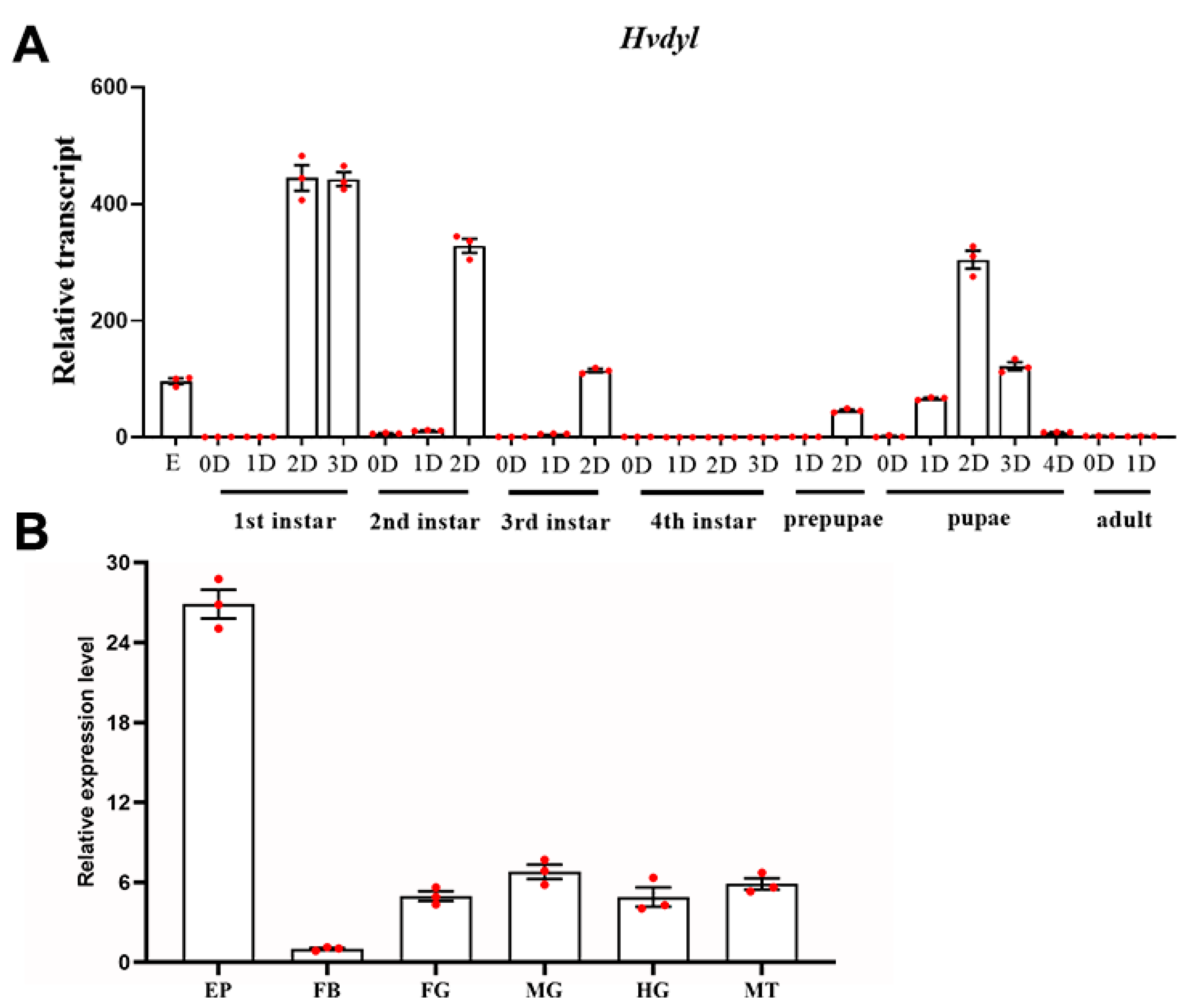
3.2. Expression Profiles of Hvdyl

The mRNA levels of Hvdyl during different developmental stages were measured using qRT-PCR. Hvdyl transcript levels could be detected in embryos, larvae, prepupae, pupae, and adults. It was highly expressed during the embryonic period, at late stages of the first-, second-, and third-instar stages, and at the pupal period. Conversely, Hvdyl had low transcription levels in fourth-instar larvae and newly eclosed adults (Figure 2A).
The tissue-based expression of Hvdyl was also investigated. Hvdyl was broadly expressed in the epidermis, foregut, midgut, hindgut, Malpighian tubules, and body fat of 1-day-old third-instar larvae. The highest expression level was found in the epidermis, followed by the midgut, Malpighian tubules, foregut, and hindgut; the lowest level was detected in the body fat (Figure 2B).

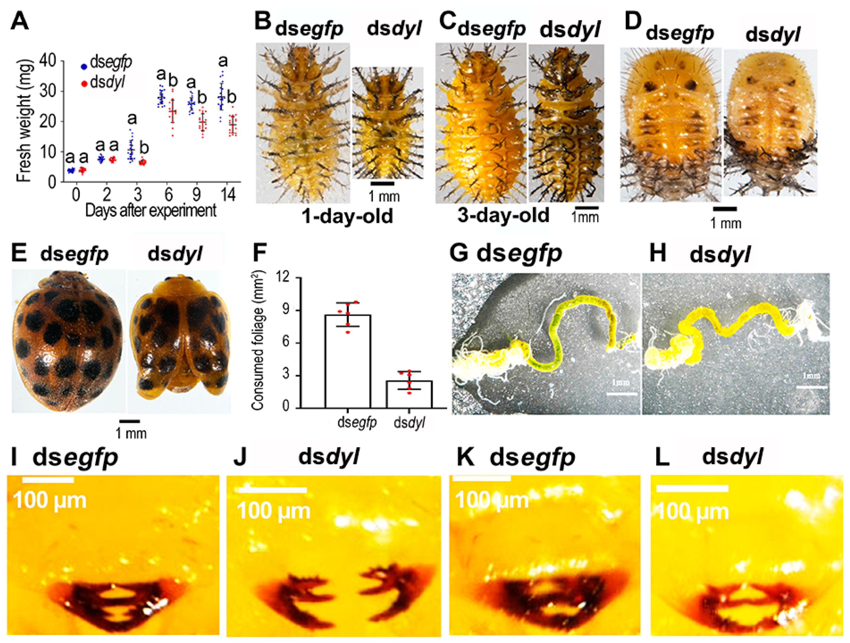
3.3. Hvdyl RNAi at the Third-Instar Stage Inhibited the Growth of Scoli and Setae

Hvdyl was knocked down with RNAi in the third-(penultimate)-instar larvae (Figure 3A). The Hvdyl RNAi larvae could molt to the fourth-instar larvae (Figure 3B). However, two obvious phenotypic defects were observed (Figure 3C–H). Firstly, 11 rows of branched spines (scoli) are located on the epidermal mastoids (struma) of an H. vigintioctopunctata larva. In dsegfp-injected beetles, the scoli were strong and highly branched, and a spine was present on the tips (Figure 3C,E). In contrast, the larval scoli were undeveloped, and the tip spines were missing in the Hvdyl RNAi larvae (Figure 3D,F). Moreover, the scoli were weak and soft, and some were bending (Figure 3D). Secondly, the setae on the larval head capsule and mouthparts did not develop, with only small dots remaining in the Hvdyl RNAi larvae (Figure 3H,G).
Approximately 80% of the fourth-instar Hvdyl RNAi larvae were pupated successfully (Figure 3I). Knockdown of Hvdyl negatively affected the growth of pupal setae. The pupal setae formed black tubercles. Moreover, the bald scoli were attached to the abdomen in the Hvdyl RNAi pupae (Figure 3K,J).
Most of the Hvdyl knockdown pupae emerged to form deformed adults (Figure 3L). About 50% of the Hvdyl-depleted adults had deformed wings, and around 20% were wrapped in the old pupal cuticles (Figure 3L,N,O). These misshapen adults hardly moved and fed and eventually died within 7 days after molting (Figure 3N,O).

3.4. Reduced Growth and Foliage Consumption in the Hvdyl RNAi Beetles

The Hvdyl-depleted beetles were weighed 0, 2, 3, 6, 9, and 14 days after injection of dsdyl at the third-instar larval stage. The fresh weights of the Hvdyl RNAi beetles were statistically significantly lower than those of the dsegfp-injected controls 3, 6, 9, and 14 days after dsdyl injection (Figure 4A). Consistently, the 1- and 3-day-old Hvdyl-silenced fourth-instar larvae, pupae, and adults were smaller than their dsegfp-injected counterparts (Figure 4B–E).
The foliage consumption of the fourth-instar Hvdyl RNAi larvae was examined. One day after ecdysis, the larvae in the dsegfp-treated group had eaten all the leaves, while the larvae in the dsdyl-injected group only ate about 30% of the leaves (Figure 4F). The guts of almost all dsegfp-treated larvae were full of food (Figure 4G). In contrast, there was almost no food in the guts of the dsdyl-treated larvae (Figure 4H).
At 30 min (Figure 4I,J) and 2 h (Figure 4K,L), after the dsdyl-injected larvae molted to fourth instars, the larval mouthparts were examined. In dsegfp-treated larvae, the mandibles on both sides met and were well pigmented (Figure 4I,K). Conversely, the mandibles of the dsdyl-injected larvae were weakly tanned and separated from each other (Figure 4J,L).

3.5. Knockdown of Hvdyl at the Fourth-Instar Stage

Two days post-injection of dsdyl into newly molted fourth-instar larvae, the target gene was markedly down-regulated (Figure 5A). Similarly to the control larvae, almost all dsdyl-treated larvae were pupated successfully (Figure 5B). However, the growth of the setae on the thoraces of pupae was completely inhibited, and black nodules were formed (Figure 5D). About 10% of the Hvdyl RNAi pupae emerged to form normal adults. The remaining 90%, however, exhibited phenotypic defects (Figure 5C,E). The elytra and hindwings of these dsdyl-treated adults were much smaller in size than those of control adults. The elytra were improperly folded and wrapped with pupal cuticles; the membranous wings were completely shrunken, and the tips of the membranous wings became withered and darkened (Figure 5F). In addition, the hairs on the elytra were suppressed in the dsdyl-treated adults (Figure 5F).

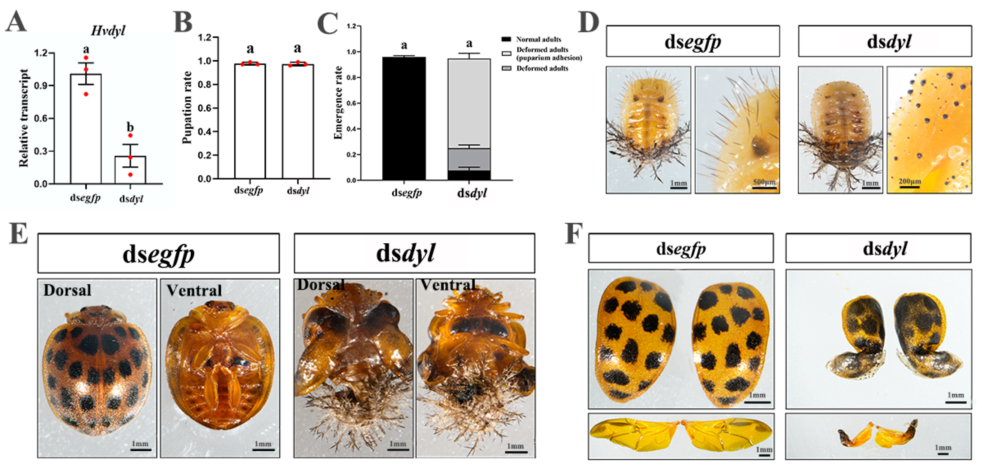
3.6. Depletion of Hvdyl at the Pupal Stage

Similarly, injection of dsdyl into newly molted pupae successfully knocked down Hvdyl (Figure 6A). All dsdyl-treated pupae formed defective adults (Figure 6B). Compared with the control adults, the dsdyl-treated adults were completely wrapped with pupal exuviae (Figure 6C,D). The elytra in the pupal exuviae were improperly folded. After the removal of the pupal cuticle, it was obvious that the development of hairs on the elytra was also inhibited. The membranous wings were shrunken, and the tips became withered and darkened (Figure 6E,F).

4. Discussion

In this study, we discovered that two characteristic Dyl RNAi phenotypes seen in D. melanogaster [3,5,8] and T. castaneum [9] were also observed in Henosepilachna vigintioctopunctata. We discuss these phenotypes below.

4.1. Dyl Is Critical for the Development of Epidermic Cellular Protuberances

In this study, we provided four pieces of experimental evidence to support the requirement for Dyl in the growth of cellular protuberances.
Firstly, the tissue-specific analysis revealed that the highest expression level of Hvdyl was in the epidermis, where the scoli and setae developed in H. vigintioctopunctata (Figure 2B). Consistently, Dyl in D. melanogaster is detected in the trichome cells of the embryonic epidermis [3,6]. During post-embryonic development, Dyl protein accumulates in wing hairs and the body hairs [5]. Tissue expression patterns suggest that Dyl functions in the cuticle, especially in the cellular protuberances.
Secondly, RNAi of Hvdyl in third-instar larvae caused undeveloped larval scoli throughout the whole body and truncated setae on the larval head capsule and mouthparts in H. vigintioctopunctata (Figure 3). Similarly, different ZP proteins localize to different parts of the first-instar larval denticles and play a specific role in the morphogenesis of denticles [3]. In D. melanogaster embryos, a mutation in Dyl causes very small, unhooked, severely split denticles [3]. Consistently, the knockdown of Dyl leads to very small denticles in embryos [5]. These findings provide conclusive experimental evidence to confirm the role of Dyl in the control of the development of insect cellular protuberances.
Thirdly, the knockdown of Hvdyl led to abnormal setae on the pupal thoraces in H. vigintioctopunctata (Figure 3K and Figure 5D). In T. castaneum, RNAi of Tcdyl at the larval stage completely blocks the larval–pupal transition, and at the pupal period, it inhibits the pupal–adult transformation [9]. Therefore, we, for the first time, have observed a pupal Dyl RNAi phenotype: living pupae with impaired thorax setae.
Fourthly, the depletion of Hvdyl brought about misshapen bristles on the adult wings of H. vigintioctopunctata (Figure 5E,F). In line with our results, Dyl function is necessary to form adult sensory bristles and wing hairs in D. melanogaster [5,8]. Knocking down Dyl in adults results in stubby bristles associated with bristle collapse [5]. Moreover, the depletion of Dyl in adults causes split, thinned, and often very short wing hairs [8].
All these pieces of experimental evidence confirm that Dyl is required for the growth of cellular protuberances in Coleoptera insects, similar to its role in D. melanogaster [5,8].
Actin filaments have been proven to be critical for the shape and elongation of the cellular protuberances [5,20,21,22,23,24,25]. Once the elongation of cellular protuberances has finished, the shape is believed to be maintained by the chitinous exoskeleton [26]. Therefore, a shortage of actin or chitin can cause misshapen epidermal cellular protuberances. Does the impairment of cellular protuberances in Hvdyl RNAi H. vigintioctopunctata beetles result from actin shortage, chitin deficiency, or both?

4.2. Dyl May Regulate Chitin Deposition

Here, we first discuss a lack of chitin in the Hvdyl RNAi beetles in H. vigintioctopunctata. It is known that Drosophila Dyl and other ZP domain proteins are associated with chitin deposition [3,5]. In our study, we provided five pieces of experimental evidence to demonstrate that Dyl is associated with the formation of the cuticle, possibly controlling chitin deposition.
Firstly, we noted that Hvdyl was highly expressed during the embryonic period, at late stages of first-, second-, and third-instar larvae and in pupae (Figure 2A). Our data suggest that Dyl is necessary for the formation of the cuticle after each ecdysis. This is consistent with observations in D. melanogaster, where the expression of Dyl increases 118-fold from the early to late stages in the process of hair morphogenesis [27]. Interestingly, the mRNA levels of UDP-N-acetylglucosamine pyrophosphorylase (UAP) and chitin synthase 1 (CHS1) involved in chitin biosynthesis are periodically raised during ecdysis and reduced in the active feeding stage in H. vigintioctopunctata [28,29] and L. decemlineata [30,31]. It appears that Dyl may be involved in the regulation of chitin production after each ecdysis, similar to D. melanogaster [8].
Secondly, the knockdown of Hvdyl caused deformed and weakly tanned larval mouthparts in H. vigintioctopunctata (Figure 4J,L). Similar phenotypic defects have been documented when chitin synthesis has been repressed in H. vigintioctopunctata, where the clypeus, labrum, mandibles, maxillae, and labium in the mouthparts were weakly tanned, and the mandibles could not meet each other, in contrast to controls [28]. In H. vigintioctopunctata [28] and L. decemlineata [31], the shortage of chitin also slightly changes the adult pigmentation in other organs, especially the coloration on the elytra. In D. melanogaster, loss of Dyl brings about abnormal pigmentation of the bristles [5,8]. Moreover, the epidermis in the Tcdyl RNAi animals in T. castaneum is poorly pigmented [9]. According to the results in other Coleoptera [28,29,31] and Diptera [5,8] species, the poorly pigmented epidermis in the Tcdyl RNAi animals in T. castaneum [9] may also be due to a lack of chitin.
Thirdly, the knockdown of Hvdyl reduced foliage consumption in H. vigintioctopunctata (Figure 4F). As a result, growth was inhibited (Figure 4B–E). In accordance with our results, disruption of the chitin biosynthesis pathway has been shown to negatively affect feeding in H. vigintioctopunctata [28,29]. Normal settling and feeding on host plant leaves depend on normal larval mouthparts [32]. Dysfunction of larval mouthparts in the Hvdyl (this study), HvCHS1, or HvUAP [28,29] RNAi larvae undoubtedly affects food ingestion.
Fourthly, the scoli were weak and soft, and some were bending in the Hvdyl RNAi larvae (Figure 3D). Similar phenotypic defects of scoli are observed when chitin biosynthesis is repressed by RNAi of UAP in H. vigintioctopunctata [29].
Lastly, injection of dsdyl at the larval and pupal stages resulted in deformed adults (Figure 3N,O, Figure 4E, Figure 5E and Figure 6D). In accordance with our results, the depletion of the HvCHS1 [28] and HvUAP [29] bring about misshapen beetles in H. vigintioctopunctata. In T. castaneum, all Tcdyl RNAi larvae fail to pupate. Moreover, injection of dsTcdyl into early-stage pupae results in developmental arrest prior to eclosion in the T. castaneum [9]. The phenotypic differences between T. castaneum and H. vigintioctopunctata may be explained by different RNAi efficacies. In fact, knockdown of HvUAP can arrest the larval development at the third- or fourth-instar larval or prepupal stages [29].
It appears that in both Coleoptera [9] and Diptera [8] species, Dyl is involved in chitin accumulation in the cuticle. Depletion of Dyl inhibits the deposition of chitin and, subsequently, the coloration of the cuticle [8,9]. In D. melanogaster, Dyl functions as a Rab11 effector for the chitin deposition [8]. Whether the same pathway is present in H. vigintioctopunctata needs further investigation to clarify.

4.3. Chitin Is Not the Only Target of Dyl

A lack of chitin in H. vigintioctopunctata only caused soft scoli but did not affect the morphogenesis of the setae [29]. Obviously, a deficiency in chitin does not mimic the Dyl-defective phenotypes of the epidermal cellular protuberances. Similarly, Drosophila Dyl mutant phenotypes in wing hairs are distinctly different from and are more severe than the phenotypes caused by a deficiency in the chitin [5,8]. Therefore, a defect in chitin deposition cannot explain all of the Hvdyl RNAi defective phenotypes. Dyl must have other targets in the morphogenesis of the cellular protuberances in H. vigintioctopunctata.
In terms of this context, the chitin-binding protein Knickkopf (Knk) is believed to play an important role in protecting the new cuticle from degradation by chitinases [33,34]. Additionally, a kind of epidermal endochitinase from T. castaneum, TcCht7, is also considered to play a role in organizing chitin in the newly forming cuticle rather than in degrading the chitin present in the old cuticle [35]. Moreover, Chitinase 6 (Cht6) is a strong candidate for a Dyl target in D. melanogaster because depletion of Cht6 brings about all of the Dyl hair phenotypes, albeit to a weaker extent [8]. Whether a similar signal pathway is involved in H. vigintioctopunctata remains to be clarified.
Given that a deficiency of chitin cannot fully explain the Dyl RNAi phenotype, the morphological abnormalities of epidermal cellular protuberances in the Hvdyl knockdown may be due to the effects on the cytoskeleton. In this context, Drosophila Dyl and other ZP domain proteins have been suggested to be able to regulate the cytoskeleton [1,2,6,8]. Specifically, the knockdown of Dyl in wing cells causes premature hair initiation [8]. Accordingly, Dyl was found to negatively control actin deposition before hair initiation in hair-forming cells [8]. However, the regulative network of actin is very complicated in Drosophila. For instance, mutations in the key general component genes cofilin and AIP1 [36,37], in F-actin bundling protein genes forked and singed [20,23,38], in non-muscle myosin encoding genes crinkled [39] and zipper [40], in regulator gene Rho kinase [41], and in F-actin-depolymerizing protein-encoding genes twin star [36] and flare [37] result in hair phenotypes. Whether the actin-related cytoskeleton components are affected in the Hvdyl RNAi beetles is another interesting question that deserves investigation in the future.

5. Conclusions

In summary, deficiency in Dyl disturbs the link between the cuticle and the plasma membrane of epidermal cells in H. vigintioctopunctata larvae. This represses the development of cellular protuberances and blocks the formation of the cuticle. As a result, food ingestion is reduced, and survival is affected.

Supplementary Materials

The following supporting information can be downloaded at: https://www.mdpi.com/article/10.3390/biology12060866/s1, Figure S1: The positions of the primary and secondary dsRNA on the mRNA; Figure S2: Knockdown of Hvdyl with secondary dsRNA at the third- and fourth-instar larval stage impairs growth of the cellular protuberances and the formation of cuticle in Henosepilachna vigintioctopunctata; Table S1: A list of primers used for RT-PCR, dsRNA synthesis and qRT-PCR.

Author Contributions

Conceptualization, Y.Z. and Q.T.; methodology, C.S.; software, Q.T. and C.S.; formal analysis, Y.Z.; investigation, Q.T. and M.L.; resources, L.J.; data curation, Q.T.; writing—original draft preparation, Y.Z.; writing—review and editing, G.L.; supervision, L.J.; project administration, G.L.; funding acquisition, G.L. All authors have read and agreed to the published version of the manuscript.

Funding

This research was funded by the National Natural Science Foundation of China (32072416), and China Agriculture Research System of MOF and MARA (CARS-09-P22).

Institutional Review Board Statement

Not applicable.

Informed Consent Statement

Not applicable.

Data Availability Statement

Data generated in association with this study are available in the Supplementary Materials published online with this article.

Conflicts of Interest

All the authors confirm they have no financial or other involvement in activities or organizations that might bias the work reported here.

References

  1. Bokel, C.; Prokop, A.; Brown, N.H. Papillote and Piopio: Drosophila ZP-domain proteins required for cell adhesion to the apical extracellular matrix and microtubule organization. J. Cell Sci. 2005, 118, 633–642. [Google Scholar] [CrossRef] [Green Version]
  2. Brodu, V.; Baffet, A.D.; Le Droguen, P.M.; Casanova, J.; Guichet, A. A Developmentally Regulated Two-Step Process Generates a Noncentrosomal Microtubule Network in Drosophila Tracheal Cells. Dev. Cell 2010, 18, 790–801. [Google Scholar] [CrossRef] [PubMed]
  3. Fernandes, I.; Chanut-Delalande, H.; Ferrer, P.; Latapie, Y.; Waltzer, L.; Affolter, M.; Payre, F.; Plaza, S. Zona Pellucida Domain Proteins Remodel the Apical Compartment for Localized Cell Shape Changes. Dev. Cell 2010, 18, 64–76. [Google Scholar] [CrossRef] [Green Version]
  4. Jazwinska, A.; Affolter, M. A family of genes encoding zona pellucida (ZP) domain proteins is expressed in various epithelial tissues during Drosophila embryogenesis. Gene Expr. Patterns 2004, 4, 413–421. [Google Scholar] [CrossRef] [PubMed]
  5. Nagaraj, R.; Adler, P.N. Dusky-like functions as a Rab11 effector for the deposition of cuticle during Drosophila bristle development. Development 2012, 139, 906–916. [Google Scholar] [CrossRef] [PubMed] [Green Version]
  6. Roch, F.; Alonso, C.R.; Akam, M. Drosophila miniature and dusky encode ZP proteins required for cytoskeletal reorganisation during wing morphogenesis. J. Cell Sci. 2003, 116, 1199–1207. [Google Scholar] [CrossRef] [Green Version]
  7. Plaza, S.; Chanut-Delalande, H.; Fernandes, I.; Wassarman, P.M.; Payre, F. From A to Z: Apical structures and zona pellucida-domain proteins. Trends Cell Biol. 2010, 20, 524–532. [Google Scholar] [CrossRef]
  8. Adler, P.N.; Sobala, L.F.; Thom, D.; Nagaraj, R. dusky-like is required to maintain the integrity and planar cell polarity of hairs during the development of the Drosophila wing. Dev. Biol. 2013, 379, 76–91. [Google Scholar] [CrossRef] [Green Version]
  9. Li, C.J.; Yun, X.P.; Li, B. Dusky-like is required for epidermal pigmentation and metamorphosis in Tribolium castaneum. Sci. Rep. 2016, 6, 20102. [Google Scholar] [CrossRef] [Green Version]
  10. Ze, L.J.; Wang, P.; Peng, Y.C.; Jin, L.; Li, G.Q. Silencing tyrosine hydroxylase or dopa decarboxylase gene disrupts cuticle tanning during larva-pupa-adult transformation in Henosepilachna vigintioctopunctata. Pest. Manag. Sci. 2022, 78, 3880–3893. [Google Scholar] [CrossRef]
  11. Casari, S.A.; Teixeira, E.P. Immatures of Epilachna Chevrolat (Coleoptera, Coccinellidae, Epilachninae). Rev. Bras. Entomol. 2015, 59, 113–120. [Google Scholar] [CrossRef] [Green Version]
  12. Wang, P.; Ze, L.J.; Jin, L.; Li, G.Q. Yellow-b, -c, -d, and -h are required for normal body coloration of Henosepilachna vigintioctopunctata. Arch. Insect Biochem. 2022, 109, e21856. [Google Scholar] [CrossRef] [PubMed]
  13. Wu, J.J.; Cheng, M.D.; Ze, L.J.; Shen, C.H.; Jin, L.; Li, G.Q. Dissecting the Isoform-Specific Roles of FTZ-F1 in the Larval-Larval and Larval-Pupal Ecdyses in Henosepilachna vigintioctopunctata. Insects 2022, 13, 228. [Google Scholar] [CrossRef]
  14. Ze, L.J.; Jin, L.; Li, G.Q. Silencing of Adc and Ebony Causes Abnormal Darkening of Cuticle in Henosepilachna vigintioctopunctata. Front. Physiol. 2022, 13, 8296755. [Google Scholar] [CrossRef] [PubMed]
  15. Zeng, J.; Kang, W.N.; Jin, L.; Anjum, A.A.; Li, G.Q. Vacuolar ATPase subunit F is critical for larval survival in Henosepilachna vigintioctopunctata. Insect Mol. Biol. 2022, 31, 177–189. [Google Scholar] [CrossRef]
  16. Wu, J.J.; Mu, L.L.; Kang, W.N.; Ze, L.J.; Shen, C.H.; Jin, L.; Anjum, A.A.; Li, G.Q. RNA interference targeting ecdysone receptor blocks the larval-pupal transition in Henosepilachna vigintioctopunctata. Insect Sci. 2021, 28, 419–429. [Google Scholar] [CrossRef]
  17. Xu, P.; Ze, L.J.; Kang, W.N.; Wu, J.J.; Jin, L.; Anjum, A.A.; Li, G.Q. Functional divergence of white genes in Henosepilachna vigintioctopunctata revealed by RNA interference. Insect Mol. Biol. 2020, 29, 466–476. [Google Scholar] [CrossRef]
  18. Ze, L.-J.; Xu, P.; Kang, W.-N.; Wu, J.-J.; Jin, L.; Anjum, A.A.; Li, G.-Q. Disruption of ommochrome biosynthesis affects eye coloration, phototaxis and climbing in Henosepilachna vigintioctopunctata. Amino Acids 2021, 53, 1091–1104. [Google Scholar] [CrossRef]
  19. Lu, J.; Chen, S.M.; Guo, M.J.; Ye, C.Y.; Qiu, B.L.; Wu, J.H.; Yang, C.X.; Pan, H.P. Selection and Validation of Reference Genes for RT-qPCR Analysis of the Ladybird Beetle Henosepilachna vigintioctomaculata. Front. Physiol. 2018, 9, 1614. [Google Scholar] [CrossRef] [Green Version]
  20. Cant, K.; Knowles, B.A.; Mooseker, M.S.; Cooley, L. Drosophila Singed, a Fascin Homolog, Is Required for Actin Bundle Formation during Oogenesis and Bristle Extension. J. Cell Biol. 1994, 125, 369–380. [Google Scholar] [CrossRef] [Green Version]
  21. Fei, X.Y.; He, B.; Adler, P.N. The growth of Drosophila bristles and laterals is not restricted to the tip or base. J. Cell Sci. 2002, 115, 3797–3806. [Google Scholar] [CrossRef] [PubMed] [Green Version]
  22. He, B.; Adler, P.N. Cellular mechanisms in the development of the Drosophila arista. Mech. Dev. 2001, 104, 69–78. [Google Scholar] [CrossRef] [PubMed]
  23. Petersen, N.S.; Lankenau, D.H.; Mitchell, H.K.; Young, P.; Corces, V.G. Forked Proteins Are Components of Fiber-Bundles Present in Developing Bristles of Drosophila melanogaster. Genetics 1994, 136, 173–182. [Google Scholar] [CrossRef] [PubMed]
  24. Tilney, L.G.; Connelly, P.S.; Vranich, K.A.; Shaw, M.K.; Guild, G.M. Actin filaments and microtubules play different roles during bristle elongation in Drosophila. J. Cell Sci. 2000, 113, 1255–1265. [Google Scholar] [CrossRef]
  25. Turner, C.M.; Adler, P.N. Distinct roles for the actin and microtubule cytoskeletons in the morphogenesis of epidermal hairs during wing development in Drosophila. Mech. Dev. 1998, 70, 181–192. [Google Scholar] [CrossRef]
  26. Tilney, L.G.; Connelly, P.; Smith, S.; Guild, G.M. F-actin bundles in Drosophila bristles are assembled from modules composed of short filaments. J. Cell Biol. 1996, 135, 1291–1308. [Google Scholar] [CrossRef] [Green Version]
  27. Ren, N.; Zhu, C.M.; Lee, H.; Adler, P.N. Gene expression during Drosophila wing morphogenesis and differentiation. Genetics 2005, 171, 625–638. [Google Scholar] [CrossRef] [Green Version]
  28. Jiang, L.H.; Mu, L.L.; Jin, L.; Anjum, A.A.; Li, G.Q. RNAi for chitin synthase 1 rather than 2 causes growth delay and molting defect in Henosepilachna vigintioctopunctata. Pestic. Biochem. Phys. 2021, 178, 104934. [Google Scholar] [CrossRef]
  29. Jiang, L.H.; Mu, L.L.; Jin, L.; Anjum, A.A.; Li, G.Q. Silencing uridine diphosphate N-acetylglucosamine pyrophosphorylase gene impairs larval development in Henosepilachna vigintioctopunctata. Pest. Manag. Sci. 2022, 78, 3894–3902. [Google Scholar] [CrossRef]
  30. Shi, J.F.; Mu, L.L.; Chen, X.; Guo, W.C.; Li, G.Q. RNA interference of chitin synthase genes inhibits chitin biosynthesis and affects larval performance in Leptinotarsa decemlineata (Say). Int. J. Biol. Sci. 2016, 12, 1319–1331. [Google Scholar] [CrossRef] [Green Version]
  31. Shi, J.F.; Fu, J.; Mu, L.L.; Guo, W.C.; Li, G.Q. Two Leptinotarsa uridine diphosphate N-acetylglucosamine pyrophosphorylases are specialized for chitin synthesis in larval epidermal cuticle and midgut peritrophic matrix. Insect Biochem. Mol. 2016, 68, 1–12. [Google Scholar] [CrossRef] [PubMed]
  32. Hillyer, J.F.; Pass, G. The Insect Circulatory System: Structure, Function, and Evolution. Annu. Rev. Entomol. 2020, 65, 121–143. [Google Scholar] [CrossRef] [PubMed] [Green Version]
  33. Chaudhari, S.S.; Arakane, Y.; Specht, C.A.; Moussian, B.; Boyle, D.L.; Park, Y.; Kramer, K.J.; Beeman, R.W.; Muthukrishnan, S. Knickkopf protein protects and organizes chitin in the newly synthesized insect exoskeleton. Proc. Natl. Acad. Sci. USA 2011, 108, 17028–17033. [Google Scholar] [CrossRef] [Green Version]
  34. Chaudhari, S.S.; Arakane, Y.; Specht, C.A.; Moussian, B.; Kramer, K.J.; Muthukrishnan, S.; Beeman, R.W. Retroactive Maintains Cuticle Integrity by Promoting the Trafficking of Knickkopf into the Procuticle of Tribolium castaneum. PLoS Genet. 2013, 9, e1003268. [Google Scholar] [CrossRef] [Green Version]
  35. Noh, M.Y.; Muthukrishnan, S.; Kramer, K.J.; Arakane, Y. A chitinase with two catalytic domains is required for organization of the cuticular extracellular matrix of a beetle. PLoS Genet. 2018, 14, e1007307. [Google Scholar] [CrossRef] [Green Version]
  36. Gunsalus, K.C.; Bonaccorsi, S.; Williams, E.; Verni, F.; Gatti, M.; Goldberg, M.L. Mutations in Twinstar, a Drosophila Gene Encoding a Cofilin Adf Homolog, Result in Defects in Centrosome Migration and Cytokinesis. J. Cell Biol. 1995, 131, 1243–1259. [Google Scholar] [CrossRef] [PubMed]
  37. Ren, N.; Charlton, J.; Adler, P.N. The flare gene, which encodes the AIP1 protein of Drosophila, functions to regulate F-actin disassembly in pupal epidermal cells. Genetics 2007, 176, 2223–2234. [Google Scholar] [CrossRef] [Green Version]
  38. Tilney, L.G.; Tilney, M.S.; Guild, G.M. F actin bundles in Drosophila bristles. I. Two filament cross-links are involved in bundling. J. Cell Biol. 1995, 130, 629–638. [Google Scholar] [CrossRef] [PubMed] [Green Version]
  39. Kiehart, D.P.; Franke, J.D.; Chee, M.K.; Montague, R.A.; Chen, T.L.; Roote, J.; Ashburner, M. Drosophila crinkled, mutations of which disrupt morphogenesis and cause lethality, encodes fly myosin VIIA. Genetics 2004, 168, 1337–1352. [Google Scholar] [CrossRef] [Green Version]
  40. Franke, J.D.; Montague, R.A.; Kiehart, D.P. Nonmuscle myosin II is required for cell proliferation, cell sheet adhesion and wing hair morphology during wing morphogenesis. Dev. Biol. 2010, 345, 117–132. [Google Scholar] [CrossRef] [Green Version]
  41. Winter, C.G.; Wang, B.; Ballew, A.; Royou, A.; Karess, R.; Axelrod, J.D.; Luo, L.Q. Drosophila Rho-associated kinase (Drok) links frizzled-mediated planar cell polarity signaling to the actin cytoskeleton. Cell 2001, 105, 81–91. [Google Scholar] [CrossRef] [PubMed] [Green Version]
Figure 1. Alignment and phylogenetic analysis of dusky-like (Dyl). (A) Sequence alignment of Dyl proteins. The ZP domain and transmembrane region are marked. Increasing background intensity (from light to dark) indicates an increase in sequence similarity. Gaps are introduced to permit alignment. (B) The tree was constructed using the neighbor-joining method. Bootstrap analyses of 1000 replications were carried out. The Dyl proteins are from seven Coleoptera [H. vigintioctopunctata (Hvdyl) (Red label), Aethina tumida (AtDyl, XM_020015731.1), Anoplophora glabripennis (AgDyl, XM_018722972.1), T. castaneum (TcDyl, XM_008201614.2), Ignelater luminosus (IlDyl, KAF2884998.1), Lamprigera yunnanaand (LyDyl, KAF5283461.1), and Photinus pyralis (PpDyl, XP_031337078.1)], three Lepidopteran [Operophtera brumata (ObDyl, KOB66544.1), Papilio machaon (PmDyl, XM_014511564.1), and Papilio polytes (PpDyl, XM_013279810.1)], three Hymenopteran [Cephus cinctus (CcDyl, XM_015745504.2), Diachasma alloeum (DaDyl, XM_015266059.1), and Fopius arisanus (FaDyl, XM_011302635.1)], and one Diptera [D. melanogaster (DmDyl, AAF47884.1)].
Biology 12 00866 g001
Figure 2. The transcription patterns of Hvdyl in Henosepilachna vigintioctopunctata. For temporal expression analysis (A), RNA templates were derived from eggs (day 3); the larvae from the first through to the fourth instars, prepupae, pupae, and adults (0D indicates newly ecdysed larvae or pupae or newly emerged adults). For tissue expression analysis (B), the relative transcripts were measured in the foregut (FG), midgut (MG), hindgut (HG), Malpighian tubules (MT), epidermis (dorsal) (EP), and body fat (FB) of the day 2 third-instar larvae. For each sample, three independent pools of 20–30 individuals were measured in technical triplicates using quantitative real-time polymerase chain reaction. The three biological replicates are marked with red dots. The values were calculated using the 2−ΔΔCT method. The lowest transcript level of mRNA at a specific developmental time point or tissue was set as 1. The columns represent averages with vertical lines indicating SE.
Biology 12 00866 g002
Figure 3. RNAi of Hvdyl impairs growth of the cellular protuberances and the formation of cuticle in Henosepilachna vigintioctopunctata. The newly molted third-instar larvae were injected with 300 ng dsegfp or dsdyl. The larvae were then transferred to potato foliage. Two days after injection, transcript levels of Hvdyl (A) were measured. Relative transcript levels are the ratios of relative copy number in treated individuals to that of dsegfp-injected controls, which was set as 1. The three biological replicates are marked with red dots. The larval molt, pupation, and emergence rates were recorded during a 2-week trial period (B,I,L). The values represent means (±SE). Different letters indicate significant differences at p < 0.05 using t-test. The phenotype of fourth-instar larvae (CH) (The red triangle indicates defective bristles), pupae (J,K), and adults (MO) are shown. M, mouthparts; MX, maxillae.
Biology 12 00866 g003
Figure 4. The reduced foliage consumption and body sizes of the Hvdyl RNAi Henosepilachna vigintioctopunctata beetles. The dsegfp- and dsdyl-injected larvae were weighed during a 2-week trial period (A). The foliage consumption at 24 h post molting was measured (F). The data are represented as values (±SE). Different letters indicate significant differences at p < 0.05 using t-test. The whole 1- and 3-day-old fourth-instar larvae, pupae, and adults were imaged (BE). The guts of the fourth-instar larvae that ingested leaves for 24 h were dissected and imaged (G,H). Dorsal views of the mouthparts of the fourth-instar larvae at 30 min (I,J) and 2 h post molting are shown (K,L).
Biology 12 00866 g004
Figure 5. Knockdown of Hvdyl at the fourth-instar larval stage causes abnormal Henosepilachna vigintioctopunctata pupae and adults. The newly molted fourth-instar larvae were injected with 300 ng dsegfp or dsdyl and then transferred to potato foliage. Two days after injection, transcript levels of Hvdyl were measured (A). Relative transcript levels are the ratios of relative copy number in treated individuals to that of dsegfp-injected controls, which was set as 1. The pupation and emergence rates were recorded during a 2-week trial period (B,C). The values represent means (±SE). Different letters indicate significant differences at p < 0.05 using t-test. The three biological replicates are marked with red dots. The phenotypes of pupae (D), adults (E), elytra, and hindwings (F) were observed and imaged.
Biology 12 00866 g005
Figure 6. Depletion of Hvdyl at pupae stage results in misshapen Henosepilachna vigintioctopunctata adults. The newly molted pupae were injected with 300 ng dsegfp or dsdyl. Two days after injection, the transcript levels of Hvdyl were measured (A). Relative transcript levels are the ratios of relative copy number in treated individuals to that of dsegfp-injected controls, which was set as 1. The emergence rates were recorded during a 1-week trial period (B). The values represent means (±SE). Different letters indicate significant differences at p < 0.05 using t-test. The three biological replicates are marked with red dots. The phenotypes of adults (C,D), elytra, and hindwings (E,F) were observed and imaged.
Biology 12 00866 g006
Disclaimer/Publisher’s Note: The statements, opinions and data contained in all publications are solely those of the individual author(s) and contributor(s) and not of MDPI and/or the editor(s). MDPI and/or the editor(s) disclaim responsibility for any injury to people or property resulting from any ideas, methods, instructions or products referred to in the content.

