Article

The Oxygen–Ozone Adjunct Medical Treatment According to the Protocols from the Italian Scientific Society of Oxygen–Ozone Therapy: How Ozone Applications in the Blood Can Influence Clinical Therapy Success via the Modulation of Cell Biology and Immunity

1
Department of Engineering for Innovation Medicine, University of Verona, 37134 Verona, Italy
2
Italian Scientific Society of Oxygen–Ozone Therapy (SIOOT), High Master School of Oxygen-Ozone Therapy, University of Pavia, 27100 Pavia, Italy
3
Tirelli Medical Group, 33170 Pordenone, Italy
4
Department of Drug Science, University of Pavia, 27100 Pavia, Italy
5
Magenta Clinical Service, 16125 Genoa, Italy
*
Author to whom correspondence should be addressed.
Biology 2023, 12(12), 1512; https://doi.org/10.3390/biology12121512
Submission received: 30 October 2023 / Revised: 27 November 2023 / Accepted: 6 December 2023 / Published: 11 December 2023

Abstract

:

Simple Summary

In this article, we show the effect of ozonated blood, i.e., ozone used via major autohemotherapy, on fighting antibiotic resistance in post-surgical infected wounds and via minor autohemotherapy (ozonated blood) for the adjunct treatment of knee osteoarthrosis. Ozone exhibited the ability to reduce bacterial inflammation and bacterial sepsis, pain, discomfort and musculoskeletal disability in the studies presented herein.

Abstract

Background. Ozone is an allotrope of oxygen whose use in medicine has rapidly grown in recent years. Ozonated blood allows for the use of ozone in a safe modality, as plasma and blood cells are endowed with an antioxidant system able to quench ozone’s pro-oxidant property and to elicit the Nrf2/Kwap1/ARE pathway. Methods. We present two clinical studies, a case-series (six patients) observational study adopting ozone as a major autohemotherapy and topical ozone to address infected post-surgical wounds with multi-drug resistant bacteria and an observational study (250 patients) using ozonated blood for treating knee osteoarthritis. Results. Ozonated blood via major autohemotherapy reduced the extent of infections in wounds, reduced the inflammatory biomarkers by more than 75% and improved patients’ QoL, whereas ozonated blood via minor autohemotherapy improved significantly (p < 0.001) WOMAC and Lequesne’s parameters in knee osteoarthritis. Conclusions. The models described, i.e., ozone autohemotherapy in wound antimicrobial treatment and ozonated blood in knee osteoarthrosis, following our protocols, share the outstanding ability of ozone to modulate the innate immune response and address bacterial clearance as well as inflammation and pain.

1. Introduction

Ozone (O3) is an allotrope of oxygen, which is usually known as a pollutant and a component of the terrestrial atmosphere [1,2,3], although the formation of ozone has also been reported in biological systems [4,5,6,7,8].
From a chemical point of view, ozone is a triatomic gas molecule composed of three atoms of oxygen linked with covalent bonds of about 1.278 Å ± 0.003 Å length and an average angle of 116° 49′ ± 30′ due to a resonance mechanism by oxygen π electrons [9]. Like oxygen, O3 behaves as a powerful pro-oxidant molecule, having yet a higher oxidizing ability than O2 [10].
However, O3 has a lower chemical stability than O2, and moreover, it is rapidly quenched by the antioxidant and scavenging systems present in the biological fluids and cells once it has entered an organism, such as during the process known as oxygen–ozone medical treatment [11]. In stable and normal conditions, the half-life of indoor gaseous O3 is between 7 and 10 min [12], whereas in water, O3 solubility depends on the temperature and the gas concentration; according to Henry’s law and in distilled water at room temperature, O3 half-life is about 28 min, whereas it rises to 53 min with 1 mM citric acid [13].
When introduced in the peripheral blood, during a process known as oxygen–ozone major autohemotherapy (O2-O3-MAHT), the gaseous mixture O2/O3, with ozone at 40 μg/mL (0.84 μmol/mL), is quenched within only 5 min by the antioxidant molecules, scavenging enzymatic systems and cell biochemical components present in blood, inasmuch as 78% O3 reacts with L-ascorbate forming dehydro-ascorbate and 20% of O3 reacts with uric acid forming allantoin, whereas only 2% of O3 targets lipids forming lipo-peroxides (LPOs), such as the alkenals 4-hydroxynonenal (4-HNE) and 4-hydroxyhexenal (4-HHE) [14].
Due to its strong oxidizing properties, which lead O3 to the formation of oxidized molecules (ozonides) from target biomolecules such as lipids (forming LPOs) and proteins (where ozone oxidates -SH groups or thiols), O3 is particularly dangerous if inhaled, as the lung is enriched with lipidic surfactants and their disruption, even by low doses of O3, completely compromises the pulmonary function [15,16,17]. In particular, ozone, even at the dose of about 2 ppm (2 μg/mL), damages the folding structure of the lung surfactant protein B (SP-B), leading to a defect in the ability to interact at the air–water interface via negatively charged glycerol phospholipids and therefore failing in sustaining the complex changes in alveolar volumes and shapes during the breathing cycle [18].
The use of ozone in the medical adjunct treatment is therefore restricted to few administration routes, i.e., parenteral injection, intramuscular or paravertebral injection, autologous blood infusion, rectal insufflation and topical treatment, despite some odd, controversial attempts via inhaled O3 was recently reported [19]. Furthermore, the doses of ozone used in the adjunct medical treatment should be within the buffer ability of the total antioxidant system (TAS) in the plasma, in the range 20–80 μg/mL O3 and below 160 μg/mL [14,20]. The various O3 administration routes and different O3 dosages used for the adjunct medical treatments have been debated and addressed in specialty sessions of experts joining a scientific society board, which deals with the better straightforward application of O3 in many medical fields [21].
A search on the medical scientific database Pubmed allows researchers and clinicians to retrieve 4504 items dealing with “ozone therapy” (on 19 October 2023), of which there are only 334 clinical trials, 237 randomized controlled trials (RCTs), 29 observational studies, 80 systematic reviews and 43 meta-analyses; the most are debating reports, extensive reviews and commentaries and in vitro or in vivo studies with cells and laboratory animals.
The interest toward ozone as an adjunct treatment in medicine has increased from 120 scientific papers/year in 2001 to 309 in 2021, whereas the general interest worldwide for ozone in the medical therapy, as reported by Google Trends, has increased and joined more and more countries from 2004 to date (Figure 1).
It is clear that the introduction of ozone in medicine is envisaged as a formidable opportunity for clinicians, and in this overview, we try to provide insights into this widespread opinion and shed light on the role of ozone in the medical field.
Ozone in the blood (ozonated blood) is the best way through which to use ozone as a successful adjunct treatment, because it is rapidly quenched by TAS, leaving only bioactive intermediates such as 4-HNE and malonyl dialdheyde (MDA) and triggering the Nrf2 signaling system. In this report, we will show how ozonated blood, either via major autohemotherapy or minor ozonated blood infusion, is able to address challenging medical painful concerns such as infected post-surgical wounds and musculoskeletal disorders, for example knee osteoarthritis.

2. Ozone in the Treatment of Infected Wounds and in Antibiotic Resistance

Use of Topical Ozone and O2-O3 Major Autohemotherapy (O2-O3-MAHT)

Ozone can be easily applied to infected and ulcerous wounds as an antimicrobial and anti-inflammatory topical formulation [22]. This ability is due, respectively, to the antimicrobial effect of the pro-oxidant potential of ozone and to the anti-inflammatory activity held by hormetic ozone at a systemic level, and this encouraged the Italian Scientific Society of Oxygen–Ozone Therapy (SIOOT) in challenging these properties to address the huge concern of antibiotic resistance [23,24].
The use of ozone in eradicating multi-resistant bacteria (MRB), or multidrug-resistant (MDR) bacteria, particularly in nosocomial contexts, has been exclusively limited so far to using this oxygen allotrope as a disinfectant for indoor environments and wastewater [25,26,27,28]. Yet, low doses of ozone, along with oxygen in a balanced mixture of oxygen/ozone, are widely used as a successful therapeutic approach to hinder microbial infections and reduce or block the consequent immunity-driven inflammation in several forms of inflammatory disorders caused by bacterial infection [29,30,31,32,33,34]. Even the way by which ozone should kill bacteria inside organisms has been assessed by demonstrating the ability of innate immune cells to produce ozone in an antibody-dependent mechanism and by amino acids assistance [7,35]. In these cases, neutrophil’s exposition to the radical singlet molecular oxygen (1O2•) allows antibodies to oxidize water, then produce H2O2 via the intermediate H2O3 (trioxidane), a soluble and highly active form of ozone [36].
Despite ozone not being commonly used by innate immune cells to remove bacteria via phagocytosis, as other reactive species such as oxygen radicals and hypochlorite ions are used, ozone is widely considered as a powerful disinfectant and its application has been used also in clinics [37,38].
The SIOOT/Multiossigen protocol to address an infected wound, even if infected by MRB or MDR bacteria such as methicillin-resistant Staphylococcus aureus (MRSA), consists of a bimodal ozone treatment using topical ozone via ozonated water and ozonated oil and O2-O3-MAHT sessions. The strategy is based on the Janus hallmark held by ozone [39], inasmuch as ozone can directly kill bacteria invading a post-surgical or traumatic wound, even ulcerous wounds, or put to an end via hormesis to the systemic antimicrobial struggle held by the innate immunity.

3. Ozone in the Treatment of Painful Inflammation, Disabilities and Fatigue—The Knee Osteoarthritis Model

Use of Ozonated Blood via O2-O3 Minor Autohemotherapy (O2-O3-mAHT)

The best performance exhibited by the adjunct medical treatment with ozone, regards, as reported also in the evidence above, its ability to reduce and often removing pain, in addition to motor disability, discomfort and fatigue, commonly associated with chronic inflammatory and/or immune pathologies [40,41,42,43,44,45,46].
The ability of ozone to reduce pain might be attributed to its ability to reduce inflammation via the Nrf2/Keap1/ARE pathway and the inhibition of NLRP3 by 4-HNE (see above). A SIOOT protocol, using O2-O3-mAHT, to reduce pain and motor disability in knee osteoarthritis (OA) has been recently addressed.
Gonarthrosis is a disused terminology, now known as knee osteoarthritis (knee OA), indicating a chronic disease developing at the joint level and with degenerative lesions affecting the articular cartilage which progressively cause pain, difficulty in movement and, in more severe cases, deformation of the joint itself [47,48]. Primary arthrosis affects all areas of the knee (panarthrosis) and is often associated with other locations of the pathology such as the hands, hips or spine. Secondary arthrosis, on the other hand, promotes a specific part of the joint [49,50,51].
Moreover, an alternative way to further classify knee osteoarthritis is also based on the location of cartilage wear (internal, external or patellar–femoral), and when the wear is limited to one side only, it is referred to as “single compartment” knee OA. Often, this is also the way in which the pathology begins, which will only later involve the other anatomic regions of the knee. In recent years, therapy of knee OA has included many infiltration methods in the knee synovial joint, using platelet-rich plasma (PRP), autologous serum or other autologous blood-derived practices [52,53,54,55,56,57].
Fundamentally, blood component infiltrations rely on the presence of various biomodulating factors, such as growth factors [52] notoriously present in PRP, e.g., platelet-derived growth factor (PDGF), epithelial growth factor (EGF) or vascular endothelial growth factor (VEGF) [58,59], in addition to immunomodulatory cytokines [52]. Humoral, cell-free components from peripheral whole blood rely on the acknowledged therapeutic activity of growth factors, cytokines, chemokines and other soluble immune mediators, whereas the cellular fraction is much less considered as a whole.
Yet, recently, chondrogenic precursors and peripheral blood-derived stem cells were put in the spotlight, promoting peripheral blood as a primary source of therapy via infiltrations in the knee OA [60,61,62]. Infiltrating autologous peripheral blood, rather than a simple autologous conditioned serum or a PRP [63], may improve the ability to involve the activity of chondrocyte mesenchymal precursors from whole peripheral blood. Finally, some authors showed that medical ozone therapy may regenerate damaged articular cartilage in osteoarthritis [64].
The use of an oxygen–ozone mixture via autologous peripheral blood infiltration (ozonated blood via a kind of minor autohemotherapy) should replace the direct intra-articular infiltration of ozone, as major bioactive components of ozone are actually represented by blood-derived electrophiles such as 4-hydroxynonenal (4-HNE) or lipo-peroxides from polyunsaturated fatty acids (LOPs) [65,66,67,68]. Furthermore, ozone may even positively affect cellular regeneration and stem cell biology [69,70], therefore suggesting its promoting, rejuvenating and reconstructing role when present in the whole blood and infiltrated in the knee.
Starting from this evidence, we addressed knee OA in a cohort of patients using ozone in the whole blood and infiltrating autologous ozonized blood (O3-WB) in the knee using the SIOOT protocol from an original protocol proposal by Prof. Luigi Valdenassi, who held the research study.

4. Materials and Methods

4.1. Patients’ Recruitment in the Ozone-MDR Study

Considering that in the present study, we expected an anticipated incidence in reducing pain and inflammation by antimicrobial therapy of 90% (group before treatment) and an anticipated incidence in reducing pain and inflammation by ozone of the same entity (90%) (group after treatment), with a Type I/II alpha error = 0.05 and a power of 80%, a number of 10 samples is the minimum to reach a sound statistic in a binomial option “ill-not ill”. Cohen’s d test = 0.45929, indicating a medium effect size, which is to be considered a possible initial limitation of this pilot study. Four samples were excluded because they did not complete the scheduled therapy.
Outpatients resorting to the Comunian Clinics (Gorle, BG, Germany) were randomly selected from within a wider population having the following major eligible criteria at the time of the study, independently from sex, race, BMI and age distribution: (a) presence of recurrent bacterial-driven inflammatory episodes failing to be treated with available antimicrobials; (b) ascertained antibiotic resistance with present bacterial-associated inflammation; (c) never being previously subjected to oxygen–ozone therapy; (d) without chronic pathologies such as cancer or neurodegenerative and behavioral disorders; (e) washed out from taking pain relief drugs for at least 72 h.
Patients signed an informed consent form according to the Declaration of Helsinki and the Ethical Committee of SIOOT.

4.2. Protocols of Ozone Therapy

The protocols entailed a minimum of five runs of two weekly O2-O3-MAHTs (50 μg/mL in 200 mL of autologous blood), supported with one microinjection/week of 5 μg/mL O3 in situ and/or by rectal ozone insufflation (100 mL of 40 μg/mL O3) and/or topical 5% ozone in a dermatologic formulation. The final amount of O3 into 200 mL of blood should consider Henry’s law; so, according to our calculations based on the Multiossigen s.r.l. (Bergamo, Italy) technology, which ensures the reliability of O3 quality production, actual O3 in 200 mL is ~1.586 mg at 37 °C, pH = 7.4. The application of rectal ozone was excluded for patients showing gut microbiome dysbiosis and rectal pathologies. Its use was recommended in cases of impairments in the mucosal immune response and to enhance the participation of gut microbiome in the immune surveillance [71,72].
The protocols followed the criteria of the International Scientific Society of Oxygen–Ozone Therapy (SIOOT), with the technological support of Multiossigen s.r.l. Gorle (BG). For the oxygen–ozone, any treatment via autologous blood venous infusion (O2-O3-MAHT) needs an ozone generator and medical-grade compressed oxygen, in addition to a CE-certified SANO3 blood collection sterile bag and a device for hemotransfusion, including a syringe. A volume of 200 mL of peripheral blood was withdrawn from any patient via venipuncture, collected in the bag and gently mixed with the prepared oxygen–ozone mixture (Multioxygen Medical 95 CPS™, from Multiossigen s.r.l., Gorle (Bergamo, Italy), containing 50 μg/mL ozone (O3) and within 2 min re-introduced it into the patient’s blood circulation via an intravenous cannula.

4.3. Evaluating Patients’ Outcomes: Inflammation and Antimicrobial Biomarkers

Each patient recruited in this study was investigated for the major plasma inflammatory biomarkers (CRP and ESR) and for blood cell counts via routinely clinical chemistry and hematological analyzers used in the various hospitals where patients were hospitalized and/or visited. Visual signs and imaging of tissue inflammation associated with traumas or other damages were also recorded. The most exemplificative visual cases are reported in the Results section and further discussed. Each patient was evaluated for the antimicrobial sensitivity/resistance by MIC assay (see also Table 1).

4.4. Evaluating Patients’ Outcomes: The McGill Quality of Life (QoL) Questionnaire

To evaluate patients’ QoL following our ozone therapy protocols, an updated form of the McGill QoL questionnaire was used [73,74,75] with patients before accessing the O2-O3-MAHT and at the end of the therapy cycle (within one week later). The questionnaire evaluated three parameters, from the minimum to the maximum, namely: the Pain Rating Index (PRI), including a scale from 0 to 78; the Pain Rating Index referring to Time (PRIT), with a scoring from 0 to 20; and the Present Pain Intensity (PPI), with a scoring scale of 0–5. Values were statistically analyzed with a Kruskal–Wallis test for p < 0.05.

4.5. Statistics

Mean ± standard deviation (SD) and medians were calculated for raw data entering our research study and analyzed with a Wilcoxon’s signed rank test for non-parametric distributions, once the Kolmogorov–Smirnov (KS) test had assessed a non-normally distributed sampling. When necessary, data were plotted (Sigma plot v.14.0 software) accordingly. For score data, a Kruskal–Wallis test (p < 0.05) was performed. An SPSS v 26.0 software was used for this purpose. The relative bias in percent, to assess if the discussion of the data could be biased, was calculated according to the following formula:
RE = i = 1 n ( P i O i ) i = 1 n O i
This bias is expressed in relative terms (relative bias) and makes it possible to evaluate the size of the bias due to under-coverage with respect to the true unknown parameter to be estimated.

4.6. Patients: Study on Knee OA

A number of 265 clinical outpatients (mean age 63.2 ± 8.3 SD years old, 71% females) asking for ozone therapy in our clinical health services were screened for enrollment in the study. Fifteen patients, for various reasons, did not reach the therapy completeness and therefore were excluded from data mining, elaboration and result reporting, while 250 entered and completed the study. Each patient signed an informed consent form and underwent any ethical criteria in compliance with the Helsinki Declaration (9 July 2018). The study was conducted by the research staff held by Prof Luigi Valdenassi, in Genoa and Gorle (BG), at SIOOT.
The major eligible criteria were the presence of knee OA according to the Ahlback classification [76]. Exclusion criteria were patients with a knee OA Ahlback score ≥ 3, articular damages or traumas not related to knee OA, both knees suffering, varus or valgus deformity ≥ 10 degrees, flexion contracture higher than 15 degrees, ligament or meniscus impairments, up-taking pharmaceutical drugs or NSAIDs in the previous 30 days, knee infiltrations in the previous 12 months, surgical interventions in the previous 6 months, physiotherapy in the previous 30 days, and further chronic systemic immune or autoimmune disorders, cancer or active infections. Upon admission, patients were interviewed for their knee OA symptomatology description and gave their signed consent to join a QoL questionnaire once the therapy program was completed.
The study was performed on 250 patients, 173 female (62.4 ± 2.4 SD years) and 77 male subjects (64.1 ± 3.1 SD years), of which 189 (134 females) were classified as grade 1 Ahlback knee OA and 61 (35 females) were classified as grade II.

4.7. Whole Blood Ozonation in Knee OA

In this study, we used the ozone-producing device Multiossigen® Medical 95 (Multiossigen s.r.l. Gorle (BG), Italy), which is an outpatient bed unit for routine oxygen–ozone major autohemotherapy (O2-O3-MAHT) that is able to ozonize biological liquid at disposal. The machinery allows physicians and operators to customize the O2-O3 gas mixture according to the clinical request, i.e., the clinical therapy protocols, and to withdraw the requested mixture of O2-O3 at disposal. The same device can ozonize autologous blood for minor autohemotherapy.
The equipment joined with the device was for medical and/or research use only. The O2-O3 mixture generator was tuned and regulated by an adaptive microprocessor, which ensures the precision of the ozone delivery, once the O3-O2 mixture amount was selected by the operator, via fluorometry detectors. The machinery can to customize the therapeutic treatment by selecting the ozone concentration in a continuous range, namely, from 1 to 100 μg of O3. Moreover, the ozone concentration can also be modulated by further varying the oxygen percentage of the mixture. Any treatment can be performed by only expert and highly trained medical personnel (II level graduated; Master’s) and can be performed in all conditions of use, even for topical treatments.
An autologous withdrawal of 2.5 mL venipuncture Na-citrate anticoagulated peripheral blood with a 5.0 mL syringe was gently mixed with 2.5 mL of a O2-O3 mixture (20 μg/mL O3 final v/v) and injected in the knee, according to the description provided in the next paragraph.

4.8. Patient’s Knee Infiltrations

The infiltration of the 5.0 mL autologous blood mixture (20 μg/mL O3) was usually performed (in compliance with our protocols) in various periarticular points, previously marked with a pencil, and by previously disinfecting the inoculation point. The operator used a frontal access for infiltrations, having the patient in a supine position and the knee bent at 90°. The reference points were represented by the patellar ligament, the lower edge of the femur condyle and the upper edge of tibia plane. The insertion of the 23 G needle was put slightly upward and parallel to the tibial plane, moderately inclined medially towards the intercondylar pit. Each patient underwent 1 infiltration a week for a total of 5 weeks.

4.9. WOMAC Index and Lequesne Algofunctional Index

Patients were interviewed before the infiltration treatment, on beginning the research study, and following one week from the last infiltration via the WOMAC (Western Ontario and McMaster University) Osteoarthritis index, which is a PRO (patient-reported outcome) questionnaire that aims to identify variations in symptoms and activity restrictions not only in subjects with hip and knee osteoarthritis but also in subjects undergoing replacement surgery [76]. The WOMAC index evaluates patient’s pain (5 items, score 1–20), functional limitations (17 items, score 0–68) and stiffness (2 items score 0–8). Furthermore, patients were followed up with a Lequesne algofunctional index [77].

4.10. Statistics

Data were expressed as mean ± standard deviation (SD) and inferred with a non-parametric Wilcoxon rank test at p < 0.05. Graphs were plotted using a Sigma plot v 14.0 and elaborated with SPSS v.24. A Kruskall–Wallis (KW) test was applied also for a comparison of score statistics. The sample size, calculated within the margin of error ≤7.0% and a default population proportion of 50%, was estimated as 196 patients, for a confidence interval of 95%; therefore, 250 patients, from January 2019 to November 2021, give a Cohen d = 0.0134 and an effect size of 0.067, meaning that no statistical confounders regarding patients’ distribution were present.

5. Results

5.1. Major Ozone Autohemotherapy along with Topical Ozone Reduces Infection and Inflammation in Post-Surgical Wounds

Table 1 summarizes the results of six cases of patients showing post-surgical wounds infected with MRSA before and after the SIOOT/Multiossigen protocol of ozone therapy. Before resorting to the specific ozone therapy approach recommended for each single individual, patients showed a previous clinical history characterized by a marked increase in the major inflammatory biomarkers, such as C-reactive protein (CRP) (41.14 ± 3.25 SD mg/dL), erythro-sedimentation rate (ESR) (56.11 ± 12.14 SD mm/h) and PCT (0.69 ± 0.32 SD μg/L), along with alterations in the white blood cells number and sometimes in the plasma level of fibrinogen.
Due to the heterogeneity in the bioanalytical panels recommended by the different clinics and hospitals, where the patients recruited in this study had received previous care, we were able to select only the major and commonest inflammatory markers to assess that any patient recommended to ozone therapy was still in active inflammation. A calculation of the relative bias was performed.
Following O2-O3-MAHT, any inflammation marker disappeared, resulting in parameters returning to their healthy normal ranges quite immediately, at the second ozone treatment, as the biomarkers rapidly normalized their plasma levels. CRP dropped down from 41.14 ± 3.25 SD mg/dL to 0.47 ± 0.02 SD mg/dL (−98.86%, p = 0.0027), ESR from 56.11 ± 12.14 SD mm/h to 11.30 ± 1.25 SD mm/h (−79.86%, p = 0.0012) and PCT to ≤ 0.05 μg/L (p < 0.001). The reduction in the inflammation impact associated with post-traumatic sepsis or bacterial colonization via a specific antimicrobial therapy failed in all of the patients treated before ozone therapy as a result of continuous relapses in inflammation events and a modest reduction in the major biomarkers (CRP = −16.16%, p = 0.00512; ESR = −10.54%, p = 0.0053) due to MRB (Table 1). The RE value for major quantitative data was −7.3% (CRP) and −10% (ESR), which is within the tolerability index for data distribution acceptance [78]. For PCT values, they were rather inhomogeneous and were not further elaborated.
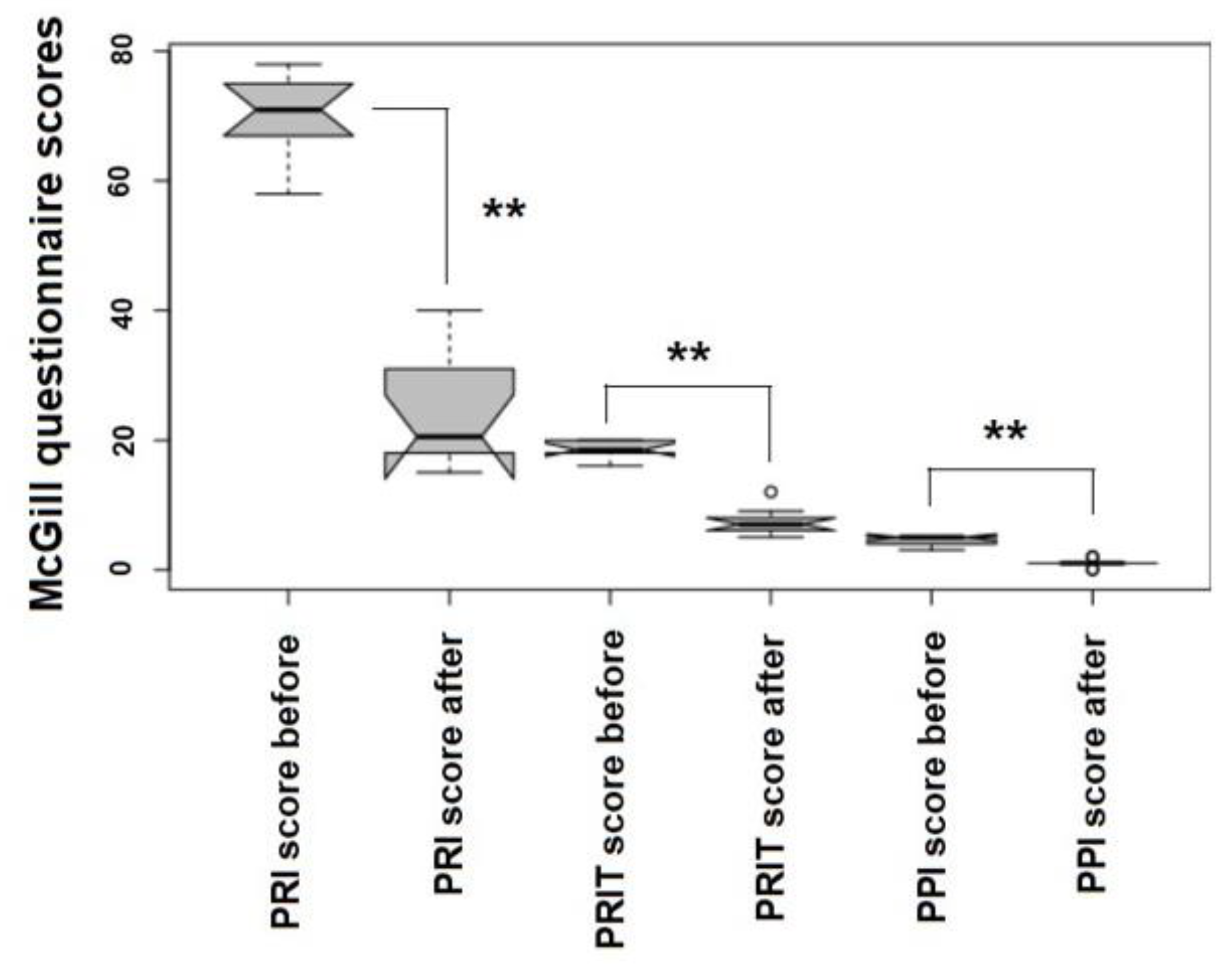
Pain and discomfort, assessed by a modified McGill questionnaire, were almost completely reduced following the ozone therapy. The Pain Rate Index (PRI) dropped down from 69.9 ± 6.66 SD to 23.6 ± 7.99 SD (−66.23%, p = 0.00016), the Pain Rate Index referrning to Time (PRIT) decreased from 18.6 ± 1.43 SD to 7.30 ± 2.11 SD (−60.75%, p = 0.00021) and the Present Pain Intensity (PPI) was reduced from 4.60 ± 0.70 SD to 1.00 ± 0.66 SD (−78.26%, p = 0.00018) (Figure 2).

5.2. The Effect of the Ozonated Blood in the Minor Autohemotherapy on Knee OA

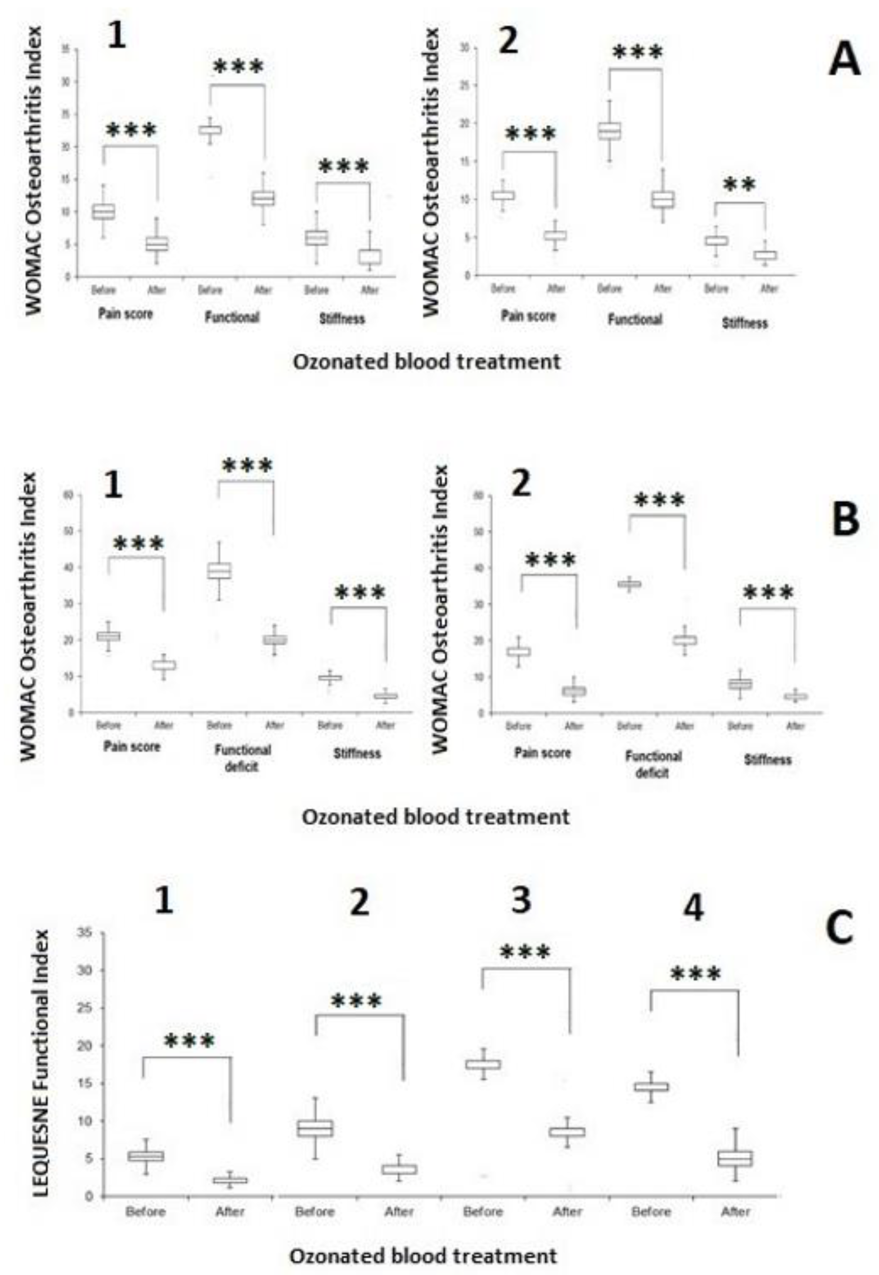
Figure 3 shows the effect of five runs of ozonized blood in knee OA-affected patients (Figure 3 panel A1: female; and Figure 3 panel A2: male) showing a type I Ahlback knee OA. In female patients, pain was reduced by five score units (from 10.45 ± 2.89 SD to 5.12 ± 1.49 SD, p < 0.0001), functional deficit by thirteen score units (from 22.60 ± 2.34 SD to 12.30 ± 2.39 SD, p < 0.0001) and stiffness by three score units (from 6.30 ± 2.18 SD to 2.85 ± 1.70 SD, p < 0.0001), i.e., ozonated blood roughly halved pain, functional deficit and stiffness (51.01%, 45.58% and 54.81%, respectively).
Male patients with type I Ahlback knee OA showed comparable results: pain was reduced by three score units (from 10.42 ± 1.25 SD to 5.00 ± 1.56 SD, =−52.03%, p < 0.0001), functional deficit by nine score units (from 19.06 ± 1.89 SD to 10.00 ± 1.60 SD, =−47.53%, p < 0.0001) and stiffness by two score units (from 4.48 ± 1.87 SD to 2.06 ± 0.75 SD, = =−54.05%, p < 0.001). Female patients showed a better response to ozonized blood therapy (delta score = +2.3 score units, KW test p = 0.00311) than males.
Figure 3 panel B shows the effect of five runs of ozonized blood in knee OA-affected patients (Figure 3, panel B1: female and Figure 3, panel B2: male) showing a type II Ahlback knee OA. In female patients, pain was reduced by nine score units (from 21.51 ± 3.42 SD to 12.51 ± 1.52 SD, p < 0.0001), functional deficit by nineteen score units (from 38.64 ± 6.65 SD to 20.42 ± 2.93 SD, p < 0.0001) and stiffness by five score units (from 9.33 ± 2.66 SD to 4.24 ± 1.68 SD, p < 0.0001), i.e., even for type II Ahlback knee OA, ozonated blood roughly halved pain, functional deficit and stiffness (41.83%, 47.13% and 54.54%, respectively). Male patients with type II Ahlback knee OA showed, as with type I, comparable results. Pain was reduced by eleven score units (from 16.81 ± 3.39 SD to 6.51 ± 2.17 SD, = −61.26%, p < 0.0001), functional deficit by sixteen score units (from 35.24 ± 6.09 SD to 19.42 ± 3.07 SD, = −44.88%, p < 0.0001) and stiffness by four score units (from 7.81 ± 3.12 SD to 4.51 ± 1.25 SD, = = −42.25%, p < 0.0001).
Apparently, males with type II Ahlback knee OA responded better in the WOMAC pain score than females, but globally, type II Ahlback knee OA females confirmed, even if in a slighter way, their best response to WOMAC ranking than males. But this difference was not significant (delta score = +1.3, KW test p = 0.45977).
Figure 3 panel C shows the Lequesne’s index for knee OA severity, as affected by ozonated blood infiltrated in the knee. In type I Ahlback knee OA, the Lequesne’s index in females dropped down from 9.42 ± 2.91 SD to 4.21 ± 2.45 SD (p < 0.0001, Figure 3, panel C1), i.e., by 55.31%, whereas in males, the ozonized blood reduced the Lequesne’s index from 9.48 ± 2.45 SD to 4.33 ± 2.38 SD (p < 0.0001, 48.82%, Figure 3, panel C2). In type II Ahlback knee OA, ozone reduced the Lequesne’s index in female patients from 17.24 ± 5.00 to 8.36 ± 2.13 SD (p < 0.0001, 51.51%, Figure 3, panel C3), whereas in males, ozone infiltrated as ozonized blood in the knee reduced the Lequesne’s index from 14.82 ± 2.69 SD to 5.28 ± 2.62 SD (p < 0.0001, 64.38%, Figure 3, panel C4).
In type II Ahlback knee OA male patients, ozone appears to be much more effective than in females and in type I Ahlback knee OA males. A total of 154 patients (85 females) were interviewed for pain, stiffness and quality of life and did not report any discomfort, pain or fatigue after a minimum period of 8 months.

6. Discussion

6.1. Ozone in Major Autohemotherapy against MDR Bacteria

Our interpretation of the reported data relies on two modalities with which ozone exerts its antimicrobial action. Ozonated formulations can directly kill bacteria in infected wounds [79,80,81,82].
In this circumstance, it is paramount to clarify that topical ozone acts via its chemical ability to cleanse wounds by killing bacteria due to lysis induced by peroxide derivatives. Furthermore, ozone in low doses via peripheral blood exerts a major role on the host’s immune response. For example, ozone used in many oxygen–ozone therapy approaches, such as major autohemotherapy (O2-O3-MAHT), targets inflammatory mechanisms by acting on more complex intracellular signaling pathways, modulating the interplay between Nrf2/NF-κB and the mitochondria-associated inflammasome NLRP3, occurring for viral diseases and other inflammatory pathologies [83,84].
Ozone activates the Nrf2/Keap1/ARE signaling pathway, which induces the activation of heme oxygenase-1 (HO-1) and the production of carbon monoxide (CO) from degraded heme groups. Carbon monoxide inhibits the activation of NF-κB, and therefore of the inflammatory signal, and promotes the macrophage skewing of M1 to M2 [85,86]. Systemic ozone, provided via O2-O3-MAHT, enhances the ability of innate immune cells, as the activation of Nrf2 contributes to shifting the presence of M1 toward skewing to the M2-phenotype, which are “trained” innate cells to improve bacterial and cell debris phagocytosis [87,88,89].
Moreover, the activation of Nrf2 by systemic ozone increases the expression of the macrophage receptor with a collagenous structure (MARCO), which, if overexpressed, enhances the ability of M2-macrophages to phagocytose bacteria and promotes the interplay between innate and acquired immunity, so as to improve the complete clearance of infectious bacteria [90,91]. In addition, ozone elicits the production of 4-hydroxynonenal (4-HNE) from ω6-PUFAs, an alkenal that inhibits the activation of the inflammasome NLRP3 and the subsequent maturation of the pro-inflammatory cytokines IL-1ß and IL-18 [92]. We reported the ability of ozone, often used in a multi-step therapy approach, to overcome the exacerbating process of inflammation in patients with chromicized wounds (usually post-surgical wounds) due to colonization from bacteria in an MDR circumstance, i.e., MRSA. All patients considered exhibited the presence of at least three antibiotic-resistant strains in their antibiograms.
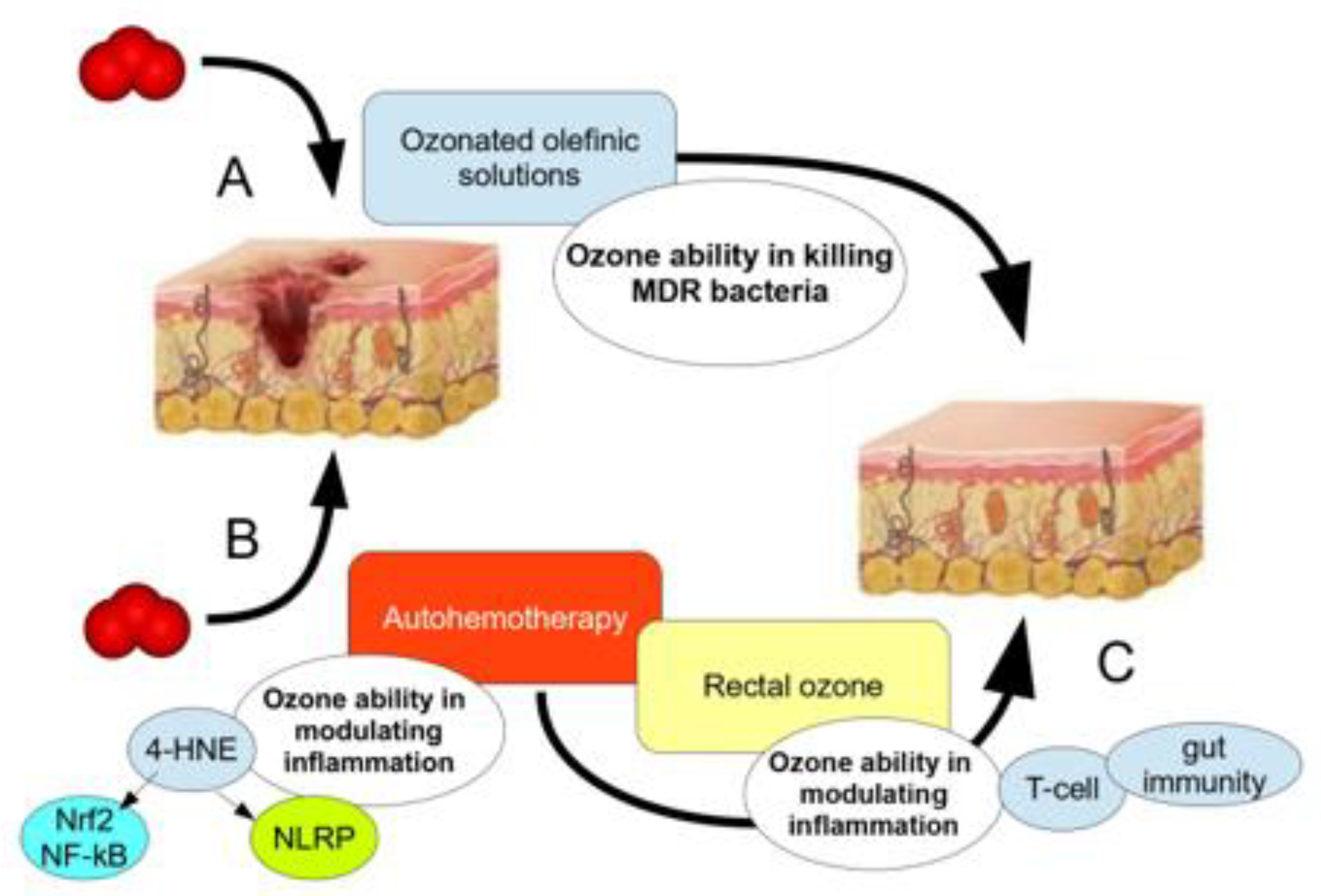
Ozone showed the ability to promote almost complete healing of these patients upon whom the recommended antibiotic therapy did not work optimally. Furthermore, the major inflammatory markers and QoL questionnaires reported a scenario of recovered health in those patients with antibiotic resistance when undergoing ozone therapy. The SIOOT protocol to eradicate bacteria-caused inflammation in post-surgical patients with antibiotic resistance patterns is grounded on two/three fundamental mechanisms (Figure 4).
When the microbial involvement of the surgical wound of any other traumatic wound is particularly widespread, chemical ozone, either in the form of ozonated injected solutions or topical formulas, rapidly quenches bacterial viability and growth, then facilitating the host’s immune system to overcome the antimicrobial resistance [93]. Medical ozone, used via the major oxygen–ozone autohemotherapy (O2-O3-MAHT), modulates the patient’s immunity, leading to the complete eradication of the septic mechanism via the immune response.
The modulating activity of O2-O3-MAHT may be implemented by adjusting the contribution of the gut microbiome/gut immunity interplay and the mucosal immunity (IgA) via the use of rectal ozone [72]. The first approach (chemical) is to be considered as an ancillary approach of the leading way of action, which is held by O2-O3-MAHT. Evidence about the ability of topical ozone solutions, ozonated oils and other topical formulas to remove antibiotic-resistant bacteria from the skin, mucosae or eyes has been widely reported in the literature to date [94,95,96,97]. In a recent paper, some authors investigated the action of an ophthalmic solution containing ozonated oil (10.5%, i.e., 5 μg/mL) against 60 bacterial strains with an MDR profile for various patients (resistance was confirmed for at least three different antibiotics, including fluoroquinolones) and showed that at 6–8 h following the treatment, at least 94% of all strains, i.e., Pseudomonas aeruginosa MDR, methicillin-resistant Staphylococcus aureus and methicillin-resistant Staphylococcus epidermis, reported a marked inhibitory zone and that more than 10% of those strains were also inhibited after 24 h [97].
However, bacterial growth may not be completely dampened by only topical or injected ozonated solutions in the neighboring skin of the wound [97]. This should assess the recommendation to include an O2-O3-MAHT as the leading therapy approach to overcome the possibility of a bacterial relapse [97]. Ozone acts in the blood in a completely different way with respect to the direct contact of the ozonated solutions and formulas [14,98,99]. As the contact of low doses of ozone in the peripheral blood activates the formation of lipo-peroxide intermediates and electrophiles, such as 4-hydroxynonenal (4-HNE) [10,92,100], this intermediate may reduce the impact of the inflammasome (NLRP3) role in the septic inflammation and inhibit innate cell pyroptosis, thus acting as an anti-inflammatory compound and immunomodulating the [10,92] inflammation process [71].
The concurrent action of the topical ozone, working in a quite direct way, and of blood-derived ozonized products, such as 4-HNE, which acts as a modulatory agent, should warrant for the complete effectiveness of ozone in the clearance of MDR bacteria in humans. Moreover, the ability of ozone via autohemotherapy to elicit a “mito-hormetic” mechanism [14,21,23,24,99] may explain how ozone has the ability to restrain inflammation by inducing a macrophage-tolerant state in an LPS-induced immune exacerbation [71].

6.2. Minor Autohemotherapy with Ozonated Blood Reduces Discomfort and Disability Markers in Knee OA

Recent reports assessed the use of ozone in knee OA [101,102,103]. Regarding the excellent clinical trial by Lopes de Jesus et al. [102], we did not use directly a gaseous oxygen/ozone mixture in the infiltrated knee but a stabilized medium made of ozonated blood instead, whilst maintaining the ozone dosage at 20 μg/mL. This approach should promote the formation of ozonized mediators, such as 4-HNE or LOPs from fatty acids, eliciting a hormetic or mito-hormetic mechanism in the knee tissue compartments [14,21,66].
Lopes de Jesus and coworkers obtained encouraging results by using ozone directly in the knee but used 1.5 fold more infiltrations (eight instead of 5five and obtained a reduction of about 50% in WOMAC or Lequesne’s scoring, the same as our results. In our hypothesis, the strong oxidative potential of ozone directly in the knee might reduce the hypoxic microenvironment, which causes severity in the knee osteoarthritis, thus relieving the concerning symptoms [102,104,105].
The hormetic mechanism triggered by ozone-caused lipid mediators and electrophiles allows the modulated reduction in the hypoxic microenvironment [106], with the paramount difference that reactive oxygen species (ROS) do not increase thanks to an improvement in the mitochondria activity, whereas in the pro-oxidant activity of gaseous ozone directly, mitochondria burst a huge increase in the oxidative activity [106]. The hormetic modulation of the Nrf2/Keap1/ARE pathway by ROS as signaling molecules can occur via modulated involvements of mitochondria biogenesis (“mito-hormesis”) [107], whereas the pro-oxidant activity of ozone directly infiltrated elicits the activation of the antioxidant enzyme system but, at the same time, triggers a pro-inflammatory response of the cellular systems, for example NF-κB and inflammasome, therefore reducing the length of positive outcomes achieved with using ozone infiltrations and/or asking for more ozone to be used.
Our results, which enlarge previous data with less patients, suggest that ozone within blood should work in a better, much more suitable, affordable and physiological way than gaseous oxygen–ozone directly infiltrated in the knee.
Further insights are needed to elucidate the way in which ozone reduces dramatically the pain, stiffness and severity of knee OA.
Autologous ozonated whole blood infiltrations in knee OA is able to reduce WOMAC indexes of pain, stiffness and functional disability, as well as Lequesne’s functional indexes, by at least 50% compared to the previous pathological condition. This evidence was attained with a reduced impact of infiltrating ozone from other current studies and represents encouraging evidence for the future of knee OA therapy.

6.3. The SIOOT Protocol of Ozonated Blood

The Italian Scientific Society of Oxygen–Ozone Therapy (SIOOT) arranged their ozone adjunct treatment protocols principally on the basis of the ozone interplay with the autologous peripheral blood, more than with other media which yet remain commendable as well. The main tenet of SIOOT methodology takes into account the fundamental action that ozone plays in the blood microenvironment, which consists of its ability in inducing PUFAs-derived alkenals (4-HNE and 4-HHE) and other LPOs, exerting a fine, modulatory activity on immunity and therefore on inflammation-related symptoms, such as pain and fatigue. In this manuscript, we collected two experimental examples of how autologous blood treated with ozone, either via O2-O3-MAHT or a minor autohemotherapy using ozonated blood, reduces inflammation-related events, such as infected wounds, chronic musculoskeletal inflammation and pain. In our opinion, an underlying shared mechanism can be highlighted.
Ozone doses should not overcome the blood ability in buffering the pro-oxidant activity of this oxygen allotrope. Doses must be produced with sharpness and a background of high-quality technology, with reliable throughput to ensure the correct O2/O3 proportion is made in compliance with SIOOT protocols, which is crucial to properly use ozone in medicine [11,108,109].
While it may be understandable that ozone can be used to sanitize and cleanse infected wounds, its application is wider than expected. By eliciting the activation of the Nrf2/Keap1/ARE signaling pathway, i.e., acting as an antioxidant molecule, ozone exerts an anti-inflammatory effect. This is true for many ailments and pathologies of musculoskeletal etiology, for example low back pain and disc herniation [11,110,111,112,113], where ozone is used as a paravertebral/intramuscular injection, rather than intradiscal, which SIOOT recently criticized [114]. It is tempting to speculate that gaseous ozone (usually 30 μg/mL), once inside the skin tissue and muscles, is rapidly quenched by the antioxidant endowment of cells and induces the formation of LPOs and ROS as signaling molecules, triggering the activation of the Nrf2/HO-1/CO axis and probably inducing a microvasculature change due to the interplay between Nrf2/angiogenesis [115,116]. The anti-inflammatory activity of ozone and its mediators LPOs (mainly 4-HNE) promotes the skewing of M1 to M2 in the intervertebral disc during a disc herniation [117]. The M2 replacement enhances the renewal of nucleus pulposus, its ECM, cell survival and the remodeling of the external fibrosus annulus, as ozone can induce the synthesis of collagen I [118,119].
The ability of ozone to work as an anti-inflammation agent via hormetic mechanisms [14,21,98,100] is closely linked to the ability and expertise of clinicians to use ozone in a more effective way; for example, high doses should be avoided [120]. In this context, the numerous scientific societies, for example SIOOT in Italy, are always engaged and encouraged in tailoring the best medical approach to successfully use ozone in the adjunct medical treatment.
The capability of ozone to modulate immunity, provided by physicians with a formidable tool, by addressing autoimmune disorders, such as multiple sclerosis [121,122], Sjögren syndrome [123], fibromyalgia [41] and ME/CFS [42], where ozone resulted in being particularly effective in reducing pain, discomfort, fatigue and other burdensome symptoms, has been shown. It is tempting to speculate that the immune-modulatory action held by ozone targets mitochondria, which are particularly involved in autoimmunity [124,125].
Once the gaseous mixture O2/O3, for example O3 = 45 μg/mL, enters the bloodstream via O2-O3-MAHT, a moderate hemolysis occurs, with release of heme byproducts (activating HO-1 and CO functions) and damaged erythrocytes, which are rapidly removed by O3-produced 4-HNE via eryptosis [126]. The gas O3 concentration into the plasma should be re-adjusted considering Henry’s law so that 45 μg/mL O3 (gas, r.t.) corresponds to about 7.165 μg/mL in water at 37 °C, a concentration which could yield 3.67 μM 4-HNE if only 2% of ozone is converted into 4-HNE [14,66,127]. A dose of 3.0 μM 4-HNE exerts an anti-inflammatory action [92].
Ozonate blood represents a straightforward possibility to apply the antioxidant and anti-inflammatory properties of ozone in medicine, yet ozone therapy should only be administered by trained and licensed practitioners in a controlled medical setting.

7. Conclusions

The use of ozone in autologous blood, which must be approached only by highly skilled experts and professionals, allows clinicians to use this oxygen allotrope as a sound therapeutical tool to address immune disorders (auto-immunity driven illnesses), musculoskeletal disorders with inflammation, infected post-surgical or traumatic wounds and many further inflammation pathologies, reducing the associated pain, fatigue, discomfort and disability, and exhibiting properties which encourage its consideration as an opportunity for medicine.

8. Acronyms

Å: angstrom; 4-HHE: 4-hydroxyhesenal; 4-HNE: 4-hydroxynonenal; knee OA: knee osteoarthritis; Nrf2: nuclear factor erythroid 2–related factor 2; O2-O3-MAHT: major autohemotherapy with oxygen–ozone; O2-O3-mAHT: minor autohemotherapy with oxygen–ozone; SIOOT: Italian Scientific Society of Oxygen–Ozone Therapy; WOMAC: Western Ontario and McMaster Universities Arthritis Index.

Author Contributions

Conceptualization, S.C., M.F., U.T., G.R. and L.V.; methodology, F.V., M.F., U.T. and L.V.; software, S.P. and T.R.; validation, M.F., L.V. and U.T.; formal analysis, M.C.; investigation, D.B., L.V. and M.F.; resources, V.S.; data curation, A.G.; writing—original draft preparation, S.C.; writing—review and editing, S.C., S.P., F.V., L.V., U.T., G.R., A.G. and F.L.; visualization, M.F.; supervision, L.V. and U.T.; project administration, M.F.; funding acquisition, M.F. All authors have read and agreed to the published version of the manuscript.

Funding

This research received no external funding.

Institutional Review Board Statement

This study was conducted in accordance with the Declaration of Helsinki and approved by the Institutional Review Board of Comunian Clinic (protocol code 345/2011).

Informed Consent Statement

Informed consent was obtained from all subjects involved in the study. Written informed consent was obtained from the patient(s) to publish this paper.

Data Availability Statement

Data can be easily requested from the corresponding author.

Acknowledgments

Special thanks are given to Francesca Turriceni for her invaluable technical support.

Conflicts of Interest

The authors declare no conflict of interest.

References

  1. Coffaro, B.; Weisel, C.P. Reactions and Products of Squalene and Ozone: A Review. Environ. Sci. Technol. 2022, 56, 7396–7411. [Google Scholar] [CrossRef] [PubMed]
  2. Zhang, J.J.; Wei, Y.; Fang, Z. Ozone Pollution: A Major Health Hazard Worldwide. Front. Immunol. 2019, 10, 2518. [Google Scholar] [CrossRef] [PubMed]
  3. Liu, Y.; Misztal, P.K.; Arata, C.; Weschler, C.J.; Nazaroff, W.W.; Goldstein, A.H. Observing ozone chemistry in an occupied residence. Proc. Natl. Acad. Sci. USA 2021, 118, e2018140118. [Google Scholar] [CrossRef]
  4. Wentworth, P., Jr.; Nieva, J.; Takeuchi, C.; Galve, R.; Wentworth, A.D.; Dilley, R.B.; DeLaria, G.A.; Saven, A.; Babior, B.M.; Janda, K.D.; et al. Evidence for ozone formation in human atherosclerotic arteries. Science 2003, 302, 1053–1056. [Google Scholar] [CrossRef]
  5. Lerner, R.A.; Eschenmoser, A. Ozone in biology. Proc. Natl. Acad. Sci. USA 2003, 100, 3013–3015. [Google Scholar] [CrossRef]
  6. Wentworth, P., Jr.; Wentworth, A.D.; Zhu, X.; Wilson, I.A.; Janda, K.D.; Eschenmoser, A.; Lerner, R.A. Evidence for the production of trioxygen species during antibody-catalyzed chemical modification of antigens. Proc. Natl. Acad. Sci. USA 2003, 100, 1490–1493. [Google Scholar] [CrossRef] [PubMed]
  7. Wentworth, P., Jr.; McDunn, J.E.; Wentworth, A.D.; Takeuchi, C.; Nieva, J.; Jones, T.; Bautista, C.; Ruedi, J.M.; Gutierrez, A.; Janda, K.D.; et al. Evidence for antibody-catalyzed ozone formation in bacterial killing and inflammation. Science 2002, 298, 2195–2199. [Google Scholar] [CrossRef]
  8. Babior, B.M.; Takeuchi, C.; Ruedi, J.; Gutierrez, A.; Wentworth, P., Jr. Investigating antibody-catalyzed ozone generation by human neutrophils. Proc. Natl. Acad. Sci. USA 2003, 100, 3031–3034. [Google Scholar] [CrossRef]
  9. Glaze, W.H. Reaction products of ozone: A review. Environ. Health Perspect. 1986, 69, 151–157. [Google Scholar] [CrossRef]
  10. Tricarico, G.; Travagli, V. The Relationship between Ozone and Human Blood in the Course of a Well-Controlled, Mild, and Transitory Oxidative Eustress. Antioxidants 2021, 10, 1946. [Google Scholar] [CrossRef]
  11. Travagli, V.; Iorio, E.L. The Biological and Molecular Action of Ozone and Its Derivatives: State-of-the-Art, Enhanced Scenarios, and Quality Insights. Int. J. Mol. Sci. 2023, 24, 8465. [Google Scholar] [CrossRef] [PubMed]
  12. Weschler, C.J. Ozone in indoor environments: Concentration and chemistry. Indoor Air 2000, 10, 269–288. [Google Scholar] [CrossRef]
  13. Hirahara, Y.; Iwata, K.; Nakamuro, K. Effect of Citric Acid on Prolonging the Half-life of Dissolved Ozone in Water. Food Saf. 2019, 7, 90–94. [Google Scholar] [CrossRef] [PubMed]
  14. Bocci, V.A.; Zanardi, I.; Travagli, V. Ozone acting on human blood yields a hormetic dose-response relationship. J. Transl. Med. 2011, 9, 66. [Google Scholar] [CrossRef] [PubMed]
  15. Groves, A.M.; Gow, A.J.; Massa, C.B.; Laskin, J.D.; Laskin, D.L. Prolonged injury and altered lung function after ozone inhalation in mice with chronic lung inflammation. Am. J. Respir. Cell Mol. Biol. 2012, 47, 776–783. [Google Scholar] [CrossRef] [PubMed]
  16. Kim, S.Y.; Kim, E.; Kim, W.J. Health Effects of Ozone on Respiratory Diseases. Tuberc. Respir. Dis. 2020, 83 (Suppl. 1), S6–S11. [Google Scholar] [CrossRef]
  17. Holm, S.M.; Balmes, J.R. Systematic Review of Ozone Effects on Human Lung Function, 2013 Through 2020. Chest 2022, 161, 190–201. [Google Scholar] [CrossRef]
  18. Hemming, J.M.; Hughes, B.R.; Rennie, A.R.; Tomas, S.; Campbell, R.A.; Hughes, A.V.; Arnold, T.; Botchway, S.W.; Thompson, K.C. Environmental Pollutant Ozone Causes Damage to Lung Surfactant Protein B (SP-B). Biochemistry 2015, 54, 5185–5197. [Google Scholar] [CrossRef]
  19. Dengiz, E.; Özcan, Ç.; Güven, Y.İ.; Uçar, S.; Ener, B.K.; Sözen, S.; Yağcı, B.; Güzel, İ.A.; Yiğit, B.; Andaç, A.; et al. Ozone gas applied through nebulization as adjuvant treatment for lung respiratory diseases due to COVID-19 infections: A prospective randomized trial. Med. Gas Res. 2022, 12, 55–59. [Google Scholar]
  20. Travagli, V.; Zanardi, I.; Bernini, P.; Nepi, S.; Tenori, L.; Bocci, V. Effects of ozone blood treatment on the metabolite profile of human blood. Int. J. Toxicol. 2010, 29, 165–174. [Google Scholar] [CrossRef]
  21. Franzini, M.; Valdenassi, L.; Chirumbolo, S. The immune mito-hormetic landscape in the oxygen-ozone therapy. Towards a novel medical approach. Int. Immunopharmacol. 2023, 121, 110598. [Google Scholar] [CrossRef] [PubMed]
  22. Wen, Q.; Liu, D.; Wang, X.; Zhang, Y.; Fang, S.; Qiu, X.; Chen, Q. Effects of ozone for treating chronically refractory wounds and ulcers: A protocol for systematic review and meta-analysis of randomized clinical trials. Medicine 2020, 99, e20457. [Google Scholar] [CrossRef] [PubMed]
  23. Chirumbolo, S.; Franzini, M.; Pandolfi, S.; Valdenassi, L. Ozone therapy in the huge concern of multidrug resistance (MDR) bacteria. The worldwide perspective. Occup. Med. Health Aff. 2023, 11, 478. [Google Scholar]
  24. Chirumbolo, S.; Valdenassi, L.; Franzini, M. Ozone adjunct treatment in facing multidrug resistant bacteria? Clin. Case Rep. 2023, in press. [Google Scholar] [CrossRef]
  25. Czekalski, N.; Imminger, S.; Salhi, E.; Veljkovic, M.; Kleffel, K.; Drissner, D.; Hammes, F.; Bürgmann, H.; von Gunten, U. Inactivation of Antibiotic Resistant Bacteria and Resistance Genes by Ozone: From Laboratory Experiments to Full-Scale Wastewater Treatment. Environ. Sci. Technol. 2016, 50, 11862–11871. [Google Scholar] [CrossRef]
  26. Lüddeke, F.; Heß, S.; Gallert, C.; Winter, J.; Güde, H.; Löffler, H. Removal of total and antibiotic resistant bacteria in advanced wastewater treatment by ozonation in combination with different filtering techniques. Water Res. 2015, 69, 243–251. [Google Scholar] [CrossRef]
  27. Rangel, K.; Cabral, F.O.; Lechuga, G.C.; Carvalho, J.P.R.S.; Villas-Bôas, M.H.S.; Midlej, V.; De-Simone, S.G. Potent Activity of a High Concentration of Chemical Ozone against Antibiotic-Resistant Bacteria. Molecules 2022, 27, 3998. [Google Scholar] [CrossRef]
  28. Roth, A.; Maruthamuthu, M.K.; Nejati, S.; Krishnakumar, A.; Selvamani, V.; Sedaghat, S.; Nguyen, J.; Seleem, M.N.; Rahimi, R. Wearable adjunct ozone and antibiotic therapy system for treatment of Gram-negative dermal bacterial infection. Sci. Rep. 2022, 12, 13927. [Google Scholar] [CrossRef]
  29. Yousefi, B.; Banihashemian, S.Z.; Feyzabadi, Z.K.; Hasanpour, S.; Kokhaei, P.; Abdolshahi, A.; Emadi, A.; Eslami, M. Potential therapeutic effect of oxygen-ozone in controlling of COVID-19 disease. Med. Gas Res. 2022, 12, 33–40. [Google Scholar] [CrossRef]
  30. Rowen, R.J. Ozone therapy as a primary and sole treatment for acute bacterial infection: Case report. Med. Gas Res. 2018, 8, 121–124. [Google Scholar] [CrossRef]
  31. Spadea, L.; Zanotto, E.; Cavallo, R.; Campagna, G.; Giannico, M.I.; Costagliola, C.; ELOOM Study Investigators. Effectiveness of liposomal ozonized oil in reducing ocular microbial flora in patients undergoing cataract surgery. J. Cataract Refract. Surg. 2021, 47, 1548–1555. [Google Scholar] [CrossRef] [PubMed]
  32. Rowen, R.J. Ozone therapy in conjunction with oral antibiotics as a successful primary and sole treatment for chronic septic prosthetic joint: Review and case report. Med. Gas Res. 2018, 8, 67–71. [Google Scholar] [CrossRef] [PubMed]
  33. Varesi, A.; Chirumbolo, S.; Ricevuti, G. Oxygen-ozone treatment and COVID-19: Antioxidants targeting endothelia lead the scenery. Intern. Emerg. Med. 2022, 17, 593–596. [Google Scholar] [CrossRef] [PubMed]
  34. Franzini, M.; Valdenassi, L.; Ricevuti, G.; Chirumbolo, S.; Depfenhart, M.; Bertossi, D.; Tirelli, U. Oxygen-ozone (O2-O3) immunoceutical therapy for patients with COVID-19. Preliminary evidence reported. Int. Immunopharmacol. 2020, 88, 106879. [Google Scholar] [CrossRef] [PubMed]
  35. Yamashita, K.; Miyoshi, T.; Arai, T.; Endo, N.; Itoh, H.; Makino, K.; Mizugishi, K.; Uchiyama, T.; Sasada, M. Ozone production by amino acids contributes to killing of bacteria. Proc. Natl. Acad. Sci. USA 2008, 105, 16912–16917. [Google Scholar] [CrossRef] [PubMed]
  36. Nyffele PTBoyle, N.A.; Eltepu LWong, C.H.; Eschenmoser, A.; Lerner, R.A.; Wentworth, P., Jr. Dihydrogen Trioxide (HOOOH) Is Generated during the Thermal Reaction between Hydrogen Peroxide and Ozone. Angew. Chem. Int. Ed. 2004, 43, 4656–4659. [Google Scholar] [CrossRef] [PubMed]
  37. Cristiano, L. Could ozone be an effective disinfection measure against the novel coronavirus (SARS-CoV-2)? J. Prev. Med. Hyg. 2020, 61, E301–E303. [Google Scholar]
  38. Sharma, M.; Hudson, J.B. Ozone gas is an effective and practical antibacterial agent. Am. J. Infect. Control 2008, 36, 559–563. [Google Scholar] [CrossRef]
  39. Bocci, V. Ozone as Janus: This controversial gas can be either toxic or medically useful. Mediat. Inflamm. 2004, 13, 3–11. [Google Scholar] [CrossRef]
  40. Yang, X.; Chen, C.; Wang, K.; Chen, M.; Wang, Y.; Chen, Z.; Zhao, W.; Ou, S. Elucidating the molecular mechanisms of ozone therapy for neuropathic pain management by integrated transcriptomic and metabolomic approach. Front. Genet. 2023, 14, 1231682. [Google Scholar] [CrossRef]
  41. Tirelli, U.; Franzini, M.; Valdenassi, L.; Pandolfi, S.; Taibi, R.; Chirumbolo, S. Fibromyalgia treated with oxygen-ozone auto-haemotherapy (O2-O3-AHT): A case study on 200 patients with a modified 10-PI-NRS evaluation. Eur. Rev. Med. Pharmacol. Sci. 2022, 26, 7974–7979. [Google Scholar] [PubMed]
  42. Tirelli, U.; Franzini, M.; Valdenassi, L.; Pandolfi, S.; Berretta, M.; Ricevuti, G.; Chirumbolo, S. Patients with Myalgic Encephalomyelitis/Chronic Fatigue Syndrome (ME/CFS) Greatly Improved Fatigue Symptoms When Treated with Oxygen-Ozone Autohemotherapy. J. Clin. Med. 2021, 11, 29. [Google Scholar] [CrossRef] [PubMed]
  43. Tirelli, U.; Franzini, M.; Valdenassi, L.; Pisconti, S.; Taibi, R.; Torrisi, C.; Pandolfi, S.; Chirumbolo, S. Fatigue in post-acute sequelae of SARS-CoV2 (PASC) treated with oxygen-ozone autohemotherapy-preliminary results on 100 patients. Eur. Rev. Med. Pharmacol. Sci. 2021, 25, 5871–5875. [Google Scholar] [PubMed]
  44. Li, X.R.; Cui, J.J.; Ge, W.P.; Wang, Z.W.; Chu, Y.C.; Zheng, G.R. Ozonated autohemotherapy combined with pulsed radiofrequency in the treatment of thoracic postherpetic neuralgia in older adults: A retrospective study. Med. Gas Res. 2024, 14, 12–18. [Google Scholar] [PubMed]
  45. Latini, E.; Nusca, S.M.; Curci, E.R.; Boaretto, D.; Santoboni, F.; Trischitta, D.; Vetrano, M.; Vulpiani, M.C. Intramuscular paravertebral oxygen-ozone therapy for chronic neck pain and low back pain: Evaluation of 6-month clinical outcomes. Med. Gas Res. 2024, 14, 6–11. [Google Scholar] [CrossRef] [PubMed]
  46. Zhang, X.T.; Zong, L.J.; Jia, R.M.; Qin, X.M.; Ruan, S.R.; Lu, L.L.; Wang, P.; Hu, L.; Liu, W.T.; Yang, Y.; et al. Ozone attenuates chemotherapy-induced peripheral neuropathy via upregulating the AMPK-SOCS3 axis. J. Cancer Res. Ther. 2023, 19, 1031–1039. [Google Scholar] [CrossRef]
  47. Primorac, D.; Molnar, V.; Rod, E.; Jeleč, Ž.; Čukelj, F.; Matišić, V.; Vrdoljak, T.; Hudetz, D.; Hajsok, H.; Borić, I. Knee Osteoarthritis: A Review of Pathogenesis and State-Of-The-Art Non-Operative Therapeutic Considerations. Genes 2020, 11, 854. [Google Scholar] [CrossRef]
  48. Mora, J.C.; Przkora, R.; Cruz-Almeida, Y. Knee osteoarthritis: Pathophysiology and current treatment modalities. J. Pain Res. 2018, 11, 2189–2196. [Google Scholar] [CrossRef]
  49. Van Manen, M.D.; Nace, J.; Mont, M.A. Management of primary knee osteoarthritis and indications for total knee arthroplasty for general practitioners. J. Am. Osteopath. Assoc. 2012, 112, 709–715. [Google Scholar]
  50. Figueiredo, C.P.; Simon, D.; Englbrecht, M.; Haschka, J.; Kleyer, A.; Bayat, S.; Hueber, A.; Pereira, R.M.; Rech, J.; Schett, G. Quantification and Impact of Secondary Osteoarthritis in Patients With Anti-Citrullinated Protein Antibody-Positive Rheumatoid Arthritis. Arthritis Rheumatol. 2016, 68, 2114–2121. [Google Scholar] [CrossRef]
  51. Samson, D.J.; Grant, M.D.; Ratko, T.A.; Bonnell, C.J.; Ziegler, K.M.; Aronson, N. Treatment of primary and secondary osteoarthritis of the knee. Evid. Rep. Technol. Assess. (Full Rep.) 2007, 157, 1–157. [Google Scholar]
  52. Mangone, G.; Orioli, A.; Pinna, A.; Pasquetti, P. Infiltrative treatment with Platelet Rich Plasma (PRP) in gonarthrosis. Clin. Cases Miner. Bone Metab. 2014, 11, 67–72. [Google Scholar] [CrossRef] [PubMed]
  53. Palco, M.; Fenga, D.; Basile, G.C.; Rizzo, P.; Cavalieri, B.; Leonetti, D.; Alito, A.; Bruschetta, A.; Traina, F. Platelet-Rich Plasma Combined with Hyaluronic Acid versus Leucocyte and Platelet-Rich Plasma in the Conservative Treatment of Knee Osteoarthritis. A Retrospective Study. Medicina 2021, 57, 232. [Google Scholar] [CrossRef] [PubMed]
  54. Tang, J.Z.; Nie, M.J.; Zhao, J.Z.; Zhang, G.C.; Zhang, Q.; Wang, B. Platelet-rich plasma versus hyaluronic acid in the treatment of knee osteoarthritis: A meta-analysis. J. Orthop. Surg. Res. 2020, 15, 403. [Google Scholar] [CrossRef] [PubMed]
  55. Dallari, D.; Stagni, C.; Rani, N.; Sabbioni, G.; Pelotti, P.; Torricelli, P.; Tschon, M.; Giavaresi, G. Ultrasound-Guided Injection of Platelet-Rich Plasma and Hyaluronic Acid, Separately and in Combination, for Hip Osteoarthritis: A Randomized Controlled Study. Am. J. Sports Med. 2016, 44, 664–671. [Google Scholar] [CrossRef] [PubMed]
  56. Kim, J.H.; Park, Y.B.; Ha, C.W.; Roh, Y.J.; Park, J.G. Adverse Reactions and Clinical Outcomes for Leukocyte-Poor Versus Leukocyte-Rich Platelet-Rich Plasma in Knee Osteoarthritis: A Systematic Review and Meta-analysis. Orthop. J. Sports Med. 2021, 9, 23259671211011948. [Google Scholar] [CrossRef] [PubMed]
  57. Fotouhi, A.; Maleki, A.; Dolati, S.; Aghebati-Maleki, A.; Aghebati-Maleki, L. Platelet rich plasma, stromal vascular fraction and autologous conditioned serum in treatment of knee osteoarthritis. Biomed. Pharmacother. 2018, 104, 652–660. [Google Scholar] [CrossRef]
  58. Civinini, R.; Nistri, L.; Martini, C.; Redl, B.; Ristori, G.; Innocenti, M. Growth factors in the treatment of early osteoarthritis. Clin. Cases Miner. Bone Metab. 2013, 10, 26–29. [Google Scholar] [CrossRef]
  59. Napolitano, M.; Matera, S.; Bossio, M.; Crescibene, A.; Costabile, E.; Almolla, J.; Almolla, H.; Togo, F.; Giannuzzi, C.; Guido, G. Autologous platelet gel for tissue regeneration in degenerative disorders of the knee. Blood Transfus. 2012, 10, 72–77. [Google Scholar]
  60. Chen, Y.R.; Yan, X.; Yuan, F.Z.; Ye, J.; Xu, B.B.; Zhou, Z.X.; Mao, Z.M.; Guan, J.; Song, Y.F.; Sun, Z.W.; et al. The Use of Peripheral Blood-Derived Stem Cells for Cartilage Repair and Regeneration In Vivo: A Review. Front. Pharmacol. 2020, 11, 404. [Google Scholar] [CrossRef]
  61. Papadopoulos, K.I.; Turajane, T. Erratum: Commentary: Autologous Peripheral Blood Stem Cells (PBSC) are Safe and Effective in Knee Osteoarthritis. Front. Pharmacol. 2021, 12, 652738. [Google Scholar] [CrossRef] [PubMed]
  62. Wang, S.J.; Yin, M.H.; Jiang, D.; Zhang, Z.Z.; Qi, Y.S.; Wang, H.J.; Yu, J.K. The Chondrogenic Potential of Progenitor Cells Derived from Peripheral Blood: A Systematic Review. Stem Cells Dev. 2016, 25, 1195–1207. [Google Scholar] [CrossRef] [PubMed]
  63. Coşkun, H.S.; Yurtbay, A.; Say, F. Platelet Rich Plasma Versus Autologous Conditioned Serum in Osteoarthritis of the Knee: Clinical Results of a Five-Year Retrospective Study. Cureus 2022, 14, e24500. [Google Scholar] [CrossRef] [PubMed]
  64. Manoto, S.L.; Maepa, M.J.; Motaung, S.K. Medical ozone therapy as a potential treatment modality for regeneration of damaged articular cartilage in osteoarthritis. Saudi J. Biol. Sci. 2018, 25, 672–679. [Google Scholar] [CrossRef] [PubMed]
  65. Bocci, V.; Valacchi, G.; Corradeschi, F.; Fanetti, G. Studies on the biological effects of ozone: 8. Effects on the total antioxidant status and on interleukin-8 production. Mediat. Inflamm. 1998, 7, 313–317. [Google Scholar] [CrossRef] [PubMed]
  66. Bocci, V.; Borrelli, E.; Travagli, V.; Zanardi, I. The ozone paradox: Ozone is a strong oxidant as well as a medical drug. Med. Res. Rev. 2009, 29, 646–682. [Google Scholar] [CrossRef] [PubMed]
  67. Travagli, V.; Zanardi, I.; Bocci, V. A realistic evaluation of the action of ozone on whole human blood. Int. J. Biol. Macromol. 2006, 39, 317–320. [Google Scholar] [CrossRef]
  68. Bocci, V.A. Scientific and medical aspects of ozone therapy. State of the art. Arch. Med. Res. 2006, 37, 425–435. [Google Scholar] [CrossRef]
  69. Tricarico, G.; Isakovic, J.; Song, M.S.; Rustichelli, F.; Travagli, V.; Mitrecic, D. Ozone influences migration and proliferation of neural stem cells in vitro. Neurosci. Lett. 2020, 739, 135390. [Google Scholar] [CrossRef]
  70. Costanzo, M.; Boschi, F.; Carton, F.; Conti, G.; Covi, V.; Tabaracci, G.; Sbarbati, A.; Malatesta, M. Low ozone concentrations promote adipogenesis in human adipose-derived adult stem cells. Eur. J. Histochem. 2018, 62, 2969. [Google Scholar] [CrossRef]
  71. Niu, M.; Chen, P. Crosstalk between gut microbiota and sepsis. Burns Trauma. 2021, 9, tkab036. [Google Scholar] [CrossRef] [PubMed]
  72. Díaz-Luis, J.; Menéndez-Cepero, S.; Macías-Abraham, C.; Fariñas-Rodríguez, L. Systemic Ozone Therapy by Rectal Insufflation for Immunoglobulin A Deficiency. MEDICC Rev. 2018, 20, 29. [Google Scholar] [CrossRef] [PubMed]
  73. Cohen, S.R.; Russell, L.B.; Leis, A.; Shahidi, J.; Porterfield, P.; Kuhl, D.R.; Gadermann, A.M.; Sawatzky, R. More comprehensively measuring quality of life in life-threatening illness: The McGill Quality of Life Questionnaire—Expanded. BMC Palliat. Care 2019, 18, 92. [Google Scholar] [CrossRef] [PubMed]
  74. Serrano, P.V.; Serrano, G.B.; Torres, I.L.S.; Graudner, R.R.; Caumo, W. The McGill Quality of Life Questionnaire-Revised (MQOL-R). Psychometric properties and validation of a Brazilian version on palliative care patients: A cross-sectional study. Health Qual. Life Outcomes 2020, 18, 368. [Google Scholar] [CrossRef] [PubMed]
  75. Szende, A.; Leidy, N.K.; Revicki, D. Health-related quality of life and other patient-reported outcomes in the European centralized drug regulatory process: A review of guidance documents and performed authorizations of medicinal products 1995 to 2003. Value Health 2005, 8, 534–548. [Google Scholar] [CrossRef] [PubMed]
  76. Weidow, J.; Cederlund, C.G.; Ranstam, J.; Kärrholm, J. Ahlbäck grading of osteoarthritis of the knee: Poor reproducibility and validity based on visual inspection of the joint. Acta Orthop. 2006, 77, 262–266. [Google Scholar] [CrossRef]
  77. Lequesne, M.G. The algofunctional indices for hip and knee osteoarthritis. J. Rheumatol. 1997, 24, 779–781. [Google Scholar]
  78. Egbewale, B.E.; Lewis, M.; Sim, J. Bias, precision and statistical power of analysis of covariance in the analysis of randomized trials with baseline imbalance: A simulation study. BMC Med. Res. Methodol. 2014, 14, 49. [Google Scholar] [CrossRef]
  79. Rangel, K.; Cabral, F.O.; Lechuga, G.C.; Carvalho, J.P.R.S.; Villas-Bôas, M.H.S.; Midlej, V.; De-Simone, S.G. Detrimental Effect of Ozone on Pathogenic Bacteria. Microorganisms 2021, 10, 40. [Google Scholar] [CrossRef]
  80. Pietrocola, G.; Ceci, M.; Preda, F.; Poggio, C.; Colombo, M. Evaluation of the antibacterial activity of a new ozonized olive oil against oral and periodontal pathogens. J. Clin. Exp. Dent. 2018, 10, e1103–e1108. [Google Scholar] [CrossRef]
  81. Higa, B.; Cintra, B.S.; Álvarez, C.M.; Ribeiro, A.B.; Ferreira, J.C.; Tavares, D.C.; Enriquez, V.; Martinez, L.R.; Pires, R.H. Ozonated oil is effective at killing Candida species and Streptococcus mutans biofilm-derived cells under aerobic and microaerobic conditions. Med. Mycol. 2022, 60, myac055. [Google Scholar] [CrossRef] [PubMed]
  82. Ugazio, E.; Tullio, V.; Binello, A.; Tagliapietra, S.; Dosio, F. Ozonated Oils as Antimicrobial Systems in Topical Applications. Their Characterization, Current Applications, and Advances in Improved Delivery Techniques. Molecules 2020, 25, 334. [Google Scholar] [CrossRef]
  83. Cenci, A.; Macchia, I.; La Sorsa, V.; Sbarigia, C.; Di Donna, V.; Pietraforte, D. Mechanisms of Action of Ozone Therapy in Emerging Viral Diseases: Immunomodulatory Effects and Therapeutic Advantages with Reference to SARS-CoV-2. Front. Microbiol. 2022, 13, 871645. [Google Scholar] [CrossRef] [PubMed]
  84. Chirumbolo, S.; Valdenassi, L.; Simonetti, V.; Bertossi, D.; Ricevuti, G.; Franzini, M.; Pandolfi, S. Insights on the mechanisms of action of ozone in the medical therapy against COVID-19. Int. Immunopharmacol. 2021, 96, 107777. [Google Scholar] [CrossRef] [PubMed]
  85. Zhang, Q.; Liu, J.; Duan, H.; Li, R.; Peng, W.; Wu, C. Activation of Nrf2/HO-1 signaling: An important molecular mechanism of herbal medicine in the treatment of atherosclerosis via the protection of vascular endothelial cells from oxidative stress. J. Adv. Res. 2021, 34, 43–63. [Google Scholar] [CrossRef] [PubMed]
  86. Alcaraz, M.J.; Vicente, A.M.; Araico, A.; Dominguez, J.N.; Terencio, M.C.; Ferrándiz, M.L. Role of nuclear factor-kappaB and heme oxygenase-1 in the mechanism of action of an anti-inflammatory chalcone derivative in RAW 264.7 cells. Br. J. Pharmacol. 2004, 142, 1191–1199. [Google Scholar] [CrossRef] [PubMed]
  87. Chen, X.; Liu, Y.; Gao, Y.; Shou, S.; Chai, Y. The roles of macrophage polarization in the host immune response to sepsis. Int. Immunopharmacol. 2021, 96, 107791. [Google Scholar] [CrossRef]
  88. Wegiel, B.; Larsen, R.; Gallo, D.; Chin, B.Y.; Harris, C.; Mannam, P.; Kaczmarek, E.; Lee, P.J.; Zuckerbraun, B.S.; Flavell, R.; et al. Macrophages sense and kill bacteria through carbon monoxide-dependent inflammasome activation. J. Clin. Investig. 2014, 124, 4926–4940. [Google Scholar] [CrossRef]
  89. Kang, I.S.; Kim, R.I.; Kim, C. Carbon Monoxide Regulates Macrophage Differentiation and Polarization toward the M2 Phenotype through Upregulation of Heme Oxygenase 1. Cells 2021, 10, 3444. [Google Scholar] [CrossRef]
  90. Harvey, C.J.; Thimmulappa, R.K.; Sethi, S.; Kong, X.; Yarmus, L.; Brown, R.H.; Feller-Kopman, D.; Wise, R.; Biswal, S. Targeting Nrf2 signaling improves bacterial clearance by alveolar macrophages in patients with COPD and in a mouse model. Sci. Transl. Med. 2011, 3, 78ra32. [Google Scholar] [CrossRef]
  91. Elomaa, O.; Sankala, M.; Pikkarainen, T.; Bergmann, U.; Tuuttila, A.; Raatikainen-Ahokas, A.; Sariola, H.; Tryggvason, K. Structure of the human macrophage MARCO receptor and characterization of its bacteria-binding region. J. Biol. Chem. 1998, 273, 4530–4538. [Google Scholar] [CrossRef] [PubMed]
  92. Hsu, C.G.; Chávez, C.L.; Zhang, C.; Sowden, M.; Yan, C.; Berk, B.C. The lipid peroxidation product 4-hydroxynonenal inhibits NLRP3 inflammasome activation and macrophage pyroptosis. Cell Death Differ. 2022, 29, 1790–1803. [Google Scholar] [CrossRef] [PubMed]
  93. Song, M.; Zeng, Q.; Xiang, Y.; Gao, L.; Huang, J.; Huang, J.; Wu, K.; Lu, J. The antibacterial effect of topical ozone on the treatment of MRSA skin infection. Mol. Med. Rep. 2018, 17, 2449–2455. [Google Scholar] [CrossRef] [PubMed]
  94. Silva, V.; Peirone, C.; Amaral, J.S.; Capita, R.; Alonso-Calleja, C.; Marques-Magallanes, J.A.; Martins, Â.; Carvalho, Á.; Maltez, L.; Pereira, J.E.; et al. High Efficacy of Ozonated Oils on the Removal of Biofilms Produced by Methicillin-Resistant Staphylococcus aureus (MRSA) from Infected Diabetic Foot Ulcers. Molecules 2020, 25, 3601. [Google Scholar] [CrossRef] [PubMed]
  95. Silva, V.; Peirone, C.; Capita, R.; Alonso-Calleja, C.; Marques-Magallanes, J.A.; Pires, I.; Maltez, L.; Pereira, J.E.; Igrejas, G.; Poeta, P. Topical Application of Ozonated Oils for the Treatment of MRSA Skin Infection in an Animal Model of Infected Ulcer. Biology 2021, 10, 372. [Google Scholar] [CrossRef]
  96. Grandi, G.; Cavallo, R.; Zanotto, E.; Cipriani, R.; Panico, C.; Protti, R.; Scapagnini, G.; Davinelli, S.; Costagliola, C. In vitro antimicrobial activity of ozonated oil in liposome eyedrop against multidrug-resistant bacteria. Open Med. 2022, 17, 1057–1063. [Google Scholar] [CrossRef]
  97. Gulmen, S.; Kurtoglu, T.; Meteoglu, I.; Kaya, S.; Okutan, H. Ozone therapy as an adjunct to vancomycin enhances bacterial elimination in methicillin resistant Staphylococcus aureus mediastinitis. J. Surg. Res. 2013, 185, 64–69. [Google Scholar] [CrossRef]
  98. Franzini, M.; Valdenassi, L.; Pandolfi, S.; Tirelli, U.; Ricevuti, G.; Simonetti, V.; Berretta, M.; Vaiano, F.; Chirumbolo, S. The biological activity of medical zone in the hormetic range and the role of full expertise professionals. Front. Public Health 2022, in press. [Google Scholar] [CrossRef]
  99. Hamilton, R.F., Jr.; Hazbun, M.E.; Jumper, C.A.; Eschenbacher, W.L.; Holian, A. 4-Hydroxynonenal mimics ozone-induced modulation of macrophage function ex vivo. Am. J. Respir. Cell Mol. Biol. 1996, 15, 275–282. [Google Scholar] [CrossRef]
  100. Timblin, G.A.; Tharp, K.M.; Ford, B.; Winchester, J.M.; Wang, J.; Zhu, S.; Khan, R.I.; Louie, S.K.; Iavarone, A.T.; Ten Hoeve, J.; et al. Mitohormesis reprogrammes macrophage metabolism to enforce tolerance. Nat. Metab. 2021, 3, 618–635. [Google Scholar] [CrossRef]
  101. Spassim, M.R.; Dos Santos, R.T.; Rossato-Grando, L.G.; Cardoso, L.; da Silva, J.S.; de Souza, S.O.; Wibelinger, L.M.; Bertol, C.D. Intra-articular ozone slows down the process of degeneration of articular cartilage in the knees of rats with osteoarthritis. Knee 2022, 35, 114–123. [Google Scholar] [CrossRef] [PubMed]
  102. Lopes de Jesus, C.C.; Dos Santos, F.C.; de Jesus, L.M.O.B.; Monteiro, I.; Sant’Ana, M.S.S.C.; Trevisani, V.F.M. Comparison between intra-articular ozone and placebo in the treatment of knee osteoarthritis: A randomized, double-blinded, placebo-controlled study. PLoS ONE 2017, 12, e0179185. [Google Scholar] [CrossRef]
  103. Giombini, A.; Menotti, F.; Di Cesare, A.; Giovannangeli, F.; Rizzo, M.; Moffa, S.; Martinelli, F. Comparison between intrarticular injection of hyaluronic acid, oxygen ozone, and the combination of both in the treatment of knee osteoarthrosis. J. Biol. Regul. Homeost. Agents 2016, 30, 621–625. [Google Scholar] [PubMed]
  104. Qing, L.; Lei, P.; Liu, H.; Xie, J.; Wang, L.; Wen, T.; Hu, Y. Expression of hypoxia-inducible factor-1α in synovial fluid and articular cartilage is associated with disease severity in knee osteoarthritis. Exp. Ther. Med. 2017, 13, 63–68. [Google Scholar] [CrossRef] [PubMed]
  105. Seyam, O.; Smith, N.L.; Reid, I.; Gandhi, J.; Jiang, W.; Khan, S.A. Clinical utility of ozone therapy for musculoskeletal disorders. Med. Gas Res. 2018, 8, 103–110. [Google Scholar] [PubMed]
  106. Oliveira, M.F.; Geihs, M.A.; França, T.F.A.; Moreira, D.C.; Hermes-Lima, M. Is “Preparation for Oxidative Stress” a Case of Physiological Conditioning Hormesis? Front. Physiol. 2018, 9, 945. [Google Scholar] [CrossRef]
  107. Onyango, A.N. Endogenous Generation of Singlet Oxygen and Ozone in Human and Animal Tissues: Mechanisms, Biological Significance, and Influence of Dietary Components. Oxid. Med. Cell Longev. 2016, 2016, 2398573. [Google Scholar] [CrossRef]
  108. Elvis, A.M.; Ekta, J.S. Ozone therapy: A clinical review. J. Nat. Sci. Biol. Med. 2011, 2, 66–70. [Google Scholar] [CrossRef]
  109. Serra, M.E.G.; Baeza-Noci, J.; Mendes Abdala, C.V.; Luvisotto, M.M.; Bertol, C.D.; Anzolin, A.P. The role of ozone treatment as integrative medicine. An evidence and gap map. Front. Public Health. 2023, 10, 1112296. [Google Scholar] [CrossRef]
  110. Costa, T.; Linhares, D.; Ribeiro da Silva, M.; Neves, N. Ozone therapy for low back pain. A systematic review. Acta Reumatol. Port. 2018, 43, 172–181. [Google Scholar]
  111. Barbosa, L.T.; Rodrigues, C.F.S.; Andrade, R.R.; Barbosa, F.T. The effectiveness of percutaneous injections of ozonotherapy in low back pain. Rev. Assoc. Med. Bras. 2020, 66, 1146–1151. [Google Scholar] [CrossRef] [PubMed]
  112. Paoloni, M.; Di Sante, L.; Cacchio, A.; Apuzzo, D.; Marotta, S.; Razzano, M.; Franzini, M.; Santilli, V. Intramuscular oxygen-ozone therapy in the treatment of acute back pain with lumbar disc herniation: A multicenter, randomized, double-blind, clinical trial of active and simulated lumbar paravertebral injection. Spine 2009, 34, 1337–1344. [Google Scholar] [CrossRef] [PubMed]
  113. Magalhaes, F.N.; Dotta, L.; Sasse, A.; Teixera, M.J.; Fonoff, E.T. Ozone therapy as a treatment for low back pain secondary to herniated disc: A systematic review and meta-analysis of randomized controlled trials. Pain Physician 2012, 15, E115–E129. [Google Scholar] [PubMed]
  114. Chirumbolo, S.; Simonetti, V.; Valdenassi, L.; Pandolfi, S.; Vaiano, F.; Franzini, M. Editorial—A practical assessment to prevent serious complications in the use of a gaseous mixture of oxygen-ozone injected by needle-mediated infiltration. Eur. Rev. Med. Pharmacol. Sci. 2022, 26, 2224–2226. [Google Scholar] [PubMed]
  115. Rossino, M.G.; Lulli, M.; Amato, R.; Cammalleri, M.; Monte, M.D.; Casini, G. Oxidative Stress Induces a VEGF Autocrine Loop in the Retina: Relevance for Diabetic Retinopathy. Cells 2020, 9, 1452. [Google Scholar] [CrossRef] [PubMed]
  116. Li, L.; Pan, H.; Wang, H.; Li, X.; Bu, X.; Wang, Q.; Gao, Y.; Wen, G.; Zhou, Y.; Cong, Z.; et al. Interplay between VEGF and Nrf2 regulates angiogenesis due to intracranial venous hypertension. Sci. Rep. 2016, 6, 37338. [Google Scholar] [CrossRef] [PubMed]
  117. Feng, P.; Che, Y.; Gao, C.; Zhu, L.; Gao, J.; Vo, N.V. Immune exposure: How macrophages interact with the nucleus pulposus. Front. Immunol. 2023, 14, 1155746. [Google Scholar] [CrossRef]
  118. Xiao, W.; Tang, H.; Wu, M.; Liao, Y.; Li, K.; Li, L.; Xu, X. Ozone oil promotes wound healing by increasing the migration of fibroblasts via PI3K/Akt/mTOR signaling pathway. Biosci. Rep. 2017, 37, BSR20170658. [Google Scholar] [CrossRef]
  119. Makita, Y.; Imamura, Y.; Masumo, K.; Tamura, I.; Fujiwara, S.; Shiota, G.; Shiba, A.; Wang, P. the effect of ozone on collagen type-I and inflammatory cytokine production in human gingival fibroblasts. Dentistry 2015, 5, 10. [Google Scholar]
  120. Franzini, M.; Valdenassi, L.; Pandolfi, S.; Ricevuti, G.; Tirelli, U.; Vaiano, F.; Chirumbolo, S. Comments on the optimal use of medical ozone in clinics versus the Ozone High Dose Therapy (OHT) approach. Transl. Med. Commun. 2022, 7, 26. [Google Scholar] [CrossRef]
  121. Masan, J.; Sramka, M.; Rabarova, D. The possibilities of using the effects of ozone therapy in neurology. Neuro Endocrinol. Lett. 2021, 42, 13–21. [Google Scholar] [PubMed]
  122. Molinari, F.; Rimini, D.; Liboni, W.; Acharya, U.R.; Franzini, M.; Pandolfi, S.; Ricevuti, G.; Vaiano, F.; Valdenassi, L.; Simonetti, V. Cerebrovascular pattern improved by ozone autohemotherapy: An entropy-based study on multiple sclerosis patients. Med. Biol. Eng. Comput. 2017, 55, 1163–1175. [Google Scholar] [CrossRef] [PubMed]
  123. Valdenassi, L.; Rossi, E.; Corsetti, M.T.; Sarli, R.; Bellardi, D.; Chirumbolo, S.; Simonetti, V.; Pandolfi, S.; Franzini, M. Sjögren syndrome successfully treated with oxygen-ozone auto-hemotherapy (O2-O3-AHT). A case report. Eur. Rev. Med. Pharmacol. Sci. 2022, 26, 5911–5917. [Google Scholar] [PubMed]
  124. Rai, P.; Janardhan, K.S.; Meacham, J.; Madenspacher, J.H.; Lin, W.C.; Karmaus, P.W.F.; Martinez, J.; Li, Q.Z.; Yan, M.; Zeng, J.; et al. IRGM1 links mitochondrial quality control to autoimmunity. Nat. Immunol. 2021, 22, 312–321. [Google Scholar] [CrossRef]
  125. Barrera, M.J.; Aguilera, S.; Castro, I.; Carvajal, P.; Jara, D.; Molina, C.; González, S.; González, M.J. Dysfunctional mitochondria as critical players in the inflammation of autoimmune diseases: Potential role in Sjögren’s syndrome. Autoimmun. Rev. 2021, 20, 102867. [Google Scholar] [CrossRef]
  126. Allegra, M.; Restivo, I.; Fucarino, A.; Pitruzzella, A.; Vasto, S.; Livrea, M.A.; Tesoriere, L.; Attanzio, A. Proeryptotic Activity of 4-Hydroxynonenal: A New Potential Physiopathological Role for Lipid Peroxidation Products. Biomolecules 2020, 10, 770. [Google Scholar] [CrossRef]
  127. Bocci, V. Autohaemotherapy after treatment of blood with ozone. A reappraisal. J. Int. Med. Res. 1994, 22, 131–144. [Google Scholar] [CrossRef]
Figure 1. World maps representing the increasing number of countries (light blue) expressing interest in the ozone therapy according to the search in Google Trend of the term “ozone therapy” for the indicated years.
Biology 12 01512 g001
Figure 2. McGill questionnaire scoring values plotted as notched box plots. Statistics: ** p < 0.01 (Stata software and SPSS v 24.1).
Biology 12 01512 g002
Figure 3. Box plot of the questionnaire scoring of patients with knee OA treated with ozonated blood. (A1) type I Ahlback knee OA female subjects: (A2): type I Ahlback knee OA male subjects; (B1): type II Ahlback knee OA female subjects; (B2): type II Ahlback knee OA, male subjects; (C1) Lequesne FI in type I Ahlback knee OA, females; (C2) Lequesne FI in type I Ahlback knee OA, males; (C3) Lequesne FI in type II Ahlback knee OA females and (C4): Lequesne FI in type II Ahlback knee OA, males. ** p < 0.001; *** p < 0.0001.
Biology 12 01512 g003
Figure 4. The ability of ozone to counteract the progression and exacerbation of infected wounds is based on three possible levels: (A) the direct chemical activity of ozonated formulations on the infected skin; (B) the immune regulation held by Nrf2 and 4-HNE, driving the M1/M2 skewing and promoting an anti-inflammatory mechanism, thus improving bacterial clearance; (C) (less investigated) the immune activity held by gut immunity.
Biology 12 01512 g004
Table 1. Oxygen–ozone therapy (O2-O3-MAHT) outcomes in the recruited outpatients with inflammatory diseases.
PatientDate of BirthClinical Condition in the EntranceMicrobiology
Before After
Antibiotic ResistanceO2-O3-MahtOutcome
1JT28 August 196414 February 2018. Pneumonia with chronic foci; left lung resistant to antimicrobial therapy; CRP = 13.50 mg/dL, ESR = 38 mm, WBC = 12.40 × 10³/μLS. aureus
3.0 × 10⁶ CFU/mL
0.5 × 10² CFU/mL
MRSA
Failure to levofloxacin 500 mg + 250 mg 7 days
Five sessions of
O2-O3-MAHT (1)
15 May 2019.
Complete eradication of parenchymal foci.
2GA24 February 19694 March 2019. Bacterial infection of shoulder prosthesis.S. aureus
4.7 × 10⁷ CFU/mL
3.2 × 101 CFU/mL
MRSAFive sessions of
O2-O3-MAHT (1)
6 May 2019.
Inflammation and infection disappeared.
3AC27 July 19758 April 2019. Osteomyelitis process following a tibial malleolar fracture with joint effusion; oedema in neighboring soft tissues; inflammation and development of pseudoarthrosis.S. aureus

2.3 × 10⁶ CFU/mL
4.3 × 10² CFU/mL
MRSA
Amoxicillin/clavulanate
CiprofloxacinColistin
Trimethoprim/sulfamethoxazole
Five sessions of
O2-O3-MAHT and ozone microinjections (3,2)
25 June 2019.
Reduction in the oedema, joint effusion and pseudo-arthrosis symptoms. Reduction in inflammatory biomarkers.
4RB4 November 196313 June 2022. Mycobacterium infection, with BAL fluid positive for alveolar macrophages and other innate cells. Grocott Ziehl–Neelsen test for cytomegalovirus was negative. The patient suffered from a cough and hot chestS. aureus
6.9 × 10⁵ CFU/mL
1.1 × 10² CFU/mL
MRSA
Amikacin
Linezolid
Moxifloxacin
Five sessions of
O2-O3-MAHT (1)
5 September 2022.
Reduction in inflammatory foci.
5ZB16 September 195418 February 2022. Post-surgery retro-peritoneum, para-aortic effusion with Gram-positive cocci due to septic infection of aortic prosthesis; CRP = 111 mg/dl.S. aureus
7.3 × 10⁶ CFU/mL
2.0 × 10³ CFU/mL
MRSA
Penicillin G
Amoxicillin/clavulanate
Five sessions of
O2-O3-MAHT (1,4)
19 May 2022.
Reduction in sepsis via PET (SUVmax = 6.93 vs. 8.48).
6AC18 October 197224 August 2020. Surgery on hip arthroprosthesis. MRSA infection; CRP = 98 mg/dL, α1-globulin = 3.2, β1-globulin = 10.2S. aureus
4.8 × 10⁸ CFU/mL
3.0 × 10³ CFU/mL
MRSAFive sessions of
O2-O3-MAHT (1)
11 November 2020.
Strong reduction in inflammation.
(1) 50 μg/mL O3 in 200 mL of autologous blood; (2) (1) + 5 μg O3 in situ microinjections; (3) 40 μg/mL O2-O3-MAHT + 10 μg/mL O3 urethral insufflation; (4) 2 02-03-MAHT/week for 5 weeks with microinjections of 5 μg O3 in 20 mL + rectal insufflation of 20 μg O3 into 100 mL and ozone cream.
Disclaimer/Publisher’s Note: The statements, opinions and data contained in all publications are solely those of the individual author(s) and contributor(s) and not of MDPI and/or the editor(s). MDPI and/or the editor(s) disclaim responsibility for any injury to people or property resulting from any ideas, methods, instructions or products referred to in the content.