Article

Comprehensive In Silico Analysis of Retrotransposon Insertions within the Survival Motor Neuron Genes Involved in Spinal Muscular Atrophy

1
Laboratory of Cytogenomics and Animal Genomics (CAG), Department of Genetics and Biotechnology (DGB), University of Trás-os-Montes and Alto Douro (UTAD), 5000-801 Vila Real, Portugal
2
BioISI-Biosystems & Integrative Sciences Institute, Faculty of Sciences, University of Lisboa, 1749-016 Lisbon, Portugal
3
Division of Neurology, Nemours Children’s Hospital Delaware, Wilmington, DE 19803, USA
4
Department of Biological Sciences, University of Delaware, Newark, DE 19716, USA
5
Department of Pediatrics, Sidney Kimmel College of Medicine, Thomas Jefferson University, Philadelphia, PA 19107, USA
*
Author to whom correspondence should be addressed.
Biology 2022, 11(6), 824; https://doi.org/10.3390/biology11060824
Submission received: 9 April 2022 / Revised: 19 May 2022 / Accepted: 25 May 2022 / Published: 27 May 2022

Abstract

:

Simple Summary

Transposable elements are DNA sequences that can move throughout the genome. They play essential roles in gene regulation and function. Spinal muscular atrophy (SMA) is a leading genetic cause of infant mortality worldwide. Since transposable elements have been linked to other genetic diseases, we examined the genomes from SMA patients as well as healthy genomes for the presence of transposable elements. We identified distinct transposable elements that may impact gene expression by affecting promoter activity or transcriptional termination of the SMN genes. These elements within the SMA genes may play key roles in understanding this early-onset neurodegenerative disease as well as how transposable elements can impact gene expression. Understanding the roles of transposable elements in SMA may provide key insights into other neurodegenerative diseases.

Abstract

Transposable elements (TEs) are interspersed repetitive and mobile DNA sequences within the genome. Better tools for evaluating TE-derived sequences have provided insights into the contribution of TEs to human development and disease. Spinal muscular atrophy (SMA) is an autosomal recessive motor neuron disease that is caused by deletions or mutations in the Survival Motor Neuron 1 (SMN1) gene but retention of its nearly perfect orthologue SMN2. Both genes are highly enriched in TEs. To establish a link between TEs and SMA, we conducted a comprehensive, in silico analysis of TE insertions within the SMN1/2 loci of SMA, carrier and healthy genomes. We found an Alu insertion in the promoter region and one L1 element in the 3′UTR that may play an important role in alternative promoter as well as in alternative transcriptional termination. Additionally, several intronic Alu repeats may influence alternative splicing via RNA circularization and causes the presence of new alternative exons. These Alu repeats present throughout the genes are also prone to recombination events that could lead to SMN1 exons deletions and, ultimately, SMA. TE characterization of the SMA genomic region could provide for a better understanding of the implications of TEs on human disease and genomic evolution.

1. Introduction

Transposable elements (TEs) are interspersed repetitive DNA sequences with the ability to mobilize in the genome. This mobility is mediated by element-encoded proteins such as DNA transposase or reverse transcriptase and occurs within the genome of virtually all walks of life, including prokaryotes, unicellular and multicellular eukaryotes and even large DNA viruses [1,2]. TEs can be divided in two major classes, retrotransposons (class 1) and DNA transposons (class 2), based on their mechanism of transposition [3,4,5]. Retrotransposons have a “copy-and-paste” mobilization mechanism where an RNA intermediate is reverse-transcribed into a cDNA copy that is integrated elsewhere in the genome [3,6,7]. On the other hand, class 2 transposons mobilize via a DNA intermediate associated with a transposase [3,4].
Retrotransposons are divided in two major subclasses based on the presence or absence of a long terminal repeat (LTR) sequence. Long and short interspersed nuclear elements (LINEs and SINEs, respectively) comprise the two types of non-LTR retrotransposons. The only active, autonomous TE family in humans is the LINE-1 family, although most of these sequences are no longer mobile due to various forms of rearrangements, mutations and 5′-truncation [3,8,9]. SINEs, on the other hand, are not autonomous but contain a 3′ tail homologous to a LINE [4,5] used to “parasitize” the enzymatic machinery of LINEs and thus, to mobilize [4]. In this way, active SINEs, such as the primate-specific Alu elements, are completely dependent on the endonuclease and reverse transcriptase of L1 elements for genome propagation [3,6,9]. Within the human genome, Alu sequences are divided into subfamilies based upon sequence variations and accordingly to their evolutionary age, with (J) standing for the oldest Alu subfamily, (S) for the intermediate and (Y) for the youngest subfamily [10]. Thus far, only some AluY subfamilies are retrotransposition-competent in the modern human genome [9].
Initially considered inert remnants of evolution and so called genomic parasites, TEs are now recognized as important players in genomic evolution, genome organization and gene regulation—due primarily to the advances in genome sequencing and better analysis tools Once co-opted by the host genomes, TEs provide important sources of new regulatory sequences that can act as alternative promoters, tissue specific enhancers, splice sites, polyadenylation signals, insulators, termination sites and transcriptional factor binding sites, thereby altering nearby gene expression in cis [6,11,12,13,14,15]. TEs can also impact mammalian development and evolution through their domestication or the domestication of their proteins. The human genome contains around 50 to 150 genes that are probably domesticated or derived from TEs [6,16,17]. Exonization of intronic TEs—i.e., intronic TEs that are incorporated into exons of coding or noncoding transcripts—also gives these elements the ability to expand the mammalian transcriptome and proteome. Intronic Alu elements are particularly prone to be captured as alternative exons through cryptic splice sites residing within key positions of their sequences [3,6,13,14,18].
While TEs play a beneficial role in genome evolution, their presence can also be detrimental to the host and cause several problems to normal gene expression and to genome organization, stability and integrity [6,8,11,19]. The ability of TEs to transpose is the main mechanism associated with TE-induced diseases, with TE insertions into genes acting as insertional mutagens and interfering with gene function [6,17,19]. Not surprisingly, the more actively propagating TE families in the human genome are the ones responsible for the onset of some of these diseases, namely (and in order of prevalence) Alu, L1 and SVA families [17]. De novo germline and somatic TE insertions disrupting normal gene function have been implicated in several human diseases, among them neurologic disorders and cancer [8,9,11,19]. Alu elements and other TE families are also capable of promoting chromosomal rearrangements between the highly homologous regions dispersed by related TEs at distant genomic positions resulting in small and large-scale deletions, duplications and inversions [3,20,21]. Another way TEs can pose a problem to the host genome stability and coding potential is through their ability to influence gene splicing. Alu elements—in particular inverted Alu repeats located within introns—are capable of influencing mRNA splicing, resulting in the formation of circular RNAs and altered splicing patterns [8]. This may result in nuclear retention of RNA and loss of protein-coding potential, making TEs potential agents of disease-causing events [8,22].
Attention to the contribution of TEs to neurodegenerative diseases has been rising in the last few years [23]. Spinal muscular atrophy (SMA) is an early-onset, autosomal recessive neurological disease characterized by degeneration of motor neurons in the anterior horn of the spinal cord and brainstem nuclei [24,25]. This motor neuron degeneration leads to progressive muscle weakness and atrophy. This neurodegenerative disease affects approximately 1/6000 to 1/10,000 individuals and is the most common inherited cause of childhood mortality [26]. The carrier frequency for SMA is highly variable between populations, ranging between 1/25 to 1/50 [26,27]. Most cases of SMA result from a complete loss of Survival Motor Neuron 1 (SMN1) but retention of the paralogous Survival Motor Neuron 2 (SMN2) gene [26,27,28]. SMN1 and SMN2 are nearly identical except for 20 single nucleotide differences, with the C to T transition in exon 7 (c.840C > T) being the most functionally relevant difference [29]. SMN1 is located in a highly unstable region of the large arm of chromosome 5 (5q13.2), a region of the genome that is enriched with repeated sequences, pseudogenes and transposable elements [30,31,32]. It has been hypothesized that TEs may be the cause behind the instability of this region, given the ability of this mobile genetic elements to promote genetic instability and large chromosomal rearrangements observed in 5q13.2 [30,31]. SMN1 and SMN2 are both highly enriched in TEs—especially Alu and L1 repeats—spanning both genes [33]. The high abundance of Alu elements and other transposable elements in SMN1 introns have an impact on the regulation of the splicing patterns as two Alu elements can give rise to new alternative SMN exons as well as on circularization events of SMN RNA that result from inverted Alu repeats [33,34,35]. Such accumulation of Alu repeats in SMN1 introns also makes this gene prone to deletion events caused by Alu/Alu recombination events [36,37,38].
Prior studies on the organization of TEs within the SMN1 and SMN2 (SMN1/2) genes, have only used the reference gene sequence in their analysis [34]. As some TE families are actively transposing and increasing in copy number within the human genome, TE insertions may not be present in the reference genome assembly. Analysis of individual genomes using next generation sequencing (NGS) technologies allows the detection of segregating structural variants within human populations and genotyping of transposable elements in healthy and diseased individuals. In this study, we will compare the location and orientation of TE insertions at the SMN1/2 loci of whole genome sequences from SMA patients, SMA carriers and healthy individuals. The results of this comprehensive bioinformatic analysis could provide important insights into the potential involvement of TEs in SMA onset as well as help understand the roles of TE dynamics in genome evolution, gene regulation and human disease.

2. Materials and Methods

2.1. SMA Genomic Sequence Cohort

Our sample database consisted of SMN1/2 gene sequences from 20 SMA carriers, 22 non-carriers and 37 SMA patients obtained from different sources. Genome sequencing data from the “1000 Genomes Project” cohort [39] were selected by their greater than 70% probability of being carriers for SMA based on a Bayesian model described [40] previously (Table 1). Genome sequencing data from the 37 SMA patients and the two healthy individuals have been previously published by the Motor Neuron Diseases Research Laboratory (MNDRL, Wilmington, DE, USA) in collaboration with Illumina, Inc. (San Diego, CA, USA) [41]. The SMA status as well as SMN1 and SMN2 copy numbers for the MNDRL cohort were confirmed by digital droplet PCR (Table 2) [42,43,44]. We also analyzed the reference SMN1 sequence (gene ID: ENSG00000172062, human genome assembly GRCh38, p13) that is available in Ensemble.

2.2. In Silico Analysis of Sequencing Data

The raw next generation sequencing (NGS) data obtained were processed and then mapped against SMN1 reference sequence by Geneious Mapper tool [45] (Geneious Prime version 2020.0.5 software, Biomatters, Ltd., Auckland, New Zealand). The mapping sensitivity was set to medium and the number of iterations was set to 10 times. Geneious Mapper generated a contig of the multiple reads mapped to SMN1 and a consensus sequence of the mapped reads which was used for predicting TE insertions.
All SMN1/2 sequences obtained and the SMN1 and SMN2 Consensus Coding Sequences (CCDS) [46] isoforms in study were screened for TEs insertions using Dfam version 3.1 [47,48]. This search comprised two parts: (1) a search of the sequence against all the Dfam models and (2) a search against the tandem repeat finder (TRF) tool, which is part of the Dfam search method. This tool allows for searching TE insertions in up to 50 kb DNA sequences against Dfam database. The source organism was specified as “Homo sapiens” to ensure that the best cut-off was applied to each model thereby ensuring more accurate predictions of TE location and orientation. Overlapping Dfam matches (nearly perfect overlaps) are automatically removed by Dfam so as to remove model redundancy.
All the TE annotated SMN1/2 sequences and CCDS were aligned using Clustal Omega version 1.2.2 multiple sequence alignment (MSA) program [49] (Geneious Prime version 2020.0.5) and the predicted TE insertion sites and subfamilies were compared between all samples. SMN1/2 transcriptional elements and motifs, including the promoter elements and other regulatory sequences were either described previously [50,51] or were computationally predicted by EMBOSS Nucleotide Analysis version 1.1.1 [52] (Geneious Prime version 2020.0.5).

3. Results

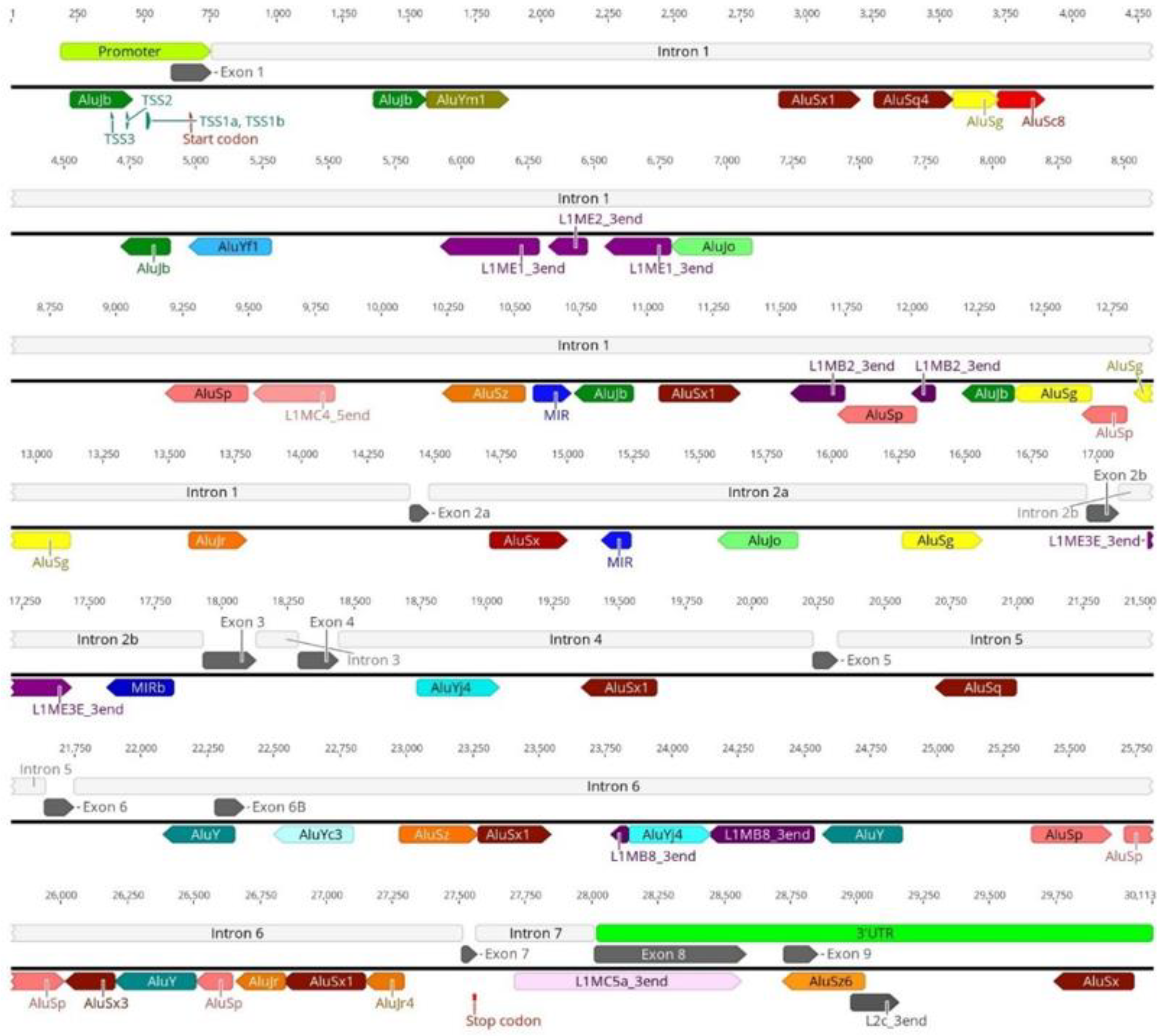
3.1. Transposable Elements and SMN1 Transcription

As previously stated, SMN1 and SMN2 are highly enriched in TEs, including in key regions for gene transcription, such as the promoter and terminator regions. Our analysis of the SMN1/2 promoter identified insertion of an AluJb repeat inside the promoter regions of both genes (Figure 1). This AluJb sequence harbors several transcription regulatory motifs upstream of the most used transcription start site (TSS1) including a fetal transcription start site (TSS2) and another transcription start site (TSS3) (Figure 1). The EMBOSS Nucleotide Analysis tool identified many other regulatory motifs within this Alu element sequence (Figure 1). Our analysis showed that all samples in study, SMA patients, carriers, non-carriers, healthy and the reference SMN1/2 sequence from Ensembl exhibit this Alu insertion in the promoter as well as harbor the two alternative transcriptional start sites and remaining regulatory motifs (TSS2 and TSS3) (Figure 2).
Interestingly, the region upstream of SMN1/2 promoter exhibits differences in TE insertion sites and subfamilies present between samples. We hypothesize that the untranslated regions may be subjected to less evolutionary pressure thereby allowing more diversity in TE insertions. With respect to the gene region downstream of exon 7, our first analysis of SMN1/2 gene reference sequence obtained from Ensembl showed several TE insertions belonging to various subfamilies (Figure 3A). The last canonical exon (exon 8) of SMN1/2 is located within the terminator region of the gene and primarily serves as the 3′UTR region of the gene [34,53]. We detected a L1 insertion within this exon. This is a truncated L1 insertion corresponding to the 3′ end of a L1MC5a subfamily retrotransposon (Figure 3B). Our analysis of the remaining samples in study showed that independently of being SMA patients (exhibiting SMN1 deletion), SMA carriers, non-carriers or healthy genomes, all samples show this L1 insertion inside exon 8 (Figure 4). Given that exon 8 serves as the 3′UTR region of the gene and that the L1 insertion within exon 8 is present in all samples regardless of disease status, we argue that the insertion of this retrotransposon in the 3′UTR region arises from a domestication event which gave this gene a novel, alternative terminator.
To determine whether the L1MC5a element inserted in exon 8 is being expressed in the SMN1 coding sequence, we conducted a deeper analysis of the SMN1 CCDS. Analysis of the longest SMN1 transcript (GenBank: BC062723.1) in Dfam showed a L1 element at the 3′ end of the CCDS, indicating that these complete SMN1 transcripts have an imbedded TE sequence derived from the L1MC5a insertion in exon 8 (Figure 5). Interestingly, the predominant transcript of SMN1, isoform d (CCDS34181.1), does not contain this L1 insertion (Figure 5). These results suggest that the alternative terminator function of the L1 element in exon 8 is correct since the L1 insertion is only detected in the longest isoform of SMN1 transcripts and effectively functions as an alternative transcription terminator to the canonical SMN1 terminator.
Since SMN1 and SMN2 share extensive sequence homology and nearly identical TE insertional patterns, we analyzed SMN2 transcripts and found that the longest isoform (BC000908.2) contains the same L1MC5a insertion in a similar 3′ location to that seen in the longest SMN1 transcript (results not shown). Similar to SMN1, the remaining and more common SMN2 transcripts isoforms (d, a, b and c) do not exhibit any L1 insertion in their sequence (results not shown).

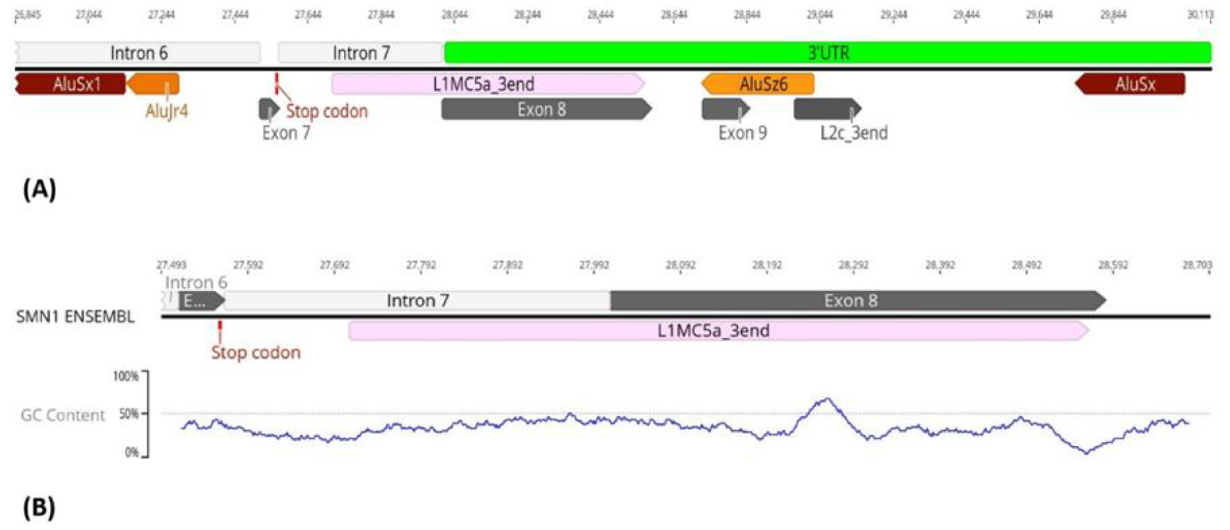
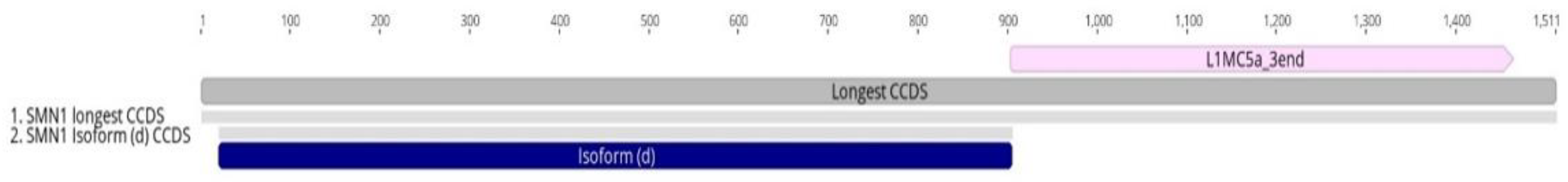
3.2. Transposable Elements and Alternative Splicing by Exonization

SMN1/2 introns are highly enriched in Alu-derived repeats with many of them in an inverted orientation (Figure 6). These inverted Alu repeats—mainly AluY, AluJr and AluSx1—span the whole locus but accumulate particularly in intron 1, 2a, 4 and 6. Our analysis of the remaining samples showed that all samples have the same pattern of Alu insertions in SMN1/2 introns with the key Alu repeats involved in the genes’ transcripts circularization being located in the same position and orientation as in the SMN1 reference sequence obtained from Ensembl (Figure 7).
Another important role of these inverted Alu repeats located in SMN1/2 introns is a complex TE domestication event that gives the genes new coding sequences, in a process termed exonization. Among the several TE families capable of forming new exons, Alu elements are particularly prone to be domesticated as alternative exons [3,18]. It has been estimated that 5% of all alternatively spliced human exons derive from the exonization of Alu elements [54,55]. In SMN1 and SMN2, two alternative exons resulting from exonization have been reported thus far, exon 6B [56,57] and exon 9 [58]. These two exonization events differ in the Alu subfamily involved, as well in the Alu arm involved in the exonization. Exon 6B results from exonization of the left arm of an inverted AluY element and exon 9 originates from the right arm of an antisense AluSz element (Figure 8).
We identified some insertional polymorphisms regarding the Alu subfamily inserted in exon 6B and exon 9 gene locations. In the case of exon 6B, most samples showed an expected inverted AluY insertion but we also found some cases where the exonization involved an inverted AluSc8 insertion instead (Figure 9). Because of the high similarity of the consensus sequences of AluY and AluSc8 (98.4% sequence identity) and the fact that AluSc8 subfamily is thought to be the evolutionary progenitor of the younger (Y) Alu subfamily [59,60], we hypothesize that the two different results obtained is due to an incorrect prediction made by the algorithm as opposed to TE insertion variability in this location. Additionally, the AluSc8 insertion was only observed in sequences obtained from the 1000 Genomes Project database. In other words, this AluSc8 insertion may be due to low sequencing read depth that is characteristic of the 1000 Genomes Project samples.
Extensive insertional polymorphisms were also detected for exon 9 in the analyzed samples. Instead of the expected AluSz TE as described by [58], we observed AluSz6 and AluSx insertions in addition to the expected AluSz, as described (Figure 10). Since there is no clear connection between this polymorphic insertion and SMA disease state, we conclude that the polymorphism of this insertion may be due to interindividual variability.

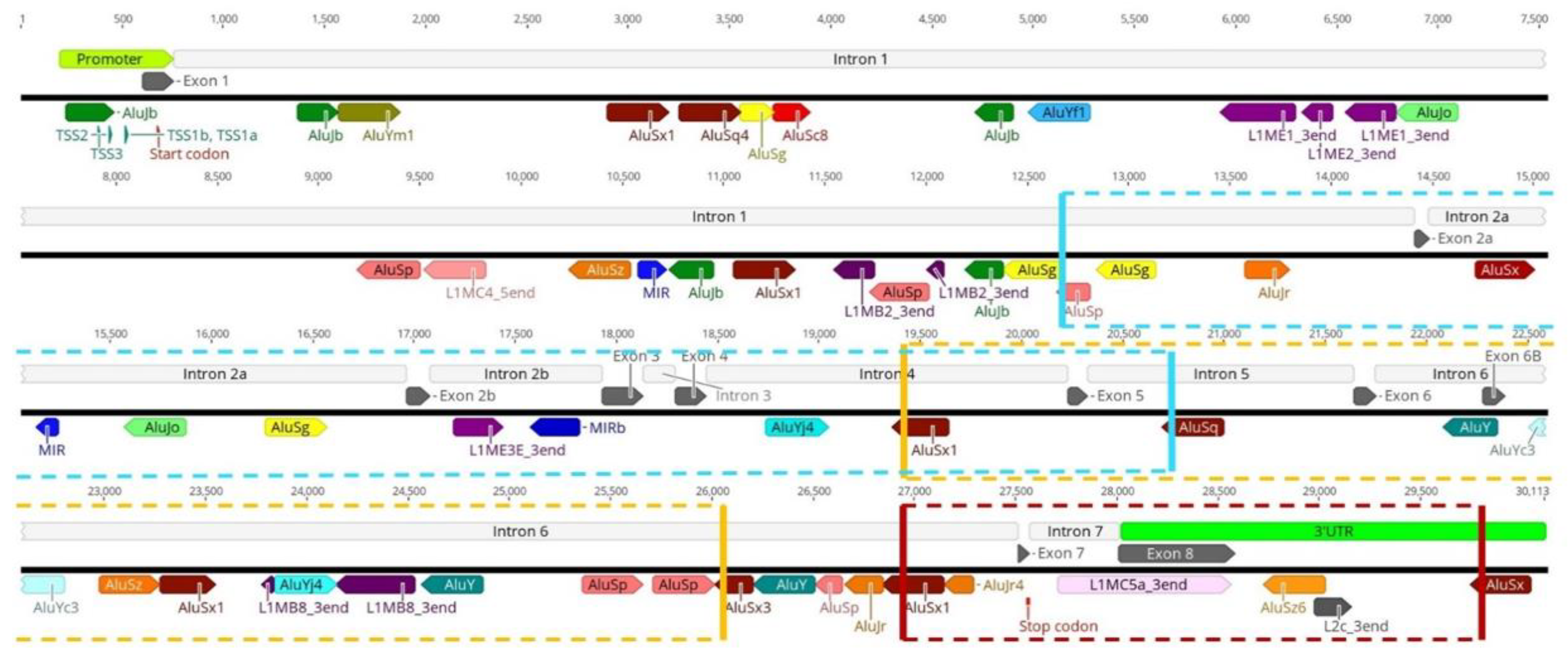
3.3. Transposable Elements and Partial Deletions of SMN1

The most direct link between activity of TEs and SMA onset stems from their ability to mediate recombination events that are known to lead to disease-associated deletions and other genomic rearrangements [8,61]. The presence of several Alu repeats within SMN1/2 that are in close proximity to each other, as previously discussed, make these genes particularly prone to Alu/Alu recombination events. The first Alu mediated deletion reported in SMN1 is a deletion involving a large sequence of the gene from intron 4 to intron 6, involving exons 5 and 6 [37]. Our analysis of this breakpoint revealed that an AluSx1 element located in intron 4 and an AluSx3 in intron 6 are the Alu subfamilies responsible for the recombination event that led to exons 5 and 6 deletion (Figure 11; yellow box). Ruhno and colleagues [36] recently reported a partial deletion of the critical exons 7 and 8 whose breakpoints were within the Alu-rich intron 6 and the gene 3′UTR [36]. Our analysis revealed an AluSx1 element in intron 6 directly upstream of exon 7-and an AluSx insertion in the 3′UTR region downstream of exon 8 (Figure 11; red box). An Alu/Alu recombination event was reported in a SMA patient with a deletion of SMN1 exons 2A, 2B, 3, 4 and 5 [38]. Our analysis of this deletion event showed that an antisense AluSp of intron 1 is the most likely element to be involved in the recombination event with the antisense AluSq of intron 5 (Figure 11; blue box).

4. Discussion

In this work we have found that the sequences of the SMN1/2 genes are enriched in TE insertions, including in key gene regions. These insertions may have important effects on regulation, splicing, expression and overall stability of the genes. TE insertions in the 5′UTR and promoter region of protein coding genes are common events with whole-genome analyses showing that up to 25% of human genes have TEs in their promoter and/or untranslated regions [62,63,64]. SINEs in particular seem to be highly represented in these regions, owing to their higher affinity to G + C rich genome regions [63,65]. The presence of TEs in 5′ regions of genes brings an evolutionary advantage for both the TE and for the host genome. For the TE, it represents an opportunity for translation as insertion in this open chromatin environment promotes its expression, and therefore its transposition [9,66]. For the host genome, the presence of TEs in these critical regions is a potential source of novel regulatory sequences by fusing with/replacing a canonical gene promoter or alternatively serving as an alternative promoter either upstream or downstream of the canonical transcription start site [15,67,68,69].
While these domesticated TEs may be integrated in the gene regulatory network, the significance of the remaining motifs inside the AluJb sequence remains to be investigated. The TSSs present inside the Alu sequence are tissue-specific and/or developmental stage-specific TSSs, with TSS2 being used as a fetal transcription start site and the use of TSS3 is still unknown [53]. This AluJb most likely serves as an alternative promoter or even as a tissue/developmental stage-specific promoter to SMN1/2. The presence of this Alu insertion in all samples in study regardless of disease status suggests a complete domestication of this element as an alternative promoter of the genes. Therefore, this insertion has led to an increased complex regulatory network capable of altering SMN expression, both in cis and trans. Promoter regions harboring Alu elements are subject to regulation in trans by long noncoding RNAs (lncRNAs) [70]. Future studies will experimentally confirm the functional presence of these alternate TSSs by using rapid amplification of cDNA 5′ ends (5′RACE) and their roles in developmental stage-specific and tissue-specific regulation of SMN expression.
The majority of human genes use alternative polyadenylation sites that are embedded in TEs, suggesting that these can influence the 3′ end processing of host gene transcripts [71,72]. L1 and other TE insertions are capable of interfering with endogenous cis regulatory elements present in 3′UTRs by introducing miRNA binding sites, promoting RNA editing and introducing polyadenylation signals [6,11,67,73,74,75]. Polyadenylation signals contained within retrotransposon sequences often lead to truncated or elongated 3′UTRs of full-length gene transcripts by providing an alternative terminator. As a result, these TE capabilities could repress transcription from the affected gene. This observation strengthens our hypothesis that the L1 present in SMN1/2 3′UTR region serves as an alternative terminator for the genes transcription by giving rise to longer transcripts. We will experimentally confirm this hypothesis in future studies using rapid amplification of the 3′cDNA ends (3′RACE) in control and SMA cells to identify alternative 3′UTRs.
Two factors may explain how this L1 element became fixed in this gene region. First, exon 8 has a lower percentage of G + C content (36.4%) when compared with the whole gene region (42.3%). The lower G + C content may have favored an L1 insertion as these elements have a bias towards lower G + C regions of the genome [76]. Second, the fact that exon 8 serves as the 3′UTR may have facilitated insertion of the L1 element as pressure against TE insertions is often relaxed in these regions [67,77,78]. We speculate, however, that such a large L1 insertion inside the gene coding region most likely had a large impact in the gene sequence and regulation and therefore should have been under negative selection pressure. Accordingly, L1 elements are especially underrepresented within genes, particularly those in the same transcriptional direction as the gene, because of their size and interference originated by retroelement regulatory motifs such as polyadenylation signals [1,3,77]. Thus, the insertion of this L1 element in the last exon of SMN1/2, that functions as the 3′UTR of the gene, is an example of a relatively rare event. Furthermore, the L1 insertion in exon 8 does not belong to the active L1 elements (also known as “hot L1 elements”) of the human genome that are composed only of the L1PA1 and L1PA2 subfamilies [3,9]. Therefore, this insertion is likely to be fixed in the human genome and not the result of a recent transposition event.
3′UTR retrotransposon insertions reduce mRNA expression [1,67,79]. The presence of this L1MC5a element in the longer SMN1 isoform transcripts could explain why these transcripts are less common than their shorter counterparts. Interestingly, weakly expressing genes were found to be rich in LINE insertions what can be explained by the ability of L1 elements to disrupt transcriptional elongation based on the presence of strong polyA signals in their sequences that possibly function as transcriptional terminators [80]. We argue that the alternative terminator provided by the L1 element is used less frequently than the canonical gene terminator located in exon 7 and may only be used in a tissue/time-specific manner. Accordingly, alternative UTRs that are often provided by TE insertions, can determine tissue-specific functions of mRNAs [81,82]. 3′UTR retrotransposons insertional events, such as the L1 insertion in the 3′ region of SMN1/2 reported in this study, are only moderately selected against and may provide a gradual mechanism of evolution by which retrotransposons alter the expression profile and influence crucial gene networks in the human genome [55,67,83].
Additionally, it is possible that this L1MC5a element has the same transcription terminator function in SMN2 as it shares the same TE insertional patterns in its sequence with SMN1. Accordingly, the longest SMN2 transcript isoform presents this L1 insertion in its 3′ region, in an identical sequence position as in SMN1 longest transcript. This hints to this retrotransposon having the same alternative terminator role in SMN2 transcription to that in SMN1 transcription. We hypothesize that this alternative terminator role can have implications in SMA severity by reducing SMN2 mRNA expression due to the presence of this L1MC5a element within SMN2. The confirmation of this hypothesis will require further analysis using a combination of experimental and in silico methodologies.
Circular RNAs (circRNAs) are a widely expressed class of non-colinear RNAs generated in a diverse set of eukaryotic organisms [84]. Due to their lack of 5′ and 3′ termini, these RNAs are extremely stable meaning that even small levels of circRNAs may affect cellular metabolism by sequestering/sponging miRNAs, sequestration and trafficking of proteins, regulation of transcription and generation of short RNA-binding proteins [84,85]. CircRNAs are important regulators of cellular physiology and also potential biomarkers of disease onset or progression [84,86]. Circular RNAs have been associated with various human diseases, particularly cancer, diabetes mellitus, cardiovascular diseases, chronic inflammatory diseases and neurological disorders [84,85,87]. The most common way in which circRNAs are generated is through backsplicing in which the 5′ splice site of a downstream exon is paired with the 3′ splice site of an upstream exon [58,88]. One of the defining features of backsplicing events appears to be the RNA secondary structure formed by inverted short repeats, especially Alu elements, within intronic sequences upstream and downstream of the 3′ and 5′ splice sites [58]. The existence of several inverted Alu repeats here detected throughout the SMN1 reference sequence explains the high levels of circularization of SMN1/2 transcripts as Alu repeats located in introns 4 and 5 are especially active in this process [35,53,58]. Pairing between the longest and highly Alu-enriched regions, introns 1 and 6, could be favored by several inverted Alu repeats thus potentially favoring backsplicing between exon 6 and exon 2A [35]. Additionally, generation of circRNAs with exons 2A, 2B, 3 and 4 requires pairing of the 5′ splice site of exon 4 with the 3′ splice site of exon 2A, which is made possible by the fact that intron 1 contains numerous Alu elements that are capable of pairing with the intron 4 Alu elements [33]. In future studies, we will experimentally confirm the presence of these putative exonization events mediated by circRNAs in SMA cells using RNA sequencing [89]. The presence of these inverted repeats that are in a favorable position to pair with each other, is often associated with alternative splicing events leading to circRNA biogenesis. Given that high levels of Alu inverted repeats in SMN1/2 introns may explain how these genes generate several circular RNAs, we believe that circRNA biogenesis occurs in healthy individuals and SMA patients to the same extent. It is possible, however, that dysregulation of these Alu repeats in SMA-affected genomes may lead to an increased formation of circRNAs coded by SMN1. These higher levels of circRNAs formation and the widespread alternative circularization of SMN1/2 pre-mRNA may have a still undiscovered role in SMA onset or may contribute to worse SMA phenotypes, owing to circRNAs ability to interfere with the coding capacity of human genes [35,58,84]. Additionally, circRNA formation in SMN1/2 may function as a potential biomarker for the genes’ overall transcriptional/splicing stability since higher circRNAs levels indicate aberrant RNA splicing events that may be linked to SMA.
The two exonization events within the SMN genes, exon 6B [56,57] and exon 9 [58], have been generated by different Alu insertions. All known Alu recombination events that led to deletions of SMN1 occurred among Alu elements of the (S) subfamily supporting the idea that sequence identity between the two elements at a locus—alongside proximity—appear to be proportional to their chances of successful recombination [61,90,91]. The high conservation of position and orientation of the Alu insertions involved in these exonization events implies that these domestication events occur identically in healthy and diseased genomes. Furthermore, they are important sources of novel exons that increase the coding capacity of the genes beyond the coding capacity of SMN1/2 canonical exons. In a wider spectrum, Alu and other TEs provide transcriptome diversity and ultimately result in the diversification of the human proteome.
Some insertional polymorphisms that were observed in these Alu repeats between the analyzed samples may be the result of normal interpersonal sequence variability. Accordingly, polymorphic Alu elements account for 17% of structural variants in the human genome, clearly establishing a link between individual TE polymorphisms and human genetic variation [92]. Since the SMA samples here analyzed are from individuals lacking the SMN1 gene, we cannot draw definitive conclusions about the involvement of Alu elements in the deletion events in a disease context. Our results show that in the analyzed SMA patients and in the remaining samples, the critical Alu elements responsible for these deletions are also present in the same position and orientation in the SMN1/2 sequence as in the gene reference sequence and as described previously [36,37,38]. Although these Alu insertions sites are most probably the reason for the complex deletion events, the high conservation of orientation and of subfamily type found in all the samples analyzed (healthy and diseased) implies that their presence per se is not the reason for the deletion events. Their presence in SMN1/2 introns is a source of sequence homology that can be responsible for genomic rearrangements and consequently disease in some genomes [8,61]. TE recombination may be responsible for the approximately 2% of SMA cases that result from de novo mutations in SMN1 and are not inherited from carrier parents [27]. It is likely that the several Alu repeats present throughout SMN1 are responsible for de novo deletions in germinative cells due to unequal recombination since TE silencing mechanisms are often relaxed in these developmental stages [3,9,19]. Alu elements may indeed, play an important role in the high instability of the SMN1/2 genomic region leading to disease-causing deletions of SMN1 exons and potentially whole gene deletions under specific circumstances. Alu elements invasion of SMN1/2 makes these genes very susceptible to Alu-mediated deletions, that have critical consequences to genome stability and host health.

5. Conclusions

Our analysis of the SMN genes revealed a pervasive invasion of its sequence by TEs that we believe may severely impact these genes’ regulation structure, expression and overall genomic stability. The several TEs present inside these genes, especially Alu and L1 elements that are highly enriched in the promoter and intronic regions of the gene, seem to play important roles in gene expression, novel exon creation, alternative splicing and deletion events known to lead to SMA. Additionally, a L1 element insertion in the 3′UTR region of the gene is also responsible for a domestication event that gave the gene an alternative terminator, therefore increasing the diversity of SMN1 transcripts and being a prime example of how a TE insertion inside a protein-coding gene can create a gradual mechanism of evolution by which retrotransposons alter the human transcriptome.
The in silico analysis of SMN1/2 completed in this work serves as a starting point for further investigations on the impacts of TEs in human disease and particularly, their role in SMA onset and severity. While the TEs identified in this in silico analysis were present in both SMN1 and SMN2, it is possible that they may affect the regulation of these genes differently. Future studies will further characterize the effects of these TEs in SMN1 and SMN2 on gene regulation under healthy conditions as well as in SMA.

Author Contributions

A.P. performed the analyses and wrote the manuscript, C.C. assisted in the analyses and manuscript writing, R.C. assisted in data interpretation and manuscript revision, M.E.R.B. provided the whole genome sequencing datasets and assisted in data interpretation as well as in manuscript writing and revision and F.A. conceptualized the study, supervised implementation and analyses, assisted in data interpretation and manuscript writing and finalization. All authors have read and agreed to the published version of the manuscript.

Funding

This work was supported by UIDB/04046/2020 and UIDP/04046/2020 Centre grants from FCT, Portugal (to BioISI); National Institutes of Health, USA (MERB; P20GM103446 and P20GM103464) and the Nemours Foundation (MERB).

Institutional Review Board Statement

This study was conducted in accordance with the Declaration of Helsinki and approved by the Institutional Review Board of Nemours Children’s Health (#764456, approved initially on 25 June 2015).

Informed Consent Statement

Informed consent was obtained from all subjects involved in this study.

Data Availability Statement

The datasets analyzed during this study are available from Butchbach ([email protected]) on reasonable request.

Acknowledgments

We deeply thank Xiao Chen, Michael Eberle and Ryan Taft at Illumina Inc. for providing the genome sequencing dataset from 37 SMA patients and the two healthy individuals.

Conflicts of Interest

The authors declare no conflict of interest.

References

  1. Hancks, D.C.; Kazazian, H.H. Roles for retrotransposon insertions in human disease. Mob. DNA 2016, 7, 9. [Google Scholar] [CrossRef] [PubMed] [Green Version]
  2. Jangam, D.; Feschotte, C.; Betrán, E. Transposable element domestication as an adaptation to evolutionary conflicts. Trends Genet. 2017, 33, 817–831. [Google Scholar] [CrossRef] [PubMed]
  3. Bourque, G.; Burns, K.H.; Gehring, M.; Gorbunova, V.; Seluanov, A.; Hammell, M.; Imbeault, M.; Izsvák, Z.; Levin, H.L.; Macfarlan, T.S. Ten things you should know about transposable elements. Genome Biol. 2018, 19, 199. [Google Scholar] [CrossRef]
  4. Platt, R.N.; Vandewege, M.W.; Ray, D.A. Mammalian transposable elements and their impacts on genome evolution. Chromosome Res. 2018, 26, 25–43. [Google Scholar] [CrossRef] [Green Version]
  5. Cosby, R.L.; Chang, N.-C.; Feschotte, C. Host-transposon interactions: Conflict, cooperation, and cooption. Genes Dev. 2019, 33, 1098–1116. [Google Scholar] [CrossRef] [PubMed] [Green Version]
  6. Garcia-Perez, J.L.; Widmann, T.J.; Adams, I.R. The impact of transposable elements on mammalian development. Development 2016, 143, 4101–4114. [Google Scholar] [CrossRef] [PubMed] [Green Version]
  7. Paço, A.; Adega, F.; Chaves, R. LINE-1 retrotransposons: From ‘parasite’ sequences to functional elements. J. Appl. Genet. 2015, 56, 133–145. [Google Scholar] [CrossRef] [PubMed]
  8. Payer, L.M.; Burns, K.H. Transposable elements in human genetic disease. Nat. Rev. Genet. 2019, 20, 760–772. [Google Scholar] [CrossRef]
  9. Burns, K.H. Our Conflict with Transposable Elements and Its Implications for Human Disease. Annu. Rev. Pathol. Mech. Dis. 2020, 15, 51–70. [Google Scholar] [CrossRef] [Green Version]
  10. Batzer, M.A.; Deininger, P.L.; Hellmann-Blumberg, U.; Jurka, J.; Labuda, D.; Rubin, C.M.; Schmid, C.W.; Zietkiewicz, E.; Zuckerkandl, E. Standardized nomenclature for Alu repeats. J. Mol. Evol. 1996, 42, 3–6. [Google Scholar] [CrossRef]
  11. Chuong, E.B.; Elde, N.C.; Feschotte, C. Regulatory activities of transposable elements: From conflicts to benefits. Nat. Rev. Genet. 2017, 18, 71. [Google Scholar] [CrossRef] [PubMed] [Green Version]
  12. Rebollo, R.; Farivar, S.; Mager, D.L. C-GATE-catalogue of genes affected by transposable elements. Mob. DNA 2012, 3, 9. [Google Scholar] [CrossRef] [PubMed] [Green Version]
  13. Friedli, M.; Trono, D. The developmental control of transposable elements and the evolution of higher species. Annu. Rev. Cell Dev. Biol. 2015, 31, 429–451. [Google Scholar] [CrossRef] [PubMed]
  14. Zeng, L.; Pederson, S.M.; Kortschak, R.D.; Adelson, D.L. Transposable elements and gene expression during the evolution of amniotes. Mob. DNA 2018, 9, 17. [Google Scholar] [CrossRef]
  15. Sundaram, V.; Wysocka, J. Transposable elements as a potent source of diverse cis-regulatory sequences in mammalian genomes. Philos. Trans. R. Soc. B 2020, 375, 20190347. [Google Scholar] [CrossRef] [Green Version]
  16. Lander, E.S.; Linton, L.M.; Birren, B.; Nusbaum, C.; Zody, M.C.; Baldwin, J.; Devon, K.; Dewar, K.; Doyle, M.; FitzHugh, W. Initial sequencing and analysis of the human genome. Nature 2001, 409, 860–921. [Google Scholar]
  17. O’Donnell, K.A.; Burns, K.H. Mobilizing diversity: Transposable element insertions in genetic variation and disease. Mob. DNA 2010, 1, 21. [Google Scholar] [CrossRef] [Green Version]
  18. Vorechovsky, I. Transposable elements in disease-associated cryptic exons. Hum. Genet. 2010, 127, 135–154. [Google Scholar] [CrossRef]
  19. Saleh, A.; Macia, A.; Muotri, A.R. Transposable elements, inflammation, and neurological disease. Front. Neurol. 2019, 10, 894. [Google Scholar] [CrossRef] [Green Version]
  20. Hedges, D.J.; Deininger, P.L. Inviting instability: Transposable elements, double-strand breaks, and the maintenance of genome integrity. Mutat. Res. Fundam. Mol. Mech. Mutagenesis 2007, 616, 46–59. [Google Scholar] [CrossRef] [Green Version]
  21. Lapp, H.E.; Hunter, R.G. Early life exposures, neurodevelopmental disorders, and transposable elements. Neurobiol. Stress 2019, 11, 100174. [Google Scholar] [CrossRef] [PubMed]
  22. Percharde, M.; Sultana, T.; Ramalho-Santos, M. What doesn’t kill you makes you stronger: Transposons as dual players in chromatin regulation and genomic variation. BioEssays 2020, 42, 1900232. [Google Scholar] [CrossRef] [PubMed] [Green Version]
  23. Jönsson, M.E.; Garza, R.; Johansson, P.A.; Jakobsson, J. Transposable elements: A common feature of neurodevelopmental and neurodegenerative disorders. Trends Genet. 2020, 36, 610–623. [Google Scholar] [CrossRef] [PubMed]
  24. Crawford, T.O.; Pardo, C.A. The neurobiology of childhood spinal muscular atrophy. Neurobiol. Dis. 1996, 3, 97–110. [Google Scholar] [CrossRef] [Green Version]
  25. Kolb, S.J.; Kissel, J.T. Spinal muscular atrophy. Neurol. Clin. 2015, 33, 831–846. [Google Scholar] [CrossRef] [Green Version]
  26. Butchbach, M.E. Genomic variability in the survival motor neuron genes (Smn1 and smn2): Implications for spinal muscular atrophy phenotype and therapeutics development. Int. J. Mol. Sci. 2021, 22, 7896. [Google Scholar] [CrossRef]
  27. Wirth, B. Spinal muscular atrophy: In the challenge lies a solution. Trends Neurosci. 2021, 44, 306–322. [Google Scholar] [CrossRef]
  28. Lefebvre, S.; Bürglen, L.; Reboullet, S.; Clermont, O.; Burlet, P.; Viollet, L.; Benichou, B.; Cruaud, C.; Millasseau, P.; Zeviani, M.; et al. Identification and characterization of a spinal muscular atrophy-determining gene. Cell 1995, 80, 155–165. [Google Scholar] [CrossRef] [Green Version]
  29. Blasco-Pérez, L.; Paramonov, I.; Leno, J.; Bernal, S.; Alias, L.; Fuentes-Prior, P.; Cuscó, I.; Tizzano, E.F. Beyond copy number: A new, rapid, and versatile method for sequencing the entire SMN2 gene in SMA patients. Hum Mutat 2021, 42, 787–795. [Google Scholar] [CrossRef]
  30. Campbell, L.; Potter, A.; Ignatius, J.; Dubowitz, V.; Davies, K. Genomic variation and gene conversion in spinal muscular atrophy: Implications for disease process and clinical phenotype. Am. J. Hum. Genet. 1997, 61, 40. [Google Scholar] [CrossRef] [Green Version]
  31. Courseaux, A.; Richard, F.; Grosgeorge, J.; Ortola, C.; Viale, A.; Turc-Carel, C.; Dutrillaux, B.; Gaudray, P.; Nahon, J.-L. Segmental duplications in euchromatic regions of human chromosome 5: A source of evolutionary instability and transcriptional innovation. Genome Res. 2003, 13, 369–381. [Google Scholar] [CrossRef] [PubMed] [Green Version]
  32. Schmutz, J.; Martin, J.; Terry, A.; Couronne, O.; Grimwood, J.; Lowry, S.; Gordon, L.A.; Scott, D.; Xie, G.; Huang, W.; et al. The DNA sequence and comparative analysis of human chromosome 5. Nature 2004, 431, 268–274. [Google Scholar] [CrossRef]
  33. Ottesen, E.W.; Singh, R.N. Characteristics of circular RNAs generated by human Survival Motor Neuron genes. Cell. Signal. 2020, 73, 109696. [Google Scholar] [CrossRef] [PubMed]
  34. Ottesen, E.W.; Seo, J.; Singh, N.N.; Singh, R.N. A multilayered control of the human survival motor neuron gene expression by Alu elements. Front. Microbiol. 2017, 8, 2252. [Google Scholar] [CrossRef] [PubMed] [Green Version]
  35. Pagliarini, V.; Jolly, A.; Bielli, P.; Di Rosa, V.; De la Grange, P.; Sette, C. Sam68 binds Alu-rich introns in SMN and promotes pre-mRNA circularization. Nucleic Acids Res. 2020, 48, 633–645. [Google Scholar] [CrossRef] [PubMed]
  36. Ruhno, C.; McGovern, V.L.; Avenarius, M.R.; Snyder, P.J.; Prior, T.W.; Nery, F.C.; Muhtaseb, A.; Roggenbuck, J.S.; Kissel, J.T.; Sansone, V.A.; et al. Complete sequencing of the SMN2 gene in SMA patients detects SMN gene deletion junctions and variants in SMN2 that modify the SMA phenotype. Hum. Genet. 2019, 138, 241–256. [Google Scholar] [CrossRef] [PubMed]
  37. Wirth, B.; Herz, M.; Wetter, A.; Moskau, S.; Hahnen, E.; Rudnik-Schöneborn, S.; Wienker, T.; Zerres, K. Quantitative analysis of survival motor neuron copies: Identification of subtle SMN1 mutations in patients with spinal muscular atrophy, genotype-phenotype correlation, and implications for genetic counseling. Am. J. Hum. Genet. 1999, 64, 1340–1356. [Google Scholar] [CrossRef] [Green Version]
  38. Jedličková, I.; Přistoupilová, A.; Nosková, L.; Majer, F.; Stránecký, V.; Hartmannová, H.; Hodaňová, K.; Trešlová, H.; Hýblová, M.; Solár, P.; et al. Spinal muscular atrophy caused by a novel Alu-mediated deletion of exons 2a-5 in SMN1 undetectable with routine genetic testing. Mol. Genet. Genom. Med. 2020, 8, e1238. [Google Scholar] [CrossRef] [Green Version]
  39. Abecasis, G.R.; Altshuler, D.; Auton, A.; Brooks, L.D.; Durbin, R.M.; Gibbs, R.A.; Hurles, M.E.; McVean, G.A. A map of human genome variation from population-scale sequencing. Nature 2010, 467, 1061–1073. [Google Scholar] [CrossRef] [Green Version]
  40. Larson, J.L.; Silver, A.J.; Chan, D.; Borroto, C.; Spurrier, B.; Silver, L.M. Validation of a high resolution NGS method for detecting spinal muscular atrophy carriers among phase 3 participants in the 1000 Genomes Project. BMC Med. Genet. 2015, 16, 100. [Google Scholar] [CrossRef] [Green Version]
  41. Chen, X.; Sanchis-Juan, A.; French, C.E.; Connell, A.J.; Delon, I.; Kingsbury, Z.; Chawla, A.; Halpern, A.L.; Taft, R.J.; Bentley, D.R. Spinal muscular atrophy diagnosis and carrier screening from genome sequencing data. Genet. Med. 2020, 22, 945–953. [Google Scholar] [CrossRef] [Green Version]
  42. Stabley, D.L.; Harris, A.W.; Holbrook, J.; Chubbs, N.J.; Lozo, K.W.; Crawford, T.O.; Swoboda, K.J.; Funanage, V.L.; Wang, W.; Mackenzie, W. SMN1 and SMN2 copy numbers in cell lines derived from patients with spinal muscular atrophy as measured by array digital PCR. Mol. Genet. Genom. Med. 2015, 3, 248–257. [Google Scholar] [CrossRef] [Green Version]
  43. Jiang, L.; Lin, R.; Gallagher, S.; Zayac, A.; Butchbach, M.E.R.; Hung, P. Development and validation of a 4-color multiplexing spinal muscular atrophy (SMA) genotyping assay on a novel integrated digital PCR instrument. Sci. Rep. 2020, 10, 19892. [Google Scholar] [CrossRef]
  44. Stabley, D.L.; Holbrook, J.; Scavina, M.; Crawford, T.O.; Swoboda, K.J.; Robbins, K.M.; Butchbach, M.E.R. Detection of SMN1 to SMN2 gene conversion events and partial SMN1 gene deletions using array digital PCR. Neurogenetics 2021, 22, 53–64. [Google Scholar] [CrossRef]
  45. Kearse, M.; Moir, R.; Wilson, A.; Stones-Havas, S.; Cheung, M.; Sturrock, S.; Buxton, S.; Cooper, A.; Markowitz, S.; Duran, C.; et al. Geneious Basic: An integrated and extendable desktop software platform for the organization and analysis of sequence data. Bioinformatics 2012, 28, 1647–1649. [Google Scholar] [CrossRef]
  46. Pujar, S.; O’Leary, N.A.; Farrell, C.M.; Loveland, J.E.; Mudge, J.M.; Wallin, C.; Girón, C.G.; Diekhans, M.; Barnes, I.; Bennett, R.; et al. Consensus coding sequence (CCDS) database: A standardized set of human and mouse protein-coding regions supported by expert curation. Nucleic Acids Res 2018, 46, D221–D228. [Google Scholar] [CrossRef] [Green Version]
  47. Hubley, R.; Finn, R.D.; Clements, J.; Eddy, S.R.; Jones, T.A.; Bao, W.; Smit, A.F.; Wheeler, T.J. The Dfam database of repetitive DNA families. Nucleic Acids Res. 2016, 44, D81–D89. [Google Scholar] [CrossRef] [Green Version]
  48. Storer, J.; Hubley, R.; Rosen, J.; Wheeler, T.J.; Smit, A.F. The Dfam community resource of transposable element families, sequence models, and genome annotations. Mob. DNA 2021, 12, 2. [Google Scholar] [CrossRef]
  49. Sievers, F.; Wilm, A.; Dineen, D.; Gibson, T.J.; Karplus, K.; Li, W.; Lopez, R.; McWilliam, H.; Remmert, M.; Söding, J.; et al. Fast, scalable generation of high-quality protein multiple sequence alignments using Clustal Omega. Mol. Syst. Biol. 2011, 7, 539. [Google Scholar] [CrossRef]
  50. Germain-Desprez, D.; Brun, T.; Rochette, C.; Semionov, A.; Rouget, R.; Simard, L.R. The SMN genes are subject to transcriptional regulation during cellular differentiation. Gene 2001, 279, 109–117. [Google Scholar] [CrossRef]
  51. Monani, U.R.; McPherson, J.D.; Burghes, A.H. Promoter analysis of the human centromeric and telomeric survival motor neuron genes (SMNC and SMNT). Biochim. Biophys. Acta 1999, 1445, 330–336. [Google Scholar] [CrossRef]
  52. Rice, P.; Longden, I.; Bleasby, A. EMBOSS: The European Molecular Biology Open Software Suite. Trends Genet. TIG 2000, 16, 276–277. [Google Scholar] [CrossRef]
  53. Singh, N.N.; Ottesen, E.W.; Singh, R.N. A survey of transcripts generated by spinal muscular atrophy genes. Biochim. Biophys. Acta Gene Regul. Mech. 2020, 1863, 194562. [Google Scholar] [CrossRef]
  54. Sorek, R.; Ast, G.; Graur, D. Alu-containing exons are alternatively spliced. Genome Res. 2002, 12, 1060–1067. [Google Scholar] [CrossRef] [Green Version]
  55. Piriyapongsa, J.; Rutledge, M.T.; Patel, S.; Borodovsky, M.; Jordan, I.K. Evaluating the protein coding potential of exonized transposable element sequences. Biol. Direct 2007, 2, 31. [Google Scholar] [CrossRef] [Green Version]
  56. Seo, J.; Singh, N.N.; Ottesen, E.W.; Lee, B.M.; Singh, R.N. A novel human-specific splice isoform alters the critical C-terminus of Survival Motor Neuron protein. Sci. Rep. 2016, 6, 30778. [Google Scholar] [CrossRef] [Green Version]
  57. Yoshimoto, S.; Harahap, N.I.F.; Hamamura, Y.; Ar Rochmah, M.; Shima, A.; Morisada, N.; Shinohara, M.; Saito, T.; Saito, K.; Lai, P.S.; et al. Alternative splicing of a cryptic exon embedded in intron 6 of SMN1 and SMN2. Hum. Genome Var 2016, 3, 16040. [Google Scholar] [CrossRef] [Green Version]
  58. Ottesen, E.W.; Luo, D.; Seo, J.; Singh, N.N.; Singh, R.N. Human Survival Motor Neuron genes generate a vast repertoire of circular RNAs. Nucleic Acids Res. 2019, 47, 2884–2905. [Google Scholar] [CrossRef] [Green Version]
  59. Bao, W.; Kojima, K.K.; Kohany, O. Repbase Update, a database of repetitive elements in eukaryotic genomes. Mob. DNA 2015, 6, 11. [Google Scholar] [CrossRef] [Green Version]
  60. Martinez-Gomez, L.; Abascal, F.; Jungreis, I.; Pozo, F.; Kellis, M.; Mudge, J.M.; Tress, M.L. Few SINEs of life: Alu elements have little evidence for biological relevance despite elevated translation. NAR Genom. Bioinform. 2020, 2, lqz023. [Google Scholar] [CrossRef] [Green Version]
  61. Jahic, A.; Erichsen, A.K.; Deufel, T.; Tallaksen, C.M.; Beetz, C. A polymorphic Alu insertion that mediates distinct disease-associated deletions. Eur. J. Hum. Genet. EJHG 2016, 24, 1371–1374. [Google Scholar] [CrossRef] [Green Version]
  62. Van de Lagemaat, L.N.; Landry, J.R.; Mager, D.L.; Medstrand, P. Transposable elements in mammals promote regulatory variation and diversification of genes with specialized functions. Trends Genet. TIG 2003, 19, 530–536. [Google Scholar] [CrossRef]
  63. Jordan, I.K.; Rogozin, I.B.; Glazko, G.V.; Koonin, E.V. Origin of a substantial fraction of human regulatory sequences from transposable elements. Trends Genet. TIG 2003, 19, 68–72. [Google Scholar] [CrossRef]
  64. Huh, J.W.; Kim, D.S.; Ha, H.S.; Lee, J.R.; Kim, Y.J.; Ahn, K.; Lee, S.R.; Chang, K.T.; Kim, H.S. Cooperative exonization of MaLR and AluJo elements contributed an alternative promoter and novel splice variants of RNF19. Gene 2008, 424, 63–70. [Google Scholar] [CrossRef]
  65. Thornburg, B.G.; Gotea, V.; Makałowski, W. Transposable elements as a significant source of transcription regulating signals. Gene 2006, 365, 104–110. [Google Scholar] [CrossRef]
  66. Wells, J.N.; Feschotte, C. A Field Guide to Eukaryotic Transposable Elements. Annu. Rev. Genet. 2020, 54, 539–561. [Google Scholar] [CrossRef]
  67. Faulkner, G.J.; Kimura, Y.; Daub, C.O.; Wani, S.; Plessy, C.; Irvine, K.M.; Schroder, K.; Cloonan, N.; Steptoe, A.L.; Lassmann, T.; et al. The regulated retrotransposon transcriptome of mammalian cells. Nat. Genet. 2009, 41, 563–571. [Google Scholar] [CrossRef]
  68. Branco, M.R.; Chuong, E.B. Crossroads between transposons and gene regulation. Philos. Trans. R. Soc. Lond. Ser. B Biol. Sci. 2020, 375, 20190330. [Google Scholar] [CrossRef] [Green Version]
  69. Marasca, F.; Gasparotto, E.; Polimeni, B.; Vadalà, R.; Ranzani, V.; Bodega, B. The Sophisticated Transcriptional Response Governed by Transposable Elements in Human Health and Disease. Int. J. Mol. Sci. 2020, 21, 3201. [Google Scholar] [CrossRef]
  70. Hu, S.; Wang, X.; Shan, G. Insertion of an Alu element in a lncRNA leads to primate-specific modulation of alternative splicing. Nat. Struct. Mol. Biol. 2016, 23, 1011–1019. [Google Scholar] [CrossRef]
  71. Lee, J.Y.; Ji, Z.; Tian, B. Phylogenetic analysis of mRNA polyadenylation sites reveals a role of transposable elements in evolution of the 3′-end of genes. Nucleic Acids Res. 2008, 36, 5581–5590. [Google Scholar] [CrossRef] [PubMed] [Green Version]
  72. Cowley, M.; Oakey, R.J. Transposable elements re-wire and fine-tune the transcriptome. PLoS Genet. 2013, 9, e1003234. [Google Scholar] [CrossRef] [PubMed]
  73. Roy-Engel, A.M.; El-Sawy, M.; Farooq, L.; Odom, G.L.; Perepelitsa-Belancio, V.; Bruch, H.; Oyeniran, O.O.; Deininger, P.L. Human retroelements may introduce intragenic polyadenylation signals. Cytogenet. Genome Res. 2005, 110, 365–371. [Google Scholar] [CrossRef] [PubMed]
  74. Sela, N.; Kim, E.; Ast, G. The role of transposable elements in the evolution of non-mammalian vertebrates and invertebrates. Genome Biol. 2010, 11, R59. [Google Scholar] [CrossRef]
  75. Chen, L.-L.; Yang, L. ALUternative regulation for gene expression. Trends Cell Biol. 2017, 27, 480–490. [Google Scholar] [CrossRef] [Green Version]
  76. Deininger, P.L.; Moran, J.V.; Batzer, M.A.; Kazazian, H.H., Jr. Mobile elements and mammalian genome evolution. Curr. Opin. Genet. Dev. 2003, 13, 651–658. [Google Scholar] [CrossRef]
  77. Medstrand, P.; van de Lagemaat, L.N.; Mager, D.L. Retroelement distributions in the human genome: Variations associated with age and proximity to genes. Genome Res. 2002, 12, 1483–1495. [Google Scholar] [CrossRef] [Green Version]
  78. Kabelitz, T.; Bäurle, I. Get the jump—Do 3′UTRs protect transposable elements from silencing? Mob. Genet. Elem. 2015, 5, 51–54. [Google Scholar] [CrossRef] [Green Version]
  79. Lavi, E.; Carmel, L. Alu exaptation enriches the human transcriptome by introducing new gene ends. RNA Biol. 2018, 15, 715–725. [Google Scholar] [CrossRef]
  80. Jjingo, D.; Huda, A.; Gundapuneni, M.; Mariño-Ramírez, L.; Jordan, I.K. Effect of the transposable element environment of human genes on gene length and expression. Genome Biol. Evol. 2011, 3, 259–271. [Google Scholar] [CrossRef] [Green Version]
  81. Hughes, T.A. Regulation of gene expression by alternative untranslated regions. Trends Genet. TIG 2006, 22, 119–122. [Google Scholar] [CrossRef] [PubMed]
  82. Sela, N.; Mersch, B.; Gal-Mark, N.; Lev-Maor, G.; Hotz-Wagenblatt, A.; Ast, G. Comparative analysis of transposed element insertion within human and mouse genomes reveals Alu’s unique role in shaping the human transcriptome. Genome Biol. 2007, 8, R127. [Google Scholar] [CrossRef] [PubMed]
  83. Petri, R.; Brattås, P.L.; Sharma, Y.; Jönsson, M.E.; Pircs, K.; Bengzon, J.; Jakobsson, J. LINE-2 transposable elements are a source of functional human microRNAs and target sites. PLoS Genet. 2019, 15, e1008036. [Google Scholar] [CrossRef] [PubMed] [Green Version]
  84. Kristensen, L.S.; Andersen, M.S.; Stagsted, L.V.W.; Ebbesen, K.K.; Hansen, T.B.; Kjems, J. The biogenesis, biology and characterization of circular RNAs. Nat. Rev. Genet. 2019, 20, 675–691. [Google Scholar] [CrossRef]
  85. Greene, J.; Baird, A.M.; Brady, L.; Lim, M.; Gray, S.G.; McDermott, R.; Finn, S.P. Circular RNAs: Biogenesis, Function and Role in Human Diseases. Front. Mol. Biosci. 2017, 4, 38. [Google Scholar] [CrossRef]
  86. Haque, S.; Harries, L.W. Circular RNAs (circRNAs) in Health and Disease. Genes 2017, 8, 353. [Google Scholar] [CrossRef] [Green Version]
  87. Chen, Y.J.; Chen, C.Y.; Mai, T.L.; Chuang, C.F.; Chen, Y.C.; Gupta, S.K.; Yen, L.; Wang, Y.D.; Chuang, T.J. Genome-wide, integrative analysis of circular RNA dysregulation and the corresponding circular RNA-microRNA-mRNA regulatory axes in autism. Genome Res. 2020, 30, 375–391. [Google Scholar] [CrossRef] [Green Version]
  88. Voellenkle, C.; Perfetti, A.; Carrara, M.; Fuschi, P.; Renna, L.V.; Longo, M.; Sain, S.B.; Cardani, R.; Valaperta, R.; Silvestri, G.; et al. Dysregulation of Circular RNAs in Myotonic Dystrophy Type 1. Int. J. Mol. Sci 2019, 20, 1938. [Google Scholar] [CrossRef] [Green Version]
  89. Florea, L.; Payer, L.; Antonescu, C.; Yang, G.; Burns, K. Detection of Alu Exonization Events in Human Frontal Cortex From RNA-Seq Data. Front. Mol. Biosci. 2021, 8, 727537. [Google Scholar] [CrossRef]
  90. Sen, S.K.; Han, K.; Wang, J.; Lee, J.; Wang, H.; Callinan, P.A.; Dyer, M.; Cordaux, R.; Liang, P.; Batzer, M.A. Human genomic deletions mediated by recombination between Alu elements. Am. J. Hum. Genet. 2006, 79, 41–53. [Google Scholar] [CrossRef] [Green Version]
  91. Kim, S.; Cho, C.-S.; Han, K.; Lee, J. Structural Variation of Alu Element and Human Disease. Genom. Inform. 2016, 14, 70–77. [Google Scholar] [CrossRef] [PubMed] [Green Version]
  92. Kryatova, M.S.; Steranka, J.P.; Burns, K.H.; Payer, L.M. Insertion and deletion polymorphisms of the ancient AluS family in the human genome. Mob. DNA 2017, 8, 6. [Google Scholar] [CrossRef] [PubMed] [Green Version]
Figure 1. AluJb element within the SMN1/2 promoter region. An AluJb element (represented by a dark green arrow) is inserted inside the promoter region of SMN1/2, upstream of the canonical transcriptional start site (TSS1a). Transcriptional start sites (TSS), two of them located inside the AluJb sequence, and the start codon are represented by green and red arrowheads, respectively.
Biology 11 00824 g001
Figure 2. Comparison of the SMN1/2 promoter regions within sample cohort. An AluJb insertion (represented by a dark green arrow) is present within the gene promotor region of all samples. Some polymorphic insertions were detected upstream of the promotor region and the 5′UTR; there were no connections, however, between these polymorphic insertions and SMA phenotype.
Biology 11 00824 g002
Figure 3. Identification of TEs within the SMN1/2 3′UTR. (A) SMN1/2 3′UTR region is enriched in transposable elements (represented in colored arrows). Exon 8 is considered part of the 3′UTR region of the gene. An L1MC5a element (pink arrow) is inserted in SMN1/2 exon 8 (grey arrow). (B) A large L1 insertion (represented by a colored arrow) was detected in SMN1/2 last canonical exon, exon 8 (represented by a grey arrow). G + C analysis of the region showed a general lower G + C content in this region compared with the adjacent gene regions, partially explaining how a L1 insertion occurred in this region.
Biology 11 00824 g003
Figure 4. Comparison of SMN1/2 exon 8 within the sample cohort. All samples in study, independently of disease status, exhibit the L1 insertion (represented by a pink arrow) inside exon 8 (represented by a grey arrow) suggesting that the L1 element inserted in exon 8 is indeed fixed in the population and that it has a biological role in SMN1/2 regulation.
Biology 11 00824 g004
Figure 5. Identification of TEs within the SMN1 Consensus Coding Sequences (CCDSs). SMN1 longest isoform CCDS represented in grey on top has a L1MC5a element (represented by a pink arrow) inserted in its sequence responsible for the extension of the CCDS. Contrarily, SMN1 most common CCDS, isoform (d; represented as a blue bar), is shorter and does not have any TE insertion in its sequence. Both sequences show 100% sequence identity within the overlapped region. We also observed the presence of a L1MC5a element within the longest CCDS for SMN2.
Biology 11 00824 g005
Figure 6. Identification of TEs within the reference SMN1/2 gene sequence. SMN1/2 exons are represented by grey arrows and introns by white boxes. SMN1/2 promoter is represented by a green box and other regulatory motifs by green arrows. Start and stop codons are represented by small red arrows. Transposable elements position and orientation is indicated by colored arrows, with the direction of the arrow indicating the orientation of the repeat element.
Biology 11 00824 g006
Figure 7. Identification of Key Alu repeats involved in RNA circularization events within SMN1/2. Comparison between key Alu repeats involved in SMN1/2 circularization events (inside the red boxes) located in introns 1, 4, 5 and 6. Independently of disease status, a conservation of position and orientation of the Alu insertions is visible. SMA patient samples (MB228 and MB358); SMA carriers (HG00281 and HG01085); healthy samples (MB109 and MB342) and Non-carrier samples (HG01341 and NA18629). Color codes for the arrows: green, AluJb; pink, AluSp; yellow, AluSg; orange, AluSz6; light green, AluJo; dark red, AluSx1; teal, AluY; light blue, AluYc and red, AluSc8.
Biology 11 00824 g007
Figure 8. Identification of SMN1/2 alternative exonization events. On top, exonization event of an intronic antisense Alu repeat (represented by a green arrow) that gave birth to alternative exon 6B. Below, another exonization event of an antisense Alu element (represented by an orange arrow) that resulted in the formation of SMN1/2 alternative exon 9. G + C content analysis of both regions shows a higher G + C content in the exonization regions when compared with the surrounding areas, which might have favored Alu insertions and the posterior exonization events.
Biology 11 00824 g008
Figure 9. Comparison of the exon 6B region within the sample cohort. An AluY insertion (represented by a teal arrow) gives rise to alternative exon 6B in all healthy and SMA samples, including the reference SMN1 ENSEMBL sequence. Contrarily, an AluSc8 (represented by a red arrow) insertion is present instead of the AluY element in the remaining samples. This Alu insertion difference is most likely the result of low sequencing read depth of the 1000 Genomes Project samples.
Biology 11 00824 g009
Figure 10. Comparison of the exon 9 region within the sample cohort. Extensive insertional polymorphisms were detected for exon 9 region in the analyzed samples. While the expected AluSz insertion (represented by a light orange arrow) was present in some samples, other AluSz6 and AluSx insertions (represented by an orange arrow and a dark red arrow, respectively) were observed in this region. This polymorphism may be due to interpersonal variability and is not associated with SMA.
Biology 11 00824 g010
Figure 11. Identification of Alu-mediated partial deletions within SMN1. The several Alu-derived repeats existent in SMN1 introns provide a fertile source of Alu/Alu recombination events, known to lead to gene deletions. To date, three Alu-mediated deletion events were reported in SMN1. The more common deletion involving exon 7 and 8 is represented by a red box. A deletion event first described by Wirth et al. [37] involving exons 5 and 6 is indicated by a yellow box. Lastly, the more recently reported Alu-mediated deletion in SMN1 is highlighted by a blue box.
Biology 11 00824 g011
Table 1. 1000 Genomes Project sample cohort.
Sample IDCarrier ProbabilityCarrier Status
HG021341Carrier
NA123831Carrier
HG017731Carrier
HG003461Carrier
HG002811Carrier
HG020871Carrier
HG010851Carrier
HG018931Carrier
HG003240.997Carrier
NA207640.982Carrier
HG022650.982Carrier
HG020790.976Carrier
HG039530.972Carrier
HG012480.935Carrier
HG014920.914Carrier
HG018920.902Carrier
HG005250.763Carrier
HG012050.756Carrier
HG010940.738Carrier
NA119320.716Carrier
HG006290.000165Non-Carrier
HG035850.000159Non-Carrier
HG013410.000155Non-Carrier
HG003250.000151Non-Carrier
HG003690.000134Non-Carrier
NA208780.000131Non-Carrier
HG040230.000127Non-Carrier
HG021360.000126Non-Carrier
NA197320.000126Non-Carrier
HG006840.000126Non-Carrier
NA186290.000126Non-Carrier
NA208640.000122Non-Carrier
HG031960.000112Non-Carrier
HG003720.000111Non-Carrier
NA128890.0000997Non-Carrier
NA197250.0000982Non-Carrier
HG037200.0000969Non-Carrier
HG007420.0000923Non-Carrier
NA203420.0000907Non-Carrier
HG039680.0000851Non-Carrier
Table 2. Phenotypic and copy number information for samples within the MNDRL Cohort.
Copy Number (Exon 7)Copy Number (Exon 8)
Sample IDPhenotypeSMN1SMN2SMN1SMN2
MB109healthy3121
MB342healthy2020
MB106SMA II0303
MB110SMA I0303
MB112SMA II0303
MB114SMA I0202
MB125SMA III0303
MB219SMA I0202
MB222SMA II0303
MB230SMA III0303
MB231SMA III0404
MB232SMA II0303
MB233SMA I0303
MB234SMA I0202
MB352SMA II0303
MB354SMA II0303
MB355SMA II0303
MB356SMA III0404
MB357SMA II0303
MB358SMA II0303
MB361SMA II0303
MB362SMA I0202
MB364SMA I0202
MB375SMA II0303
MB377SMA III0404
MB378SMA II0303
MB388SMA III0303
MB488SMA I0202
MB489SMA I0202
MB501SMA I0202
MB503SMA III0404
MB507SMA I0202
MB509SMA I0202
MB510SMA I0202
MB511SMA II0303
MB513SMA III0303
MB691SMA I0202
MB692SMA I0202
MB693SMA I0202
Publisher’s Note: MDPI stays neutral with regard to jurisdictional claims in published maps and institutional affiliations.

