
Nonoperative Treatment of Finger Flexor Tenosynovitis in Sport Climbers—A Retrospective Descriptive Study Based on a Clinical 10-Year Database


 ,
,
Abstract
:Simple Summary
Abstract
1. Introduction
2. Materials and Methods
2.1. Study Design and Participants
2.2. Clinical and Radiographical Diagnosis
2.3. Treatment Algorithm
2.4. Follow-Up Questionnaire
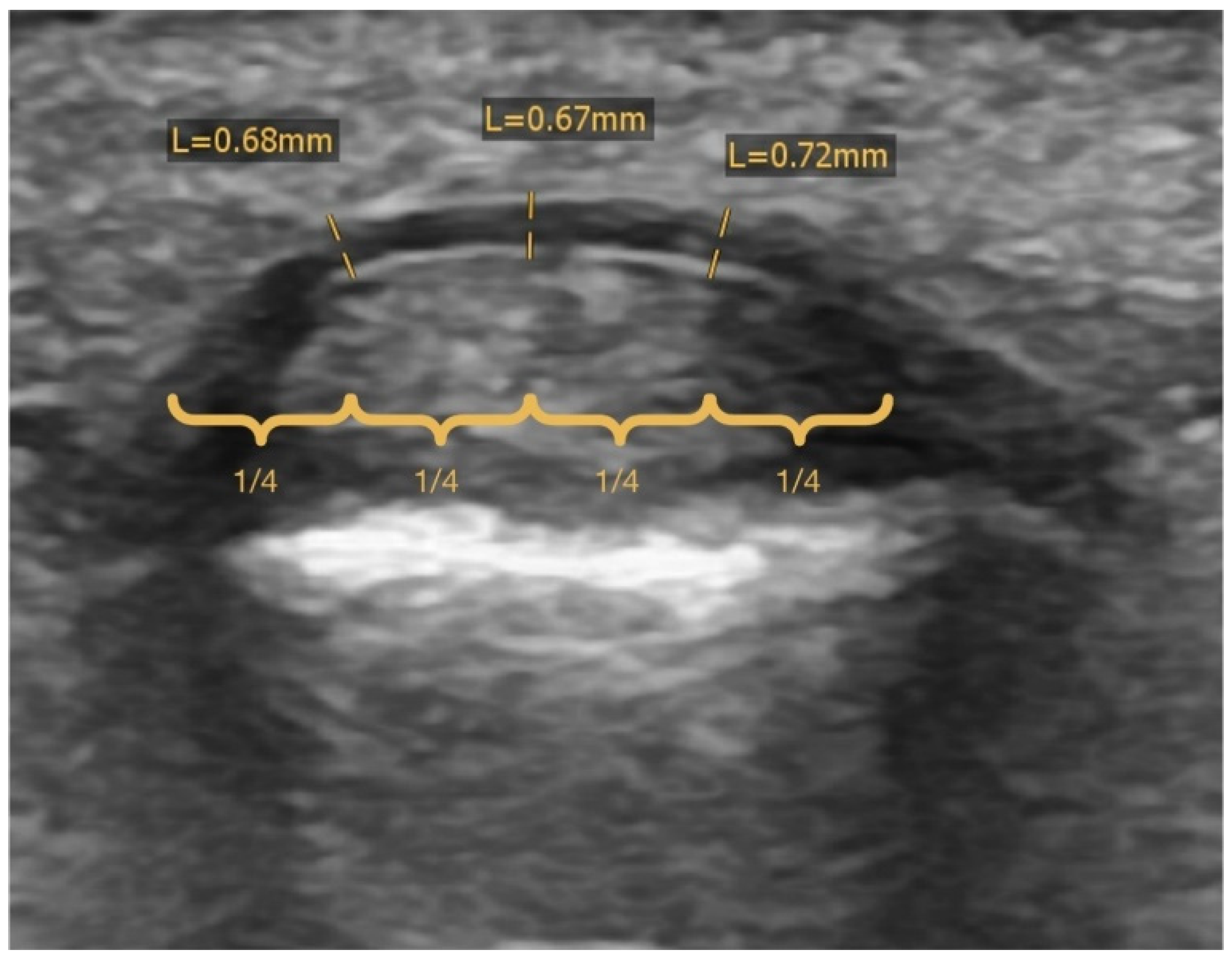
2.5. Radiographic Analysis
2.6. Statistical Analysis
3. Results
3.1. Baseline Characteristics
3.2. Injury Characteristics, Perceived Injury Triggers and Training Attitudes
3.3. Content and Outcome of Therapy
4. Discussion
4.1. Injury Characteristics and Etiology
4.2. Treatment and Outcome
4.3. Limitations
5. Conclusions
Author Contributions
Funding
Institutional Review Board Statement
Informed Consent Statement
Data Availability Statement
Conflicts of Interest
References
- Lutter, C.; El-Sheikh, Y.; Schöffl, I.; Schöffl, V. Sport Climbing: Medical Considerations for This New Olympic Discipline. Br. J. Sports Med. 2017, 51, 2–3. [Google Scholar] [CrossRef] [PubMed] [Green Version]
- Jones, G.; Johnson, M.I. A Critical Review of the Incidence and Risk Factors for Finger Injuries in Rock Climbing. Curr. Sports Med. Rep. 2016, 15, 400–409. [Google Scholar] [CrossRef] [PubMed]
- Buzzacott, P.; Schöffl, I.; Chimiak, J.; Schöffl, V. Rock Climbing Injuries Treated in US Emergency Departments, 2008–2016. Wilderness Environ. Med. 2019, 30, 121–128. [Google Scholar] [CrossRef] [PubMed]
- Schöffl, V.R.; Schöffl, I. Finger Pain in Rock Climbers: Reaching the Right Differential Diagnosis and Therapy. J. Sports Med. Phys. Fitness 2007, 47, 70–78. [Google Scholar] [PubMed]
- Schöffl, V.; Popp, D.; Küpper, T.; Schöffl, I. Injury Trends in Rock Climbers: Evaluation of a Case Series of 911 Injuries between 2009 and 2012. Wilderness Environ. Med. 2015, 26, 62–67. [Google Scholar] [CrossRef] [PubMed] [Green Version]
- Lutter, C.; Tischer, T.; Hotfiel, T.; Frank, L.; Enz, A.; Simon, M.; Schöffl, V. Current Trends in Sport Climbing Injuries after the Inclusion into the Olympic Program. Analysis of 633 Injuries within the Years 2017/18. Muscles. Ligaments Tendons J. 2020, 10, 201–210. [Google Scholar] [CrossRef]
- Schöffl, V.; Schöffl, I.; Frank, L.; Küpper, T.; Simon, M.; Lutter, C. Tendon Injuries in the Hands in Rock Climbers: Epidemiology, Anatomy, Biomechanics and Treatment-An Update. Muscles. Ligaments Tendons J. 2020, 10, 233–243. [Google Scholar] [CrossRef]
- Schweizer, A. Biomechanical Properties of the Crimp Grip Position in Rock Climbers. J. Biomech. 2001, 34, 217–223. [Google Scholar] [CrossRef]
- Lutter, C.; Tischer, T.; Schöffl, V.R. Olympic Competition Climbing: The Beginning of a New Era—A Narrative Review. Br. J. Sports Med. 2021, 55, 857–864. [Google Scholar] [CrossRef]
- Schöffl, V.; Strohm, P.; Lutter, C. Efficacy of Corticosteroid Injection in Rock Climber’s Tenosynovitis. Hand Surg. Rehabil. 2019, 38, 317–322. [Google Scholar] [CrossRef]
- Schweizer, A. Sport Climbing from a Medical Point of View. Swiss Med. Wkly. 2012, 142, w13688. [Google Scholar] [CrossRef] [PubMed] [Green Version]
- Callegari, L.; Spanò, E.; Bini, A.; Valli, F.; Genovese, E.; Fugazzola, C. Ultrasound-Guided Injection of a Corticosteroid and Hyaluronic Acid. Drugs R&D 2011, 11, 137–145. [Google Scholar] [CrossRef]
- Fleisch, S.B.; Spindler, K.P.; Lee, D.H. Corticosteroid Injections in the Treatment of Trigger Finger: A Level I and II Systematic Review. J. Am. Acad. Orthop. Surg. 2007, 15, 166–171. [Google Scholar] [CrossRef] [PubMed] [Green Version]
- Shakeel, H.; Ahmad, T.S. Steroid Injection versus NSAID Injection for Trigger Finger: A Comparative Study of Early Outcomes. J. Hand Surg. Am. 2012, 37, 1319–1323. [Google Scholar] [CrossRef]
- Schreiber, T.; Allenspach, P.; Seifert, B.; Schweizer, A. Connective Tissue Adaptations in the Fingers of Performance Sport Climbers. Eur. J. Sport Sci. 2015, 15, 696–702. [Google Scholar] [CrossRef]
- Draper, N.; Giles, D.; Schöffl, V.; Konstantin Fuss, F.; Watts, P.; Wolf, P.; Baláš, J.; Espana-Romero, V.; Blunt Gonzalez, G.; Fryer, S.; et al. Comparative Grading Scales, Statistical Analyses, Climber Descriptors and Ability Grouping: International Rock Climbing Research Association Position Statement. Sport. Technol. 2015, 8, 88–94. [Google Scholar] [CrossRef] [Green Version]
- Gire, J.D.; Koltsov, J.C.B.; Segovia, N.A.; Kenney, D.E.; Yao, J.; Ladd, A.L. Single Assessment Numeric Evaluation (SANE) in Hand Surgery: Does a One-Question Outcome Instrument Compare Favorably? J. Hand Surg. Am. 2020, 45, 589–596. [Google Scholar] [CrossRef]
- Bee Wah, Y.; Mohd Razali, N. Power Comparisons of Shapiro-Wilk, Kolmogorov-Smirnov, Lilliefors and Anderson-Darling Tests. J. Stat. Model. Anal. 2011, 2, 21–33. [Google Scholar]
- West, S.G.; Finch, J.F.; Curran, P.J. Structural Equation Models with Non- Normal Variables: Problems and Remedies; Hoyle, R.H., Ed.; Sage Publications, Inc.: Thousand Oaks, CA, USA, 1995. [Google Scholar]
- Schöffl, I.; Oppelt, K.; Jüngert, J.; Schweizer, A.; Neuhuber, W.; Schöffl, V. The Influence of the Crimp and Slope Grip Position on the Finger Pulley System. J. Biomech. 2009, 42, 2183–2187. [Google Scholar] [CrossRef]
- Miro, P.H.; vanSonnenberg, E.; Sabb, D.M.; Schöffl, V. Finger Flexor Pulley Injuries in Rock Climbers. Wilderness Environ. Med. 2021, 32, 247–258. [Google Scholar] [CrossRef]
- Dy, C.J.; Daluiski, A. Flexor Pulley Reconstruction. Hand Clin. 2013, 29, 235–242. [Google Scholar] [CrossRef] [PubMed]
- Schöffl, I.; Meisel, J.; Lutter, C.; Schöffl, V. Feasibility of a New Pulley Repair: A Cadaver Study. J. Hand Surg. Am. 2018, 43, e1–e380. [Google Scholar] [CrossRef]
- Cole, K.P.; Uhl, R.L.; Rosenbaum, A.J. Comprehensive Review of Rock Climbing Injuries. J. Am. Acad. Orthop. Surgeons. 2020, 28, e501–e509. [Google Scholar] [CrossRef] [PubMed]
- Wolfe, S.W.; Hotchkiss, R.N.; Pederson, W.C.; Kozin, S.H.; Cohen, M.S. Green’s Operative Hand Surgery; Elsevier: Amsterdam, The Netherlands; pp. 183–230.
- Josephsen, G.; Shinneman, S.; Tamayo-Sarver, J.; Josephsen, K.; Boulware, D.; Hunt, M.; Pham, H. Injuries in Bouldering: A Prospective Study. Wilderness Environ. Med. 2007, 18, 271–280. [Google Scholar] [CrossRef] [PubMed] [Green Version]
- Schweizer, A. Biomechanical Effectiveness of Taping the A2 Pulley in Rock Climbers. J. Hand Surg. Am. 2000, 25, 102–107. [Google Scholar] [CrossRef]
- Schneeberger, M.; Schweizer, A. Pulley Ruptures in Rock Climbers: Outcome of Conservative Treatment with the Pulley-Protection Splint—A Series of 47 Cases. Wilderness Environ. Med. 2016, 27, 211–218. [Google Scholar] [CrossRef] [Green Version]
- Bovard, R. Pulley Injuries in Rock Climbers. Wilderness Environ. Med. 2004, 15, 70. [Google Scholar] [CrossRef]
- Schöffl, V.; Lutter, C.; Woollings, K.; Schöffl, I. Pediatric and Adolescent Injury in Rock Climbing. Res. Sport. Med. 2018, 26 (Suppl. S1), 91–113. [Google Scholar] [CrossRef]
- Woollings, K.Y.; McKay, C.D.; Emery, C.A. Risk Factors for Injury in Sport Climbing and Bouldering: A Systematic Review of the Literature. Br. J. Sports Med. 2015, 49, 1094–1099. [Google Scholar] [CrossRef]
- Matzon, J.L.; Lebowitz, C.; Graham, J.G.; Lucenti, L.; Lutsky, K.F.; Beredjiklian, P.K. Risk of Infection in Trigger Finger Release Surgery Following Corticosteroid Injection. J. Hand Surg. Am. 2020, 45, 310–316. [Google Scholar] [CrossRef]

| Climbing Group | IRCRA | YDS | French/Sport | UIAA | |
|---|---|---|---|---|---|
| Lower Grade (Level 1) Male and Female | 1 | 5.1 | 1 | I | |
| 2 | 5.2 | 2 | II | ||
| 3 | 5.3 | 2+ | III/III+ | ||
| 4 | 5.4 | 3- | III+/IV | ||
| 5 | 5.5 | 3 | IV/IV+ | ||
| 6 | 5.6 | 3+ | IV/V- | ||
| 7 | 5.7 | 4 | V-/V | ||
| 8 | 5.8 | 4+ | V+ | ||
| 9 | 5.9 | 5 | VI- | ||
| Intermediate (Level 2) Male | Intermediate (Level 2) Female | 10 | 5.10a | 5+ | VI |
| 11 | 5.10b | 6a | VI+ | ||
| 12 | 5.10c | 6a+ | VII- | ||
| 13 | 5.10d | 6b | VII-/VII | ||
| 14 | 5.11a | 6b+ | VII/VII+ | ||
| Advanced (Level 3) Female | 15 | 5.11b | 6c | VII+/VIII- | |
| 16 | 5.11c | 6c+ | VIII- | ||
| 17 | 5.11d | 7a | VIII | ||
| Advanced (Level 3) Male | 18 | 5.12a | 7a+ | VIII/VIII+ | |
| 19 | 5.12b | 7b | VIII+/IX- | ||
| 20 | 5.12c | 7b+ | IX-/IX | ||
| Elite (Level 4) Female | 21 | 5.12d | 7c | IX | |
| 22 | 5.13a | 7c+ | IX+ | ||
| 23 | 5.13b | 8a | IX+/X- | ||
| Elite (Level 4) Male | 24 | 5.13c | 8a+ | X-/X | |
| 25 | 5.13d | 8b | X/X+ | ||
| 26 | 5.14a | 8b+ | X+ | ||
| Higher Elite (Level 5) Female | 27 | 5.14b | 8c | XI- | |
| Higher Elite (Level 5) Male | 28 | 5.14c | 8c+ | XI-/XI | |
| 29 | 5.14d | 9a | XI/XI+ | ||
| 30 | 5.15a | 9a+ | XI+/XII- | ||
| 31 | 5.15b | 9b | XII- | ||
| 32 | 5.15c | 9b+ | XII | ||
| Overall (N = 65) | Female (N = 16) | Male (N = 49) | |
|---|---|---|---|
| age [year] | 34.1 (32.0, 36.2) | 33.3 (28.9, 37.7) | 34.4 (32.0, 36.8) |
| climbing years before therapy [year] | 11.1 (8.9, 13.2) c | 7.7 (4.4, 11.0) b | 12.1 (9.5, 14.7) b |
| average pulley thickness [cm] | 1.06 (0.95, 1.17) h | 0.97 (0.70, 1.25) d | 1.08 (0.92, 1.25) g |
| climbing level higher than 7b (redpoint) [%] | 63.1 a | 56.3 a | 65.3 a |
| pain intensity during climbing before therapy [VAS] | 5.9 (5.4, 6.4) f | 6.1 (4.9, 7.4) b | 5.8 (5.1, 6.5) e |
| pain intensity in daily life before therapy [VAS] | 3.1 (2.7, 3.5) f | 3.9 (2.7, 5.1) b | 2.8 (2.3, 3.3) e |
| Hands | |
| dominant hand affected [%] | 50.8 |
| nondominant hand affected [%] | 60.0 |
| Digit | |
| digit III affected [%] | 58.5 |
| digit IV affected [%] | 55.4 |
| digit V affected [%] | 1.5 |
| Pulley | |
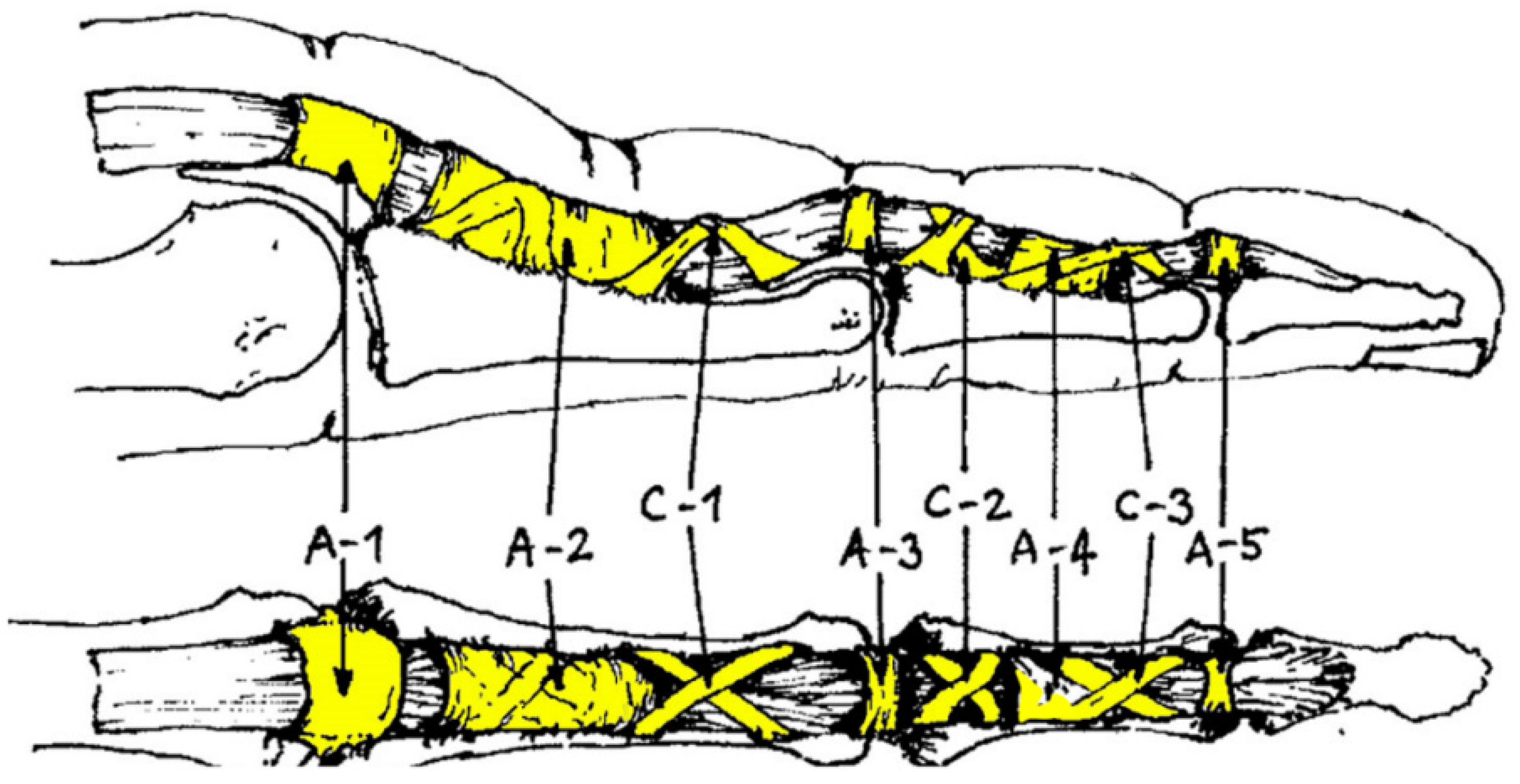
| A2 pulley affected [%] | 84.6 |
| A4 pulley affected [%] | 23.1 |
| multiple pulleys affected [%] | 27.7 |
| Perceived Injury Triggers | |
| hard and intensive training [%] | 63.1 |
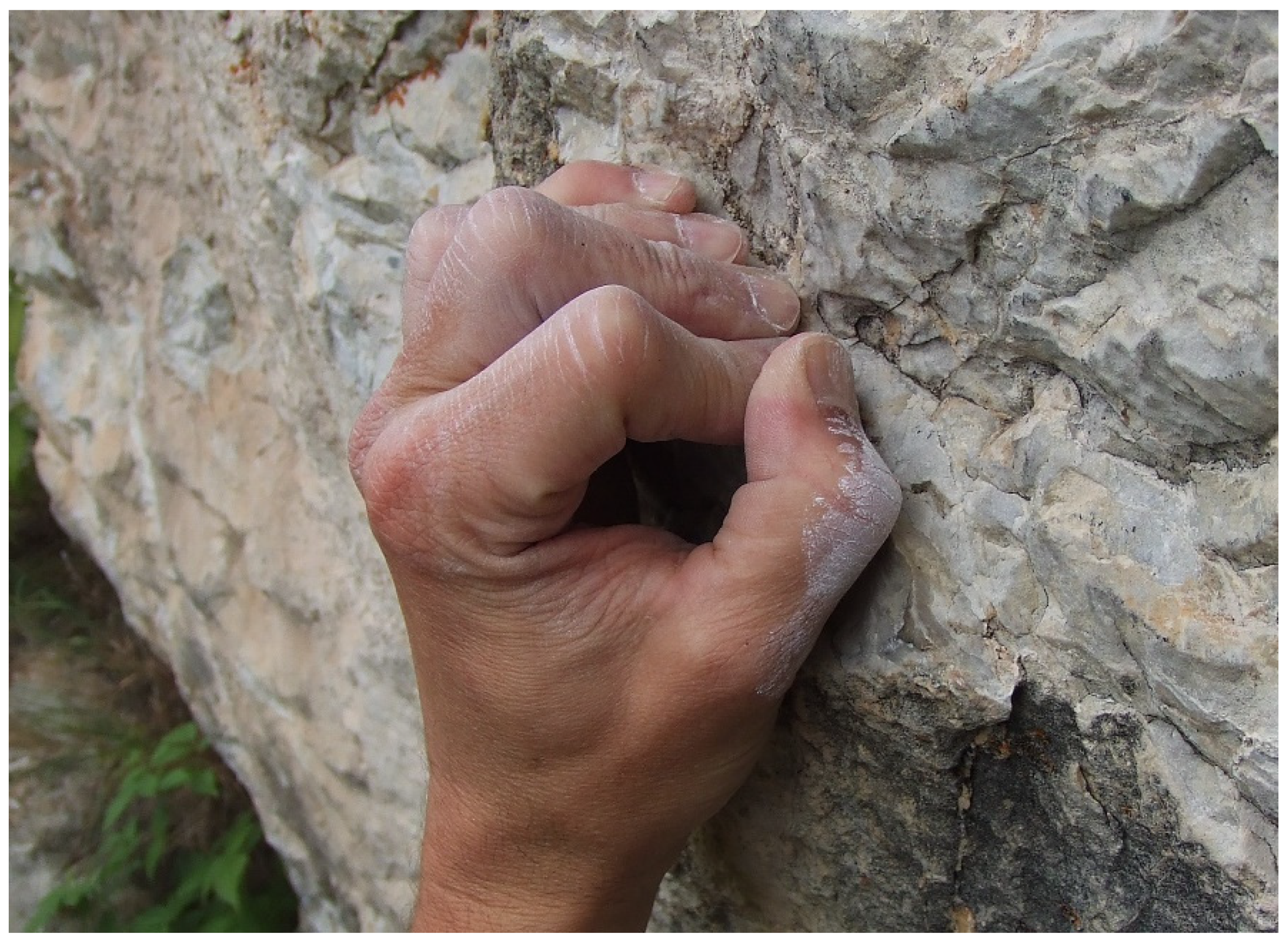
| extensive crimping [%] | 30.8 |
| extensive use of pockets [%] | 6.2 |
| foot slipped off [%] | 13.8 |
| repeated attempts of a hard and/or dynamic single pull [%] | 41.5 |
| other [%] | 16.9 |
| Training Attitudes | |
| regular warm-up [%] | 41.5 |
| training on campus board [%] | 32.3 |
| training on hang board [%] | 43.1 |
| specific training on small holds [%] | 33.8 |
| more than 3 training and/or climbing sessions per week [%] | 30.8 |
| dynamo training [%] | 20.0 |
| Therapy Content | |
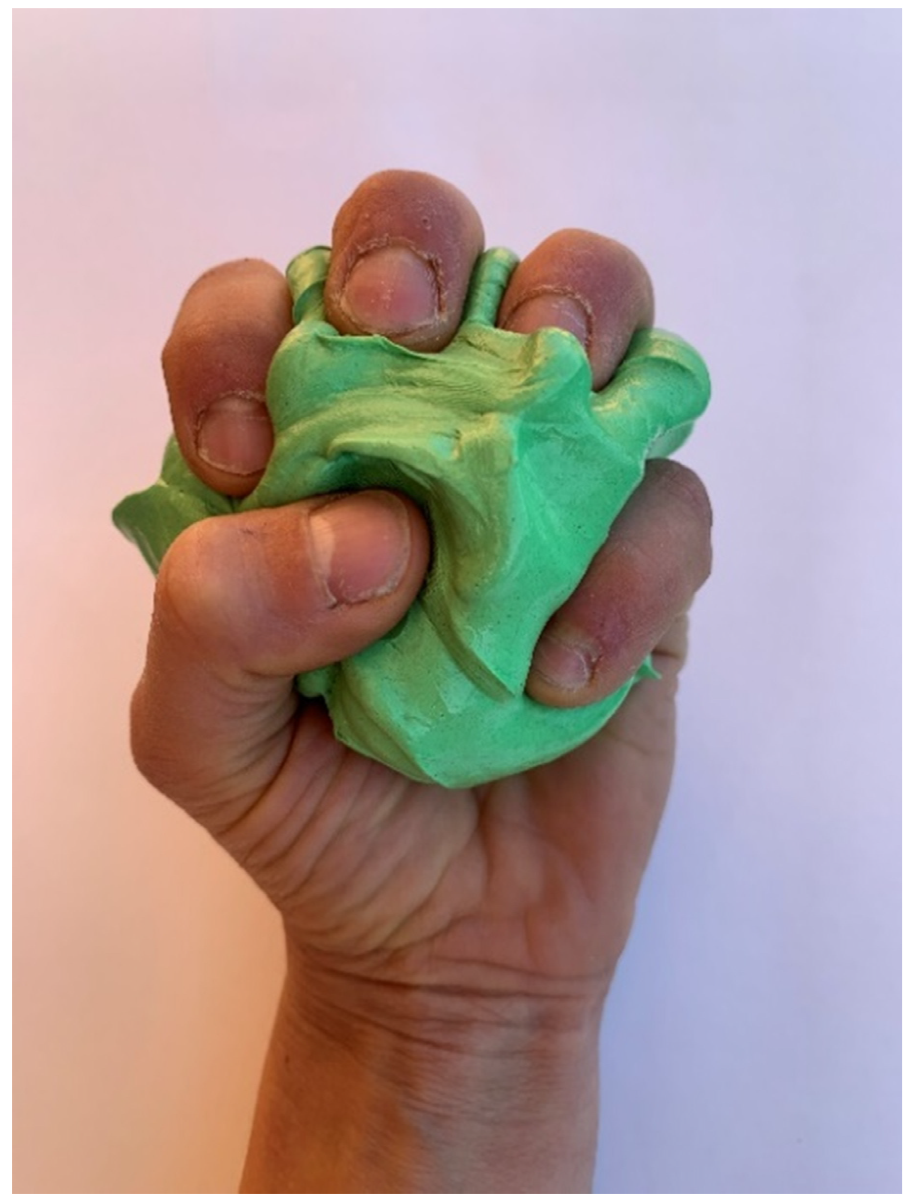
| modelling clay [%] | 75.4 |
| compression fingerling [%] | 35.4 |
| finger stretching [%] | 32.3 |
| ergotherapy [%] | 12.3 |
| medication [%] | 13.8 |
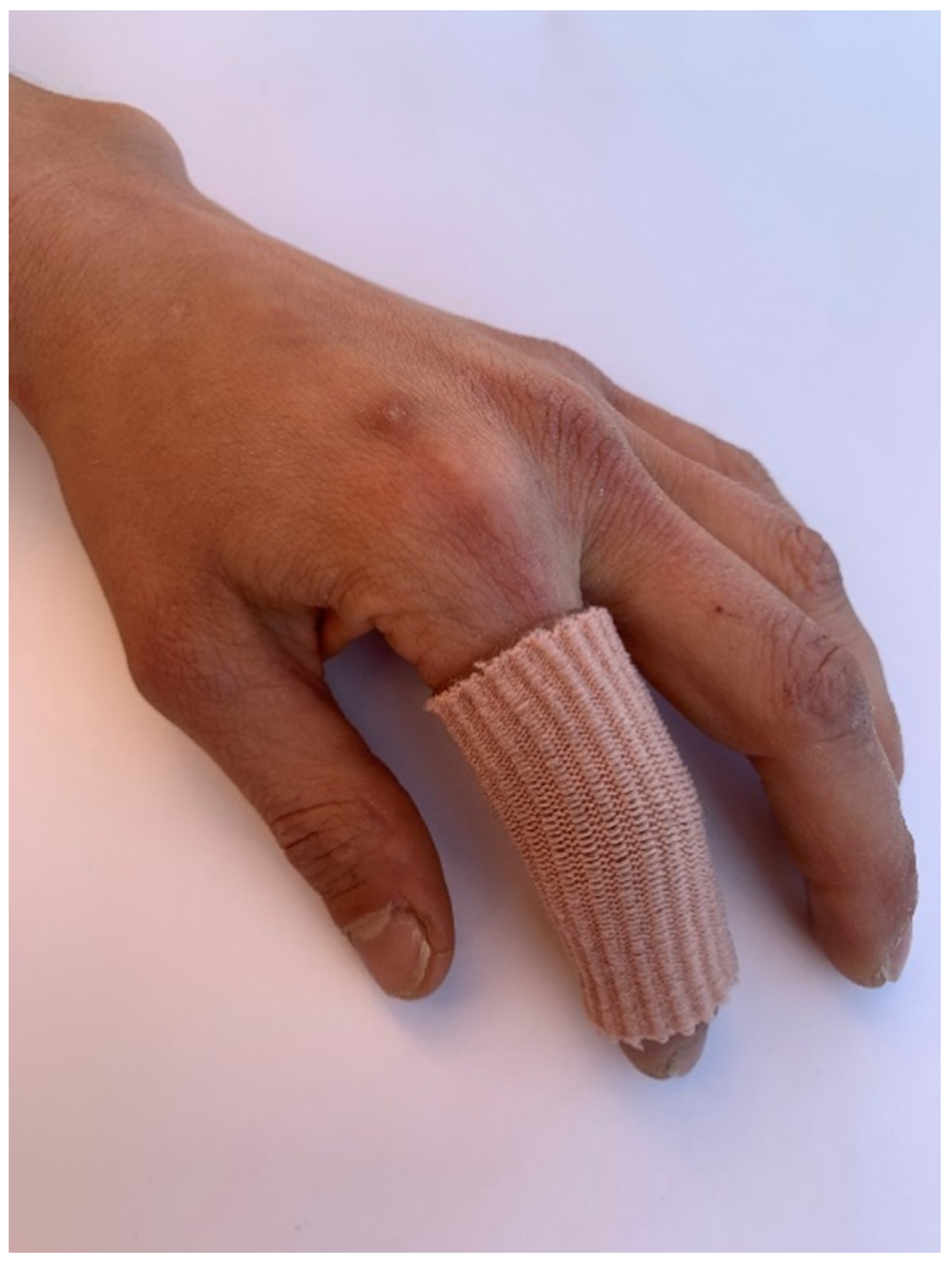
| taping [%] | 64.6 |
| climbing-related load reduction [%] | 90.8 |
| Therapy Outcome | |
| symptom duration [weeks] | 30.5 (24.3, 36.8) a,b |
| before/after therapy ratio in pain intensity during climbing [VAS/VAS] | 0.62 (0.55, 0.68) a,c |
| Model Parameter | Dependent Variable | Predictor | B | SEB | β Weight | p Value a |
|---|---|---|---|---|---|---|
| Model 1 b F = 6.613, p = 0.014, Adjusted R2 = 0.120, Cohen f2 = 0.140 n = 42 | before/after therapy ratio in pain intensity during climbing [VAS/VAS] | pain intensity in daily life before therapy [VAS] | 0.060 | 0.023 | 0.377 | 0.014 * |
| excluded variables by stepwise method | ||||||
| climbing years before therapy [year] | - | - | - | 0.769 | ||
| average pulley thickness [cm] | - | - | - | 0.296 | ||
| climbing level higher than 7b (redpoint) [0;1] | - | - | - | 0.247 | ||
| pain intensity during climbing before therapy [VAS] | - | - | - | 0.057 | ||
| therapy content: modelling clay [0;1] | - | - | - | 0.315 | ||
| therapy content: compression fingerling [0;1] | - | - | - | 0.702 | ||
| therapy content: finger stretching [0;1] | - | - | - | 0.781 | ||
| therapy content: ergotherapy [0;1] | - | - | - | 0.822 | ||
| therapy content: medication [0;1] | - | - | - | 0.987 | ||
| therapy content: taping [0;1] | - | - | - | 0.286 | ||
| therapy content: climbing-related load reduction [0;1] | - | - | - | 0.846 | ||
| Model 2 b F = 4.642, p = 0.037, Adjusted R2 = 0.082, Cohen f2 = 0.089 n = 42 | symptom duration [weeks] | climbing level higher than 7b (redpoint) [0;1] | −15.821 | 7.343 | −0.332 | 0.037 * |
| excluded variables by stepwise method | ||||||
| climbing years before therapy [year] | - | - | - | 0.906 | ||
| average pulley thickness [cm] | - | - | - | 0.447 | ||
| pain intensity during climbing before therapy [VAS] | - | - | - | 0.206 | ||
| pain intensity in daily life before therapy [VAS] | - | - | - | 0.282 | ||
| therapy content: modelling clay [0;1] | - | - | - | 0.144 | ||
| therapy content: compression fingerling [0;1] | - | - | - | 0.244 | ||
| therapy content: finger stretching [0;1] | - | - | - | 0.580 | ||
| therapy content: ergotherapy [0;1] | - | - | - | 0.168 | ||
| therapy content: medication [0;1] | - | - | - | 0.904 | ||
| therapy content: taping [0;1] | - | - | - | 0.176 | ||
| therapy content: climbing-related load reduction [0;1] | - | - | - | 0.301 |
Publisher’s Note: MDPI stays neutral with regard to jurisdictional claims in published maps and institutional affiliations. |
© 2022 by the authors. Licensee MDPI, Basel, Switzerland. This article is an open access article distributed under the terms and conditions of the Creative Commons Attribution (CC BY) license (https://creativecommons.org/licenses/by/4.0/).
Share and Cite
Mohn, S.; Spörri, J.; Mauler, F.; Kabelitz, M.; Schweizer, A. Nonoperative Treatment of Finger Flexor Tenosynovitis in Sport Climbers—A Retrospective Descriptive Study Based on a Clinical 10-Year Database. Biology 2022, 11, 815. https://doi.org/10.3390/biology11060815
Mohn S, Spörri J, Mauler F, Kabelitz M, Schweizer A. Nonoperative Treatment of Finger Flexor Tenosynovitis in Sport Climbers—A Retrospective Descriptive Study Based on a Clinical 10-Year Database. Biology. 2022; 11(6):815. https://doi.org/10.3390/biology11060815
Chicago/Turabian StyleMohn, Sabrina, Jörg Spörri, Flavien Mauler, Method Kabelitz, and Andreas Schweizer. 2022. "Nonoperative Treatment of Finger Flexor Tenosynovitis in Sport Climbers—A Retrospective Descriptive Study Based on a Clinical 10-Year Database" Biology 11, no. 6: 815. https://doi.org/10.3390/biology11060815
APA StyleMohn, S., Spörri, J., Mauler, F., Kabelitz, M., & Schweizer, A. (2022). Nonoperative Treatment of Finger Flexor Tenosynovitis in Sport Climbers—A Retrospective Descriptive Study Based on a Clinical 10-Year Database. Biology, 11(6), 815. https://doi.org/10.3390/biology11060815