Article

Cmcrf1, a Putative Zn2Cys6 Fungal Transcription Factor, Is Involved in Conidiation, Carotenoid Production, and Fruiting Body Development in Cordyceps militaris

1
Tianjin Institute of Industrial Biotechnology, Chinese Academy of Sciences, Tianjin 300308, China
2
National Center of Technology Innovation for Synthetic Biology, Tianjin 300308, China
3
College of Plant Protection, Jilin Agricultural University, Changchun 130118, China
*
Authors to whom correspondence should be addressed.
These authors contributed equally to this work.
Biology 2022, 11(10), 1535; https://doi.org/10.3390/biology11101535
Submission received: 5 September 2022 / Revised: 3 October 2022 / Accepted: 16 October 2022 / Published: 19 October 2022

Abstract

:

Simple Summary

Cordyceps militaris produce a wide variety of bioactive components, such as cordycepic acid, cordycepin, polysaccharides, pentostatin, ergosterol, and carotenoids. In particular, natural carotenoids from C. militaris are attracting increasing attention in human healthy and food coloring. Investigating the genetic regulatory mechanism of carotenoid biosynthesis will help to increase the carotenoid content of C. militaris through genetically engineering. This study focuses on the role of a putative Zn2Cys6 fungal transcription factor Cmcrf1 on carotenoid biosynthesis and fruiting body formation. Deletion of Cmcrf1 exhibited drastically reduced carotenoid biosynthesis and failed to generate fruiting bodies. In addition, the ΔCmcrf1 mutant exhibited significantly increased conidiation and increased hypersensitivity to cell-wall-perturbing agents. This study is helpful to deepen our knowledge of the regulatory mechanism of carotenoid biosynthesis in C. militaris.

Abstract

Cordyceps militaris is a high-value medicinal and edible fungus that produces many bioactive compounds, including carotenoid, and thus, improving the carotenoid productivity of C. militaris will increase its commercial value. However, little is known about the genetic regulatory mechanism of carotenoid biosynthesis in C. militaris. To further understanding the regulatory mechanism of carotenoid biosynthesis, we performed a large-scale screen of T-DNA insertional mutant library and identified a defective mutant, denoted T111, whose colonies did not change color from white to yellow upon exposure to light. Mutation analysis confirmed that a single T-DNA insertion occurred in the gene encoding a 695-amino-acid putative fungal-specific transcription factor with a predicted Zn2Cys6 binuclear cluster DNA-binding domain found uniquely in fungi. Targeted deletion of this gene, denoted C. militaris carotenogenesis regulatory factor 1 (Cmcrf1), generated the ΔCmcrf1 mutant that exhibited drastically reduced carotenoid biosynthesis and failed to generate fruiting bodies. In addition, the ΔCmcrf1 mutant showed significantly increased conidiation and increased hypersensitivity to cell-wall-perturbing agents compared with the wild-type strain. However, the Cmcrf1 gene did not have an impact on the mycelia growth of C. militaris. These results show that Cmcrf1 is involved in carotenoid biosynthesis and is required for conidiation and fruiting body formation in C. militaris.

1. Introduction

Cordyceps militaris is an entomopathogenic fungus that parasitizes the larvae or pupae of lepidopteran insects. It is also an edible fungus with medicinal properties and has been widely used in traditional Chinese medicine as a substitute for C. sinensis, as both species produce a variety of similar bioactive ingredients. C. militaris produce a wide variety of bioactive components, such as cordycepic acid, cordycepin, polysaccharides, pentostatin, ergosterol, and carotenoids [1], compounds that possess anti-fatigue, anti-tumor, anti-oxidant, and anti-inflammatory activities [2]. Particular attention has been paid to the carotenoids produced by C. militaris due to their strong antioxidant activities [3].
Natural carotenoids are widely used in health food industries. More than 700 natural carotenoids have been identified so far [4]. C. militaris has in recent years been investigated for carotenoid production [5].The productivity of carotenoid in C. militaris is relatively low and cannot meet the market demands. With the rapid development of synthetic biology, genetic engineering is an effective method to increase the carotenoid productivity of C. militaris [6,7,8]. However, the insufficient understanding of the carotenoid biosynthesis pathways in C. militaris prevents efficient genetic engineering of this fungus [9,10,11].
Most studies have focused on elucidating the key genes involved in the various metabolic pathways that affect carotenoid biosynthesis in C. militaris. For example, recent studies have revealed that Cmhyd1, which encodes a class II hydrophobin, may influence the yellow pigment accumulation in C. militaris. In particular, a Cmhyd1-null mutant formed white colonies on potato dextrose agar (PDA) plates even after light irradiation [12]. Moreover, Cmfhp encodes flavohemoprotein, a nitric oxide (NO) dioxygenase, that was found to significantly affect carotenoid biosynthesis, as the carotenoid content of the Cmfhp-null mutant decreased compared with the WT strain in C. militaris [13]. Cmtns encoding a terpenoid synthase gene was reported to have a similar effect as Cmfhp on the carotenoid production of C. militaris [14]. Defective primordium and fruiting bodies were also found in the null mutants of these three genes. Furthermore, high concentrations of carotenoids accumulate in the mycelia and fruiting bodies of C. militaris upon exposure to light during cultivation. Seven photoreceptors related to photobiological processes have been identified through genome-wide analysis of C. militaris, and the functions of three of these seven photoreceptors (CmWC-1, CmVVD, and CmCry-DASH) have been comprehensively investigated in C. militaris. Deletion of Cmwc-1 led to a drastically decrease in carotenoid production and defects in fruiting body development [15]. Deletion of Cmvvd, which encodes a blue-light receptor, led to abnormal development of fruiting bodies and a significant increase in carotenoid content [16]. In contrast to the phenotypes of ΔCmwc-1 mutant, the Cmcry-DASH-null mutant exhibited more carotenoid accumulation than the WT strain. However, despite the ΔCmcry-DASH strain exhibiting normal primordium formation, its fruiting bodies did not elongate normally [17]. In addition, although several studies have performed genome-wide transcriptional analyses and genome-level metabolic network-driven analyses to probe carotenoid biosynthetic pathways in C. militaris, only a few differentially expressed genes (DEGs) that might be involved in carotenoid biosynthesis have been identified [18,19]. The functions of these DEGs in C. militaris have yet to be elucidated, and thus, delineating the functions of genes involved in carotenoid biosynthesis in this fungus remains an important research topic.
T-DNA insertion mutation is an effective method to identify novel genes participated in the different metabolic pathways. Many novel pathogenicity-related genes have been discovered through large-scale screening of T-DNA mutant libraries of phytopathogenic fungi, such as Verticillium dahliae, Ustilaginoidea virens, and Sporisorium scitamineum [20,21,22]. In the industrial cultivation of filamentous fungi, the ATMT method has been proven an efficient tool for screening random T-DNA insertion mutants to identify those with improved production of certain metabolic products [23,24]. In the current study, we performed a large-scale screen of T-DNA tagged mutants of C. militaris and found that the mutation of a gene encoding a fungal-specific transcription factor resulted in a white colony phenotype upon exposure to light. This gene, which we designated C. militaris carotenogenesis regulatory factor 1 (Cmcrf1), encodes a 695-amino-acid protein with a predicted Zn2Cys6 binuclear cluster DNA-binding domain, demonstrating that Cmcrf1 is a Zn2Cys6 transcription factor. We also functionally characterized Cmcrf1 in C. militaris using a targeted gene-deletion strategy. Effects of Cmcrf1 on fungal growth, conidiation, cell wall integrity, fruiting body formation, and carotenoid production were extensively investigated. This study might shed light on the regulatory mechanisms involved in carotenoid biosynthesis and fruiting body development in C. militaris.

2. Materials and Methods

2.1. Strains, Media, and Cultivation Conditions

C. militaris strain CM20-Xi (Edible Fungi Research Institute, Ankang Academy of Agricultural Sciences, Shaanxi Province) purified by single-spore isolation was used in all experiments. All of the C. militaris strains used or generated in this study were cultivated on modified PDA medium (20.0% potato, 1.0% dextrose, 0.1% KH2PO4, 0.05% MgSO4, peptone 0.2%, and 1.5% agar, w/v) at 25 °C for 20 days. For long-term conservation, strains were maintained as stocks on wheat seed medium at 4 °C. Agrobacterium tumefaciens was cultivated in Luria–Bertani (LB) broth or LB agar. Escherichia coli DH-5α was used for plasmid constructions.

2.2. Disruption of Cmcrf1 and Transformation of C. militaris

Cmcrf1 was knocked out using a homologous recombination gene-deletion strategy. Briefly, the targeted gene-deletion cassettes were constructed using a double-joint polymerase chain reaction (PCR) method [25]. The 5′- and 3′-flanking sequences of Cmcrf1 were amplified from the genomic DNA of the WT strain with primers CRF15F/CRF15R and CRF13F/CRF13R, respectively, the sequences of which are provided in Table S1. The hygromycin B phosphotransferase gene (hph) was generated by PCR from the pBSHPH1 vector using the primer pair hphF/hphR [26]. All of the generated PCR products were purified using a gel extraction kit (Omega Bio-tek, USA) for the subsequent procedure. The 5′-flanking fragment, the hph gene, and the 3′-flanking fragment were fused together in this order, and the resulting fused deletion cassettes were then cloned into the binary vector pCAMBIA1300 to produce the targeted gene-deletion vector pCAMBIA1300-yprKO (Figure S1A). The constructed vectors were then transformed into C. militaris through the Agrobacterium tumefaciens-mediated transformation (ATMT) method as previously described, with some modifications [27]. For Southern blotting analysis, Genomic DNA extraction and restriction enzyme digestion were performed as described [28]. The 3′ flanking sequence of Cmcrf1 was amplified from genomic DNA and labeled as a probe. Hybridization processes were conducted following the manufacturer’s instructions.

2.3. Complementation of the Cmcrf1-Null Mutant

The Cmcrf1-null mutant was complemented with Cmcrf1 from the WT strain. The complementation gene sequences including the native promoter region, the full-length DNA sequence of Cmcrf1, and the 0.5 kb downstream sequence were amplified from the genomic DNA of WT and cloned into pCAMBIA3301 to generate the complementary vector pCAMBIA3301-yprC (Figure S1B). This vector was then transformed into the null mutant using the ATMT method. All the obtained transformants were screened on PDA with 300 μg/mL glufosinate ammonium. The complemented transformants were verified through PCR and quantitative reverse transcription (qRT)-PCR.

2.4. Fungal Development Assays

Mycelia disks were inoculated onto PDA plates and incubated at 25 °C for 3 weeks. The diameters of colonies were measured every 2 days. For conidial production, cultivated mycelia from 14-day-old plates were removed with a sterilized spreader and added into 50 mL centrifuge tubes containing 5 mL double-distilled (dd) H2O. The resulting conidial suspensions were filtered through four layers of lens paper, and the numbers of conidia in the filtered suspensions were counted with a hemocytometer under a microscope and then diluted to appropriate concentrations. For conidial germination, 10 μL droplets of the conidial suspensions (resuspended and diluted to 1 × 10⁵/mL with sterilized dd H2O) were placed on glass slides. The droplet-bearing slides were then incubated in a moisture chamber at 25 °C, and the conidial germination rates were examined under a microscope at 12, 14, and 16 h post inoculation (hpi). Three independent experiments were performed with three replicates each time.

2.5. Cell Wall Integrity Assays

Mycelial disks were inoculated onto minimal medium (MM) plates containing various chemicals, such as Calcofluor White (CFW) and Congo Red (CR), for sensitivity tests on these chemicals. The resulting inoculated plates were incubated at 25 °C for 20 days, and the diameters of colonies were examined to enable comparison between all the tested strains. The test of each strain’s sensitivity to each chemical was performed in triplicate.

2.6. Determination of Carotenoids

Mycelia from cultures of different strains were filtered, lyophilized, and used for carotenoid determination using the acid-heating method, as described previously [29]. Determination of the carotenoid content was performed by measuring the absorbance at 445 nm.

2.7. Quantitative Real-Time PCR (qRT-PCR)

Total RNA of mycelia was extracted using TRIzol reagent (Invitrogen Life Technologies, USA) and treated with DNase. All qRT-PCR assays were performed as described [30]. Three biological replicates were carried out with three technical replicates.

3. Results

3.1. Identification and Expression Pattern of T-DNA-Tagged Cmcrf1 in C. militaris

We constructed a T-DNA insertion-mutant library of the wild-type C. militaris using a binary vector on PDA plates, which afforded mutants with abnormal morphology. A mutant that did not form yellow-pigmented colonies when exposed to light was picked out for further functional analysis and was designated as T111 (Figure 1A). To investigate whether single T-DNA insertion caused the mutation in the T111 mutant, the genomic DNA of T111 was digested with SalI and HindIII for Southern blotting. The result showed that a single-copy T-DNA was integrated into the genome of the T111 mutant (Figure 1B). High-efficiency thermal asymmetrical interlaced PCR (hiTAIL-PCR) was performed to amplify the genomic DNA sequences flanking the integrated T-DNA [31]. Comparison between the sequences obtained by hiTAIL-PCR and the related sequences in the C. militaris genome database showed that the T-DNA insertion in the T111 mutant was located in the fourth exon of the predicted gene, CCM_07998 (Figure 1C), which was annotated to encode a fungal-specific transcription factor in the database. This gene encodes a putative fungal transcription factor containing a Zn2Cys6 binuclear cluster DNA-binding domain found exclusively in fungi and a middle homology domain (Figure S2A). Thus, this gene was designated Cmcrf1 due to the T111 mutant exhibiting defective carotenoid production. Alignment of Cmcrf1 with its homologs from other fungal species revealed that Cmcrf1 had 63.2%, 41.1%, 38%, and 36.9% similarities with homologs from Akanthomyces lecanii, Neurospora crassa, Aspergillus terreus, and Metarhizium robertsii, respectively (Figure S2B). Figure S1C showed the results of the phylogenetic analysis of the putative homologs of Cmcrf1 from several filamentous fungal species. The results indicate that Cmcrf1 is most closely related to the homologs expressed in A. lecanii and N. crassa and that it is highly conserved in filamentous fungi (Figure S2).
To investigate the expression pattern of Cmcrf1, the transcription profiles of Cmcrf1 in different developmental stages were analyzed. The results of qPCR showed that Cmcrf1 was constitutively expressed during the entire fungal-cultivation process (Figure 2). Cmcrf1 expression under light exposure was higher than that under dark conditions, demonstrating that light promotes its expression. It was noteworthy that the levels of Cmcrf1 expression were higher in the plate cultivation and fruiting body stages than in the other stages, which implies that Cmcrf1 might play a role in vegetative growth on plates and in fruiting body development.

3.2. Disruption of Cmcrf1

To characterize the function of Cmcrf1 in C. militaris, a targeted gene-deletion strategy was applied (Figure 3A). Thus, the targeted gene-deletion vector pCAMBIA1300-yprKO was transformed into the WT strain using the ATMT method. All of the hygromycin-B-resistant transformants were screened by PCR using primers CRF1checkF/R. The suspected mutant was confirmed to be the Cmcrf1 null (ΔCmcrf1) mutant by Southern blot analysis. The hybridized 3.8 kb band and an 11 kb band were detected in the ΔCmcrf1 mutant and the WT strain, respectively (Figure 3B). The Cmcrf1 mRNA expression was analyzed by qRT-PCR in and the ΔCmcrf1 mutant. The results revealed that there was no Cmcrf1 expressed by the ΔCmcrf1 mutant (Figure 3C), and this confirmed ΔCmcrf1 strain was used for further investigations.
For the generation of complemented transformants, the constructed complementary vector pCAMBIA3301-yprC was transformed into the ΔCmcrf1 mutant. The complemented mutants grown on PDA containing glufosinate ammonium were identified by PCR and qRT-PCR. The qRT-PCR results showed that Cmcrf1 was expressed in the WT strain and in the complemented mutant ΔCmcrf1c, indicating that Cmcrf1 was successfully complemented in the ΔCmcrf1 mutant (Figure 3C).

3.3. Deletion of Cmcrf1 Affects the Growth Characteristics of C. militaris

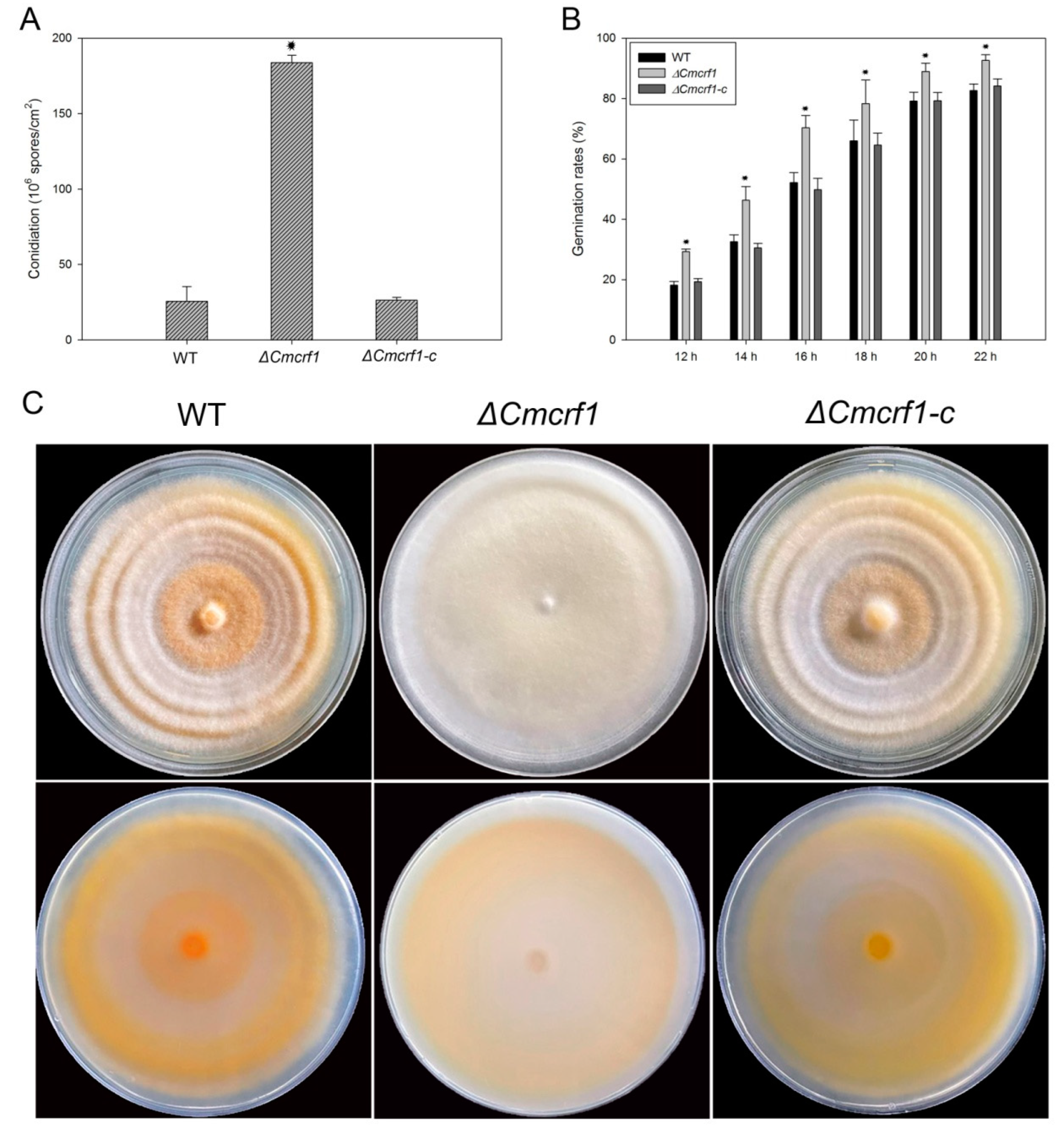
The growth characteristics of the WT strain, the ΔCmcrf1 mutant, and the complemented mutant ΔCmcrf1c were performed on PDA at 25 °C for 3 weeks. There were no significant differences between the WT strain and the ΔCmcrf1 mutant in terms of radial growth (Figure S3A). However, conidial production was seven-fold higher in the ΔCmcrf1 mutant than in the WT strain (Figure 4A). The conidiation was restored to that of the WT strain when Cmcrf1 was reintroduced to the null mutant. Furthermore, the conidial germination rates were analyzed to investigate whether Cmcrf1 is required for conidial germination. The conidial suspensions of all of the C. militaris strains were inoculated on slides, which were then incubated in a moisture chamber at 25 °C. The conidia began to germinate at 12 h after inoculation. From 12 hpi to 22 hpi, the germination rate of the ΔCmcrf1 mutant was significantly higher than that of the WT strain (Figure 4B).
To determine whether Cmcrf1 affects the morphology of C. militaris, all of the strains were cultivated at 25 °C for 20 days in the dark. When the cultivated plates were transferred from dark to light conditions, the colonies of the ΔCmcrf1 mutant remained white to light yellow, while those of the WT strain and the complemented mutant changed to yellow-orange after 12 h of light exposure (Figure 4C).
Furthermore, the carbon source utilization of all of the strains was examined on MM containing various carbon sources (Figure S3B). The results revealed that no significant differences were found between the carbon source utilization of the ΔCmcrf1 mutant and that of the WT strain.

3.4. Effect of Cmcrf1 on Cell-Wall Integrity

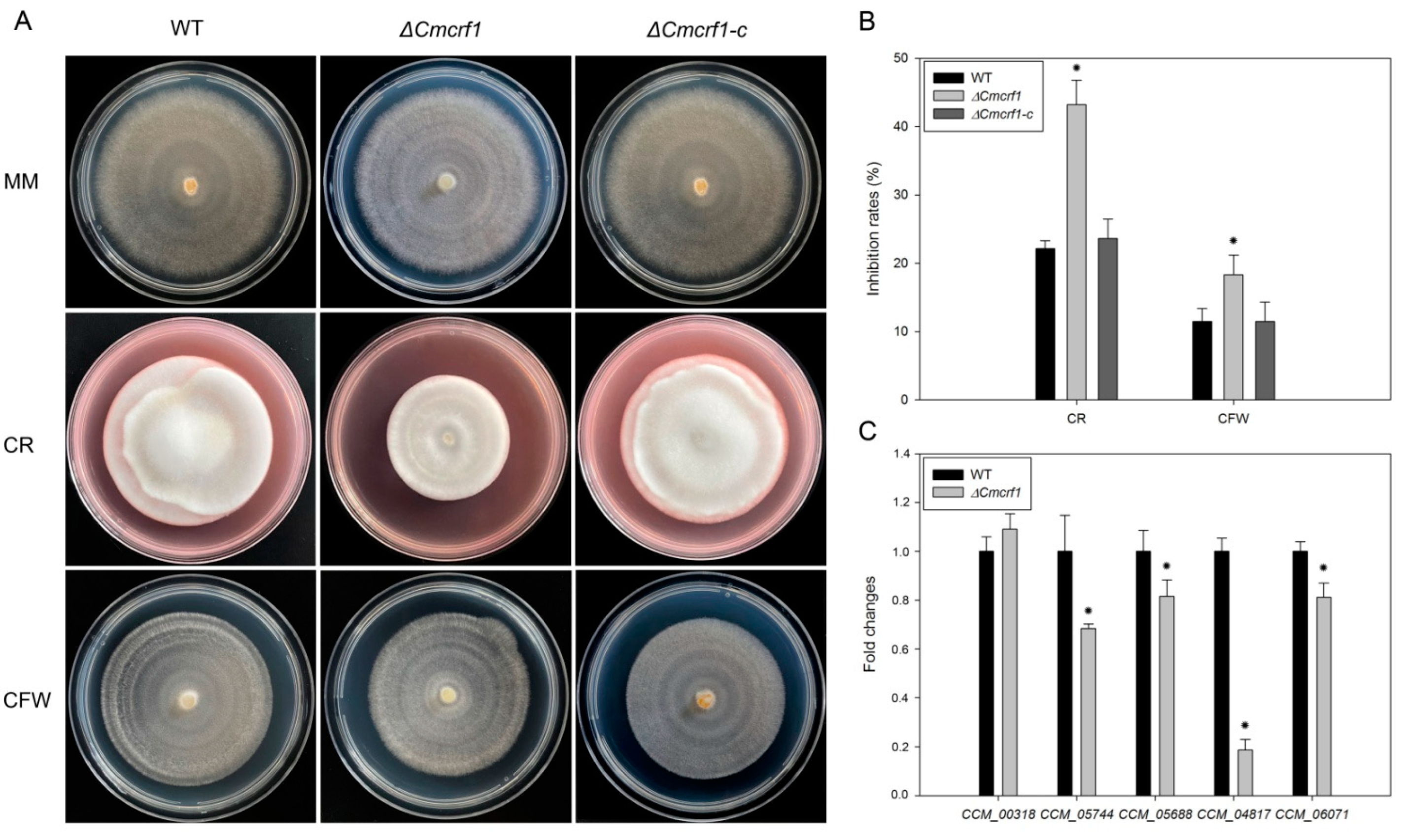
The fungal cell wall is vital to the growth, survival, pathogenesis, and morphogenesis of a fungus, as it provides a protective barrier against a wide range of environmental stressors [32]. To examine effect of Cmcrf1 on cell wall integrity, the sensitivity of the WT strain, the ΔCmcrf1 mutant, and the complemented ΔCmcrf1c mutant to CFW and CR was compared. While all of the strains exhibited reduced growth in the presence of CFW or CR (Figure 5A,B), the growth inhibition rate of the ΔCmcrf1 mutant was higher than that of the WT strain, indicating that Cmcrf1 deletion increases hypersensitivity to substances that interfere with fungal cell-wall synthesis.
Chitin is one of the major components of fungal cell wall, which is synthesized by chitin synthases in ascomycetes. The transcription levels of five genes encoding chitin synthases (CCM_00318, CCM_05744, CCM_05688, CCM_04817, and CCM_06071) were examined in the WT strain and the ΔCmcrf1 mutant. As seen in Figure 5C, transcript abundance of four chitin synthase genes were significantly decreased, whereas the transcript abundance of one chitin synthase gene CCM_00318 was not altered in the ΔCmcrf1 mutant. The results suggested that Cmcrf1 is involved in cell wall synthesis, which further influences cell wall integrity via affecting the expression of genes that encode chitin synthases.

3.5. The ΔCmcrf1 Mutant Showed Defective Development of Fruiting Bodies

The fruiting body development of the WT strain, the ΔCmcrf1 mutant, and the ΔCmcrf1c mutant on wheat-grain medium was compared. The WT strain produced normal, orange-colored fruiting bodies. However, the mycelia of the ΔCmcrf1 mutant were white to light yellow and unable to produce even primordia for fruiting body formation (Figure 6A). The capacity to produce fruiting bodies was recovered in ΔCmcrf1c. These results indicate that Cmcrf1 plays an essential role in fruiting body development in C. militaris.

3.6. Cmcrf1 Affects the Carotenoid Content of C. militaris

For the determination of carotenoid content, the WT strain, the ΔCmcrf1 mutant, and the ΔCmcrf1c mutant were cultivated on PDA plates, in potato dextrose broth in flasks, and on wheat-seed medium under different light-exposure conditions. As seen in Figure 6B, the carotenoid content in the hyphae of the ΔCmcrf1 mutant was up to 3.51-fold lower than that in the hyphae of the WT strain after 12 h of light exposure. Moreover, the carotenoid content in the primordia and fruiting bodies of the ΔCmcrf1 mutant could not be determined, as it did not form either of these structures. These results are consistent with the observed colony colors, indicating that Cmcrf1 influences pigment synthesis and is thus a regulatory genetic factor in C. militaris.
Recently, a putative carotenoid biosynthetic pathway was proposed, and seven genes were reported to participating in this putative pathway [18]. We then examined the expression levels of these seven genes (CCM_00646, CCM_08845, CCM_01982, CCM_04017, CCM_03203, CCM_06728, and CCM_09155) by qRT-PCR. The expression levels of four genes (CCM_01982, CCM_03203, CCM_06728, and CCM_09155) were significantly down-regulated, while the expression levels of the other three genes (CCM_00646, CCM_08845, and CCM_04017) were not altered in the ΔCmcrf1 mutant (Figure 6C). The results revealed that Cmcrf1 has a role in carotenoids biosynthesis through influencing the expression of genes involved in this putative carotenoid biosynthetic pathway.

4. Discussion

The Zn2Cys6 zinc cluster proteins are a family of transcription factors found exclusively in fungi [33]. They function as transcriptional regulators and are involved in a wide variety of biological processes, including asexual and sexual development, secondary metabolite biosynthesis, carbon and nitrogen metabolism, stress responses, pleiotropic drug resistance, and fungal pathogenesis [34,35,36,37,38]. However, the functions of Zn2Cys6 TFs in different fungi are diverse. In the rice blast fungus Magnaporthe oryzae, the functions of 104 genes encoding Zn2Cys6 transcription factors were systematically analyzed. Deletion of these genes resulted in phenotypic changes in fungal development and pathogenicity, such as changes in conidiation, mycelial growth, pigmentation, appressorium formation, response to abiotic stresses, and colony morphology [39]. In the model filamentous fungus N. crassa, 135 genes encoding Zn2Cys6 transcription factors were found to be involved in basal hyphal growth, asexual sporulation, and sexual development [40]. When comparing the phenotypes of the Zn2Cys6 TFs homologs of M. oryzae and N. crassa, the results seems to be consistent with those previously reported that evolve differently in how to regulate fungal growth and asexual development rather than maintaining the same function in different fungi [41]. In this study, a color-changing-defective mutant T111 was isolated using T-DNA insertional mutagenesis, and Cmcrf1, which putatively encodes a Zn2Cys6 transcription factor, was identified in C. militaris. Our results showed that compared with the C. militaris WT strain, the ΔCmcrf1 mutant exhibited excessive conidiation, a decreased conidial germination rate, and a higher sensitivity to cell-wall-perturbing agents. Furthermore, compared with the WT strain, the two most prominent important phenotypic characteristics of the ΔCmcrf1 mutant were its drastically reduced carotenoid content and halted fruiting body formation. These results indicate that Cmcrf1 is involved in fungal development, stress responses, secondary metabolite biosynthesis, and fruiting body development in C. militaris.
Carotenoids are massively accumulated in the mycelia and fruiting bodies of C. militaris upon light irradiation during the fungal cultivation process. However, little is known about the genetic regulatory mechanism of carotenoid biosynthesis in C. militaris [9,10,11]. In this study, the most remarkable phenotypic differences between the WT strain and the ΔCmcrf1 mutant was that the ΔCmcrf1 mutant contained significantly lower concentrations of carotenoids than the WT strain. Recently, a putative carotenoid biosynthetic pathway was depicted by integrating with DEGs and genome-scale metabolic network [18]. Seven genes were reported to participate in this putative pathway. In the ΔCmcrf1 mutant, the transcript abundance of four genes were significantly down-regulated, while the other three genes were similar with the WT strain, indicating that reduction in carotenoids contents are probably led by the comprehensive effect of the decreased expression level of genes involved in the putative carotenoid biosynthetic pathway.
Cmcrf1 is also involved in fungal development and fruiting body development. To date, seven photoreceptors have been identified in C. militaris, and three photoreceptors—Cmwc-1, Cmvvd, and Cmcry-DASH—were reported to regulate fungal development, such as conidiation and fruiting body formation, in C. militaris [15,16,17]. In addition, all three genes affect carotenoid production. Genes participating in other metabolic pathways, such as Cmhyd1 encoding a class II hydrophobin and Cmfhp encoding an NO dioxygenase, have also been reported to regulate carotenoid accumulation and fungal development simultaneously [12,13]. The regulation of growth and development in response to environmental stimuli is a basic biological process in all organisms. Many filamentous fungi synthesize carotenoid pigments in response to several internal and external cues, including fungal development and light exposure [42]. These findings suggest that genes involved in carotenoid synthesis seem to also participate in the development of C. militaris. Nevertheless, whether carotenoid production plays an important biological role in fungal development in C. militaris and that of filamentous fungi in general remains to be determined.
Chitin is an essential component of fungal cell wall, and its synthesis depends on the activity of chitin synthases [43]. In this study, the ΔCmcrf1 mutant showed defects in cell wall integrity and a higher sensitivity to cell-wall-perturbing agents CR and CFW than the WT strain. Previous studies reported that chitin synthases coding genes play crucial roles in the growth, conidiation, and cell wall integrity through regulating chitin contents in phytopathogenic fungi [44,45,46,47]. In the present study, the expression levels of four chitin synthases were significantly down-regulated in the ΔCmcrf1 mutant compared to the WT strain, implying that Cmcrf1 probably directly or indirectly regulates chitin synthesis, which further influences the response to different cell wall stressors.

5. Conclusions

Our study found that Cmcrf1 is involved in carotenoid biosynthesis and fruiting body development in C. militaris, as it negatively regulates conidiation and cell wall integrity. These findings broaden our understanding of the functions of Zn2Cys6-type transcription factors in this edible and medicinal fungus. Future studies are warranted to further elucidate how carotenoid biosynthesis is regulated in C. militaris and to determine the relationship between fungal development and the role of Cmcrf1 in carotenoid biosynthesis.

Supplementary Materials

The following supporting information can be downloaded at: https://www.mdpi.com/article/10.3390/biology11101535/s1, Figure S1: Construction of the knockout vector pCAMBIA1300-yprKO (A) and the complementary vector pCAMBIA3301-yprC (B); Figure S2: Amino acid sequence alignment and phylogenetic analysis of Cmcrf1 with the homologues from other fungal species; Figure S3: Growth of C. militaris strains on different carbon sources; Table S1: Primers used in this study.

Author Contributions

Conceptualization, R.H.; methodology, R.H. and L.Z.; formal analysis, L.Z., J.L. and S.M.; investigation, L.Z., J.L. and S.M.; resources, R.H. and Y.L.; writing—original draft preparation, L.Z. and R.H.; writing—review and editing, R.H.; supervision, R.H. and Y.L.; project administration, R.H. and Y.L.; funding acquisition, Y.L. All authors have read and agreed to the published version of the manuscript.

Funding

This work was supported by Tianjin Synthetic Biotechnology Innovation Capacity Improvement Project (No. TSBICIP-CXRC-006) and Engineering Research Center of Edible and Medicinal Fungi, Ministry of Education (No. SYJ2020004).

Institutional Review Board Statement

Not applicable.

Informed Consent Statement

Not applicable.

Data Availability Statement

Not applicable.

Conflicts of Interest

The authors declare no conflict of interest.

References

  1. Jędrejko, K.J.; Lazur, J.; Muszyńska, B. Cordyceps militaris: An overview of its chemical constituents in relation to biological activity. Foods 2021, 10, 2634. [Google Scholar] [CrossRef] [PubMed]
  2. Rupa, E.J.; Li, J.F.; Arif, M.H.; Yaxi, H.; Puja, A.M.; Chan, A.J.; Hoang, V.A.; Kaliraj, L.; Yang, D.C.; Kang, S.C. Cordyceps militaris fungus extracts-mediated nanoemulsion for improvement antioxidant, antimicrobial, and anti-inflammatory activities. Molecules 2020, 25, 5733. [Google Scholar] [CrossRef] [PubMed]
  3. Zhao, Y.; Li, S.L.; Chen, H.Y.; Zou, Y.; Zheng, Q.W.; Guo, L.Q.; Wu, G.H.; Lu, J.; Lin, J.F.; Ye, Z.W. Enhancement of carotenoid production and its regulation in edible mushroom Cordyceps militaris by abiotic stresses. Enzyme Microb. Technol. 2021, 148, 109808. [Google Scholar] [CrossRef] [PubMed]
  4. Stahl, W.; Sies, H. Bioactivity and protective effects of natural carotenoids. Biochim. Biophys. Acta. 2005, 1740, 101–107. [Google Scholar] [CrossRef] [PubMed] [Green Version]
  5. Zhang, J.J.; Wang, F.F.; Li, K.B.; Liu, Q.; Yang, Y.; Dong, C.H. Heat and light stresses affect metabolite production in the fruit body of the medicinal mushroom Cordyceps militaris. Appl. Microbiol. Biotechnol. 2018, 102, 4523–4533. [Google Scholar]
  6. Zheng, P.; Xia, Y.; Zhang, S.; Wang, C. Genetics of Cordyceps and related fungi. Appl. Microbiol. Biotechnol. 2013, 97, 2797–2804. [Google Scholar] [CrossRef]
  7. Gao, Y.L.; Yu, C.; Li, L.J. Heterologous expression of a natural product biosynthetic gene cluster from Cordyceps militaris. Antibiot. 2022, 75, 16–20. [Google Scholar] [CrossRef]
  8. Meng, G.; Wang, X.; Liu, M.; Wang, F.; Liu, Q.; Dong, C. Efficient CRISPR/Cas9 system based on autonomously replicating plasmid with an AMA1 sequence and precisely targeted gene deletion in the edible fungus, Cordyceps militaris. Microb. Biotechnol. 2022, 15, 2594–2606. [Google Scholar] [CrossRef]
  9. Zheng, P.; Xia, Y.; Xiao, G.; Xiong, C.; Hu, X.; Zhang, S.; Zheng, H.; Huang, Y.; Zhou, Y.; Wang, S.; et al. Genome sequence of the insect pathogenic fungus Cordyceps militaris, a valued traditional Chinese medicine. Genome Biol. 2011, 12, R116. [Google Scholar] [CrossRef] [Green Version]
  10. Sung, G.H. Complete mitochondrial DNA genome of the medicinal mushroom Cordyceps militaris (Ascomycota, Cordycipitaceae). Mitochondrial DNA 2015, 26, 789–790. [Google Scholar] [CrossRef]
  11. Kramer, G.J.; Nodwell, J.R. Chromosome level assembly and secondary metabolite potential of the parasitic fungus Cordyceps militaris. BMC Genomics 2017, 18, 912. [Google Scholar] [CrossRef] [PubMed]
  12. Li, X.; Wang, F.; Liu, M.Q.; Dong, C.H. Hydrophobin CmHYD1 is involved in conidiation, infection and primordium formation, and regulated by GATA transcription factor CmAreA in edible fungus, Cordyceps militaris. J. Fungi 2021, 7, 674. [Google Scholar] [CrossRef] [PubMed]
  13. Lou, H.W.; Zhao, Y.; Chen, B.X.; Yu, Y.H.; Tang, H.B.; Ye, Z.W.; Lin, J.F.; Guo, L.Q. Cmfhp gene mediates fruiting body development and carotenoid production in Cordyceps militaris. Biomolecules 2020, 10, 410. [Google Scholar] [CrossRef] [PubMed] [Green Version]
  14. Lou, H.W.; Zhao, Y.; Tang, H.B.; Ye, Z.W.; Wei, T.; Lin, J.F.; Guo, L.Q. Transcriptome analysis of Cordyceps militaris reveals genes associated with carotenoid synthesis and identification of the function of the Cmtns gene. Front. Microbiol. 2019, 10, 2105. [Google Scholar] [CrossRef] [Green Version]
  15. Yang, T.; Guo, M.M.; Yang, H.J.; Guo, S.P.; Dong, C.H. The blue-light receptor CmWC-1 mediates fruit body development and secondary metabolism in Cordyceps militaris. Appl. Microbiol. Biotechnol. 2016, 100, 743–755. [Google Scholar] [CrossRef]
  16. Zhang, J.J.; Wang, F.F.; Yang, Y.; Wang, Y.; Dong, C.H. CmVVD is involved in fruiting body development and carotenoid production and the transcriptional linkage among three blue-light receptors in edible fungus Cordyceps militaris. Environ. Microbiol. 2020, 22, 466–482. [Google Scholar] [CrossRef]
  17. Wang, F.; Song, X.H.; Dong, X.M.; Zhang, J.J.; Dong, C.H. DASH-type cryptochromes regulate fruiting body development and secondary metabolism differently than CmWC-1 in the fungus Cordyceps militaris. Appl. Microbiol. Biotechnol. 2017, 101, 4645–4657. [Google Scholar] [CrossRef]
  18. Thananusak, R.; Laoteng, K.; Raethong, N.; Zhang, Y.; Vongsangnak, W. Metabolic responses of carotenoid and cordycepin biosynthetic pathways in Cordyceps militaris under light-programming exposure through genome-wide transcriptional analysis. Biology 2020, 9, 242. [Google Scholar] [CrossRef]
  19. In-On, A.; Thananusak, R.; Ruengjitchatchawalya, M.; Vongsangnak, W.; Laomettachit, T. Construction of light-responsive gene regulatory network for growth, development and secondary metabolite production in Cordyceps militaris. Biology 2022, 11, 71. [Google Scholar] [CrossRef]
  20. Deng, S.; Wang, C.Y.; Zhang, X.; Lin, L. Bidirectional promoter trapping T-DNA for insertional mutagenesis in Verticillium dahliae. Can. J. Microbiol. 2014, 60, 445–454. [Google Scholar] [CrossRef]
  21. Lv, B.; Zheng, L.; Liu, H.; Tang, J.T.; Hsiang, T.; Huang, J.B. Use of random T-DNA Mutagenesis in identification of gene UvPRO1, a regulator of conidiation, stress response, and virulence in Ustilaginoidea virens. Front. Microbiol. 2016, 7, 2086. [Google Scholar] [CrossRef] [PubMed]
  22. Lu, S.; Guo, F.; Wang, Z.; Shen, X.; Deng, Y.; Meng, J.; Jiang, Z.; Chen, B. Genetic dissection of T-DNA insertional mutants reveals uncoupling of dikaryotic filamentation and virulence in sugarcane smut fungus. Phytopathology 2021, 111, 2303–2308. [Google Scholar] [CrossRef] [PubMed]
  23. Zhong, Y.H.; Wang, X.L.; Wang, T.H.; Jiang, Q. Agrobacterium-mediated transformation (AMT) of Trichoderma reesei as an efficient tool for random insertional mutagenesis. Appl. Microbiol. Biotechnol. 2007, 73, 1348–1354. [Google Scholar] [CrossRef] [PubMed]
  24. Chen, X.; Wang, Y.; He, C.Y.; Wang, G.L.; Zhang, G.C.; Wang, C.L.; Wang, D.H.; Zou, X.; Wei, G.Y. Improved production of β-glucan by a T-DNA-based mutant of Aureobasidium pullulans. Appl. Microbiol. Biotechnol. 2021, 105, 6887–6898. [Google Scholar] [CrossRef]
  25. Yu, J.H.; Hamari, Z.; Han, K.H.; Seo, J.A.; Reyes-Domínguez, Y.; Scazzocchio, C. Double-joint PCR: A PCR-based molecular tool for gene manipulations in filamentous fungi. Fungal Genet. Biol. 2004, 41, 973–981. [Google Scholar] [CrossRef]
  26. Liu, X.H.; Lu, J.P.; Zhang, L.; Dong, B.; Min, H.; Lin, F.C. Involvement of a Magnaporthe grisea serine/threonine kinase gene, MgATG1, in appressorium turgor and pathogenesis. Eukaryot. Cell 2007, 6, 997–1005. [Google Scholar] [CrossRef] [Green Version]
  27. de Groot, M.J.A.; Bundock, P.; Hooykaas, P.J.J.; Beijersbergen, A.G.M. Agrobacterium tumefaciens-mediated transformation of filamentous fungi. Nat. Biotechnol. 1998, 16, 839–842. [Google Scholar] [CrossRef]
  28. Green, M.R.; Sambrook, J. Molecular Cloning: A Laboratory Manual (Fourth Edition): Three-Volume Set; Cold Spring Harbor Laboratory: Long Island, NY, USA, 2012; ISBN 978-7-03-038606-9. [Google Scholar]
  29. Yang, T.; Sun, J.; Lian, T.T.; Wang, W.Z.; Dong, C.H. Process optimization for extraction of carotenoids from medicinal caterpillar fungus, Cordyceps militaris (Ascomycetes). Int. J. Med. Mushrooms 2014, 16, 125–135. [Google Scholar] [CrossRef]
  30. He, R.; Ma, L.; Li, C.; Jia, W.; Li, D.; Zhang, D.; Chen, S. Trpac1, a pH response transcription regulator, is involved in cellulase gene expression in Trichoderma reesei. Enzyme Microb. Technol. 2014, 67, 17–26. [Google Scholar]
  31. Liu, Y.G.; Chen, Y. High-efficiency thermal asymmetric interlaced PCR for amplification of unknown flanking sequences. BioTechniques 2007, 43, 649–650. [Google Scholar] [CrossRef]
  32. Free, S.J. Fungal cell wall organization and biosynthesis. Adv. Genet. 2013, 81, 33–82. [Google Scholar] [PubMed]
  33. MacPherson, S.; Larochelle, M.; Turcotte, B. A fungal family of transcriptional regulators: The zinc cluster proteins. Microbiol. Mol. Biol. Rev. 2006, 70, 583–604. [Google Scholar] [CrossRef] [PubMed]
  34. Long, N.B.; Orasch, T.; Zhang, S.Z.; Gao, L.; Xu, X.L.; Hortschansky, P.; Ye, J.; Zhang, F.L.; Xu, K.; Gsaller, F.; et al. The Zn2Cys6-type transcription factor LeuB cross-links regulation of leucine biosynthesis and iron acquisition in Aspergillus fumigatus. PLoS Genet. 2018, 14, 10. [Google Scholar] [CrossRef] [PubMed] [Green Version]
  35. Huang, Z.L.; Ye, W.; Zhu, M.Z.; Kong, Y.L.; Li, S.N.; Liu, S.; Zhang, W.M. Interaction of a novel Zn2Cys6 transcription factor DcGliZ with promoters in the gliotoxin biosynthetic gene cluster of the deep-sea-derived fungus Dichotomomyces cejpii. Biomolecules 2019, 10, 56. [Google Scholar] [CrossRef] [Green Version]
  36. Liao, L.S.; Li, C.X.; Zhang, F.F.; Yan, Y.S.; Luo, X.M.; Zhao, S.; Feng, J.X. How an essential Zn2Cys6 transcription factor PoxCxrA regulates cellulase gene expression in ascomycete fungi? Biotechnol. Biofuels 2019, 12, 105. [Google Scholar] [CrossRef] [Green Version]
  37. Porquier, A.; Moraga, J.; Morgant, G.; Dalmais, B.; Simon, A.; Sghyer, H.; Collado, I.G.; Viaud, M. Botcinic acid biosynthesis in Botrytis cinerea relies on a subtelomeric gene cluster surrounded by relics of transposons and is regulated by the Zn2Cys6 transcription factor BcBoa13. Curr. Genet. 2019, 65, 965–980. [Google Scholar] [CrossRef]
  38. Zhang, Z.X.; Jia, H.; Liu, N.; Li, H.X.; Meng, Q.J.; Wu, N.; Cao, Z.Y.; Dong, J.G. The zinc finger protein StMR1 affects the pathogenicity and melanin synthesis of Setosphaeria turcica and directly regulates the expression of DHN melanin synthesis pathway genes. Mol. Microbiol. 2022, 117, 261–273. [Google Scholar] [CrossRef]
  39. Lu, J.P.; Cao, H.J.; Zhang, L.L.; Huang, P.Y.; Lin, F.C. Systematic analysis of Zn2Cys6 transcription factors required for development and pathogenicity by high-throughput gene knockout in the rice blast fungus. PLoS Pathog. 2014, 10, 10. [Google Scholar] [CrossRef]
  40. Carrillo, A.J.; Schacht, P.; Cabrera, I.E.; Blahut, J.; Prudhomme, L.; Dietrich, S.; Bekman, T.; Mei, J.; Carrera, C.; Chen, V.; et al. Functional profiling of transcription factor genes in Neurospora crassa. G3 Genes Genomes Genet. 2017, 7, 2945–2956. [Google Scholar]
  41. Son, H.; Seo, Y.S.; Min, K.; Park, A.R.; Lee, J.; Jin, J.M.; Lin, Y.; Cao, P.Y.; Hong, S.Y.; Kim, E.K.; et al. A phenome-based functional analysis of transcription factors in the cereal head blight fungus, Fusarium graminearum. PLoS Pathog. 2011, 7, e1002310. [Google Scholar] [CrossRef]
  42. Moise, A.R.; Al-Babili, S.; Wurtzel, E.T. Mechanistic aspects of carotenoid biosynthesis. Chem. Rev. 2014, 114, 164–193. [Google Scholar] [CrossRef] [Green Version]
  43. Bowman, S.M.; Free, S.J. The structure and synthesis of the fungal cell wall. Bioessays 2006, 28, 799–808. [Google Scholar] [CrossRef]
  44. Kong, L.A.; Yang, J.; Li, G.T.; Qi, L.L.; Zhang, Y.J.; Wang, C.F.; Zhao, W.S.; Xu, J.R.; Peng, Y.L. Different chitin synthase genes are required for various developmental and plant infection processes in the rice blast fungus Magnaporthe oryzae. PLoS Pathog. 2012, 8, e1002526. [Google Scholar] [CrossRef] [PubMed]
  45. Soulie, M.C.; Perino, C.; Piffeteau, A.; Choquer, M.; Malfatti, P.; Cimerman, A.; Kunz, C.; Boccara, M.; Vidal-Cros, A. Botrytis cinerea virulence is drastically reduced after disruption of chitin synthase class III gene (Bcchs3a). Cell Microbiol. 2006, 8, 1310–1321. [Google Scholar] [CrossRef] [PubMed]
  46. Werner, S.; Sugui, J.A.; Steinberg, G.; Deising, H.B. A chitin synthase with a myosin-like motor domain is essential for hyphal growth, appressorium differentiation, and pathogenicity of the maize anthracnose fungus Colletotrichum graminicola. Mol. Plant. Microbe Interact. 2007, 20, 1555–1567. [Google Scholar] [CrossRef] [PubMed] [Green Version]
  47. Larson, T.M.; Kendra, D.F.; Busman, M.; Brown, D.W. Fusarium verticillioides chitin synthases CHS5 and CHS7 are required for normal growth and pathogenicity. Curr. Genet. 2011, 57, 177–189. [Google Scholar] [CrossRef]
Figure 1. Identification of carotenogenesis-defective mutant T111 and the location of T-DNA insertion in this mutant. (A) Morphology of the WT strain and T-DNA insertion mutant T111 on PDA plates. (B) Southern blotting of the WT strain and the T111 mutant. Lane 1, genomic DNA of the WT strain digested with SalI; lanes 2–3, genomic DNA of the T111 mutant digested with SalI and HindIII. Genomic DNA was hybridized with the digoxigenin (DIG)-labeled 1.4 kb hygromycin resistance gene probe. (C) Positions of T-DNA insertions in the T111 mutant. Filled arrows indicated the positions T-DNA insertion in the predicted gene CCM_07998. Filled boxes represent the coding regions, and the thin line connecting these filled boxes indicates the introns.
Biology 11 01535 g001
Figure 2. Transcription profiles of Cmcrf1 in different stages of fungal cultivation. MPD, mycelia grown on PDA in the dark; MPL, mycelia grown on PDA in the dark and then exposed to light for 12 h; MPBD, mycelia grown in potato dextrose broth (PDB) in the dark; MPBL, mycelia grown in PDB in the dark and then exposed to light for 12 h; MBD, mycelia grown in a bottle in the dark; MBL, mycelia grown in a bottle in the dark and then exposed to light for 12 h; PR, primordium; FB, fruiting body. Tef1 expression was used for normalization. Three biological replicates were carried out with three technical replicates. The values reported are the averages of three independent experiments ± standard deviations.
Biology 11 01535 g002
Figure 3. Disruption of Cmcrf1. (A) Targeted deletion of Cmcrf1. X, XbaI. (B) Southern blotting analysis of the WT strain and the ΔCmcrf1 mutant. All Genomic DNA samples were digested with XbaI and probed with a 5′ flanking sequence of Cmcrf1 labeled by digoxigenin. (C) qRT-PCR analysis of Cmcrf1 in the WT strain, the ΔCmcrf1 mutant, and the complemented mutant ΔCmcrf1c. Cmcrf1 expression was not detected in the ΔCmcrf1 mutant.
Biology 11 01535 g003
Figure 4. Growth characteristics of the WT strain and the mutants. (A) Conidiation of the WT strain and the mutants. (B) Conidial germination rates of the WT strain and the mutants. (C) Colony characteristics of the WT strain and the mutants. All of the strains were cultivated in the dark at 25 °C for 20 days and then exposed to light for 12 h. The error bars represent the standard deviations of three replicates. The data were statistically analyzed using Student’s t-test. Asterisks indicate significant differences between the WT strain and the ΔCmcrf1 mutant (p < 0.05).
Biology 11 01535 g004
Figure 5. Sensitivity of the WT strain and the mutants to the cell-wall-perturbing chemicals. (A) Growth of all of the strains on minimal medium (MM) plates supplemented with 200 mg/L CR and 200 mg/L CFW. (B) The growth inhibition rate of mycelia in MM supplemented with CR and CFW. (C) Transcription abundance of genes encoding chitin synthases in the WT strain and the ΔCmcrf1 mutant. The error bars represent the standard deviations of three replicates. The data were statistically analyzed using Student’s t-test. Asterisks indicate significant differences between the WT strain and the ΔCmcrf1 mutant (p < 0.05).
Biology 11 01535 g005
Figure 6. Fruiting body production and carotenoid concentration of the WT and mutant strains. (A) Fruiting body production of strain. All of the strains were cultivated on wheat-seed medium for 45 days. (B) Carotenoid concentration of the WT strain and the ΔCmcrf1 mutant. MPBD, mycelia grown in PDB in the dark; MPBL, mycelia grown in PDB in the dark and then exposed to light for 12 h; MBD, mycelia grown in bottles in the dark; MBL, mycelia grown in bottles in the dark and then exposed to light for 12 h; FB, fruiting body. (C) Relative expression levels of seven genes that participated in carotenoids biosynthesis pathway. The error bars indicate the standard deviations of three replicates. The data were analyzed using Student’s t-test. Asterisks indicate significant differences between the WT strain and the ΔCmcrf1 mutant (p < 0.05).
Biology 11 01535 g006
Publisher’s Note: MDPI stays neutral with regard to jurisdictional claims in published maps and institutional affiliations.