Article

Can Symbiotic Bacteria (Xenorhabdus and Photorhabdus) Be More Efficient than Their Entomopathogenic Nematodes against Pieris rapae and Pentodon algerinus Larvae?

by
Hanaa Elbrense
1,*,
Amr M. A. Elmasry
2,
Mahmoud F. Seleiman
3,4,*,
Mohammad S. AL-Harbi
5 and
Ahmed M. Abd El-Raheem
6
1
Zoology Department, Faculty of Science, Tanta University, Tanta 31527, Egypt
2
Botany Department, Faculty of Agriculture, Menoufia University, Shibin El-Kom 32514, Egypt
3
Plant Production Department, College of Food and Agriculture Sciences, King Saud University, P.O. Box 2460, Riyadh 11451, Saudi Arabia
4
Department of Crop Sciences, Faculty of Agriculture, Menoufia University, Shibin El-Kom 32514, Egypt
5
Department of Biology, College of Science, Taif University, P.O. Box 11099, Taif 21944, Saudi Arabia
6
Department of Economic Entomology and Agricultural Zoology, Faculty of Agriculture, Menoufia University, Shibin El-Kom 32514, Egypt
*
Authors to whom correspondence should be addressed.
Biology 2021, 10(10), 999; https://doi.org/10.3390/biology10100999
Submission received: 10 September 2021 / Revised: 28 September 2021 / Accepted: 2 October 2021 / Published: 4 October 2021
(This article belongs to the Special Issue Biological Control in Agroecosystems)

Abstract

:

Simple Summary

Food security is the people’s main concern, and agricultural crops play a significant role in ensuring it. Agricultural pests, on the other hand, are regarded one of the most serious threats to cause a significant problem for food security. Entomopathogenic nematodes of the genera Herterorhabditids and Sterinernematids fulfil the fundamental requirements of perfect bio-control agents; however, their efficacy mostly dependent on their symbiotic bacteria. As a result, this study aimed to investigate the ability of the isolated symbiotic bacteria (Photorhabdus and Xenorhabdus) to control Pieris rapae and Pentodon algerinus larvae in comparison with their own nematodes, Heterorhabditis bacteriophora and Steinernema riobravis, respectively. The results showed that both nematode species and their symbiotic bacteria were able to suppress both insect species. However, both bacterial genera were more efficient than the investigated nematode species against P. rapae, although nematodes were superior against P. algerinus. Gas chromatography–mass spectrophotometry of Xenorhabdus sp. and Photorhabdus sp. identified the key components with the insecticidal properties. The two bacteria genera were proven to be safe and had no significant effect on normal WI-38 human cells. In conclusion, the symbiotic bacteria can be employed safely and effectively against the tested insects independently on their own entomopathogenic nematodes.

Abstract

Pieris rapae and Pentodon algerinus are considered a global threat to agricultural crops and food security; hence, their control is a critical issue. Heterorhabditid and Steinernematid nematodes, along with their symbiotic bacteria, can achieve the optimal biocontrol agent criterion. Therefore, this study aimed to evaluate the efficacy of Heterorhabditis bacteriophora, Steinernema riobravis, and their symbiotic bacteria (Xenorhabdus and Photorhabdus) against P. rapae and P. algerinus larvae. The virulence of entomopathogenic nematodes (EPNs) was determined at different infective juvenile concentrations and exposure times, while the symbiotic bacteria were applied at the concentration of 3 × 10⁷ colony-forming units (CFU)/mL at different exposure times. Gas chromatography–mass spectrophotometry (GC-MS) analysis and the cytotoxic effect of Photorhabdus sp. and Xenorhabdus sp. were determined. The results indicated that H. bacteriophora, S. riobravis, and their symbiotic bacteria significantly (p ≤ 0.001) induced mortality in both insect species. However, H. bacteriophora and its symbiont, Photorhabdus sp., were more virulent. Moreover, the data clarified that both symbiotic bacteria outperformed EPNs against P. rapae but the opposite was true for P. algerinus. GC-MS analysis revealed the main active compounds that have insecticidal activity. However, the results revealed that there was no significant cytotoxic effect. In conclusion, H. bacteriophora, S. riobravis, and their symbiotic bacteria can be an optimal option for bio-controlling both insect species. Furthermore, both symbiotic bacteria can be utilized independently on EPNs for the management of both pests, and, hence, they can be safely incorporated into biocontrol programs and tested against other insect pests.

Graphical Abstract

1. Introduction

The cabbage worm, Pieris rapae L. (Lepidoptera: Pieridae), and the scarab beetle, Pentodon algerinus dispar (Coleoptera: Scarabaeidae), are considered to be among the most important pests that threaten agricultural crops and food security globally. P. rapae is considered the most common pest of the cruciferous crops, including cabbage, cauliflower, broccoli, and brussel sprouts [1]. P. algerinus is an endemic in Egypt and the Middle East, and their larvae are called white grubs. Furthermore, they are polyphagous and considered basic pests of different crops, turfgrasses, nurseries, and ornamentals worldwide [1]. They also live in the soil and feed on plant roots [2]. Chemical methods have been used to control both insect pests, but they have not achieved the desired results [3]. Therefore, biocontrolling these pests has become an important priority.
Entomopathogenic nematodes (EPNs) of the Steinernematid and Heterorhabditid genera are considered among the most important biocontrol agents because of their effectiveness and low cost, as well as their high levels of safety to nontargets. EPNs carry symbiotic bacteria, which have a major role in insect death [4,5,6,7]. Infective juveniles (IJs) of Heterorhabditid and Steinernematid nematodes actively seek insect hosts, penetrating through an insect’s openings to reach the hemocoel, where symbiotic bacteria in the genera Photorhabdus sp. and/or Xenorhabdus sp., respectively, are released [8]. Liu et al. [9] reported that the symbiotic bacteria associated with Steinernematid and Heterorhabditid nematodes were successfully isolated and classified taxonomically both by phenotypic-biochemical criteria and the sequencing of 16S rDNA to Xenorhabdus sp. and Photorhabdus sp., respectively. They were also identified as Gram-negative bacteria of the family Enterobacteriaceae, having rod shapes and peritrichous flagella. These bacteria can colonize insect hemolymph and degrade insect tissues. They also release several virulence factors, including toxin complexes, hydrolytic enzymes, hemolysins, and antimicrobial compounds that kill insect hosts typically within 48 h [10,11,12]. However, this process provides nutrients for nematode development and reproduction within the insect cadaver. Most of the recent studies have focused on evaluating the efficacy of EPNs in controlling agricultural insect pests [13,14,15,16]. Nevertheless, only a few of these studies have shed light on using the isolated symbiotic bacteria alone for pest control [17,18,19].
The main goal of this study was to find a new approach instead of pesticides to mitigate the hazard impact of both P. rapae and P. algerinus, which attack agricultural crops. This aim was achieved by evaluating the activity of S. riobravis and H. bacteriophora against P. rapae and P. algerinus in comparison to the activity of their symbiotic bacteria (Xenorhabdus and Photorhabdus), thus determining whether these symbiotic bacteria can fight the insects independently of their nematodes.

2. Materials and Methods

2.1. Insects Used in the Current Investigation

Third-instar larvae (2 days old) of Pieris rapae and Pentodon algerinus were used in this study. P. rapae was reared in the Entomology Lab, Faculty of agriculture Menoufia University according to Webb and Shelton [20], where butterfly adults were kept in oviposition cages (100 × 100 × 100 cm3). Then, they were provided with 10% sucrose solution, and fresh cabbage leaves were continuously provided to favor egg laying. For colony maintenance, egg collection was carried out daily. Subsequently, hatched larvae were provided with fresh cabbage leaves, and emerged pupae were transferred to new rearing cages. Additionally, P. algerinus third-instar larvae were obtained from the Plant Protection Institute, Dokki, Egypt, where they were reared on potato tubers. Both insects were reared at 30 °C and 12D:12L photoperiods.

2.2. Entomopathogenic Nematodes (EPNs)

The two EPNs, namely, Steinernema riobravis and Heterorhabditis bacteriophora, used in this study were obtained from the Plant Protection Institute, Dokki, Egypt. Nematodes were then mass-reared in the Entomology Lab, Faculty of Science, Tanta University according to Kotchofa and Baimey [21]. Following their protocol, Galleria mellonella larvae were exposed to nematode juveniles at a concentration of five juveniles per larva. Then, dead Galleria larvae were transferred to white traps for harvesting juveniles [22]. The harvested juveniles were used for the subsequent experiments.

2.3. Susceptibility of Third-Instar Larvae of P. rapae and P. algerinus to EPNs, S. riobravis, and H. bacteriophora

Following Yuksel et al. [23], suspensions of 10, 25, 50, 100, 150, and 200 IJs/mL distilled water of each EPN species were prepared. One milliliter of each suspension was applied to a Whatman’s No. 2 filter paper in a plastic container (9 × 5 cm2). Then, ten third-instar larvae of P. rapae were collected from the colony and introduced into the plastic container containing the treated filter paper. Cabbage leaf discs were provided as food. A distilled water treatment was used as control. Each treatment was replicated five times.
For P. algerinus, the previous procedures were followed. However, equal potato tubers were provided as food. Subsequently, P. rapae and P. algerinus larval mortalities were recorded daily, and the dead larvae were dissected to ensure the infections. Next, the mortality data from this bioassay were used to estimate the response curve (Slope, LC50, and LC90 values) using Probit analysis according to Finney [24].

2.4. Isolation of the Symbiotic Bacteria, Photorhabdus sp. and Xenorhabdus sp.

Entomopathogenic bacteria (EB), namely, Xenorhabdus sp. and Photorhabdus sp., were isolated from the G. mellonella larval hemolymph infected with S. riobravis and H. bacteriophora, respectively, in the Microbiology Lab, Faculty of agriculture Menoufia University according to the method of Poinar and Thomas [25] modified by Vitta et al. [18]. All work was practiced in an air laminar flow cabinet that was cleaned with 70% alcohol, and the fan motor was left on for 15 min at high speed. Briefly, G. mellonella larvae were infected with S. riobravis or H. bacteriophora at a concentration of five IJs per larva in a plastic Petri dish (15 × 3 cm2) at 28 ± 2 °C and 12D:12L photoperiod. After 48 h, the infected G. mellonella larvae were withdrawn, washed with 70% ethanol and then with distilled water, and finally dried on a filter paper. Subsequently, treated larvae prolegs were incised by a sterile sharp needle to create an influx of the hemolymph that contains Xenorhabdus or Photorhabdus bacteria. Then, the hemolymph samples were distributed on nutrient agar media in Petri dishes (9 × 3 cm2). After 24 h, bacterial colonies were plated on NBTA (i.e., nutrient agar with 0.004% triphenyl tetrazolium chloride and 0.025% bromothymol blue) [26], and the process was repeated every 24 h until the pure isolated colonies were obtained. For the bioassays, the isolated bacterial colonies were inoculated in Luria–Bertani (LB) broth and left to multiply for 48 h at a temperature ranging from 28–30 °C in a shaking incubator at 220 rpm. Finally, the cell concentration was adjusted to 3 × 10⁷ colony-forming units (CFU) per mL [27].

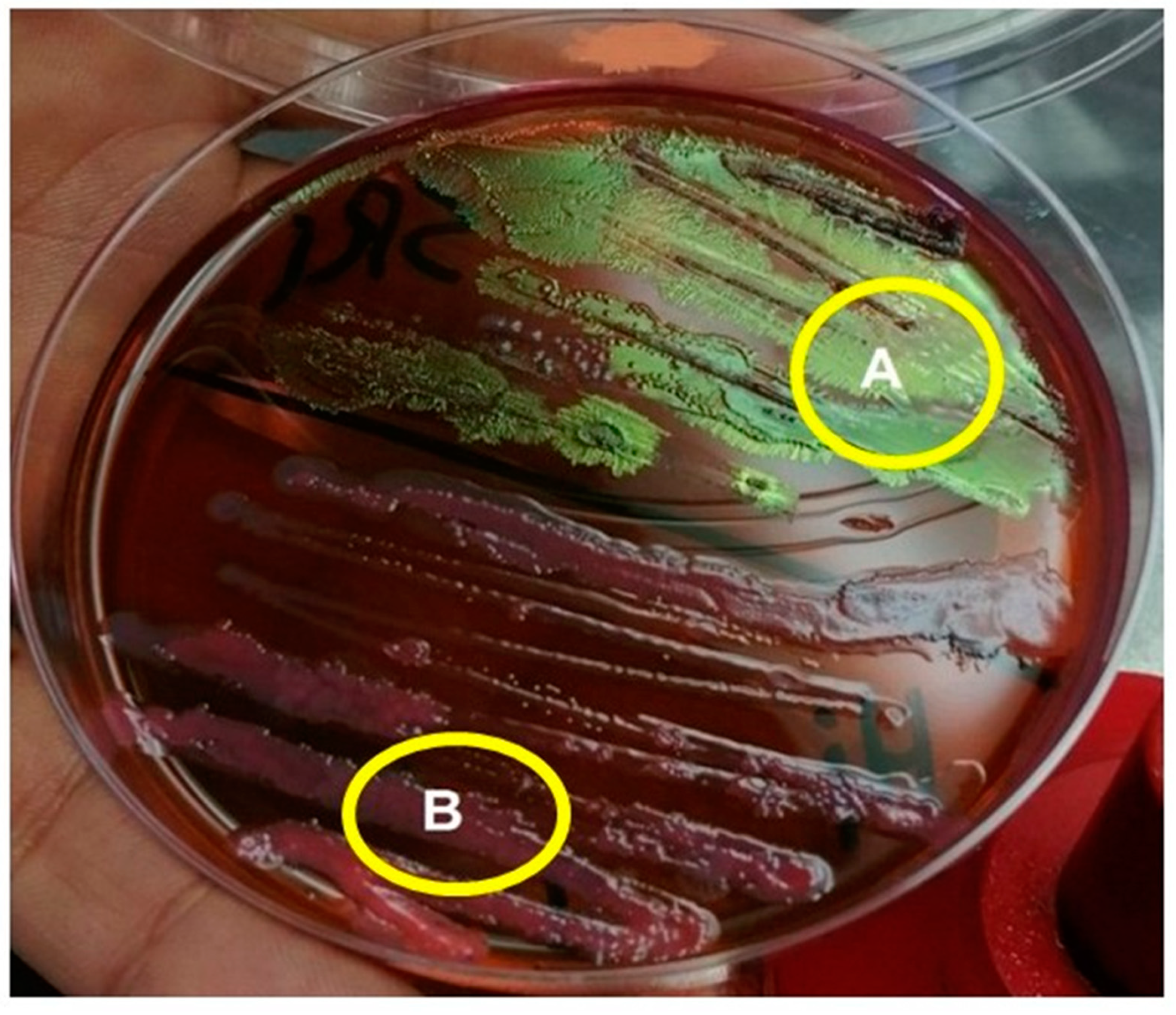
2.5. Morphological Differentiation between the Two Types of Symbiotic Bacteria

The primary bacterial cells of Xenorhabdus sp. and Photorhabdus sp. were stained with a Gram stain to describe them. Then, using the streaking approach described by Fukruksa et al. [27], bacterial colonies were distinguished based on their shape and color change on NBTA and eosin methylene blue (EMB) media.

2.6. Susceptibility of the Third-Instar Larvae of P. rapae and P. algerinus to Symbiotic Bacteria Xenorhabdus sp. and Photorhabdus sp.

This experiment was performed as described by Adithya et al. [28], in which cabbage leaves were cleaned, dried, and cut into equal leaf discs. Then, 10 of these leaf discs were impregnated in 2 mL of each bacterial suspension at concentration of 3 × 10⁷ CFU/mL. The treated cabbage leaf discs were then picked up and placed in a plastic container (9 × 5 cm2) with filter paper (Whatman number 2). Following that, 10 P. rapae larvae were put into the plastic container, which was then covered with a porous lid. In addition, cabbage leaf discs treated simply with bacterial medium were employed in a parallel control. Each treatment was replicated five times. Similar approaches were used for P. algerinus, with the exception that equal potato tuber pieces were used as food. Finally, daily mortalities of P. rapae and P. algerinus larvae were recorded for 96 h following treatment.

2.7. Efficacy and Time-Course Viability of Symbiotic Bacteria (Xenorabdus sp. and Photorabdus sp.) against the Third-Instar Larvae of P. rapae under Field Conditions

A small trial was undertaken during the winter season of 2019 in a cabbage field at the Agricultural Research Farm, Faculty of Agriculture, Menoufia University, Egypt, to assess the efficacy and time-course viability of Photorhabdus sp. and Xenorhabdus sp. bacteria against P. rapae third-instar larvae. Four randomized experimental plots were designed in the field. There were five cabbage plantations in each plot. The first plot’s cabbage plantations were treated with a bacterial suspension of Photorhabdus sp. at a concentration of 3 × 10⁷ CFU/mL. Following that, Xenorhabdus sp. was used to treat the plantations in the second plot at a concentration of 3 × 10⁷ CFU/mL. The plantations in the third plot, however, were just treated with bacterial medium (positive control). Finally, plantations in the fourth plot served as the untreated negative control group. For bioassay, five cabbage leaves were obtained independently from each plot after one hour of the treatment, transferred to the lab, and then cut into equal discs (3 × 3 cm2). Then, ten leaf discs from each plot were added to the 20 starved third-instar larvae of P. rapae in a plastic container (15 × 10 cm2). This step was replicated five times, and P. rapae larval mortality was recorded 48 h post exposure to leaf discs from each plot. The dead larvae were then sterilized in 70% ethyl alcohol, and a hemocoel sample from the dead insects was taken and streaked onto a nutrient agar media to determine whether the mortality was due to the presence of bacteria or not. Finally, to estimate the time-course viability of both bacteria, the same procedures described above were followed on the second (24 h), third (48 h), and fourth days (72 h) post treatment.

2.8. Gas Chromatography–Mass Spectrophotometry (GC-MS) of Photorhabdus sp. and Xenorhabdus sp. Bacteria

The chemical compositions of Photorhabdus sp. and Xenorhabdus sp. bacteria were determined using a Trace GC-ISQ mass spectrometer (Thermo Scientific, Austin, TX, USA) with a direct capillary column TG–5MS (30 m × 0.25 mm × 0.25 m film thickness) and a direct capillary column TG–5MS (30 m × 0.25 mm × 0.25 m film thickness). The temperature in the column oven was initially maintained at 50 °C, then increased at a rate of 5 °C/min to 200 °C, and maintained for 2 min. After that, the temperature was raised to 300 °C and kept for 2 min. The injector and MS transfer line temperatures were also kept at 270 and 260 °C, respectively. At a constant flow rate of 1 mL/min, helium was also used as a carrier gas. The solvent delay was 4 min, and diluted samples of 1 µL were automatically injected using an Autosampler AS1300 and a split mode GC. EI mass spectra were also taken in full scan mode at 70 eV ionization voltages spanning the m/z 50–650 range. The temperature of the ion source was fixed to 250 °C. Finally, the main components were identified by comparing their retention durations and mass spectra to the mass spectral databases WILEY 09 and NIST 14.

2.9. Cytotoxicity of the Symbiotic Bacteria, Xenorhabdus sp. and Photorhabdus sp.

2.9.1. Cell Lines and Chemical Reagents

The cell line human lung fibroblast (WI-38) was obtained from ATCC via a holding company for biological products and vaccines (VACSERA), Cairo, Egypt. Moreover, RPMI-1640 medium, MTT, and dimethyl sulfoxide (DMSO) (Sigma Co., St. Louis, MO, USA), as well as fetal bovine serum (GIBCO, Loughborough, UK) reagents, were used.

2.9.2. MTT Assay

The purpose of this assay was to see if Xenorhabdus sp. and Photorhabdus sp. bacteria had any effect on the viability of human lung fibroblast (WI-38) cells. This colorimetric assay is based on the conversion of yellow tetrazolium bromide to a purple formazan derivative by mitochondrial succinate dehydrogenase in viable cells. Cell lines were cultured in RPMI-1640 medium with 10% fetal bovine serum. The antibiotics added were 100 units/mL penicillin and 100µg/mL streptomycin at 37 °C in a 5% CO2 incubator. The cell lines were seeded in a 96-well plate at a density of 104 cells/well at 37 °C for 48 h under 5% CO2. After incubation, the cells were treated with bacteria and/or medium and incubated for 24 h. Subsequently, 20 µL of MTT solution at 5 mg/mL was added and incubated for 4 h. Dimethyl sulfoxide (DMSO) in a volume of 100 µL was added into each well to dissolve the purple formazan formed by mitochondrial succinate dehydrogenase in viable cells. The colorimetric assay was measured and recorded at an absorbance of 570 nm using a plate reader (ELX 800, BioTek® Instruments, Inc. Winooski, VT, USA). Thus, the intensity of the colored product was directly proportional to the number of viable cells present in the culture. The percentage cell viability was calculated as T h e   p e r c e n t a g e   c e l l   v i a b i l i t y = A 570   o f   t r e a t e d   s a m p l e s A 570   o f   t h e   u n t r e a t e d   s a m p l e × 100

2.10. Statistical Analysis

Data obtained were expressed as mean ± standard error (M ± SE). The Shapiro–Wilk and Bartlett tests for homogeneity of variances were also used to ensure that response variables were normal. The mortality percentage of the larvae was analyzed using a two-way analysis of variance (ANOVA). Furthermore, the data on the inhibitory effect of Xenorhabdus sp. and Photorhabdus sp. bacteria on the viability of human lung fibroblast (WI-38) cells were analyzed using a one-way ANOVA. All analyses were conducted using the Minitab program [29]. Then, the p-value was adjusted according to Bonferroni correction to control the family-wise error rate, where p ≤ 0.05 means significance.

3. Results

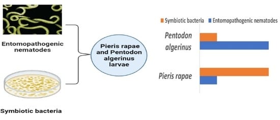
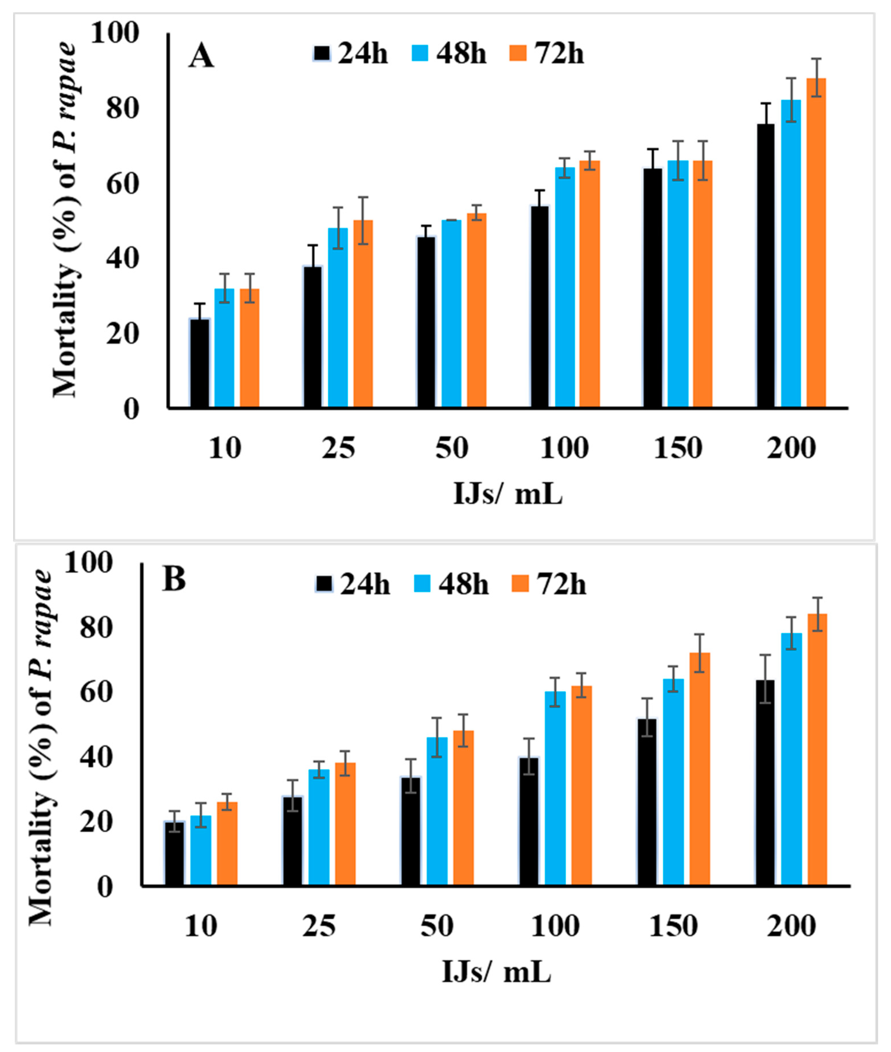
3.1. Susceptibility of the Third-Instar Larvae of P. rapae to EPNs, H. bacteriphora and S. riobravis

The data in Figure 1A, B show that both H. bacteriophora and S. riobravis had a highly significant effect on the mortality of P. rapae larvae (p < 0.001). The results showed that both nematode species induced a close percentage of mortality in P. rapae larvae (p < 0.05). Hence, H. bacteriophora induced 88% mortality, and S. riobravis induced 84% mortality at 200 IJs/mL distilled water and 72 h post exposure. The results also showed that a direct relationship existed between the percentage mortality and IJs’ concentration (p < 0.001). Thus, as the IJs’ concentration increased, the percentage of mortality increased. By contrast, exposure time did not significantly affect the percentage of mortality (p > 0.05).

3.2. Susceptibility of the Third-Instar Larvae of P. algerinus to EPNs, H. bacteriophora and S. riobravis

The data in Figure 2A,B show that the third-instar larvae of P. algerinus were highly susceptible (p < 0.001) to both H. bacteriophora and S. riobravis. From the results, H. bacteriophora surpassed S. riobravis in inducing mortality in P. algerinus. As observed, H. bacteriophora induced 100% larval mortality compared with 83% induced by S. riobravis at 200 infective juveniles/mL distilled water and 72 h post exposure. The data also indicated that the mortality percentage had a direct relationship with the exposure time and IJs’ concentration (p < 0.001).

3.3. Lethal Concentration Values of EPNs, H. bacteriophora and S. riobravis, on the Third-Instar Larvae of P. rapae

The data in Table 1 show the LC50 and LC90 values of H. bacteriophora and S. riobravis against the third-instar larvae of P. rapae. The data show that at 24 and 48 h, H. bacteriophora was more effective against P. rapae larvae than S. riobravis, as it recorded lower LC50 and LC90 values of 56.88 and 1178.41 IJs/mL distilled water, respectively, at 24 h and; 35.52 and 948.28 IJs/mL distilled water, respectively at 48 h compared with S. riobravis, which recorded 125.39 and 4325.11 IJs/mL distilled water, respectively at 24 h, and; 50.15 and 1580.56 IJs/mL distilled water, respectively, at 48 h. However, at 72 h, no significant difference was observed between the LC50 and LC90 values of both nematode species. H. bacteriophora recorded 32.19 and 647.84 IJ/mL, respectively, compared with 35.14 and 606.22, respectively, for S. riobravis.

3.4. Lethal Concentration Values of EPNs, H. bacteriophora and S. riobravis, on the Third-Instar Larvae of P. algerinus

The data in Table 2 show that H. bacteriophora was more efficient against P. algerinus than S. riobravis, as it recorded a lower LC50 and LC90 of 22.79 and 365.36 IJs/mL distilled water at 24 h, 19.15 and 264.28 IJs/mL distilled water at 48 h, and at 72 h, it recorded 19.00 and 162.53 IJs/mL, respectively. S. riobravis, however, recorded 91.50 and 1927.89 IJs/mL distilled water at 24 h, 55.02 and 829.61 IJs/mL distilled water at 48 h, and at 72 h it recorded 43.50 and 547.12 IJs/mL distilled water, respectively. P. algerinus was more vulnerable to both nematode species than P. rapae according to the toxicity data in Table 1 and Table 2. In addition, the LC50 and LC90 values decreased with an increase in the time.

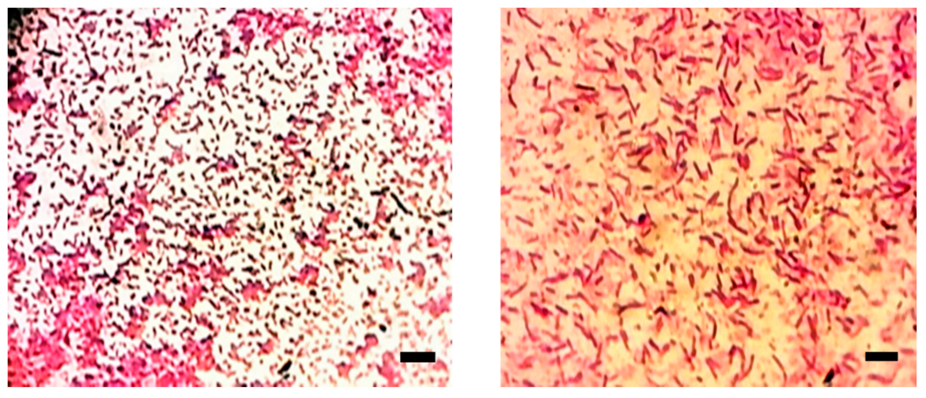
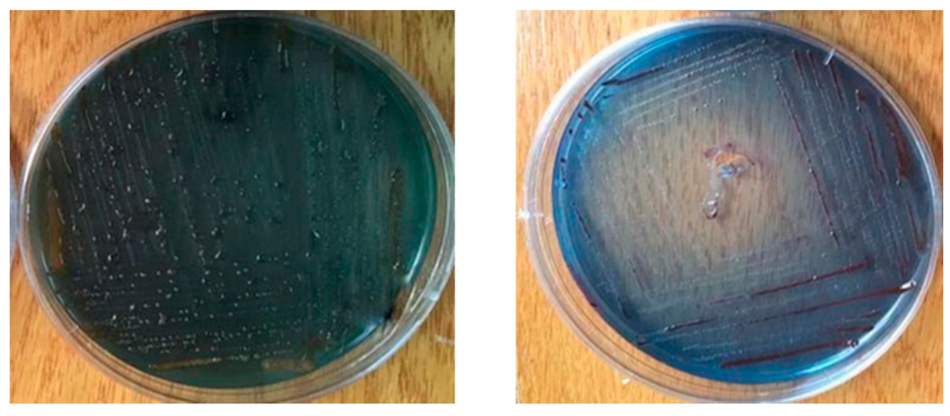
3.5. Morphological Characterization of the Isolated Symbiotic Bacteria, Photorhabdus sp. and Xenorhabdus sp.

Based on the staining of the bacterial cells with Gram stain, it was obvious that both Xenorhabdus and Photorhabdus (Figure 3) bacterial cells had purple coloration. Meanwhile, the Xenorhabdus cells (left graph) were smaller than the Photorhabdus ones (right graph). Furthermore, on the basis of the growth of the tested bacteria on NBTA medium, the colony morphology of the Xenorhabdus bacterium was characterized as a dark blue, convex, and umbonate colony (Figure 4; left graph). However, Photorhabdus bacterium appeared as a dark red, convex, and umbonate colony (Figure 4; right graph). Additionally, using the EMB medium, Xenorhabdus bacterium was shown as flat, with a green metallic sheen colony (Figure 5A). By contrast, the Photorhabdus bacterium was demonstrated as a rose convex colony (Figure 5B).

3.6. Efficacy of the Symbiotic Bacteria, Xenorhabdus sp. and Photorhabdus sp., against Pieris rapae Larvae

The data in Figure 6A show that both Photorhabdus sp. and Xenorhabdus sp. bacteria significantly affected P. rapae larvae (p < 0.001). Both bacterial species induced 100% larval mortality at 96 h of treatment. Moreover, the obtained data indicated that time had a significant effect (p < 0.01) on the percentage of mortality.

3.7. Efficacy of the Symbiotic Bacteria, Xenorhabdus sp. and Photorhabdus sp., against P. algerinus Larvae

As shown in Figure 6B, both Photorhabdus sp. and Xenorhabdus sp. bacteria successfully induced mortality in P. algerinus larvae (p ˂ 0.001). Photorhabdus sp. bacterium caused 80% mortality and Xenorhabdus sp. induced 42% mortality at 96 h post exposure.

3.8. Efficacy and Time-Course Viability of Entomopathogenic Bacteria, Xenorhabdus sp. and Photorhabdus sp. against the Third-Instar Larvae of P. rapae under Field Conditions

The data in Figure 7 show that both Photorhabdus sp. and Xenorhabdus sp. significantly controlled the third-instar larvae of P. rapae under field conditions (p < 0.001). However, the time-course viability of both bacteria was decreased by time. The highest mortality percentages were recorded in the sets where the larvae were exposed to cabbage leaves that were collected after one hour of application, and the lowest ones were recorded in the sets where the larvae were exposed to cabbage leaves that were collected 72 h post application, and this was for both bacterial genera. The data also indicated that Photorhabdus sp. bacterium was more effective than Xenorhabdus sp. bacterium, as it had the ability to induce 78, 59, 38, and 21% mortalities of the third-instar larvae of P. rapae at 1, 24, 48, and 72 h of application, respectively, compared with 64, 53, 29, and 17% at 1, 24, 48, and 72 h of application, respectively, for Xenorhabdus sp. bacterium. Furthermore, the mortality rates of the positive control (the leaf discs treated with media alone) were 1.00, 1, 0, and 0% at 1, 24, 48, and 72 h of application, respectively. The negative control (untreated leaves) mortalities were 0, 1, 0, and 0% at 1, 24, 48, and 72 h of the beginning of the experiment.

3.9. Gas Chromatography–Mass Spectrophotometry of Xenorhabdus sp. and Photorhabdus sp. Bacteria

3.9.1. Xenorhabdus sp. Bacterium

GC-MS analysis of Xenorhabdus sp. bacterium revealed 14 components (Table 3). The main constituent was 2-pyrrolidinone (35.04%), followed by 9-octadecenoic acid (z)-(oleic acid) (13.86%), 1,4-benzenediol, 2-(1,1-dimethylethyl)-5-(2-propenyl) (4.92%), 2,2-dideutero octadecanal (4.53%), octadecanoic acid (3.42%), 4-octadecenal (3.19%), cyclopentane tridecanoic acid, methyl ester (2.87%), 1,2-benzenedicarboxylic acid (2.80%), hexadecanoic acid (2.72%), 2,3-dihydroxypropyl ester paromomycin (2.63%), 1-tetradecanol (2.62%), 2,8,9-Trioxa-5-aza-1-silabicyclo [3.3.3]undecane, 1-methyl (2.37%), 7-nonenoic-7,8-d2 acid, methyl ester (2.11%), and docosanoic acid-1,2,3-propanetriyl ester (2.00%).

3.9.2. Photorhabdus sp. Bacterium

The GC-MS of Photorhabdus sp. bacterium showed 12 components (Table 4). The main constituent was 2-piperidinone (44.09%), followed by pentadecanoic acid, 14-methyl-methyl ester (14.43%), 1,2-benzenedicarboxylic acid (13.20%), 1-eicosanol (5.57%), 4-trifluoroacetoxytetradecane (4.66%), bacteriochlorophyll-c-stearyl (2.91%), 15-methyltricyclo[6.5.2(13,14).0(7,15)] pentadeca-1,3,5,7,9,11,13-heptene (4.25%), octadecanoic acid, methyl ester (3.92%), bacteriochlorophyll-c-stearyl (2.91%), 1-tetradecanol (2.66%), 2(1h)-naphthalenone, octahydro-1-methyl-1-(2-p ropenyl)-, (1à,4aá,8aà) (2.28%), erucic acid (2.26%), and acetic acid, octyl ester (1.42%).

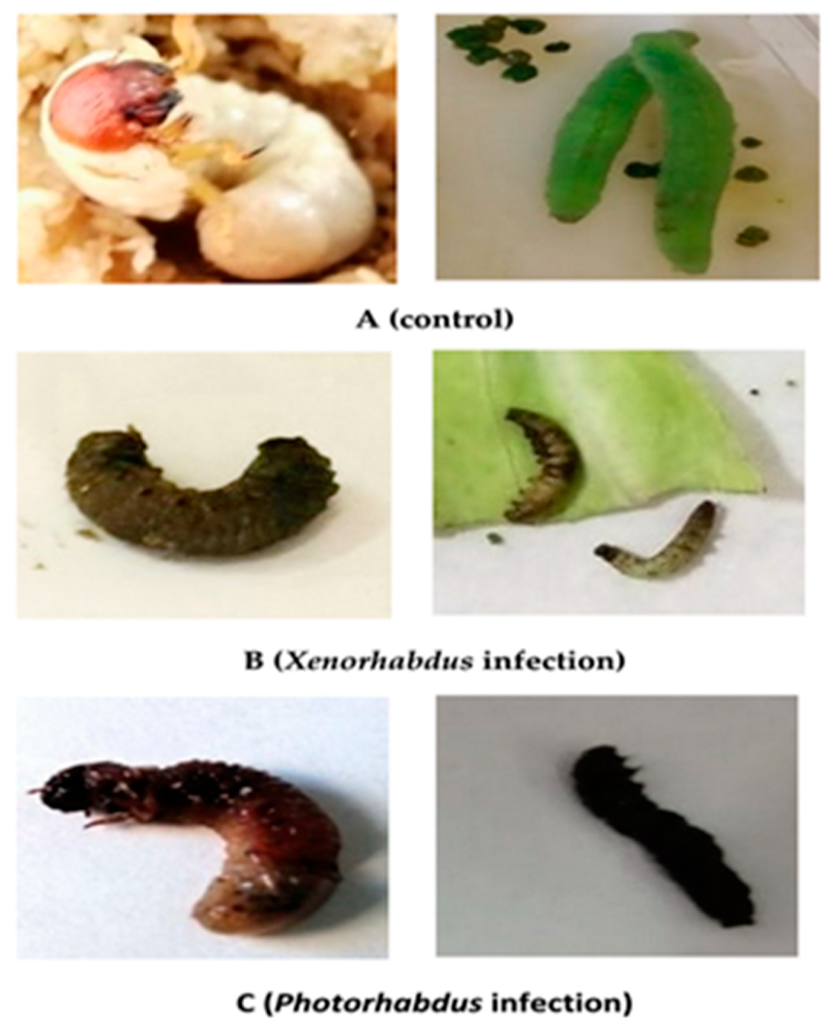
3.10. Morpho-Pathological Alterations in P. rapae and P. algerinus Larvae Caused by the Symbiotic Bacteria, Xenorhabdus sp. and Photorhabdus sp.

Figure 8 shows the morpho-pathological alterations of P. rapae and P. algerinus caused by Xenorhabdus sp. and Photorhabdus sp. bacteria. The control of P. algerinus larvae showed a creamy white coloration with a large brown head. However, P. rapae larvae had a bright green coloration (Figure 8A). Upon infection with Xenorhabdus sp. bacterium, the color of both insect species turned grayish (Figure 8B). Meanwhile, the color of P. algerinus and P. rapae larvae turned into a somewhat reddish color due to infection with the Photorhabdus bacterium (Figure 8C).

3.11. Cytotoxicity of the Isolated Symbiotic Bacteria, Xenorhabdus sp. and Photorhabdus sp.

In vitro, an MTT assay was conducted to evaluate the inhibitory effect of Xenorhabdus sp. and Photorhabdus sp. bacteria on normal WI-38 human cell viability. The results revealed a percentage cell viability of 85.3% for Xenorhabdus and 81.7% for Photorhabdus compared with 88.0% for the control (Table 5). Thus, these results reveal weak in vitro cytotoxicity of the tested bacteria on WI-38 cells (p ˃ 0.05).

4. Discussion

Various governments give special attention to the agricultural economy, because it is one of the most important sources of national income. Therefore, there is a great interest in agricultural pests and the damage they cause. Combating these pests has also become one of the most important priorities of people. For example, previous studies have been concerned with controlling P. rapae; however, they did not solve the problem. In addition, most of these studies focused on the use of chemical pesticides. Alternatively, studies on the biocontrol of P. algerinus remain scarce. Therefore, the present study aimed to evaluate the efficacy of H. bacteriophora and S. riobravis, including their symbiotic bacteria Photorhabdus sp. and Xenorhabdus sp., respectively, against P. rapae and P. algerinus larvae. The results revealed that both H. bacteriophora and S. riobravis nematodes successfully induced mortality in P. rapae and P. algerinus larvae. These results were in accordance with those of Ali et al. [30], who reported the efficacy of Steinernema masoodi, Steinernema seemae, Steinernema carpocapsae, Steinernema glaseri, and Steinernema thermophilum against Helicoverpa armigera, G. mellonella, and Corcyra cephalonica. Additionally, Reda et al. [16] reported that S. carpocapsae induced mortality in fourth-instar larvae and the pupae of P. rapae, with LC50 values of 18.148 and 38.96 IJs/larva and pupa, respectively. Recently, Askary and Ahmad [31] also recorded the efficacy of Heterorhabditis pakistanensis for controlling Pieris brassicae. Likewise, Grewal et al. [32] and Kleim et al. [33] improved the susceptibility of Japanese beetle, Popillia japonica, to EPNs infecting turf in the USA. WU [34] also reported the efficacy of H. bacteriophora and H. megidis against masked chafer white grubs, Cyclocephala spp. Similarly, Kajuga et al. [35] reported that both H. bacteriophora and S. carpocapsae killed up to 58% of white grubs. Another study also reported that Steinernema abbasi and Heterorhabditis indica had the capability to control the white grub Leucopholis lepidophora [36]. The obtained data also revealed that H. bacteriophora was more effective than S. riobravis against both P. rapae and P. algerinus. Shapiro-Ilan et al. [37,38] attributed the discrepancy in the infectivity and virulence of different EPN strains to different foraging behavior, host specificity, morphological characterization of the ENs, and the tolerance to host immune defenses.
Based on foraging behavior, EPNs have been classified into cruisers (active searchers) and ambushers (sit-and-wait foragers) [39]. Previous studies classified Heterorhabditids as cruisers and Steinernematids as ambushers [39]. Hence, the superiority of H. bacteriophora over S. riobravis in this study may be attributed to its foraging behavior as a cruiser. Grewal et al. [40] attributed the higher effect of H. bacteriophora and H. megidis than that of S. carpocapsae and Steinernema scapterisci to their different foraging behaviors. Additionally, Dillon et al. [41,42] reported that S. carpocapsae was less effective than the classic cruiser–foraging species, Heterorhabditis downesi.
EPNs’ morphological characterization is an important factor in determining their virulence toward insect hosts. The greater virulence of H. bacteriophora larvae compared to S. riobravis larvae may be attributable to the Heterorhabditid IJs’ distinctive buccal cuticular teeth, which facilitate their penetration into the host. Bedding et al. [43], who attributed the quick invasion rate of Heterorhabditid nematodes in several insect hosts to the dorsal tooth of their IJs, backed up this theory. This assumption could explain why Gouge et al. [44] and Menti et al. [45] discovered that Heterorhabditid nematodes infect insect hosts more quickly than Steinernematid nematodes. Furthermore, because Heterorhabditid nematodes are hermaphrodites, only one IJ is required to complete the life cycle and settle inside the insect host. Steinernematids, on the other hand, are amphimictic. As a result, for effective reproduction and establishment, both a male and a female would need to enter the host.
The variations in virulence between H. bacteriophora and S. riobravis against P. rapae and P. algerinus could potentially be attributed to their tolerance of host immune responses. This finding agrees with that of Silva et al. [46], who reported that in Manduca sexta, P. luminescens cells accompanied with H. bacteriophora secreted antiphagocytic and anti-encapsulation factors that permitted nematodes to overcome the insect’s immune defenses.
The obtained data also revealed that the third-instar larvae of P. algerinus were more susceptible to H. bacteriophora and S. riobravis infestation than those of P. rapae. The appreciable differences in the susceptibility of the two insect hosts may be attributed to different host morphological structures, sizes, behaviors, and immune defense mechanisms. This opinion is in agreement with that of Medeiros et al. [47]; they attributed the differences in the susceptibility of different stages of Pseudaletia unipuncta to S. carpocapsae, S. glaseri, and H. bacteriophora to different insect sizes, behaviors, and immune defense mechanisms. As a result, P. algerinus’ larger size may be the explanation for its superiority as a nematode host over P. rapae. Similarly, Lewis et al. [48] found that large larvae are more attractive to EPNs than smaller larvae. Boff et al. [49] also found that as larval size rose, the amount of invading H. megidis IJs and the production of IJs from infected wax moth and vine weevil larvae increased. Another reason for the P. algerinus larvae’s higher vulnerability to nematodes than that of the P. rapae larvae is that the P. algerinus larvae reside deep in the soil, where nematodes live. As a result, infection is thought to have occurred once or more, and the nematodes recognized the insect’s immunity map. The P. rapae larvae, however, reside on the surface of the cabbage plant, so it is probable that the infestation occurred for the first time; thus, the immune system’s tools worked together to combat the nematode onslaught.
As shown in the above result, H. bacteriophora and S. riobravis to some extent succeed in the control of both P. algerinus and P. rapae. However, it is known that symbiotic bacteria have a major role in killing insects. Hence, we isolated the symbiotic bacteria of H. bacteriophora and S. riobravis and then applied them to control both insect species. Subsequently, the symbiotic Xenorhabdus sp. and Photorhabdus sp. from S. riobravis and H. bacteriophora, respectively, were isolated, mass cultured, and applied at a concentration of 3 × 10⁷ CFU/mL against P. algerinus and P. rapae. The obtained data revealed that both Xenorhabdus sp. and Photorhabdus sp. significantly affected P. algerinus and P. rapae larvae. Some studies have also emphasized the ability of Xenorhabdus spp. and Photorhabdus spp. to induce mortality in different insect species [8,18,50,51,52,53].
The data obtained also revealed that Photorhabdus sp. was more effective than Xenorhabdus sp. against both P. algerinus and P. rapae; however, P. rapae was more susceptible. This higher lethality of Photorhabdus sp. than that of Xenorhabdus sp. correlates with the better efficacy of H. bacteriophora than that of S. riobravis. These results were in line with those of Rahoo et al. [51], who reported that the mortality caused by P. luminescens was significantly higher than that of X. bovienii. Moreover, ref. [8] reported that Photorhabdus species produced 75–96% mortality in S. frugiperda larvae. In contrast, Xenorhabdus bacteria were less active, with mortality rates in the range of 33–57%.
The insecticidal activity of Photorhabdus sp. and Xenorhabdus sp. bacteria may be attributed to the fact that both produce toxin complexes, proteases, lipases, lipopolysaccharides, and other active components [46,54,55,56,57]. These components make caterpillars floppy [58], induce apoptosis, inhibit hemocyte motility, and inhibit cellular and humoral immunity [59,60].
The GC-MS analysis of Xenorhabdus sp. and Photorhabdus sp. bacteria revealed that Xenorhabdus sp. bacterium possessed 14 main components, whereas Photorhabdus sp. bacterium had 12 main components as shown in Table 3 and Table 4, respectively. Five of these compounds (2-Piperidinone, 1,2-benzenedicarboxylic acid, tetradecanol, and octadecanoic acid) were commonly detected in the two bacterial genera. However, the ratios in Photorhabdus sp. were higher than those in Xenorhabdus sp.
The piperidinone compound was the highest ever in both Photorhabdus sp. and Xenorhabdus sp. bacteria. Piperidinone is an organic chemical that is a derivative of piperidine. Piperidine, on the other hand, is a colorless fuming liquid with an ammoniacal, peppery odor. Piperidine is a common chemical reagent and building block in the production of organic molecules, including pharmaceuticals. The piperidine structural motif is present in numerous natural alkaloids. [59,60]. Vivekanandhan et al. [61] emphasized the role of piperidinone in the insecticidal activity of Beauveria bassiana against Cx. quinquefasciatus mosquito.
Several other studies have detected similar compounds from different strains of Xenorhabdus and Photorhabdus bacteria [62,63,64,65]. These compounds may be responsible for the insecticidal activity of Xenorhabdus and Photorhabdus bacteria in this study. This assumption may be supported by the opinion of Ullah et al. [62], who attributed the insecticidal and antimicrobial activity of P. temperate against G. mellonella larvae to 1,2-benzenedicarboxylic acid, which plays a crucial role in the inhibition of insect phenoloxidase (the key mediator of insect immune systems). Similarly, Hemalatha et al. [66] attributed the insecticidal activity of X. nematophilus against Ferrisia virgata to 1,2-Benzenedicarboxylic acid and cosine groups. Hasan et al. [64] also attributed the virulence of six X. nematophila strains against Spodoptera exigua to active secondary compounds, such as benzeneacetic acid, n-Decanoic acid, Tetradecane,1-Decene, and 3-Benzylidene-hexahydro-pyrrolo, which inhibit the insect immune system. Later, Mollah and Kim [65] detected fatty alcohol, 1-ecosine, heptadecane, octadecanes, and methyl-12-tetradecen-1-ol acetate in different strains of Xenorhabdus and Photorhabdus bacteria. The authors suggested that these compounds inhibited the insect’s phospholipase A2, thereby eradicating the insect immune system. The phospholipase A2 enzyme catalyzes fatty acids that are later oxygenated by cyclooxygenase and lipoxygenase enzymes to produce prostaglandins and leukotrienes, respectively, which are mediators of the immune response in insects [67]. This was supported by the findings of [68], who reported that X. nematophila and P. temperata were responsible for suppressing the phospholipase A2 enzyme. Another compound identified from the GC-MS analysis of Photorhabdus sp. in this study was uric acid, which plays a crucial role as a food inhibitor in order to prevent infected insects from feeding, thus inducing insect death.
In the continuation of this study and in an attempt to model an integrated idea regarding the efficacy of the tested EPNs and their symbiotic bacteria, we evaluated the efficacy of Xenorhabdus sp. and Photorhabdus sp. bacteria to control P. rapae in the field. The data obtained showed that both bacterial species significantly decreased the population of P. rapae in the field. The percentage mortality reached 78% by Photorhabdus sp. and 64% by Xenorhabdus sp. Although there are several studies documenting the use of EPNs for insect control in the field [31,69,70,71,72,73,74,75,76], those that document the efficacy of Xenorhabdus sp. and Photorhabdus sp. bacteria in the field are scarce. Gerritsen et al. [77] recorded the efficacy of Photorhabdus and Xenorhabdus strains against Frankliniella occidentalis and Thrips tabaci after sucking the bacteria from treated leaves. Therefore, these results from the efficacy of Xenorhabdus sp. and Photorhabdus sp. in the field confirm the results at the laboratory scale and are further proof of the effectiveness of these bacteria.

5. Conclusions

From this study, we concluded that H. bacteriophora, S. riobravis, and their symbiotic bacteria (Photorhabdus sp. and Xenorhabdus sp., respectively) are effective candidates for biocontrolling P. rapae and P. algerinus, either in experimental or field studies. The results also clarified that both symbiotic bacteria can be utilized separately from their nematodes. Thus, we can recommend these EPNs and their symbiotic bacteria to be certified alternatives for chemical pesticides in the control programs of P. rapae and P. algerinus and to be tested against other insect pests.

Author Contributions

Conceptualization: H.E., A.M.A.E., M.F.S., M.S.A.-H., and A.M.A.E.-R. Data curation: H.E., A.M.A.E., M.F.S., M.S.A.-H., and A.M.A.E.-R. Formal analysis: H.E., A.M.A.E., M.F.S., M.S.A.-H., and A.M.A.E.-R. Investigation: H.E., A.M.A.E., M.F.S., M.S.A.-H., and A.M.A.E.-R. Methodology: H.E., A.M.A.E., and A.M.A.E.-R. Resources: H.E., A.M.A.E., M.F.S., M.S.A.-H., and A.M.A.E.-R. Software: H.E., A.M.A.E., M.F.S., M.S.A.-H., and A.M.A.E.-R. Writing—original draft: H.E., A.M.A.E., and A.M.A.E.-R. Writing—review and editing: H.E., A.M.A.E., M.F.S., M.S.A.-H., and A.M.A.E.-R. All authors have read and agreed to the published version of the manuscript.

Funding

The authors are thankful to the Taif University Researchers Supporting Project number (TURSP-2020/64), Taif University, Taif, Saudi Arabia, for providing the financial support and research facilities.

Institutional Review Board Statement

Not applicable.

Informed Consent Statement

Not applicable.

Data Availability Statement

All data are presented within the article.

Conflicts of Interest

The authors declare that they have no conflict of interest.

References

  1. Abd El-Salam, A.M.E. Field evaluation of some eco-friendly formulations against strawberry white grubs in Egypt. Spec. J. Biol. Sci. 2019, 5, 1–6. [Google Scholar]
  2. Abd-Rabou, S.; Saadia, A.A. New records of scarabaid white grub species and diptrean genus in sugar cane soil in upper Egypt (Coleoptera: Scarabidae). Egypt. J. Agric. Res. 2006, 84, 797–801. [Google Scholar]
  3. Aktar, W.; Sengupta, D.; Chowdhury, A. Impact of pesticides use in agriculture: Their benefits and hazards. Interdiscip. Toxicol. 2009, 2, 1–12. [Google Scholar] [CrossRef] [Green Version]
  4. Campos-Herrera, R.; Tailliez, P.; Pagès, S.; Ginibre, N.; Gutiérrez, C.; Boemare, N. Characterization of Xenorhabdus isolates from La Rioja (Northern Spain) and virulence with and without their symbiotic entomopathogenic nematodes (Nematoda: Steinernematidae). J. Invertebr. Pathol. 2009, 102, 173–181. [Google Scholar] [CrossRef] [PubMed]
  5. Pietri, J.E.; Liang, D. Virulence of entomopathogenic bacteria in the bed bug, Cimex lectularius. J. Invertebr. Pathol. 2018, 151, 1–6. [Google Scholar] [CrossRef] [PubMed]
  6. Stock, S.P. Partners in crime: Symbiont-assisted resource acquisition in Steinernema entomopathogenic nematodes. Curr. Opin. Insect Sci. 2019, 32, 22–27. [Google Scholar] [CrossRef]
  7. Sheetal, P.; Geetha, B.; Dandapani, V.; Devaraj, B. Mosquitocidal efficacy of lecithinase derived from entomopathogenic bacteria Xenorhabdus sp. strain PBU1755 against filarial vector Culex quinquefasciatus. Biocatal. Agric. Biotechnol. 2019, 17, 492–498. [Google Scholar] [CrossRef]
  8. Salvadori, J.D.M.; Defferrari, M.S.; Ligabue-Braun, R.; Lau, E.Y.; Salvadori, J.R.; Carlini, C.R. Characterization of ento-mopathogenic nematodes and symbiotic bacteria active against Spodoptera frugiperda (Lepidoptera: Noctuidae) and contribu-tion of bacterial urease to the insecticidal effect. Biol. Control. 2012, 63, 253–263. [Google Scholar] [CrossRef] [Green Version]
  9. Liu, J.; Berry, R.E.; Blouin, M.S. Identification of Symbiotic Bacteria (Photorhabdus and Xenorhabdus) from the Entomopathogenic Nematodes Heterorhabditis marelatus and Steinernema oregonense Based on 16S rDNA Sequence. J. Invertebr. Pathol. 2001, 77, 87–91. [Google Scholar] [CrossRef] [PubMed]
  10. Eleftherianos, I.; Joyce, S.A.; Ffrench-Constant, R.H.; Clarke, D.J.; Reynolds, S.E.; Ffrench-Constant, R. Probing the tritrophic interaction between insects, nematodes and Photorhabdus. Parasitology 2010, 137, 1695–1706. [Google Scholar] [CrossRef]
  11. Fang, X.; Li, Z.; Wang, Y.; Zhang, X. In vitro and in vivo antimicrobial activity of Xenorhabdus bovienii YL002 against Phytophthora capsici and Botrytis cinerea. J. Appl. Microbiol. 2011, 111, 145–154. [Google Scholar] [CrossRef]
  12. Shi, D.; An, R.; Zhang, W.; Zhang, G.; Yu, Z. Stilbene Derivatives from Photorhabdus temperata SN259 and Their Antifungal Activities against Phytopathogenic Fungi. J. Agric. Food Chem. 2017, 65, 60–65. [Google Scholar] [CrossRef]
  13. Bortoluzzi, L.; Alves, L.F.A.; Alves, V.S.; Holz, N. Entomopathogenic nematodes and their interaction with chemical insecticide aiming at the control of banana weevil borer, Cosmopolites sordidus Germar (Coleoptera: Curculionidae). Arq. Inst. Biol. 2013, 80, 183–192. [Google Scholar] [CrossRef] [Green Version]
  14. Abdolmaleki, A.H.R.; Maafi, Z.t.; Naseri, B. Virulence of two entomopathogenic nematodes through their interaction with Beauveria bassiana and Bacillus thuringiensis against Pieris brassicae (Lepidoptera: Pieridae). J. Crop. Prot. 2017, 6, 287–299. [Google Scholar]
  15. Gorgadze, O.; Bakhtadze, G.; Nebieridze, D. Efficacy of local entomopathogenic nematodes against Pieris brassicae (L, 1758) (Lepidoptera: Pieridae). Int. J. Dev. Res. 2018, 8, 23900–23903. [Google Scholar]
  16. Reda, E.S.; Sallam, A.M.; Ibrahim, H.Y.E.; Eid, S.M.E. Efficacy of Entomopathogenic Nematode, Steinernema carpocapsae and its Interaction with Beauveria bassiana against Pieris rapae L. (Lepidoptera: Pieridae). J. Plant Prot. Pathol. 2018, 9, 795–798. [Google Scholar] [CrossRef] [Green Version]
  17. Hinchliffe, S.J. Insecticidal Toxins from the Photorhabdus and Xenorhabdus Bacteria. Open Toxinol. J. 2010, 3, 83–100. [Google Scholar] [CrossRef]
  18. Vitta, A.; Thimpoo, P.; Meesil, W.; Yimthin, T.; Fukruksa, C.; Polseela, R.; Mangkit, B.; Tandhavanant, S.; Thanwisai, A. Larvicidal activity of Xenorhabdus and Photorhabdus bacteria against Aedes aegypti and Aedes albopictus. Asian Pac. J. Trop. Biomed. 2018, 8, 31. [Google Scholar] [CrossRef]
  19. Da Silva, W.J.; Júnior, H.L.P.; Heermann, R.; Da Silva, O.S. The great potential of entomopathogenic bacteria Xenorhabdus and Photorhabdus for mosquito control: A review. Parasites Vectors 2020, 13, 1–14. [Google Scholar] [CrossRef]
  20. Webb, S.; Shelton, A. Laboratory Rearing of the Imported Cabbage Worm. N. Y. Food Life Sci. Bull. 1988, 122. [Google Scholar]
  21. Kotchofa, R.; Baimey, H. In vivo production of entomopathogenic nematodes using Galleria mellonella: Costs and effect of diets on nematode pathogenicity. J. Nematol. 2019, 51, 1–15. [Google Scholar] [CrossRef] [PubMed] [Green Version]
  22. White, G.F. A method for obtaining infective nematode larvae from cultures. Science 1927, 66, 302–303. [Google Scholar] [CrossRef]
  23. Yuksel, E.; Taskesen, Y.E.; Erarslan, D.; Canhilal, R. Effectiveness of different entomopathogenic nematode species against the variegated cutworm, Peridroma saucia (Hubner) (Lepidoptera: Noctuidae). Egypt. J. Biol. Pest Control. 2018, 28. [Google Scholar] [CrossRef] [Green Version]
  24. Finney, D.J. Probit Analysis, 3rd ed.; Cambridge University Press: London, UK, 1971. [Google Scholar]
  25. Poinar, G.G.; Thomas, G.M. Significance of Achromobacter nematophilus Poinar and Thomas (Achromobacteraceae: Eubacteriales) in the development of the nematode, DD-136 (Neoaplectana sp., Steinernematidae). Parasitology 1966, 56, 385–390. [Google Scholar] [CrossRef]
  26. Yooyangket, T.; Muangpat, P.; Polseela, R.; Tandhavanant, S.; Thanwisai, A.; Vitta, A. Identification of entomopathogenic nematodes and symbiotic bacteria from Nam Nao National Park in Thailand and larvicidal activity of symbiotic bacteria against Aedes aegypti and Aedes albopictus. PLoS ONE 2018, 13, e0195681. [Google Scholar] [CrossRef] [PubMed]
  27. Fukruksa, C.; Yimthin, T.; Suwannaroj, M.; Muangpat, P.; Tandhavanant, S.; Thanwisai, A.; Vitta, A. Isolation and identification of Xenorhabdus and Photorhabdus bacteria associated with entomopathogenic nematodes and their larvicidal activity against Aedes aegypti. Parasites Vectors 2017, 10, 440. [Google Scholar] [CrossRef] [Green Version]
  28. Adithya, S.; Shivaprakash, M.; Sowmya, E. Evaluation of insecticidal activity of entomopathogenic bacteria Photorhabdus and Xenorhabdus against shoot and fruit borer Earias vittella (Lepidoptera: Noctuidae) of vegetable crops. J. Èntomol. Zoöl. Stud. 2020, 8, 2343–2348. [Google Scholar] [CrossRef]
  29. Minitab, Inc. Minitab Statistical Software, Release 14 for Windows; Minitab, Inc.: State College, PA, USA, 2003. [Google Scholar]
  30. Ali, S.S.; Pervez, R.; Hussain, M.A.; Ahmad, R. Susceptibility of three lepidopteran pests to five entomopathogenic nematodes and in vivo mass production of these nematodes. Arch. Phytopathol. Plant Prot. 2008, 41, 300–304. [Google Scholar] [CrossRef]
  31. Askary, T.H.; Ahmad, M.J. Efficacy of entomopathogenic nematodes against the cabbage butterfly (Pieris brassicae (L.) (Lepidoptera: Pieridae) infesting cabbage under field conditions. Egypt. J. Biol. Pest Control. 2020, 30, 1–7. [Google Scholar] [CrossRef]
  32. Grewal, P.; Power, K.; Grewal, S.; Suggars, A.; Haupricht, S. Enhanced consistency in biological control of white grubs (Coleoptera: Scarabaeidae) with new strains of entomopathogenic nematodes. Biol. Control. 2004, 30, 73–82. [Google Scholar] [CrossRef]
  33. Klein, M.G.; Grewal, P.S.; Jackson, T.A.; Koppenhöfer, A.M. Lawn, turf and grassland pests. In Field Manual of Techniques in Invertebrate Pathology: Application and Evaluation of Pathogens for Control of Insects and Other Invertebrate Pests, 2nd ed.; Lacey, L.A., Kaya, H.K., Eds.; Springer: Dordrecht, The Netherlands, 2007; pp. 655–675. [Google Scholar]
  34. Wu, S. Efficacy of Entomopathogenic Nematodes and Entomopathogenic Fungi against Masked Chafer White Grubs, Cyclocephala spp.(Coleoptera: Scarabaeidae). PhD Thesis, Virginia Tech, Blacksburg, VA, USA, 2013. [Google Scholar]
  35. Kajuga, J.; Hategekimana, A.; Yan, X.; Waweru, B.; Li, H.; Li, K.; Yin, J.; Cao, L.; Karanja, D.; Umulisa, C.; et al. Management of white grubs (Coleoptera: Scarabeidae) with entomopathogenic nematodes in Rwanda. Egypt. J. Biol. Pest Control. 2018, 28, 1–13. [Google Scholar] [CrossRef] [Green Version]
  36. Patil, J.; Rangasamy, V. Field evaluation of the entomopathogenic nematodes against the white grub, Leucopholis lepidophora Blanchard (Coleoptera: Scarabaeidae). Egypt. J. Biol. Pest Control. 2018, 28, 28–41. [Google Scholar] [CrossRef]
  37. Shapiro-Ilan, D.; Stuart, R.; McCoy, C.W. A Comparison of Entomopathogenic Nematode Longevity in Soil under Laboratory Conditions. J. Nematol. 2006, 38, 119–129. [Google Scholar]
  38. Shapiro-Ilan, D.I.; Gouge, D.H.; Piggott, S.J.; Fife, J.P. Application technology and environmental considerations for use of entomopathogenic nematodes in biological control. J. Biol. Control. 2006, 38, 124–133. [Google Scholar] [CrossRef]
  39. Bal, H.K.; Grewal, P.S. Lateral Dispersal and Foraging Behavior of Entomopathogenic Nematodes in the Absence and Presence of Mobile and Non-Mobile Hosts. PLoS ONE 2015, 10, e0129887. [Google Scholar] [CrossRef] [PubMed]
  40. Grewal, P.S.; Lewis, E.E.; Gaugler, R.; Campbell, J.F. Host finding behaviour as a predictor of foraging strategy in ento-mopathogenic nematodes. Parasitology 1994, 108, 207–215. [Google Scholar] [CrossRef]
  41. Dillon, A.B.; Ward, D.; Downes, M.J.; Griffin, C.T. Suppression of the large pine weevil Hylobius abietis (L.) (Coleoptera: Curculionidae) in pine stumps by entomopathogenic nematodes with different foraging strategies. Biol. Control. 2006, 38, 217–226. [Google Scholar] [CrossRef] [Green Version]
  42. Dillon, A.B.; Downes, M.J.; Ward, D.; Griffin, C.T. Optimizing application of entomopathogenic nematodes to manage large pine weevil, Hylobius abietis L. (Coleoptera:Curculionidae) populations developing in pine stumps, Pinus sylvestris. Biol. Control. 2007, 40, 253–263. [Google Scholar] [CrossRef] [Green Version]
  43. Bedding, R.A.; Molynou, A.S.; Åkhurst, R.J. Heterorhabditis spp., Neoaplectana spp. and Steinernema kraussei: Interspecific and intraspecific differences in infectivity for insects. Exp. Parasitol. 1983, 55, 249–257. [Google Scholar] [CrossRef]
  44. Gouge, D.H.; Lee, L.L.; Henneberry, T.J. Effect of temperature and lepidopteran host species on entomopathogenic nematode (Nematoda: Steinernemtidae, Heterorhabditidae) infection. Environ. Entomol. 1999, 28, 876–883. [Google Scholar] [CrossRef]
  45. Menti, H.; Wright, D.J.; Perry, R.N. Infectivity of populations of entomopathogenic nematodes Steinernema feltiae and Het-erorhabditis megidis in relation to temperature, age and lipid content. Nematology 2000, 2, 515–521. [Google Scholar] [CrossRef]
  46. Silva, C.P.; Waterfield, N.R.; Daborn, P.J.; Dean, P.; Chilver, T.; Au, C.P.Y.; Sharma, S.; Potter, U.; Reynolds, S.E.; Ffrench-Constant, R.H. Bacterial infection of a model insect: Photorhabdus luminescens and Manduca sexta. Cell. Microbiol. 2002, 4, 329–339. [Google Scholar] [CrossRef] [Green Version]
  47. Medeiros, J.; Rosa, J.S.; Tavares, J.; Simões, N. Susceptibility of Pseudaletia unipuncta (Lepidoptera: Noctuidae) to ento-mopathogenic nematodes (Rhabditida: Steinernematidae and Heterorhabditidae) isolated in the Azores: Effect of nematode strain and host age. J. Econ. Entomol. 2000, 93, 1403–1408. [Google Scholar] [CrossRef]
  48. Lewis, E.E.; Gaugler, R.; Harrison, R. Response of cruiser and ambusher entomopathogenic nematodes (Steinernematidae) to host volatile cues. Can. J. Zoöl. 1993, 71, 765–769. [Google Scholar] [CrossRef]
  49. Boff, M.I.C.; Wiegers, G.I.; Smits, P.H. Influences of host size and host species on the infectivity and development of Het-erorhabditis megidis (strain NLH-E87.3). BioControl 2000, 45, 469–482. [Google Scholar] [CrossRef]
  50. Mahar, A. Studies of Different Application Methods of Xenorhabdus and Photorhabdus Cells and Their Toxin in Broth Solution to Control Locust (Schistocerca gregaria). Asian J. Plant Sci. 2004, 3, 690–695. [Google Scholar] [CrossRef]
  51. Rahoo, A.M.; Mukhtar, T.; Gowen, S.R.; Pembroke, B. Virulence of entomopathogenic bacteria Xenorhabdus bovienii and Photorhabdus luminescens against Galleria mellonella larvae. Pak. J. Zool. 2011, 43, 543–548. [Google Scholar]
  52. Da Silva, O.S.; Prado, G.R.; Da Silva, J.L.R.; Silva, C.E.; Da Costa, M.; Heermann, R. Oral toxicity of Photorhabdus lumi-nescens and Xenorhabdus nematophila (Enterobacteriaceae) against Aedes aegypti (Diptera: Culicidae). Parasitol Res. 2013, 112, 2891–2896. [Google Scholar] [CrossRef] [PubMed]
  53. Kim, I.-H.; Aryal, S.K.; Aghai, D.T.; Casanova-Torres, Á.M.; Hillman, K.; Kozuch, M.P.; Mans, E.J.; Mauer, T.J.; Ogier, J.-C.; Ensign, J.C.; et al. The insect pathogenic bacterium Xenorhabdus innexi has attenuated virulence in multiple insect model hosts yet encodes a potent mosquitocidal toxin. BMC Genom. 2017, 18, 927. [Google Scholar] [CrossRef] [Green Version]
  54. Forst, S.; Dowds, B.; Boemare, N.; Stackebrandt, E. Xenorhabdus and Photorhabdus spp.: Bugs that kill bugs. Annu. Rev. Microbiol. 1997, 51, 47–72. [Google Scholar] [CrossRef] [PubMed]
  55. Ffrench-Constant, R.H.; Bowen, D.J. Novel insecticidal toxins from nematode symbiotic bacteria. Cell. Mol. Life Sci. 2000, 57, 828–833. [Google Scholar] [CrossRef]
  56. Ffrench-Constant, R.H.; Dowling, A.; Waterfield, N. Insecticidal toxins from Photorhabdus bacteria and their potential use in agriculture. Toxicon 2007, 49, 436–451. [Google Scholar] [CrossRef]
  57. Dillman, A.R.; Guillermin, M.L.; Lee, J.H.; Kim, B.; Sternberg, P.W.; Hallem, E.A. Olfaction shapes host-parasite interactions in parasitic nematodes. Proc. Natl. Acad. Sci. USA 2012, 109, E2324–E2333. [Google Scholar] [CrossRef] [PubMed] [Green Version]
  58. Daborn, P.J.; Waterfield, N.; Silva, C.P.; Au, C.P.Y.; Sharma, S.; Ffrench-Constant, R.H. A single Photorhabdus gene, makes caterpillars floppy (mcf), allows Escherichia coli to persist within and kill insects. Proc. Natl. Acad. Sci. USA 2002, 99, 10742–10747. [Google Scholar] [CrossRef] [Green Version]
  59. Dowling, A.J.; Daborn, P.J.; Waterfield, N.R.; Wang, P.; Streuli, C.H.; Ffrench-Constant, R.H. The insecticidal toxin Makes caterpillars floppy (Mcf) promotes apoptosis in mammalian cells. Cell. Microbiol. 2004, 6, 345–353. [Google Scholar] [CrossRef] [PubMed]
  60. Dowling, A.J.; Waterfield, N.R.; Hares, M.C.; Le Goff, G.; Streuli, C.H.; Ffrench-Constant, R.H. The Mcf1 toxin induces apoptosis via the mitochondrial pathway and apoptosis is attenuated by mutation of the BH3-like domain. Cell. Microbiol. 2007, 9, 2470–2484. [Google Scholar] [CrossRef] [PubMed]
  61. Vivekanandhan, P.; Kavitha, T.; Karthi, S.; Senthil-Nathan, S.; Shivakumar, M.S. Toxicity of Beauveria bassiana-28 Mycelial Extracts on Larvae of Culex quinquefasciatus Mosquito (Diptera: Culicidae). Int. J. Environ. Res. Public Health 2018, 15, 440. [Google Scholar] [CrossRef] [Green Version]
  62. Ullah, I.; Khan, A.L.; Ali, L.; Khan, A.R.; Waqas, M.; Lee, I.J.; Shin, J.H. An insecticidal compound produced by an in-sect-pathogenic bacterium suppresses host defenses through phenoloxidase inhibition. Molecules 2014, 19, 20913–20928. [Google Scholar] [CrossRef] [Green Version]
  63. Ullah, I.; Khan, A.L.; Ali, L.; Khan, A.R.; Waqas, M.; Hussain, J.; Lee, I.-J.; Shin, J.-H. Benzaldehyde as an insecticidal, antimicrobial, and antioxidant compound produced by Photorhabdus temperata M1021. J. Microbiol. 2015, 53, 127–133. [Google Scholar] [CrossRef]
  64. Hasana, A.; Ahmeda, S.; Mollaha, M.M.I.; Leeb, D.; Kima, Y. Variation in pathogenicity of different strains of Xenorhabdus nematophila; Differential immunosuppressive activities and secondary metabolite production. J. Invertebr. Pathol. 2019, 166, 107221. [Google Scholar] [CrossRef]
  65. Mollah, M.I.; Kim, Y. Virulent secondary metabolites of entomopathogenic bacteria genera, Xenorhabdus and Photorhabdus, inhibit phospholipase A2 to suppress host insect immunity. BMC Microbiol. 2020, 20, 359. [Google Scholar] [CrossRef]
  66. Hemalatha, D.; Prabhu, S.; Rani, W.B.; Anandham, R. Isolation and characterization of toxins from Xenorhabdus nematophilus against Ferrisia virgata (Ckll.) on tuberose, Polianthes tuberosa. Toxicon 2018, 146, 42–49. [Google Scholar] [CrossRef]
  67. Burke, J.E.; Dennis, E.A. Phospholipase A2 structure/function, mechanism, and signaling. J. Lipid Res. 2009, 50, S237–S242. [Google Scholar] [CrossRef] [PubMed] [Green Version]
  68. Seo, S.; Lee, S.; Hong, Y.; Kim, Y. Phospholipase A 2 Inhibitors Synthesized by Two Entomopathogenic Bacteria, Xenorhabdus nematophila and Photorhabdus temperata subsp. temperata. Appl. Environ. Microbiol. 2012, 78, 3816–3823. [Google Scholar] [CrossRef] [PubMed] [Green Version]
  69. Wright, R.J.; Villani, M.G.; Agudelo-Silva, F. Steinernematid and Heterorhabditid Nematodes for Control of Larval European Chafers and Japanese Beetles (Coleoptera: Scarabaeidae) in Potted Yew. J. Econ. Èntomol. 1988, 81, 152–157. [Google Scholar] [CrossRef]
  70. Shanks, C.H., Jr.; Fernando, A. Field Pathogenicity and Persistence of Heterorhabditid and Steinernematid Nematodes (Nematoda) Infecting Black Vine Weevil Larvae (Coleoptera: Curculionidae) in Cranberry Bogs. J. Econ. Entomol. 1990, 83, 107–110. [Google Scholar] [CrossRef]
  71. Klein, M.G.; Georgis, R. Persistence of Control of Japanese Beetle (Coleoptera: Scarabaeidae) Larvae with Steinernematid and Heterorhabditid Nematodes. J. Econ. Èntomol. 1992, 85, 727–730. [Google Scholar] [CrossRef]
  72. Shields, E.J.; Testa, A.; Miller, J.M.; Flanders, K.L. Field Efficacy and Persistence of the Entomopathogenic Nematodes Heterorhabditis bacteriophora ‘Oswego’ and H. bacteriophora ‘NC’ on Alfalfa Snout Beetle Larvae (Coleoptera: Curculionidae). Environ. Entomol. 1999, 28, 128–136. [Google Scholar] [CrossRef]
  73. Susurluk, A.; Ehlers, R.-U. Field persistence of the entomopathogenic nematode Heterorhabditis bacteriophora in different crops. BioControl 2008, 53, 627–641. [Google Scholar] [CrossRef]
  74. Ferguson, C.S.; Schroeder, P.C.; Shields, E.J. Vertical Distribution, Persistence, and Activity of Entomopathogenic Nematodes (Nematoda: Heterorhabditidae and Steinernematidae) in Alfalfa Snout Beetle-(Coleoptera: Curculionidae) Infested Fields. Environ. Èntomol. 1995, 24, 149–158. [Google Scholar] [CrossRef] [Green Version]
  75. Wright, R.J.; Agudelo-Silva, F.; Georgis, R. Soil Applications of Steinernematid and Heterorhabditid Nematodes for Control of Colorado Potato Beetles, Leptinotarsa decemlineata (Say). J. Nematol. 1987, 19, 201–206. [Google Scholar] [PubMed]
  76. Sikora, R.A.; Coyne, D.; Hallmann, J.; Timper, P. (Eds.) Plant parasitic nematodes in subtropical and tropical agriculture; CABI: Cambridge, UK, 2018. [Google Scholar]
  77. Gerritsen, L.J.; Georgieva, J.; Wiegers, G.L. Oral toxicity of Photorhabdus toxins against thrips species. J. Invertebr. Pathol. 2005, 88, 207–211. [Google Scholar] [CrossRef] [PubMed]
Figure 1. Mortality percentage (mean ± SE) of third-instar larvae of P. rapae exposed to different concentrations of H. bacteriophora (A) and S. riobravis (B) infective juveniles at different exposure periods. IJs/mL = infective juveniles/mL distilled water.
Biology 10 00999 g001
Figure 2. Mortality percentage (mean ± SE) of third-instar larvae of P. algerinus exposed to different concentrations of H. bacteriophora (A) and S. riobravis (B) infective juveniles at different exposure periods. IJs/mL = infective juveniles/mL distilled water.
Biology 10 00999 g002
Figure 3. Photomicrograph of Xenorhabdus sp. cells (right graph) and Photorhabdus sp. (left graph) cells stained with Gram stain. Bar = 10 µm.
Biology 10 00999 g003
Figure 4. Photomicrograph of Xenorhabdus sp. cells (right graph) and Photorhabdus sp. (left graph) cells on NBTA medium.
Biology 10 00999 g004
Figure 5. Photomicrograph of Xenorhabdus sp. cells (A) and Photorhabdus sp. cells (B) on EMB medium.
Biology 10 00999 g005
Figure 6. Mortality percentage (mean ± SE) of third-instar larvae of P. rapae (A) and P. algerinus (B) exposed to 3 × 10⁷ CFU/mL of symbiotic bacteria Photorhabdus sp. and Xenorhabdus sp. at different exposure times.
Biology 10 00999 g006
Figure 7. Mortality response (mean ± SE) of third-instar larvae of P. rapae fed on cabbage leaves treated with Xenorhabdus and/or Photorhabdus bacterial suspension at concentration of 3 × 10⁷ CFU/mL at different time intervals post application.
Biology 10 00999 g007
Figure 8. Photomicrograph of P. algerinus and P. rapae larvae control (A), infected with Xenorhabdus sp. bacterium (B), and infected with Photorhabdus sp. bacterium (C).
Biology 10 00999 g008
Table 1. Response of third-instar larvae of P. rapae to EPNs, H. bacteriophora, and S. riobravis.
EPNsExposure Period (h)LC50 (95%FL)LC90 (95%FL)Slope
Heterorhabditis bacteriophora2456.88 (26.26–123.25)1178.41 (543.86–2553.30)0.90
4835.52 (15.43–81.75)948.28 (412.03–2182.44)0.97
7232.19 (15.11–68.57)647.84 (304.18–1379.76)1.01
Steinernema riobravis24125.39 (50.63–310.56)4325.11 (1746.38–10711.65)0.83
4850.15 (20.96–119.98)1580.56 (660.61–3781.61)0.86
7235.14 (16.95–72.83)606.22 (292.51–1256.36)1.05
LC50 and LC90: lethal concentration kills 50 and 90%, respectively, of insect host. Concentration expressed as infective juveniles/mL distilled water.
Table 2. Response of third-instar larvae of P. algerinus to EPNs, H. bacteriophora, and S. riobravis.
EPNsExposure Period (h)LC50LC90Slope
Heterorhabditis bacteriophora2422.79 (10.89–47.68)365.36 (174.67–764.23)1.06
4819.15 (9.37–39.12)264.28 (129.37–539.90)1.12
7219.00 (9.82–35.24)162.53 (90.09–293.22)1.43
Steinernema riobravis2491.50 (41.93–199.68)1927.89 (883.43–4207.20)0.974
4855.02 (27.37–110.61)829.61 (412.69–1667.74)1.09
7243.50 (22.59–83.77)547.12 (284.10–1053.61)1.17
LC50 and LC90: lethal concentration kills 50 and 90%, respectively, of insect host. Concentration expressed as infective juveniles/mL distilled water.
Table 3. Gas chromatography–mass spectrophotometry analysis of Xenorhabdus sp. bacterium.
Peak No.Rentation TimeArea%Compound NameMolecular Formula
15.632.117-NONENOIC-7,8-D2 ACID, METHYL ESTERC10H16D2O2
25.842.63ParomomycinC23H45N5O14
37.4835.042-PYRROLIDINONEC4H7NO
48.874.532,2-DIDEUTERO OCTADECANALC18H34D2O
512.812.621-TETRADECANOLC14H30O
613.192.372,8,9-Trioxa-5-aza-1-silabicyclo[3 .3.
3]undecane, 1-methyl-
C7H15NO3Si
715.664.921,4-benzenediol, 2-(1,1-dimethylethyl)-5-(2-propenyl)-C13H18O2
816.893.194-OctadecenalC18H34O
922.922.87CYCLOPENTANETRIDECANOIC ACID, METHYL ESTERC19H36O2
1023.9713.869-OCTADECENOIC ACID (Z)-(Oleic Acid)C18H34O2
1124.062.72hexadecanoic acid, 2,3-dihydroxypropyl esterC19H38O4
1227.063.42OCTADECANOIC ACIDC18H36O2
1331.982.801,2-benzenedicarboxylic acidC24H38O4
1435.282.00Docosanoic acid, 1,2,3-propanetriyl esterC69H134O6
Table 4. Gas chromatography–mass spectrophotometry analysis of Photorhabdus sp. bacterium.
Peak NumberRentation TimeArea%Compound NameMolecular Formula
16.391.42ACETIC ACID, OCTYL ESTERC10H20O2
27.5144.092-PiperidinoneC5H9NO
38.3013.201,2-benzenedicarboxylic acidC8H6O4
412.812.661-TETRADECANOLC14H30O
515.634.2515-METHYLTRICYCLO[6.5.2(13,14).0(7,15)]PENTADECA-1,3,5,7,9,11,13-HEPTENEC16H14
616.312.282(1H)-NAPHTHALENONE, OCTAHYDRO-1-METHYL-1-(2-P
ROPENYL)-, (1à,4Aá,8Aà)-
C14H22O
716.894.664-TrifluoroacetoxytetradecaneC16H29F3O2
820.305.571-EICOSANOLC20H42O
922.222.91Bacteriochlorophyll-c-stearylC52H72MgN4O4
1022.9314.43PENTADECANOIC ACID,14-METHYL-, METHYL ESTERC17H34O2
1126.103.92OCTADECANOIC ACID, METHYL ESTERC19H38O2
1227.062.26Erucic acidC22H42O2
Table 5. Percentage viability of WI-38 human cells treated with the isolated Xenorhabdus sp. and Photorhabdus sp. bacteria.
TreatmentsPercentage Viability of WI-38 Human Cells (%)
Xenorhabdus sp.85.33 ± 1.52
Photorhabdus sp.81.66 ± 3.05
Control (samples treated only with medium)88.00 ± 4.00
Publisher’s Note: MDPI stays neutral with regard to jurisdictional claims in published maps and institutional affiliations.

