Review

Multicomponent Crystals of Phthalocyanines–A Possibility of Fine-Tuning Properties

by
David O. Oluwole
* and
Nikoletta B. Báthori
*
Department of Chemistry, Cape Peninsula University of Technology, P.O. Box 652, Cape Town 8000, South Africa
*
Authors to whom correspondence should be addressed.
Colorants 2023, 2(2), 405-425; https://doi.org/10.3390/colorants2020018
Submission received: 14 April 2023 / Revised: 24 May 2023 / Accepted: 2 June 2023 / Published: 7 June 2023

Abstract

:
Phthalocyanines (Pcs) are 18-electron π-conjugated macrocyclic ring systems with proven activities in diverse fields, including pharmaceuticals and catalysis. These demonstrated activities are often alluded to as their fascinating photophysical and photochemical dispositions, which are usually dependent on their molecular structures. However, many of these molecules suffer from aggregation due to π–π stacking and have limited insolubility in hydrophilic media, which limits their extensive utilisation in pharmaceutical applications. This review will explore the possibility of fine-tuning the physicochemical properties of phthalocyanines when designed as multicomponent crystals. Among the proven and SMART approaches that have been shown to enhance drug solubility without altering the molecular structure is co-crystallisation. This protocol involves the design and formation of non-covalent interactions between two or more molecular entities to create a supramolecular assembly and subsequently afford multicomponent crystals (MCCs). A systematic review of the Cambridge Structural Database repository yielded several single and multicomponent crystals of Pcs; however, most of them were either salts or solvates, with only a few reports on their co-crystals.

1. Introduction

Cancer and infectious diseases remain daunting challenges for the scientific community, with an increasing number of cases exhibiting multidrug resistance and a shortage of efficacious medications in some instances [1,2]. Moreover, cancer is one of the leading illnesses responsible for high global morbidity and mortality, with adverse effects on the economies of affected nations [1]. In 2018, approximately 18.1 million new cases of cancer occurred worldwide, resulting in 9.6 million fatalities. More than 4 in 10 cancers recorded globally are found in continents with a low or medium Human Development Index (HDI), including Africa, South America, and Asia [1]. The prevalence of this disease is often associated with factors such as increasing population, ageing, and social and economic development [1]. Interestingly, several treatment options, including chemotherapy, surgery, and radiotherapy [1,3,4,5], have been explored for the long-term management or cure of cancer. However, most of these treatments suffer from drawbacks, such as lack of selectivity and specificity for tumorigenic cells, leading to the passive targeting of healthy cells alongside the elimination of tumour cells, which is often evidenced by nausea, appetite loss, alopecia, anaemia, and bleeding, among others [6]. Moreover, there is a growing concern regarding the failure of antineoplastic agents in chemotherapeutic interventions for cancer cells due to drug resistance [7].
On the other hand, microbial infections have always been a burden to humanity, causing significant morbidities and negatively impacting economies. The recent coronavirus outbreak has laid credence to the devastating effects of a viral infection and its tremendous impact on lives and economies, which led to its acknowledgement and declaration as a global pandemic [8]. In the past, microbial infections were known to be endemic to tropical continents such as Africa, Asia, and South America. However, the recent unexpected pandemic has demonstrated that they are a global threat. For instance, outbreaks have been observed, such as listeriosis in South Africa, Ebola in sub-Saharan Africa, Yellow Fever in Brazil, MERS/bird flu in Asia and the Middle East, that have had a limited impact on the economies of the affected regions [8,9,10]. Furthermore, the growing antimicrobial resistance often associated with conventional antibiotics in the treatment of pathogenic microbes necessitates the need for effective interventions [9,10,11,12]. Unfortunately, chemotherapeutic interventions have not been entirely helpful in the treatment of microbial infections and tumorigenic cells.
There is an exigent need for concerted efforts in the development of more efficacious and dependable therapeutic options and agents for the long-term management and eradication of these diseases [11,12]. Photodynamic therapy (PDT) has been demonstrated as an alternative option for the treatment of certain cancers and microbial infections due to its rapid pharmacological action [2,5,13,14]. Moreover, the efficacy of this treatment option is dependent on several factors, including the nature of the photosensitisers. Photosensitisers such as phthalocyanines (Pcs) have been explored for various applications, including PDT. However, the efficacy of this treatment option is dependent on the photosensitiser, biological microenvironment (its localisation and accumulation in the target tissue), and the light source. Moreover, the physicochemical properties of the photosensitiser are crucial (in our case, Pcs). Pcs with poor physicochemical properties are undesirable for biological applications due to the possibility of aggregation, which can deactivate the efficient photosensitisation of the Pcs. Crystal engineering could be explored as a potential strategy for the improvement of Pcs with poor physicochemical properties. This approach is known to enhance drug solubility without altering the molecular structure by forming multicomponent crystals, including solvates, salts, and co-crystals.

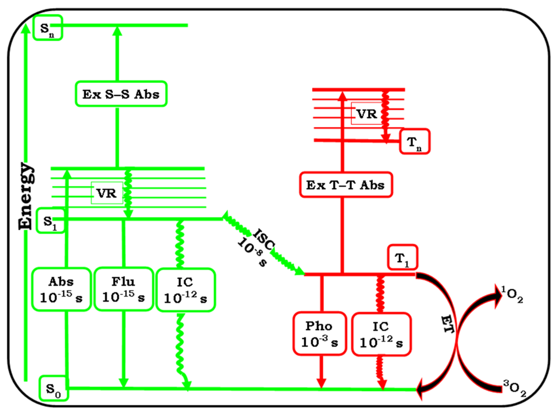
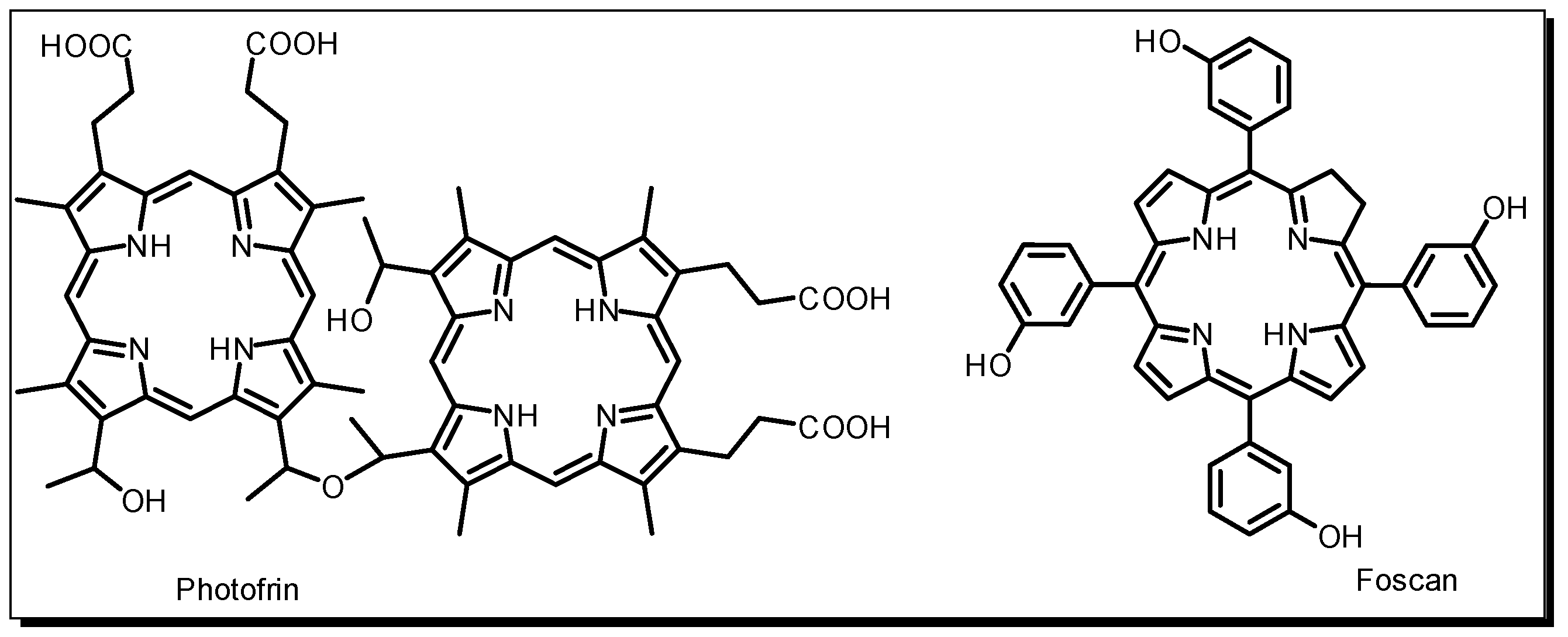
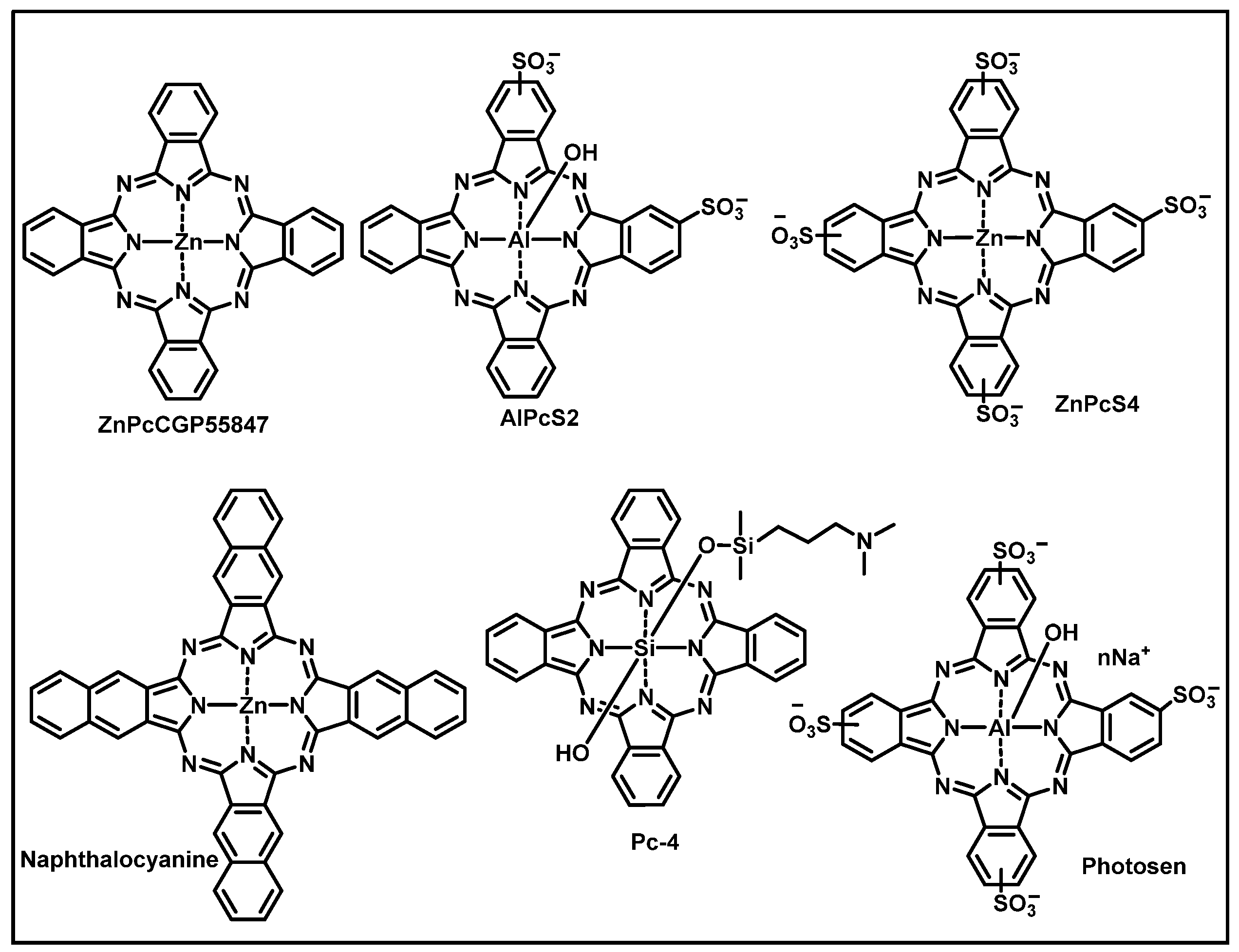
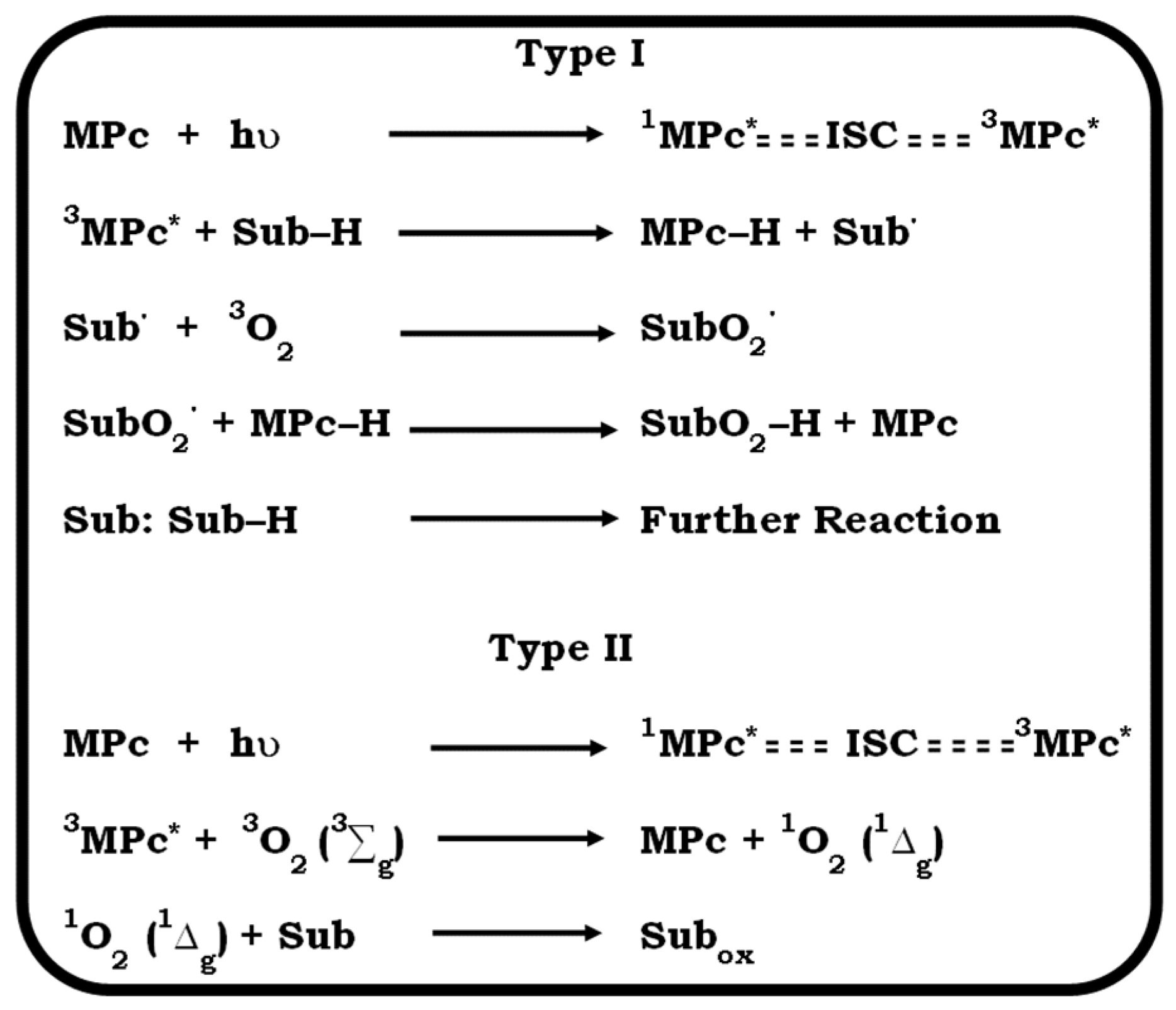
2. Photodynamic Therapy and Photosensitiser

A relatively novel non-invasive therapeutic protocol, known as photodynamic therapy (PDT), is now gaining traction in the scientific community. Growing clinical data have proven its imperative efficacy as a treatment modality, with its photosensitisers demonstrating the potential for dual functionality as photoantimicrobial and photoanticancer therapeutics [13,14]. PDT is a reliable and dependable targeting treatment strategy for the selective elimination of microbial infections and tumorigenic cells [1,2,3,4] and has been successfully utilised in several medical fields, including dermatology, dentistry, and urology [15,16,17]. It is known for its typical selectivity and specificity in its mode of action [18,19]. Interestingly, this modality can be used as a (neo)adjunctive therapy or a substitute for conventional treatment options, including chemotherapy and radiotherapy [20,21]. PDT treatment exploits the synergistic triad system involving the reaction of a functional dye molecule known as photosensitisers (PS) with laser light in the presence of tissue oxygen. This generates reactive oxygen species (ROS) such as singlet oxygen, which are used to eliminate microbial species (e.g., bacteria) or tumorigenic cells via apoptotic or necrotic death [2,16]. PS are light-absorbing compounds with the ability to convert the absorbed energy to a photochemical reaction or transfer it to a ground state molecular oxygen (3O2) and generate ROS (1O2), the main agent for the selective destruction of tumorigenic cells or microbial species [2,14,18], as shown in Figure 1.
The role of the PS in the success of PDT is indispensable due to their crucial function in the photosensitisation process. For optimum therapeutic activity, suitable PS are expected to possess several characteristics, including good solubility in biological media, deep tissue penetration, minimal dark toxicity, intense Q-band absorption within the therapeutic window (600 nm–800 nm), purity, photothermal stability, and high singlet-oxygen quantum yields [18,22,23,24]. However, there have been cases where pristine PS exhibited low purity, undesirable side effects (skin irritation), limited efficacy in deep tissue lesions, and reduced specificity, necessitating the need for PS with precise selectivity and specificity, high purity, and deep tissue penetration [18]. These drawbacks are typical of first-generation PS, including the hematoporphyrin derivatives (HpD) Foscan and Photofrin II, as shown in Figure 2.
Recently, several PS have gained prominence in clinical trials and applications with moderate success [25,26,27,28]. Some of the leading PS in clinical trials and administration include Pc–4® and Photosens® (Figure 3), as well as metallophthalocyanine (MPc) derivatives (second-generation PS), which are known for their extraordinary photoanticancer activity against breast, gastrointestinal, colon, and lung cancers, as well as skin lesions [25,26,27]. Photofrin® and Foscan®, on the other hand, are analogues of porphyrin, with proven photoanticancer efficacy against superficial tumorigenic cells [16]. Although Pc–4® and Photosens® are mixtures of compounds, making their purity unrealistic, Photofrin® and Foscan® lack deep tissue penetration and exhibit minimal efficacy due to their weak Q-band absorption within the therapeutic window, resulting in their application against superficial melanoma, such as skin cancer [15,17].
They are also known for having some undesirable adverse effects, including phototoxicity, as a result of their long half-life and prolonged accumulation in patients [16,27,28,29]. These PS often bioaccumulate in patients for a prolonged period post-treatment, causing cutaneous irritation due to phototoxicity from patients’ exposure to sunlight [15,16]. As a result of these drawbacks, it is crucial to develop an ideal PS with proven therapeutic efficacy and safety.
Most PS are known to be subcellularly localised in tumorigenic cells after administration via topical or parenteral routes [29,30,31,32,33,34,35]. The extensive localisation of ROS in tumorigenic cells often leads to oxidative stress, which can affect cellular components such as proteins, DNA, and lipids, resulting in oxidation and subsequent damage [36]. Moreover, it has been proven that ROS are intracellularly located in the mitochondria of the tumour and can inhibit tumorigenic cell proliferation, which ultimately leads to senescence and cytocidal activity. However, the response of ROS is dependent on several factors such as the cell type, cellular uptake, and dosimetry parameters, including the laser wavelength and duration [37,38]. The success of PDT is due in part to the optimal conditions, including the excitation of the PS at the intense Q-band absorption wavelength using a specific laser wavelength in the presence of tissue oxygen. This leads to the generation of reactive oxygen species, which are known to be the main agents responsible for the killing of malignant cells or microbes via type I or type II mechanism pathways. In the type I mechanism, the excited triplet state of the PS reacts with substrates (biomolecules such as protein, and lipids), leading to the abstraction of a hydrogen atom, which initiates the formation of free radicals and cytotoxic species such as hydrogen peroxide, hydroxyl radicals, and superoxide. In the type II mechanism, the excited triplet state of the MPcs undergoes direct energy transfer to the ground-state molecular oxygen to form singlet oxygen, which is the reactive oxygen species responsible for the tumourigenic cytocidal activity, as shown in Figure 4 [18,27]. However, the type II mechanism seems to be the more prevalent mechanism for PDT activity.
A number of Pcs have been explored for their PDT activity in clinical trials, with most exhibiting efficacious tumorigenic cell clearance, as shown in Figure 3 [14,27,39,40,41,42,43,44,45,46,47]. As stated earlier, the drawbacks related to some of the commercially available PS have led to extensive research focusing on the modification of MPcs. These MPcs are known for their structural flexibility, which has enabled their tremendous applicability in diverse fields. They have been used in pharmaceutics as photosensitisers, medicine as biosensors, optics as nonlinear optical (NLO) absorbers, catalysis as photocatalysts, and colourants in the cosmetic industry, among others [48,49,50,51,52,53,54,55,56,57]. However, the focus of our review is on the possibility of fine-tuning the physicochemical properties of phthalocyanines as multicomponent crystals. Porphyrin is a naturally occurring pigment similar to haem, a well-known precursor for haemoglobin, which is responsible for transporting dioxygen and plays a role in the destruction of peroxides and other biological functions. The fine-tuning of the physicochemical properties of phthalocyanines as multicomponent crystals is considered in this review due to their potential for various biological applications. Pcs are structurally related to porphyrin, making them useful for biological applications due to their biocompatibility and physicochemical behaviours [58]. Moreover, it is possible to modify their molecular structures to allow for interaction with other molecules or coformers. Crystal engineering provides insights into the design and development of molecular crystals with fine-tuned properties and functionalities. This could be in the form of supramolecular synthons, i.e., the assembly of molecules via non-covalent interactions, such as hydrogen bonding or van der Waals forces, resulting in the formation of multicomponent crystals, including solvates, salts, and co-crystals.

3. Approaches for Improving Compounds’ Physicochemical Properties

Several strategies have been explored for enhancing the physicochemical properties of both organic and inorganic compounds. Some of these methods include molecular modification by altering the molecular structures or incorporating nanomaterials and crystal engineering by manipulating the solid-state architecture of the compound. The latter option is straightforward, with reliable outcomes in tuning the compounds’ physicochemical properties and possible biological activity. On the other hand, the former approach involves the modification of the compounds’ molecular structures, which can be difficult to isolate or purify. Moreover, this option could lead to the loss or reduction of biological activity.

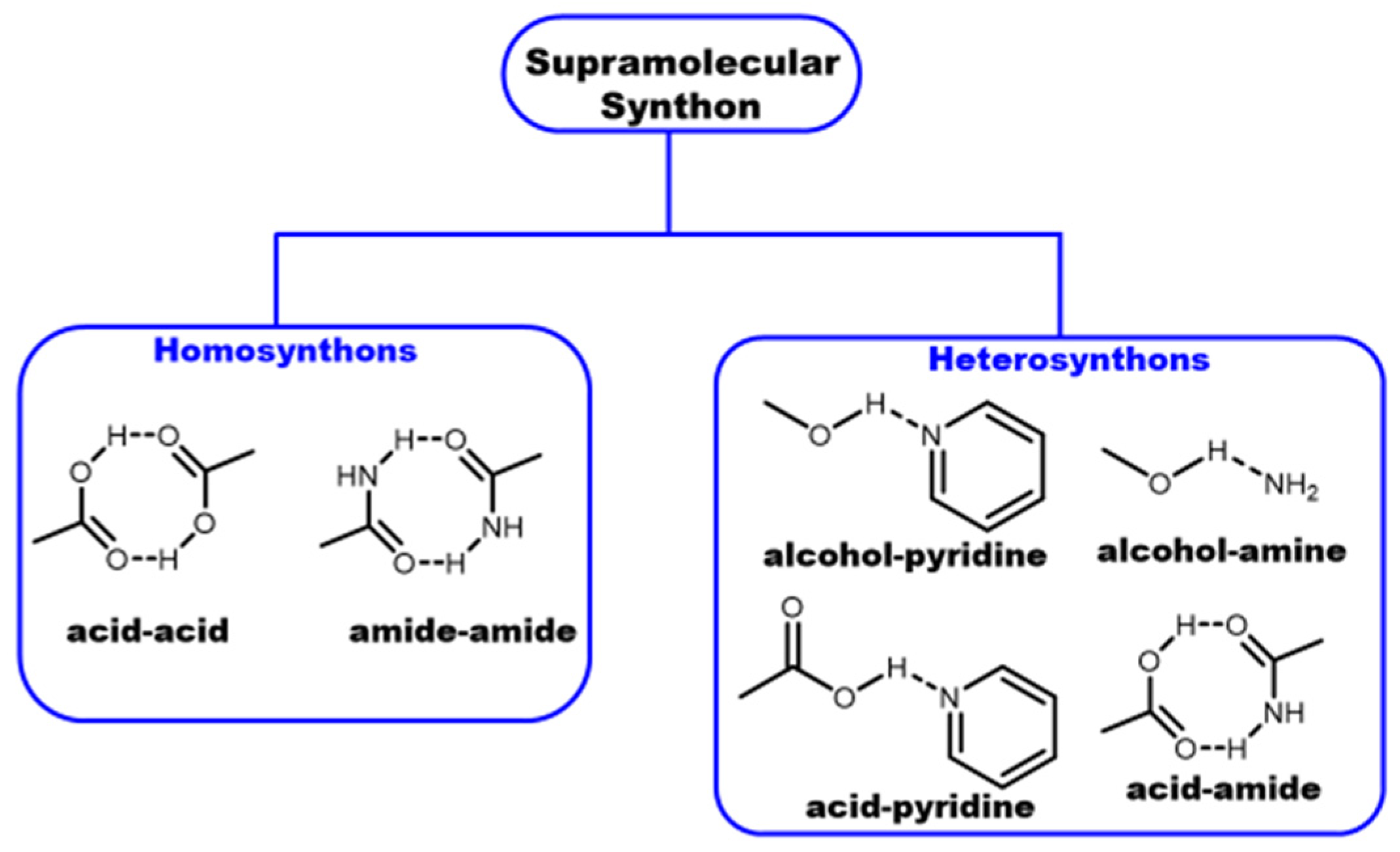
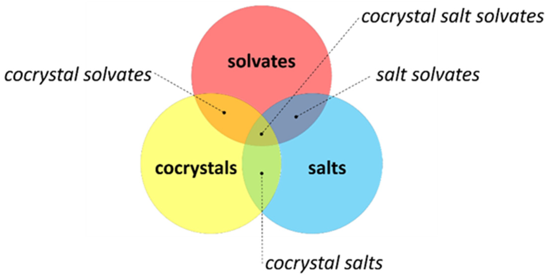
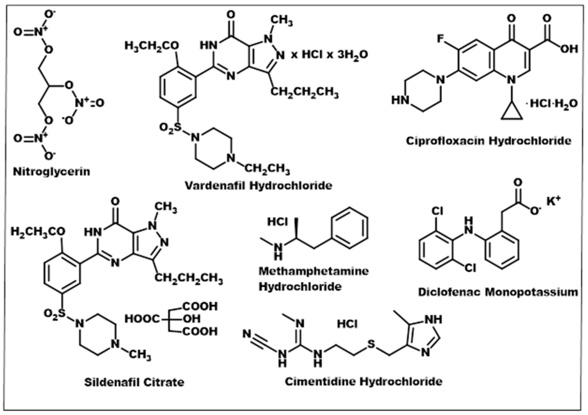
3.1. Crystal Engineering, Supramolecular Synthons, and Multicomponent Crystals

Crystal engineering is the understanding of intermolecular interactions in the context of crystal packing, and the usefulness of this knowledge lies in the design of new solid materials with the intended physicochemical properties [59]. Over the years, significant research efforts have been devoted to improving the physicochemical properties of active pharmaceutical ingredients (API), including solubility, while retaining their inherent molecular structures and pharmacological responses without compromising their stability and safety [60,61]. Compounds with poor physicochemical properties are undesirable for pharmaceutical applications, as they tend to lack the capacity to be well-expressed in biological systems. This shortcoming is known to account for the disapproval of at least 80% of new chemical entities by the United States Food and Drug Administration (FDA) [62,63,64]. Approximately 30% of new drug molecules and commercially available drug products are categorised as class II in the Biopharmaceutics Classification System due to their low solubility and high permeability [62,63,64]. This is generally a problem because it limits their extensive pharmaceutical applications. Drug molecules with good physicochemical properties often possess broad administration routes, including oral ingestion with dissolution in the gastrointestinal tract or fluid formulations with optimal pharmacological outcomes [65]. Several efforts have been explored for the enhancement of drug products with low solubility, and co-crystallisation has proven to be a viable alternative for improving the physicochemical disposition of these drug compounds [61,66,67,68]. This could be attributed to the fact that co-crystallisation can be applied to drug molecules with both ionisable and non-ionisable functional groups, using a suitable coformer to form multicomponent crystals [61]. Multicomponent crystals (MCCs) are composed of two or more individual substances that interact via non-covalent intermolecular interactions to form a crystal structure. This supramolecular assembly formation is based on supramolecular synthons [60,61]. A clear understanding of the intermolecular interactions involved in MCCs’ formation is crucial for their design. Some of the intermolecular forces include ionic bonds, hydrogen bonding, π–π stacking, and Van der Waals or halogen interactions. Hydrogen bonding seems to be the most prevalent in the formation of MCCs. This is because most APIs have amide, pyridine, carboxylic acid, and alcohol moieties in their molecular structures [69,70]. On the other hand, there are complementary coformers that can interact with suitable APIs to obtain MCCs. Coformers are small molecules usually obtained from a class of substances generally regarded as safe (GRAS) [71], with the propensity to interact with other molecules to form multicomponent crystals [61]. The first step in MCC formation is the selection of an appropriate coformer (CF) molecule. The suitability of the CF is judged by the possibility of synthon formation between an API and the CF. Synthons are supramolecular units formed between functional groups of two or more molecules assembled via non-covalent interactions and can be classified as homo- or hetero-synthons, as shown in Figure 5 [69,70]. Homosynthons are formed between two identical functional groups, and heterosynthons are obtained when non-identical but complementary functional groups interact. Some supramolecular synthons, such as acid–acid or acid–pyridine, can be formed with a high probability. Thus, when an API with a carboxylic acid substituent is considered to form an MCC, a CF should be selected with carboxylic acid or pyridine moieties to maximise the possibility of successful co-crystallisation, as shown in Figure 5.
Multicomponent crystals can be classified into three major groups, including co-crystals, salts, and solvates, as shown in Figure 6 [60,72,73]. These MCCs can be further divided into seven subclasses: (1) true salt, (2) true solvate, (3) true co-crystal, (4) salt solvate, (5) co-crystal solvate, (6) co-crystal salt, and (7) co-crystal salt solvate, as shown in Figure 6 [73]. Solvates can be described as any crystal having one or more solvent residues crystallised with the compound of interest. Salts consist of ionic moieties, whereas co-crystals contain at least one additional neutral residue that is not a solvent. The compositions of the seven subclasses are derived from the combination of the main classes [73].
Co-crystals have many advantages compared to salts. Salt formation is possible only if the molecular building blocks have ionisable functional groups. Subsequently, the stoichiometry of salt crystals is fixed. In co-crystals, the building blocks of the crystal are neutral; thus, the ratio between the target compound and the CF can be varied. Co-crystals can also be formed with molecules that have ionisable functional groups, but no proton transfer is observed between the complementary functional groups. In these cases, the ΔpKa values of the co-crystallised compounds will determine the outcome of the crystallisation, i.e., the formation of a co-crystal or a salt. The so-called pKa rule states that an ΔpKa > 2 or 3 often favours the formation of a salt. This empirical rule was quantified by Cruz Cabeza [74,75,76] and states that (i) the formation of co-crystals is likely if the ΔpKa is below −1, and (ii) salt formation is preferred if the ΔpKa is higher than 4. Between these boundaries lies the so-called ‘salt–co-crystal continuum’ (−1 < ΔpKa < 4), where the plausibility of either obtaining a salt or a co-crystal depends on many factors, one of them being the nature of the solvent employed. Pharmaceutical co-crystals are a subset of MCCs, with one of their components being an active pharmaceutical ingredient (API) [72]. Previously, the salt formation has been proven to be the preferred method for the improvement of drug substances’ physicochemical behaviour, including solubility, dissolution, purity, and solid-state properties. However, this is only applicable to ionisable molecules and thus has limited applicability. At least half of the orally administered pharmaceutical drugs in circulation are often marketed in salt forms [77], as shown in Figure 7.
Various methods have been reported to be useful in the formation of MCCs, including mechanochemistry (liquid-assisted grinding or neat grinding), solvent evaporation, melt cooling, gel crystallisation, supercritical fluid extraction, pressurisation, and sublimation [78,79,80,81,82]. The API and the appropriate coformer are often mixed using a predetermined stoichiometric ratio. MCCs are known to exhibit different physicochemical behaviour compared to their starting materials (i.e., API and coformers) because of their modified crystal structure [83,84,85,86,87,88,89].
Interestingly, co-crystallisation can help in the formation of an API with improved solubility, pharmacokinetic disposition (such as liberation, absorption, distribution, and excretion), and bioavailability [83,84,85,86,87,88,89]. For instance, the lipophilicity and hydrophilicity of the API can be balanced by the choice of coformer, i.e., a highly hydrophilic API can be co-crystallised with a lipophilic coformer, thereby imparting its crystal structure with an ideal amphiphilicity, which advertently impacts the good release and high bioavailability of the API [83].
The solubility of MCCs often correlates with their coformers, with long alkyl chain coformers tending to be well-expressed in non-polar solvents, and hydrophilic coformers showing good solubility in protic polar solvents, including ethanol and water [83]. This is attributed to the improved solvation with a higher solubility coformer [83,84]. Highly lipophilic drugs are known to sometimes precipitate out in the aqueous intracellular interface, which is undesirable for drug molecules intended for biological application, as only a few or none of the APIs will reach the target site, potentially leading to therapeutic failure and drug resistance [65,66]. It is a known fact that dosage forms can be utilised to control the rate and location of drug release, making co-crystallisation a plausible choice for evoking the immediate or modified release of APIs to foster quick-onset or delayed/timed therapeutic responses [84].
The choice of coformer is imperative for the successful formation of MCCs with improved physicochemical disposition. Advances in knowledge-based methods using the Cambridge Structural Database (CSD) [90], with a comprehensive crystal structure repository of over 1 million entries, have facilitated the selection of a suitable coformer, reducing the need for laborious laboratory experiments involving several trial and error attempts to form an appropriate synthon [91,92,93]. However, the coformer selected for a pharmaceutical co-crystal must be chosen from the list of generally regarded as safe (GRAS) compounds [71]. It is crucial to note that the coformer must have functional groups complementary to the active pharmaceutical ingredient to foster intermolecular interaction. The most widely explored supramolecular-forming functional groups are the carboxylic acid and aromatic amine groups because they have both hydrogen bond donor and acceptor sites, making them suitable for co-crystal formation.
The nature of the solvents used for co-crystallisation is important because the two substances, i.e., the drug molecule and the coformer, must be soluble in the selected solvents for optimum co-crystal formation. These can be a mono-component solvent or a mixture of solvents, depending on the polarity of the reactants. If one or both reactants are not soluble for the chosen solvent(s), the reactant(s) will precipitate out of the solution, producing a recrystallised compound rather than a co-crystal, which is undesirable for MCC formation [93].
The slow evaporation crystallisation method is known to enable the formation of various types of MCCs, including salts, co-crystals, solvates, or hydrates. The behaviour of co-crystals, solvates, and hydrates differs in terms of their physical properties at ambient temperatures, which is often observed in the nature of the isolated pure crystal, with solvates showing liquid disposition and co-crystals exhibiting solid-state features [73]. As stated earlier, it is important to note that a co-crystal differs from salt because its formation is not dependent on the charge transfer between its two or more distinct neutral components [74]. In essence, there is no need for ionisable functional moieties for the successful formation of co-crystals unlike salt, which requires molecules with ionisable functional groups. Once these considerations have been implemented, various methods can be applied, including mechanochemistry via neat or liquid-assisted grinding, slurry methods, slow evaporation, and other protocols. The mechanochemistry approach is a viable and easy method to achieve MCCs with a rapid formation time. It is often employed in screening for the potential formation of MCCs between the API and coformers and involves grinding the components with/without a solvent (neat grinding). However, single crystals cannot be obtained using this method due to the relatively small grains generated. Although the structural details of the microcrystalline material can be obtained using powder X-ray diffraction, this process is not straightforward [81,94]. Recent studies have demonstrated the efficiency of slurry or slow evaporation crystallisation methods due to the reliability of MCC formation [95,96]. The exploration of crystal engineering could be a good choice for fine-tuning the physicochemical properties of phthalocyanines through the formation of multicomponent crystals. This strategy has shown promising performance in improving the physicochemical properties of active pharmaceutical ingredients when they interact with suitable coformers [60].

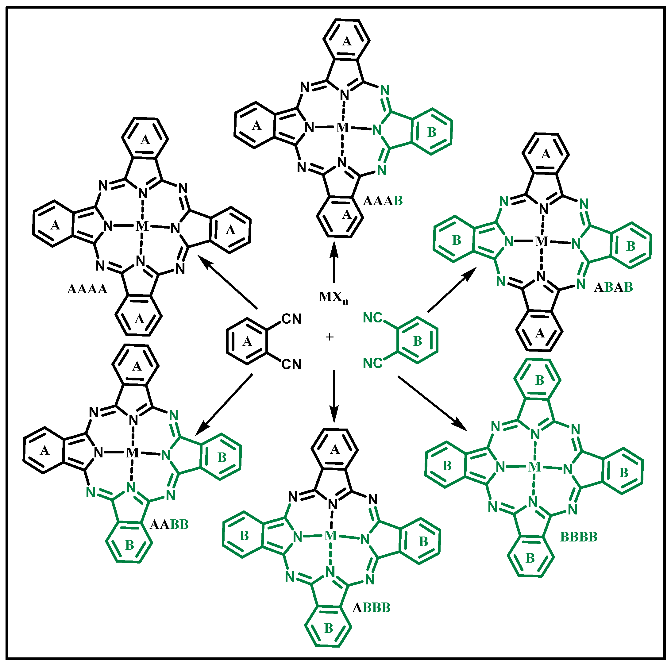
3.2. Phthalocyanines: Molecular Design and the Possibility of Fine-Tuning Their Physicochemical Properties through Crystal Engineering

The molecular structural flexibility of phthalocyanines makes them potential compounds for crystal engineering. This property allows for the modification of their molecular structures with other (in)organic substituents in various positions, including the alpha (α) and beta (β) positions [97], as shown in Figure 8. Points 1, 4, 8, 11, 15, 18, 22, and 25 on the Pcs molecular structure correspond to the α position, and points 2, 3, 9, 10, 16, 17, 23, and 24 correspond to the β position. Moreover, the central cavity of Pcs exhibits a coordination affinity for up to 70 different elements from the periodic table, including many main-group elements and d10 metals, which confers on them metallophthalocyanine (MPc) properties with remarkable redox chemistry [55,56,57,58,97,98].
Moreover, they can be tailored to be free base ligands, otherwise known as metal-free phthalocyanines (unmetalated Pcs), as shown in Figure 8. However, the activity of Pc molecules is dependent on several factors such as the central atoms, symmetry, position of substitution, nature of ring substituents/axial ligation, and extent of macrocycle conjugation [22,55,97]. The insertion of diamagnetic metals, such as Zn, Al, Ga, In, Si, Ru, and Pt, into the central cavity of the Pc ring system has been shown to exhibit excellent photochemical and PDT disposition [18,22]. Concerning the point of substitution, alpha (α)-substituted Pc complexes are known to exhibit a larger bathochromic shift in contrast to their beta (β)-substituted analogues, with the former exhibiting superior photochemical properties [22,97,99]. Solvents play a crucial role in the spectroscopic behaviour of Pc complexes. This was exemplified in a study conducted by Ogunsipe and Nyokong, where they evaluated the effect of solvents (acids, including sulfuric and trifluoroacetic acids) on the photochemical properties of Pcs. It was observed that the solvents could protonate Pc complexes [100]. This phenomenon might be useful for the design and fabrication of their multicomponent crystals. Furthermore, most organic solvents have the tendency to influence the physicochemical behaviour of Pc molecules, depending on their refractive index [100]. Kobayashi and co-workers demonstrated that symmetry does influence the photo-physicochemical disposition of Pcs, with asymmetrical Pcs exhibiting improved photo-physicochemical behaviour in comparison to their symmetrical analogues [101]. Symmetry is imperative in the functionality of Pcs, and this characteristic is sometimes dependent on their starting materials and the synthetic pathway used to produce them [22,97]. Symmetrical Pcs are often obtained through the cyclo-condensation of mono or di-substituted phthalonitriles as precursors to obtain tetra- or octa-substituted Pcs, as shown in Figure 8 [97,102]. On the other hand, asymmetrical Pcs are usually synthesised using the statistical mixed condensation route [103,104,105], subphthalocyanine ring expansion route [103,105], or polymeric support-based route [105]. Among these pathways, statistical mixed condensation has been the most widely utilised approach for fabricating asymmetrical Pcs, particularly with three identical isoindole subunits (A3B), due to its simplicity and satisfactory outcomes [103,105]. The statistical cross-condensation route is known to be non-selective due to the reaction of two different substituted phthalonitrile or 1,3–diiminoisoindoline precursors, as shown in Figure 9 [103,105]. In most cases, the purification of symmetrical Pcs is straightforward; however, the isolation and purification of asymmetrical Pcs are challenging due to the presence of diverse products in their crude mixtures [105]. For instance, if the statistical cross-condensation synthetic approach is employed for the synthesis of asymmetrical Pcs, theoretically, six possible products could be obtained, as shown in Figure 9 [105].
The absorption spectra of metal-free Pcs are known for their weak soret (or B) band in the range of 250–350 nm and strong split Q-bands due to their D2h molecular symmetry. Their metalated analogues exhibit weak B-bands and a monomeric symmetrically intense Q-band due to their D2h symmetry, as shown in Figure 10 [106,107]. Of note are the four possible constitutional structural isomeric mixtures possessed by symmetrical Pcs, particularly for the tetrasubstituted analogues with molecular symmetries of C4h, C2v, Cs, and D2h, making their isolation cumbersome due to their close similarities.
The variation in the electronic absorption properties of H2Pcs and MPcs can be attributed to the energy transitions in their molecular orbitals. In unmetalated Pcs, the split Q-bands, namely Q1 and Q2, resulting from the transitions from a1u of the highest occupied molecular orbital (HOMO) to b2g and b3g of the lowest unoccupied molecular orbital (LUMO), respectively. Conversely, metalated Pcs have a symmetrical Q-band due to their D4h symmetry resulting from the transitions from a1u of the HOMO to the degenerate eg of the LUMO, as shown in Figure 11. MPcs are often characterised by two B-bands, B1 and B2, resulting from the transitions from a2u and b2u of the HOMO to eg of the LUMO. Moreover, compounds with a small HOMO-LUMO energy gap are likely to absorb higher-energy photons, as demonstrated by their low UV-visible absorption. On the other hand, compounds with a larger HOMO-LUMO energy gap are likely to absorb lower-energy photons, exhibiting near-infrared absorption [107].
Aggregation is a known phenomenon in the chemistry of Pc complexes and is often reflected in their electronic absorption spectra [99,108]. There are two known possible aggregations for Pcs, including the H and J aggregates [99]. The H aggregate is often blue-shifted with Q-band absorption at around 630 nm compared to the bathochromic J aggregate with Q-band absorption at around 750 nm. This phenomenon can be attributed to the electronic interactions between rings of two or more molecules [99]. The formation of dimers and aggregates has been reported to lower the photosensitisation efficiency of Pcs, which is undesirable for these compounds when applied in PDT [99,108]. Aggregation always reduces the lifetimes of the Pc’s excited state parameters, which can be attributed to enhanced radiationless excited-state dissipation [98,108]. The lowering of the excited triplet-state population leads to inefficient energy transfer to the ground-state molecular oxygen and results in minimal singlet-oxygen generation. This limitation is ubiquitous in aqueous solutions of Pcs with hydrophilic substituents, including carboxylic and sulfonic acid moieties [108].
Multicomponent crystals in phthalocyanine chemistry are a relatively new aspect, with limited reports on their development [109,110,111]. Moreover, single crystal X-ray diffraction studies on Pcs or their analogues as MCCs are scarce. This can be attributed to their strong intermolecular π–π stacking, which limits their solubility and makes their co-crystallisation challenging [109,110,111]. However, these phenomena are often more prevalent with planar phthalocyanine molecules [109]. A study conducted by Sosa-Sanchez and co-workers investigated the axial hydrocarbon tail length needed to obtain enough spacing to avoid π–π stacking forces and increase the solubility of SiPcs. It was established that the use of longer alkyl chains has a major influence on the spacing, resulting in improved solubility [109]. However, this option is mostly suitable for the design of lipophilic Pcs crystals, which are undesirable for several biological applications [66]. Moreover, tert–butyl or long alkyl chain-substituted Pcs are not suitable for co-crystallisation via hydrogen bond formation due to the absence of suitable hydrogen bond donor groups (Figure 12). In contrast, other functionalities support the formation of MCCs via hydrogen bonding, including sulfonic acid, carboxylic acid, amine, and pyridine, as shown in Figure 13. Moreover, most bulky, long alkyl chain hydrocarbons and phenyl substituents have been reported to exhibit poor aqueous solubility, making them undesirable for most biological applications [66].
Many Pcs lack aqueous solubility or ideal amphiphilicity, which limits their broad application in biological environments via conventional routes. Due to their high lipophilicity, they are mostly administered via parenteral or topical routes [112]. It has been proven that MCCs of Pcs with suitable coformers lead to improved physicochemical behaviour with better and broader applicability, as opposed to their pristine analogues [113]. Moreover, the high lipophilicity of Pcs can be modified with a suitable aqueous coformer, thereby improving their physicochemical behaviour. Although Pcs’ solubility can be improved through the direct modification of their ring substituents or axial ligands using moieties such as sugars, amino acids, or cholesterol, the covalent modification of the molecular structure is often difficult due to the extensive purification process and low yield. In this case, MCC formation via non-covalent interactions is a more appealing approach due to its easier purification and higher yield of products compared to covalent assembly [114].

4. Cambridge Structural Database Repository Analysis of Multicomponent Crystals of Phthalocyanines

Molecular design plays an indispensable role in determining the overall properties of phthalocyanines, including their photo-physicochemical properties such as stability, solubility, optical and electrochemical behaviour, and processability (ease of interaction with other molecules or coformers). Pcs’ molecular design is often dependent on their intended application. For instance, Pcs intended for biological applications require diamagnetic metals in their central cavity and suitable ring substituents, making them ideal for biological microenvironments. The efficient design of Pcs can result in improved physicochemical properties, including enhanced solubility and reduced aggregation, as well as high photophysical and photochemical properties. Moreover, it improves the versatility of Pcs for various applications by establishing structure-property relationships and incorporating suitable ring substituents. However, molecular design may require complex synthetic routes, which could result in difficulties with isolating and purifying the target Pcs. Moreover, certain synthetic routes or components may not be feasible for industrial scale-up.
A search of the Cambridge Structural Database (CSD Version 5.41 + 2) revealed that the current mono- and multicomponent crystals of Pcs are either salt or solvates, with few co-crystals, inferring that these MCCs were formed using Pcs and coformers with ionisable functional groups. However, little data exist on the use of Pcs and coformers for co-crystal formation. This observation has highlighted the need to design co-crystals of Pcs. Moreover, the axial ligands attached to the central atom of Pcs have been found to play a significant role in the formation of monocomponent crystals. Interestingly, diamagnetic atoms, such as silicon, aluminium, ruthenium, indium, and zinc, are suitable for interaction, as shown in Figure 14.
As stated earlier, it has been shown that Pcs can be designed to have various functional moieties, such as carboxylic acid, amine, pyridine, sulfonic acid, and alcohol, making their intermolecular interaction with complementary coformers feasible, as shown in Figure 13. Several Pcs MCCs have been explored for diverse applications, including sensing, and photophysical and photochemical activities. In a study by Konarev and co-workers, the formed Pcs MCCs showed improved photophysical properties [113].
Currently, the CSD hosts over 1 million crystal structure repositories [90]; however, only a small number of Pcs single-crystal structures were found, which further underscores the limited exploration of these important compounds for MCC formation. Considering the immense benefits that phthalocyanines can provide to the pharmaceutical and healthcare industries, particularly in sensing and PDT for the management or elimination of microbes and cancerous cells, it becomes crucial to develop a wide range of MCCs of Pcs with improved physicochemical disposition and therapeutic efficacy [112,113]. In particular, the formation of their co-crystals will be ideal because there are fewer limitations in the selection of the coformers compared to salt formation. In addition, in terms of safety, stability, cost, and marketing, co-crystals are more appealing compared to other MCCs [120].
A knowledge-based protocol was employed in this study using the Cambridge Structural Database (CSD Version 5.41 + 2) with the aid of ConQuest [90]. The database scan was performed based on the molecular structural properties of the phthalocyanines (Pcs), including unsubstituted metal-free Pcs (H2Pc), substituted metal-free Pcs (RH2Pc), unsubstituted metalated Pcs (MPc), and substituted metalated Pcs (RMPc), as shown in Figure 15.
A total of 538 hits were recorded that contained Pcs moieties. Among these, H2Pc accounted for 14 (2.6%) hits, RH2Pc provided 27 (5.02%) structures, MPcs yielded 190 (35.32%) hits, and RMPcs provided 306 (57.06%) crystal structures. Furthermore, 296 (55.02%) MCCs were obtained from the 538 hits, with H2Pc accounting for 8 (2.70%), RH2Pc accounting for 10 (3.38%), MPcs accounting for 109 (36.82%), and RMPcs accounting for 169 (57.10%), as shown in Figure 16. The values in brackets were obtained by comparing each class against the overall hits for Pcs crystal structures and MCCs, respectively.
From our findings, it can be deduced that the substituted Pcs (62.08%) provided more hits compared to the unsubstituted Pcs (37.92%). This trend can be attributed to an increase in the number of sites for intermolecular interaction (e.g., hydrogen bonding), which enhances the possibility of crystallisation of the substituted Pcs compared to the unsubstituted Pcs. Moreover, it can be inferred that the metalated Pcs (92.38%) accounted for more hits, indicating reduced intermolecular π–π stacking and improved solubility due to minimal aggregation, thereby making their crystallisation more feasible and achievable. The low occurrence of the metal-free Pcs (7.62%) can be attributed to the reduced solubility and aggregation in most protic polar solvents because of the strong π–π stacking of the molecules, as shown in Figure 17.
Interestingly, the Pcs MCCs accounted for 55.02% of the total number of Pcs single-crystal structures. Comparing the hits for the MCCs against the sub-total hits for each class, H2Pc accounted for 57.14% of hits, RH2Pc accounted for 37.04%, MPcs accounted for 57.37%, and RMPcs accounted for 55.05%. The unsubstituted Pcs yielded more hits compared to the substituted Pcs, whereas the metalated Pcs (55.99%) yielded more hits compared to the unmetalated analogues (43.90%), which is consistent with the trend observed earlier for the overall hits. Moreover, the high number of hits reported for the substituted metalated Pcs can be attributed to the higher number of possible sites for intermolecular interactions with other molecules. Furthermore, the 296 (55.02%) MCCs of Pcs that were obtained from the 538 hits were mostly solvates or salts. The incorporation of solvents with Pcs can improve their solubility and influence their optoelectronic properties of Pcs. However, the inclusion of solvents in Pcs structures can also affect their long-term physicochemical properties, including their stability and crystal structure, due to possible defects in their crystal structures over a long duration.

5. Conclusions

Phthalocyanines have demonstrated tremendous usefulness in several applications, including bioimaging and photodynamic therapy of cancer and microbes. However, their limited applicability in photodynamic therapy as photosensitisers have been attributed to their limited insolubility in hydrophilic environments. The incorporation of suitable coformers with Pcs to form multicomponent crystals (MCCs) can improve their physicochemical properties, including solubility and stability. However, the most common MCCs of Pcs are usually in either salt or solvate forms. The former is dependent on molecules with ionisable groups. On the other hand, co-crystals are more appealing due to their versatility, as their formation is not dependent on compounds with ionisable functionalities. A co-crystal of Pcs designed as a photosensitiser for photodynamic therapy may offer improved photo-physicochemical properties, including solubility in the bilayer of the biological microenvironment and high singlet-oxygen quantum yields. These parameters are crucial in the photosensitisation of the photosensitisers designed for PDT. A CSD repository search yielded 538 hits for Pcs single-crystal data and 296 hits for their multicomponent crystals, including salts, solvates, and co-crystals. Notably, a greater number of Pcs solvates, simple salts, and molecular salts were observed compared to the relatively fewer Pcs co-crystals. In addition, the role of substitution and metalation in the formation of Pcs MCCs is imperative. In the design of Pcs MCCs, it is imperative to have Pcs that have complementary functional groups to the coformers. The slurry or slow evaporation crystallisation method may be a useful protocol to explore for making these MCCs.

Author Contributions

Conceptualisation: D.O.O. and N.B.B.; software: D.O.O. and N.B.B.; formal analysis, D.O.O. and N.B.B.; writing—original draft preparation: D.O.O.; writing—review and editing: D.O.O. and N.B.B. All authors have read and agreed to the published version of the manuscript.

Funding

This work was supported by the Cape Peninsula University of Technology Postdoctoral Fellowship grant.

Conflicts of Interest

The authors declare no conflict of interest.

References

  1. Bray, F.; Ferlay, J.; Soerjomataram, I.; Siegel, R.L.; Torre, L.A.; Jemal, A. Global cancer statistics 2018: GLOBOCAN estimates of incidence and mortality worldwide for 36 cancers in 185 countries. CA Cancer J. Clin. 2018, 68, 394–424. [Google Scholar] [CrossRef] [PubMed] [Green Version]
  2. Wainwright, M.; Maisch, T.; Nonell, S.; Plaetzer, K.; Almeida, A.; Tegos, G.P.; Hamblin, M.R. Photoantimicrobials-are we afraid of the light? Lancet Infect. Dis. 2017, 17, e49–e55. [Google Scholar] [CrossRef] [PubMed]
  3. Dougherty, T.J.; Kaufman, J.E.; Goldfarb, A.; Weishaupt, K.R.; Boyle, D.; Mittleman, A. Photoradiation therapy for the treatment of malignant tumors. Cancer Res. 1978, 38, 2628–2635. [Google Scholar] [PubMed]
  4. Triesscheijn, M.; Baas, P.; Schellens, J.H.M.; Stewart, F.A. Photodynamic Therapy in Oncology. Oncologist 2006, 11, 1034–1044. [Google Scholar] [CrossRef]
  5. Huang, Z. A Review of Progress in Clinical Photodynamic Therapy. Technol. Cancer Res. Treat. 2005, 4, 283–293. [Google Scholar] [CrossRef] [Green Version]
  6. Housman, G.; Byler, S.; Heerboth, S.; Lapinska, K.; Longacre, M.; Snyder, N.; Sarkar, S. Drug Resistance in Cancer: An Overview. Cancers 2014, 6, 1769–1792. [Google Scholar] [CrossRef] [Green Version]
  7. Moudi, M.; Go, R.; Yien, C.Y.S.; Nazre, M. Vinca alkaloids. Int. J. Prev. Med. 2013, 4, 1231–1235. [Google Scholar]
  8. Boutayeb, A. The Burden of Communicable and Non-Communicable Diseases in Developing Countries. In Handbook of Disease Burdens and Quality of Life; Preedy, V.R., Watson, R.R., Eds.; Springer: New York, NY, USA, 2010; pp. 531–546. [Google Scholar] [CrossRef]
  9. European Commission Staff Working Document. Progress Report on the Action Plan against the Rising Threats from Antimicrobial Resistance. 2015. Available online: http://ec.europa.eu/health/antimicrobial_resistance/docs/2015_amr_progress_report_en.pdf (accessed on 16 October 2020).
  10. ECDC Surveillance Report. Annual Epidemiological Report. Antimicrobial Resistance and Healthcare-Associated Infections. The European Union Summary Report on Antimicrobial Resistance in Zoonotic and Indicator Bacteria from Humans, Animals and Food in 2017/2018. Available online: https://www.ecdc.europa.eu/en/publications/EU-summary-report-antimicrobial-resistance-zoonoses-2017-2018 (accessed on 20 May 2021).
  11. Oluwole, D.O.; Coleman, L.; Buchanan, W.; Chen, T.; La Ragione, R.M.; Liu, L.X. Antibiotics-Free Compounds for Chronic Wound Healing. Pharmaceutics. 2022, 5, 1021. [Google Scholar] [CrossRef]
  12. US CDC. Available online: http://www.cdc.gov/drugresistance/pdf/carb_national_strategy.pdf (accessed on 18 May 2021).
  13. Lo, P.-C.; Rodríguez-Morgade, M.S.; Pandey, R.K.; Ng, D.K.P.; Torres, T.; Dumoulin, F. The unique features and promises of phthalocyanines as advanced photosensitisers for photodynamic therapy of cancer. Chem. Soc. Rev. 2019, 49, 1041–1056. [Google Scholar] [CrossRef]
  14. Zhang, J.; Jiang, C.; Figueiró Longo, J.P.; Azevedo, R.B.; Zhang, H.; Muehlmann, L.A. An updated overview on the development of new photosensitizers for anticancer photodynamic therapy. Acta Pharm. Sin. B 2018, 8, 137–146. [Google Scholar] [CrossRef]
  15. Babilas, P.; Schreml, S.; Landthaler, M.; Szeimies, R.-M. Photodynamic therapy in dermatology: State-of-the-art. Photodermatol. Photoimmunol. Photomed. 2010, 26, 118–132. [Google Scholar] [CrossRef] [PubMed]
  16. Konopka, K.; Goslinski, T. Photodynamic Therapy in Dentistry. J. Dent. Res. 2007, 86, 694–707. [Google Scholar] [CrossRef] [PubMed]
  17. Bozzini, G.; Colin, P.; Betrouni, N.; Nevoux, P.; Ouzzane, A.; Puech, P.; Villers, A.; Mordon, S. Photodynamic therapy in urology: What can we do now and where are we heading? Photodiagnosis Photodyn. Ther. 2012, 9, 261–273. [Google Scholar] [CrossRef] [PubMed]
  18. Bonnett, R. Chemical Aspects of Photodynamic Therapy; Gordon and Breach Science Publishers: Amsterdam, The Netherlands, 2000. [Google Scholar] [CrossRef]
  19. Shi, X.; Zhang, C.Y.; Gao, J.; Wang, Z. Recent advances in photodynamic therapy for cancer and infectious diseases. WIREs Nanomed. Nanobiotechnol. 2019, 11, e1560. [Google Scholar] [CrossRef]
  20. Deng, W.; McKelvey, K.J.; Guller, A.; Fayzullin, A.; Campbell, J.M.; Clement, S.; Habibalahi, A.; Wargocka, Z.; Liang, L.; Shen, C.; et al. Application of Mitochondrially Targeted Nanoconstructs to Neoadjuvant X-ray-Induced Photodynamic Therapy for Rectal Cancer. ACS Central Sci. 2020, 6, 715–726. [Google Scholar] [CrossRef]
  21. Savoia, P.; Deboli, T.; Previgliano, A.; Broganelli, P. Usefulness of Photodynamic Therapy as a Possible Therapeutic Alternative in the Treatment of Basal Cell Carcinoma. Int. J. Mol. Sci. 2015, 16, 23300–23317. [Google Scholar] [CrossRef] [Green Version]
  22. Nyokong, T. Desired properties of new phthalocyanines for photodynamic therapy. Pure Appl. Chem. 2011, 83, 1763–1779. [Google Scholar] [CrossRef]
  23. Methfessel, C.D.; Volland, M.; Brunner, K.; Wibmer, L.; Hahn, U.; De La Torre, G.; Torres, T.; Hirsch, A.; Guldi, D.M. Exfoliation of Graphene by Dendritic Water-Soluble Zinc Phthalocyanine Amphiphiles in Polar Media. Chem. A Eur. J. 2018, 24, 18696–18704. [Google Scholar] [CrossRef]
  24. Oluwole, D.O.; Manoto, S.L.; Malabi, R.; Maphanga, C.; Ombinda-Lemboumba, S.; Mthunzi-Kufa, P.; Nyokong, T. Evaluation of the photophysicochemical properties and photodynamic therapy activity of nanoconjugates of zinc phthalocyanine linked to glutathione capped Au and Au 3 Ag 1 nanoparticles. Dye. Pigment. 2018, 150, 139–150. [Google Scholar] [CrossRef]
  25. Love, W.G.; Duk, S.; Biolo, R.; Jori, G.; Taylor, P.W. Liposome-Mediated Delivery of Photosensitizers: Localization of Zinc (11)-Phthalocyanine within Implanted Tumors after Intravenous Administration. Photochem. Photobiol. 1996, 63, 656–661. [Google Scholar] [CrossRef]
  26. Brilkina, A.A.; Dubasova, L.V.; Sergeeva, E.A.; Pospelov, A.; Shilyagina, N.Y.; Shakhova, N.M.; Balalaeva, I.V. Photobiological properties of phthalocyanine photosensitizers Photosens, Holosens and Phthalosens: A comparative in vitro analysis. J. Photochem. Photobiol. B Biol. 2018, 191, 128–134. [Google Scholar] [CrossRef] [PubMed]
  27. Miller, J.D.; Baron, E.D.; Scull, H.; Hsia, A.; Berlin, J.C.; McCormick, T.; Colussi, V.; Kenney, M.E.; Cooper, K.; Oleinick, N.L. Photodynamic therapy with the phthalocyanine photosensitizer Pc 4: The case experience with preclinical mechanistic and early clinical–translational studies. Toxicol. Appl. Pharmacol. 2007, 224, 290–299. [Google Scholar] [CrossRef] [PubMed] [Green Version]
  28. Hamblin, M.R. Photodynamic Therapy for Cancer: What’s Past is Prologue. Photochem. Photobiol. 2019, 96, 506–516. [Google Scholar] [CrossRef] [PubMed] [Green Version]
  29. Robertson, C.A.; Evans, D.H.; Abrahamse, H. Photodynamic therapy (PDT): A short review on cellular mechanisms and cancer research applications for PDT. J. Photochem. Photobiol. B 2009, 96, 1–8. [Google Scholar] [CrossRef]
  30. Skupin-Mrugalska, P.; Sobotta, L.; Kucinska, M.; Murias, M.; Mielcarek, J.; Duzgunes, N. Cellular Changes, Molecular Pathways and the Immune System Following Photodynamic Treatment. Curr. Med. Chem. 2014, 21, 4059–4073. [Google Scholar] [CrossRef]
  31. Chandel, N.S. Mitochondria and cancer. Cancer Metab. 2014, 2, 8. [Google Scholar] [CrossRef] [Green Version]
  32. Wang, X.; Peralta, S.; Moraes, C.T. Mitochondrial Alterations During Carcinogenesis: A review of metabolic transformation and targets for anticancer treatments. Adv. Cancer Res. 2013, 119, 127–160. [Google Scholar] [CrossRef]
  33. Wallace, D.C. Mitochondria and cancer. Nat. Rev. Cancer 2012, 12, 685–698. [Google Scholar] [CrossRef] [Green Version]
  34. Tarhouni, M.; Durand, D.; Önal, E.; Aggad, D.; Işci, Ü.; Ekineker, G.; Brégier, F.; Jamoussi, B.; Sol, V.; Gary-Bobo, M.; et al. Triphenylphosphonium-substituted phthalocyanine: Design, synthetic strategy, photoproperties and photodynamic activity. J. Porphyr. Phthalocyanines 2018, 22, 552–561. [Google Scholar] [CrossRef]
  35. Zhao, X.; Huang, Y.; Yuan, G.; Zuo, K.; Huang, Y.; Chen, J.; Li, J.; Xue, J. A novel tumor and mitochondria dual-targeted photosensitizer showing ultra-efficient photodynamic anticancer activities. Chem. Commun. 2018, 55, 866–869. [Google Scholar] [CrossRef]
  36. Zhang, S.; Yang, L.; Ling, X.; Shao, P.; Wang, X.; Edwards, W.B.; Bai, M. Tumor mitochondria-targeted photodynamic therapy with a translocator protein (TSPO)-specific photosensitizer. Acta Biomater. 2015, 28, 160–170. [Google Scholar] [CrossRef] [Green Version]
  37. Gong, N.; Ma, X.; Ye, X.; Zhou, Q.; Chen, X.; Tan, X.; Yao, S.; Huo, S.; Zhang, T.; Chen, S.; et al. Carbon-dot-supported atomically dispersed gold as a mitochondrial oxidative stress amplifier for cancer treatment. Nat. Nanotechnol. 2019, 14, 379–387. [Google Scholar] [CrossRef]
  38. Lv, W.; Zhang, Z.; Zhang, K.Y.; Yang, H.; Liu, S.; Xu, A.; Guo, S.; Zhao, Q.; Huang, W.A. Mitochondria-targeted photosensitizer showing improved photodynamic therapy effects under hypoxia. Angew. Chem. Int. Ed. 2016, 55, 9947–9951. [Google Scholar] [CrossRef] [PubMed]
  39. Shao, J.; Xue, J.; Dai, Y.; Liu, H.; Chen, N.; Jia, L.; Huang, J. Inhibition of human hepatocellular carcinoma HepG2 by phthalocyanine photosensitiser PHOTOCYANINE: ROS production, apoptosis, cell cycle arrest. Eur. J. Cancer. 2012, 13, 2086–2096. [Google Scholar] [CrossRef] [PubMed]
  40. Sounik, J.R.; Rihter, B.D.; Ford, W.E.; Rodgers, M.A.J.; Kenney, M.E. Naphthalocyanines relevant to the search for second-generation PDT sensitizers. Proc. SPIE 1991, 1426, 340–349. [Google Scholar] [CrossRef]
  41. Dolmans, D.E.; Fukumura, D.; Jain, R.K. Photodynamic therapy for cancer. Nat. Rev. Cancer 2003, 3, 380–387. [Google Scholar] [CrossRef]
  42. Uspenskiĭ, L.V.; Chistov, L.V.; Kogan, E.A.; Loshchenov, V.B.; Ablitsov, I.A.; Rybin, V.K.; Zavodnov, V.I.; Shiktorov, D.I.; Serbinenko, N.F.; Semenova, I.G. Endobronchial laser therapy in complex preoperative preparation of patients with lung diseases. Khirurgiia 2000, 2, 38–40. [Google Scholar]
  43. Stranadko, E.F.; Garbuzov, M.I.; Zenger, V.G.; Nasedkin, A.N.; Markichev, N.A.; Riabov, M.V.; Leskov, I.V. Photodynamic therapy of recurrent and residual oropharyngeal and laryngeal tumors. Vestn. Otorinolaringol. 2001, 3, 36–39. [Google Scholar]
  44. Filonenko, E.; Sokolov, V.V.; Chissov, V.I.; Lukyanets, E.A.; Vorozhtsov, G.N. Photodynamic therapy of early esophageal cancer. Photodiagnosis Photodyn. Ther. 2008, 5, 187–190. [Google Scholar] [CrossRef]
  45. Ghazal, B.; Machacek, M.; Shalaby, M.A.; Novakova, V.; Zimcik, P.; Makhseed, S. Phthalocyanines and Tetrapyrazinoporphyrazines with Two Cationic Donuts: High Photodynamic Activity as a Result of Rigid Spatial Arrangement of Peripheral Substituents. J. Med. Chem. 2017, 60, 6060–6076. [Google Scholar] [CrossRef]
  46. Huang, Z. An update on the regulatory status of PDT photosensitizers in China. Photodiagnosis Photodyn. Ther. 2008, 5, 285–287. [Google Scholar] [CrossRef] [PubMed]
  47. Ben-Hur, E.; Kol, R.; Marko, R.; Riklis, E.; Rosenthal, I. Combined Action of Phthalocyanine Photosensitization and Gamma-radiation on Mammalian Cells. Int. J. Radiat. Biol. 1988, 54, 21–30. [Google Scholar] [CrossRef]
  48. Sakamoto, K.; Okumura, E.; Hirohashi, R. Phthalocyanine as Functional Dyes; IPC: Tokyo, Japan, 2004. [Google Scholar]
  49. de la Torre, G.; Vázquez, P.; Agulló-López, F.; Torres, T. Role of Structural Factors in the Nonlinear Optical Properties of Phthalocyanines and Related Compounds. Chem. Rev. 2004, 104, 3723–3750. [Google Scholar] [CrossRef] [PubMed]
  50. Oluwole, D.O.; Yagodin, A.V.; Mkhize, N.C.; Sekhosana, K.E.; Martynov, A.G.; Gorbunova, Y.G.; Tsivadze, A.Y.; Nyokong, T. First Example of Nonlinear Optical Materials Based on Nanoconjugates of Sandwich Phthalocyanines with Quantum Dots. Chem.—A Eur. J. 2017, 23, 2820–2830. [Google Scholar] [CrossRef] [PubMed]
  51. Cid, J.-J.; Yum, J.-H.; Jang, S.-R.; Nazeeruddin, M.K.; Martínez-Ferrero, E.; Palomares, E.; Ko, J.; Grätzel, M.; Torres, T. Molecular Cosensitization for Efficient Panchromatic Dye-Sensitized Solar Cells. Angew. Chem. Int. Ed. 2007, 46, 8358–8362. [Google Scholar] [CrossRef]
  52. Gu, D.; Chen, Q.; Tang, X.; Gan, F.; Shen, S.; Liu, K.; Xu, H. Application of phthalocyanine thin films in optical recording. Opt. Commun. 1995, 121, 125–129. [Google Scholar] [CrossRef]
  53. Campidelli, S.; Ballesteros, B.; Filoramo, A.; Díaz, D.D.; de la Torre, G.; Torres, T.; Rahman, G.M.A.; Ehli, C.; Kiessling, D.; Werner, F.; et al. Facile Decoration of Functionalized Single-Wall Carbon Nanotubes with Phthalocyanines via “Click Chemistry”. J. Am. Chem. Soc. 2008, 130, 11503–11509. [Google Scholar] [CrossRef]
  54. Okura, I. Photosensitization of Porphyrins and Phthalocyanines; Gordon and Breach: Amsteldijk, The Netherlands, 2001; p. 151. [Google Scholar]
  55. Dini, D.; Hanack, M. Phthalocyanines Properties and Materials. In The Porphyrin Handbook; Kadish, K.M., Smith, K.M., Guilard, R., Eds.; Academic Press: New York, NY, USA, 2003; Volume 17, p. 107. [Google Scholar]
  56. Ben-Hur, E.; Chan, W.S. Phthalocyanines in Photobiology and their Medical Applications. In The Porphyrin Handbook; Kadish, K.M., Smith, K.M., Guilard, R., Eds.; Academic Press: New York, NY, USA, 2003; Volume 19, p. 1. [Google Scholar]
  57. Gregory, P. Industrial applications of phthalocyanines. J. Porphyr. Phthalocya 2000, 4, 432–437. [Google Scholar] [CrossRef]
  58. Sorokin, A.B. Phthalocyanine Metal Complexes in Catalysis. Chem. Rev. 2013, 113, 8152–8191. [Google Scholar] [CrossRef]
  59. Desiraju, G.R. Crystal Engineering: The Design of Organic Solids; Elsevier: Amsterdam, The Netherlands, 1989. [Google Scholar]
  60. Duggirala, N.K.; Perry, M.L.; Almarsson, O.; Zaworotko, M.J. Pharmaceutical cocrystals: Along the path to improved medicines. Chem. Commun. 2015, 52, 640–655. [Google Scholar] [CrossRef]
  61. Desiraju, G.R. Crystal engineering. From molecules to materials. J. Mol. Struct. 2003, 656, 5–15. [Google Scholar] [CrossRef]
  62. Lipinski, C.A. Solubility in water and DMSO: Issues and potential solutions. In Pharmaceutical Profiling in Drug Discovery for Lead Selection; RBorchardt, T., Kerns, E.H., Lipinski, C.A., Thakker, D., Wang, B., Eds.; AAPS Press: Arlington, VA, USA, 2004; pp. 93–125. [Google Scholar]
  63. Hens, B.; Brouwers, J.; Corsetti, M.; Augustijns, P. Supersaturation and Precipitation of Posaconazole Upon Entry in the Upper Small Intestine in Humans. J. Pharm. Sci. 2016, 105, 2677–2684. [Google Scholar] [CrossRef] [PubMed] [Green Version]
  64. Takagi, T.; Ramachandran, C.; Bermejo, M.; Yamashita, S.; Yu, L.X.; Amidon, G.L. A Provisional Biopharmaceutical Classification of the Top 200 Oral Drug Products in the United States, Great Britain, Spain, and Japan. Mol. Pharm. 2006, 3, 631–643. [Google Scholar] [CrossRef] [PubMed]
  65. Bolla, G.; Nangia, A. Pharmaceutical cocrystals: Walking the talk. Chem. Commun. 2016, 52, 8342–8360. [Google Scholar] [CrossRef]
  66. Whalen, K.; Feild, C.; Radhakrishnan, R. Lippincott® Illustrated Reviews: Pharmacology, 7th ed.; Lippincott Williams & Wilkins (Wolters Kluwer): Philadelphia, PA, USA, 2019. [Google Scholar]
  67. Babu, N.J.; Nangia, A. Solubility advantage of amorphous drugs and pharmaceutical cocrystals. Cryst. Growth Des. 2011, 11, 2662–2679. [Google Scholar] [CrossRef]
  68. Shiraki, K.; Takata, N.; Takano, R.; Hayashi, Y.; Terada, K. Dissolution improvement and the mechanism of the improvement from cocrystallization of poorly water-soluble compounds. Pharm. Res. 2008, 25, 2581–2592. [Google Scholar] [CrossRef]
  69. Desiraju, G.R. Supramolecular synthons in crystal engineering—A New Organic Synthesis. Angew. Chem. Int. Ed. 1995, 34, 2311–2327. [Google Scholar] [CrossRef]
  70. Walsh, R.D.B.; Bradner, M.W.; Fleischman, S.; Morales, L.A.; Moulton, B.; Rodríguez-Hornedo, N.; Zaworotko, M.J. Crystal engineering of the composition of pharmaceutical phases. Chem. Commun. 2003, 2, 186–187. [Google Scholar] [CrossRef]
  71. United State Food and Drug Administration (USFDA). Generally Recognized as Safe (GRAS) Food Additive Status List; USFDA: Maryland, MD, USA, 2022. Available online: https://www.fda.gov/food/generally-recognized-safe-gras/gras-substances-scogs-database (accessed on 20 May 2021).
  72. Aitipamula, S.; Banerjee, R.; Bansal, A.K.; Biradha, K.; Cheney, M.L.; Choudhury, A.R.; Desiraju, G.R.; Dikundwar, A.G.; Dubey, R.; Duggirala, N.; et al. Polymorphs, Salts, and Cocrystals: What’s in a Name? Cryst. Growth Des. 2012, 12, 2147–2152. [Google Scholar] [CrossRef]
  73. Grothe, E.; Meekes, H.; Vlieg, E.; ter Horst, J.H.; de Gelder, R. Solvates, Salts, and Cocrystals: A Proposal for a Feasible Classification System. Cryst. Growth Des. 2016, 16, 3237–3243. [Google Scholar] [CrossRef] [Green Version]
  74. Cruz-Cabeza, A.J. Acid–base crystalline complexes and the pKa rule. Crystengcomm 2012, 14, 6362–6365. [Google Scholar] [CrossRef]
  75. Stahl, P.H.; Wermuth, C.G. Handbook of Pharmaceutical Salts: Properties, Selection, and Use, Wiley-vch. Org. Proc. Res. Dev. 2003, 7, 222–223. [Google Scholar]
  76. Bhogala, B.R.; Basavoju, S.; Nangia, A. Tape and layer structures in cocrystals of some di- and tricarboxylic acids with 4,4′-bipyridines and isonicotinamide. From binary to ternary cocrystals. Crystengcomm 2005, 7, 551–562. [Google Scholar] [CrossRef]
  77. Paulekuhn, G.S.; Dressman, J.B.; Saal, C. Trends in Active Pharmaceutical Ingredient Salt Selection based on Analysis of the Orange Book Database. J. Med. Chem. 2007, 50, 6665–6672. [Google Scholar] [CrossRef]
  78. Desiraju, G.R.; Vittal, J.J.; Ramanan, A. Crystal Engineering: A Textbook; World Scientific: Singapore, 2011. [Google Scholar]
  79. Shan, N.; Toda, F.; Jones, W. Mechanochemistry and co-crystal formation: Effect of solvent on reaction kinetics. Chem. Commun. 2002, 20, 2372–2373. [Google Scholar] [CrossRef]
  80. James, S.L.; Adams, C.J.; Bolm, C.; Braga, D.; Collier, P.; Friščić, T.; Grepioni, F.; Harris, K.D.M.; Hyett, G.; Jones, W.; et al. Mechanochemistry: Opportunities for new and cleaner synthesis. Chem. Soc. Rev. 2011, 41, 413–447. [Google Scholar] [CrossRef] [Green Version]
  81. Kaupp, G. Mechanochemistry: The varied applications of mechanical bond-breaking. CrystEngComm 2009, 11, 388–403. [Google Scholar] [CrossRef]
  82. Karagianni, A.; Malamatari, M.; Kachrimanis, K. Pharmaceutical Cocrystals: New Solid Phase Modification Approaches for the Formulation of APIs. Pharmaceutics 2018, 10, 18. [Google Scholar] [CrossRef] [Green Version]
  83. Aakeröy, C.B.; Forbes, S.; Desper, J. Using Cocrystals To Systematically Modulate Aqueous Solubility and Melting Behavior of an Anticancer Drug. J. Am. Chem. Soc. 2009, 131, 17048–17049. [Google Scholar] [CrossRef] [Green Version]
  84. Good, D.J.; Rodríguez-Hornedo, N. Solubility Advantage of Pharmaceutical Cocrystals. Cryst. Growth Des. 2009, 9, 2252–2264. [Google Scholar] [CrossRef]
  85. Schultheiss, N.; Newman, A. Pharmaceutical Cocrystals and Their Physicochemical Properties. Cryst. Growth Des. 2009, 9, 2950–2967. [Google Scholar] [CrossRef] [PubMed] [Green Version]
  86. Wang, J.-R.; Wang, X.; Yang, Y.; Chen, X.; Mei, X. Solid-state characterization of 17β-estradiol co-crystals presenting improved dissolution and bioavailability. Crystengcomm 2016, 18, 3498–3505. [Google Scholar] [CrossRef]
  87. Zhang, Z.-H.; Zhang, Q.; Zhang, Q.-Q.; Chen, C.; He, M.-Y.; Chen, Q.; Song, G.-Q.; Xuan, X.-P.; Huang, X.-F. From a binary salt to salt co-crystals of antibacterial agent lomefloxacin with improved solubility and bioavailability. Acta Crystallogr. Sect. B Struct. Sci. 2015, 71, 437–446. [Google Scholar] [CrossRef] [PubMed]
  88. D’aertrycke, J.D.M.; Robeyns, K.; Willocq, J.; Leyssens, T. Cocrystallization as a tool to solve deliquescence issues: The case of l-lactic acid. J. Cryst. Growth 2017, 472, 3–10. [Google Scholar] [CrossRef]
  89. Trask, A.V.; Motherwell, W.D.S.; Jones, W. Pharmaceutical Cocrystallization: Engineering a Remedy for Caffeine Hydration. Cryst. Growth Des. 2005, 5, 1013–1021. [Google Scholar] [CrossRef]
  90. Groom, C.R.; Bruno, I.J.; Lightfoot, M.P.; Ward, S.C. The Cambridge Structural Database. Acta Crystallogr. Sect. B Struct. Sci. 2016, 72, 171–179. [Google Scholar] [CrossRef]
  91. Musumeci, D.; Hunter, C.A.; Prohens, R.; Scuderi, S.; McCabe, J.F. Virtual cocrystal screening. Chem. Sci. 2011, 2, 883–890. [Google Scholar] [CrossRef]
  92. Fábián, L. Cambridge Structural Database Analysis of Molecular Complementarity in Cocrystals. Cryst. Growth Des. 2009, 9, 1436–1443. [Google Scholar] [CrossRef]
  93. Abramov, Y.A.; Loschen, C.; Klamt, A. Rational Coformer or Solvent Selection for Pharmaceutical Cocrystallization or Desolvation. J. Pharm. Sci. 2012, 101, 3687–3697. [Google Scholar] [CrossRef] [Green Version]
  94. Friščić, T.; Childs, S.L.; Rizvi, S.A.A.; Jones, W. The role of solvent in mechanochemical and sonochemical cocrystal formation: A solubility-based approach for predicting cocrystallisation outcome. Crystengcomm 2008, 11, 418–426. [Google Scholar] [CrossRef]
  95. Malapile, R.J.; Nyamayaro, K.; Nassimbeni, L.R.; Báthori, N.B. Multicomponent crystals of baclofen with acids and bases—Conformational flexibility and synthon versatility. Crystengcomm 2020, 23, 91–99. [Google Scholar] [CrossRef]
  96. Odounga, J.-E.O.; Báthori, N.B. Systematic comparison of racemic and enantiopure multicomponent crystals of phenylsuccinic acid—The role of chirality. Crystengcomm 2020, 22, 2208–2218. [Google Scholar] [CrossRef]
  97. McKeown, N.B. Phthalocyanine Materials–Synthesis, Structure and Function; Cambridge University Press: Cambridge, UK, 1998. [Google Scholar]
  98. Nxele, S.R.; Oluwole, D.O.; Nyokong, T. Electrocatalytic activity of a push pull Co(II) phthalocyanine in the presence of graphitic carbon nitride quantum dots. Electrochim. Acta 2019, 326, 134978. [Google Scholar] [CrossRef]
  99. Nyokong, T.; Isago, H. The renaissance in optical spectroscopy of phthalocyanines and other tetraazaporphyrins. J. Phthalocya Porphyr. 2004, 8, 1083–1090. [Google Scholar] [CrossRef]
  100. Ogunsipe, A.; Nyokong, T. Effects of substituents and solvents on the photochemical properties of zinc phthalocyanine complexes and their protonated derivatives. J. Mol. Struct. 2003, 689, 89–97. [Google Scholar] [CrossRef]
  101. Kobayashi, N.; Muranaka, A.; Ishii, K. Symmetry-Lowering of the Phthalocyanine Chromophore by a C2 Type Axial Ligand. Inorg. Chem. 2000, 39, 2256–2257. [Google Scholar] [CrossRef]
  102. Nyokong, T.; Antunes, E. The Handbook of Porphyrin Science; Kadish, K.M., Smith, K.M., Guilard, R., Eds.; Academic Press: New York, NY, USA, 2010; Volume 7, p. 247. [Google Scholar]
  103. Liu, Y.; Zhu, D.; Wada, T.; Yamada, A.; Sasabe, H. Synthesis and characterization of a novel unsymmetrical metal-free phthalocyanine with donor-acceptor substituents. J. Heterocycl. Chem. 1994, 31, 1017–1020. [Google Scholar] [CrossRef]
  104. Oluwole, D.O.; Yagodin, A.V.; Britton, J.; Martynov, A.G.; Gorbunova, Y.G.; Tsivadze, A.Y.; Nyokong, T. Optical limiters with improved performance based on nanoconjugates of thiol substituted phthalocyanine with CdSe quantum dots and Ag nano-particles. Dalton Trans. 2017, 46, 16190–16198. [Google Scholar] [CrossRef] [Green Version]
  105. Rodríguez-Morgade, M.S.; de la Torre, G.; Torres, T. The Porphyrin Handbook; Kadish, K.M., Smith, K.M., Guilard, R., Eds.; Academic Press: Cambridge, MA, USA; Elsevier Science: Amsterdam, The Netherlands, 2003; Volume 15, p. 99. [Google Scholar]
  106. Kadish, K.; Smith, K.M.; Guilard, R. (Eds.) . The Porphyrin Handbook; Academic Press: San Diego, CA, USA, 2003; Volume 15. [Google Scholar]
  107. Gouterman, M. The Porphyrins, Part A. Physical Chemistry, Optical Spectra and Electronic Structure of Porphyrins and Related Rings; Dolphin, D., Ed.; Chapter 1; Academic Press: New York, NY, USA, 1978; Volume 3, pp. 1–29. [Google Scholar]
  108. Darwent, J.R.; McCubbin, I.; Phillips, D. Excited singlet and triplet state electron-transfer reactions of aluminium(III) sulphonated phthalocyanine. J. Chem. Soc. Faraday Trans. 2 1982, 78, 347–357. [Google Scholar] [CrossRef]
  109. Sosa-Sánchez, J.L.; Sosa-Sánchez, A.; Farfan, N.; Zamudio-Rivera, L.S.; López-Mendoza, G.; Flores, J.P.; Beltrán, H.I. Novel Phthalocyaninatobis(alkylcarboxylato)silicon(IV) Compounds: NMR Data and X-ray Structures To Study the Spacing Provided by Long Hydrocarbon Tails That Enhance Their Solubility. Chem.—A Eur. J. 2005, 11, 4263–4273. [Google Scholar] [CrossRef]
  110. Ikeue, T.; Mori, S.; Fujishiro, R.; Sonoyama, H.; Ide, Y.; Sugimori, T.; Nagai, A. Molecular Structure and Spectroscopic Properties of [2,3,9,10,16,17,23,24-Octakis(3-carboxyphenoxy)phthalocyaninato-κ4N](pyridine-κN)zinc(II) Pyridine Octasolvate. Heterocycles 2017, 94, 131–139. [Google Scholar] [CrossRef]
  111. Engel, M.K. Single-Crystal and Solid-State Molecular Structures of Phthalocyanine Complexes. Kawamura Rikagaku Kenkyusho Hokoku 1997, 8, 11–54. [Google Scholar]
  112. Tedesco, A.C.; Preto, R.; Beltrame, M. Phthalocyanines: Synthesis, Characterisation and Biological Applications of Photo-Dynamic Therapy (PDT), Nanobiotechnology, Magnetohyperthermia and Photodiagnosis (Theranostics); Elsevier Inc.: Amsterdam, The Netherlands, 2016. [Google Scholar]
  113. Konarev, D.V.; Khasanov, S.S.; Lyubovskaya, R.N. Fullerene complexes with coordination assemblies of metalloporphyrins and metal phthalocyanines. Coord. Chem. Rev. 2013, 262, 16–36. [Google Scholar] [CrossRef]
  114. Feng, X.; Liu, C.; Wang, X.; Jiang, Y.; Yang, G.; Wang, R.; Zheng, K.; Zhang, W.; Wang, T.; Jiang, J. Functional Supramolecular Gels Based on the Hierarchical Assembly of Porphyrins and Phthalocyanines. Front. Chem. 2019, 7, 336. [Google Scholar] [CrossRef] [Green Version]
  115. Galstyan, A.; Block, D.; Niemann, S.; Grüner, M.C.; Abbruzzetti, S.; Oneto, M.; Daniliuc, C.G.; Hermann, S.; Viappiani, C.; Schäfers, M.; et al. Labeling and Selective Inactivation of Gram-Positive Bacteria Employing Bimodal Photoprobes with Dual Readouts. Chem. A Eur. J. 2016, 22, 5243–5252. [Google Scholar] [CrossRef]
  116. Lapkina, L.A.; Larchenko, V.E.; Kirakosyan, G.A.; Tsivadze, A.Y.; Troyanov, S.I.; Gorbunova, Y.G. Cation-Induced Dimerization of Crown-Substituted Phthalocyanines by Complexation with Rubidium Nicotinate As Revealed by X-ray Structural Data. Inorg. Chem. 2017, 57, 82–85. [Google Scholar] [CrossRef] [PubMed]
  117. Konarev, D.V.; Kuzmin, A.; Khasanov, S.S.; Fatalov, A.M.; Yudanova, E.I.; Lyubovskaya, R.N. Coordination Complexes of Titanium(IV) and Indium(III) Phthalocyanines with Carbonyl-Containing Dyes: The Formation of Singly Bonded Anionic Squarylium Dimers. Chem.—A Eur. J. 2018, 24, 8415–8423. [Google Scholar] [CrossRef]
  118. Li, X.; He, X.; Chen, Y.; Fan, X.; Zeng, Q. Solid-state supramolecular chemistry of zinc tetraphenylporphyrin and zinc phthalo-cyanine with bis(pyridyl) ligands. J. Mol. Struct. 2011, 1002, 145–150. [Google Scholar] [CrossRef]
  119. Fujishiro, R.; Sonoyama, H.; Ide, Y.; Fujimura, T.; Sasai, R.; Nagai, A.; Mori, S.; Kaufman, N.E.M.; Zhou, Z.; Vicente, M.G.H.; et al. Synthesis, photodynamic activities, and cytotoxicity of new water-soluble cationic gallium(III) and zinc(II) phthalocyanines. J. Inorg. Biochem. 2019, 192, 7–16. [Google Scholar] [CrossRef]
  120. Guo, Y. Impact of solid-state characteristics to the physical stability of drug substance and drug product. In Handbook of Stability Testing in Pharmaceutical Development; Huynh-Ba, K., Ed.; Springer: New York, NY, USA, 2009; pp. 241–261. [Google Scholar] [CrossRef]
Figure 1. A modified Jablonski diagram, illustrating the transition between the ground-state singlet (S0) and electronic excited (Ex) states (S1 and T1). Excited state singlet-singlet absorption (Ex S-S abs), Excited state triplet-triplet absorption (Ex T-T abs), fluorescence (Flu), phosphorescence (Pho), internal conversion (IC), vibrational relaxation (VR), intersystem crossing (ISC), and energy transfer (ET).
Colorants 02 00018 g001
Figure 2. Molecular structures of Photofrin II and Foscan.
Colorants 02 00018 g002
Figure 3. Molecular structures of some MPcs in clinical trials.
Colorants 02 00018 g003
Figure 4. Photoreaction mechanism of PDT. MPc* = Excited triplet state of metallophthalocyanine, Sub = Substrate, ISC = intersystem crossing.
Colorants 02 00018 g004
Figure 5. Non-covalent interaction of molecules depicting typical homosynthons and heterosynthons.
Colorants 02 00018 g005
Figure 6. Graphical representation of multicomponent crystal classification, adapted from Grothe et al. with permission from the ACS [73], https://pubs.acs.org/doi/10.1021/acs.cgd.6b00200. Further permission related to the material excerpted should be directed to the ACS.
Colorants 02 00018 g006
Figure 7. Molecular structures of active pharmaceutical ingredients obtained via salt formation.
Colorants 02 00018 g007
Figure 8. Molecular structures of phthalocyanines, showing their alpha (α = 1, 4, 8, 11, 15, 18, 22, and 25) and beta (β = 2, 3, 9, 10, 16, 17, 23, and 24) positions for substitution, central cavity for metal insertion, and axial ligands for axial ligation with other molecules or coformers.
Colorants 02 00018 g008
Figure 9. Synthetic route for the fabrication of asymmetrical Pcs via the statistical cross-condensation approach.
Colorants 02 00018 g009
Figure 10. Representative electronic absorption spectra of metallophthalocyanines (MPcs) and unmetalated Pcs (H2Pcs).
Colorants 02 00018 g010
Figure 11. Ground state electronic transition of molecular orbital representation of mononuclear Pcs.
Colorants 02 00018 g011
Figure 12. Molecular structures of substituted phthalocyanines with phenyl or alkyl moieties.
Colorants 02 00018 g012
Figure 13. Molecular structures of phthalocyanine with diverse hydrogen-bonding functionalities.
Colorants 02 00018 g013
Figure 14. Crystal structure of Pcs with axial ligands, labelled with their CSD refcodes [115,116,117,118,119].
Colorants 02 00018 g014
Figure 15. Representative crystal structures of the classified Pcs with their CSD refcodes. PHTHCY 14 = H2Pc, JUBPUT = RH2Pc, AGICIF = MPc, MORTOD = RMPc.
Colorants 02 00018 g015
Figure 16. Chart showing the occurrence of each class of compounds.
Colorants 02 00018 g016
Figure 17. Single-crystal structures of some multicomponent crystals.
Colorants 02 00018 g017
Disclaimer/Publisher’s Note: The statements, opinions and data contained in all publications are solely those of the individual author(s) and contributor(s) and not of MDPI and/or the editor(s). MDPI and/or the editor(s) disclaim responsibility for any injury to people or property resulting from any ideas, methods, instructions or products referred to in the content.

