Article

Can X-ray Diffraction Distinguish Natural from Anthropogenic Hematite? Replication of the Conversion of Natural Goethite in Both Furnace and Campfire

Department of Chemistry, Hobart and William Smith Colleges, Geneva, NY 14456, USA
*
Author to whom correspondence should be addressed.
Colorants 2022, 1(3), 363-375; https://doi.org/10.3390/colorants1030022
Submission received: 16 May 2022 / Revised: 22 July 2022 / Accepted: 1 September 2022 / Published: 6 September 2022
(This article belongs to the Special Issue Colorants: Ancient and Modern)

Abstract

:
Hematite, the mineral that gives color to bright red iron ochres, occurs naturally, but there is much evidence that early humans sometimes artificially produced hematite by heating a related mineral, goethite, in wood fires. This represents an important cognitive and technological advance in early human prehistory. Thus, there is a need to distinguish natural hematite from hematite generated by heating goethite in a wood fire. Measuring the line widths of powder X-ray diffraction (XRD) in hematite has been explored, and synthetic goethite heated in a modern furnace has been used as a model system for studying this process. We now show that to be an inappropriate model. Although chemically identical, natural goethite is physically different from and much more variable than goethite produced in a laboratory. Furthermore, by replicating the process using Stone Age technology, we show that heating goethite in a wood fire complicates the interpretation of XRD line widths of the resulting hematite. We conclude that strategies other than powder XRD are necessary to draw conclusions about the ancient processing of iron ochres.

1. Introduction

The human use of ochre began long ago, and the technology of its processing and use is documented globally [1,2,3,4,5,6,7,8,9,10,11,12,13,14,15,16,17,18,19,20,21,22,23]. Iron ochres are quintessentially pigments (although other uses have been inferred, including as a polishing agent, sunblock, and a component of glue [2,3,4]), and there is considerable evidence that they were among the earliest pigments used by humans. Provenance, mining, processing, practical use, Paleolithic cave art, and symbolic use are of special interest to archaeologists [4,5,6,7,8,9,10,11,12,13,14,15,16,17,18,19,20,21,22,23].
While the term “ochre” is used in a variety of contexts, an excellent definition for the context of this study is “a geological product containing iron oxide and/or oxyhydroxides”, which “leaves a colored streak when rubbed against a harder material” [23] (p. 1). A range of color terms are often used, including red, yellow, orange, and brown, [2,20,22,23], and the presence of other minerals, especially silicates, carbonates, and/or quartz is often noted.
The two most common minerals in ochre are goethite (FeO(OH)), which forms a yellowish-brown pigment, and hematite (Fe2O3), which forms a bright red pigment. A frequent observation by archaeologists is that humans often preferred red ochres, and several explanations for this have been offered [1,13,16]. Historical documentation establishes that humans have known at least since early Roman times that yellowish-brown ochre can be converted to a bright red one by heating [24]. Material scientists demonstrated that yellowish-brown goethite, FeO(OH), is converted to red hematite Fe2O3, by heating above 300 °C (Equation (1)).
FeO(OH)(s) → Fe2O3(s) + H2O(l)
For how long have humans intentionally converted goethite to hematite? As hematite occurs naturally, archaeologists need to be able to distinguish natural hematite from the hematite produced by heating goethite. They are chemically identical, but powder X-ray diffraction (XRD) can detect differences in crystallinity, so this is one strategy that has been pursued.
During the transformation of goethite, water is lost, and the remaining iron and oxide ions must rearrange themselves into the crystalline pattern of hematite. As the rearrangement occurs in the solid state, the movement of ions and the release of water are impeded by the rigid crystalline matrix. Thus, if goethite is heated to 1000 °C, complete recrystallization occurs [24,25,26,27]. However, if goethite is heated at temperatures from 300 to 900 °C, hematite is formed, but the crystals are partially disordered, resulting in the selective or non-uniform broadening of the XRD lines [14]. More specifically, in the hematite produced by heating goethite, four XRD lines (012, 104, 024, and 214) may be broad, while other lines (e.g., 113, 116, etc.) are not. As ancient campfires were typically limited to no more than 750 °C [25,26], in 1985, Onoratini and Perinet [27] recognized that the selective broadening of XRD peaks may offer a clue as to whether ancient hematite pigments were natural or resulted from artists heating goethite. In their study, 25% of the red ochre samples found at archaeological sites in France contained hematite with non-uniform peak broadening. This is especially interesting in Upper Paleolithic cave art because of the presence of goethite and hematite in the caves; for example, in the paintings in Lascaux Cave, ochres range in color from yellow to purplish red (5, 14, 15, 27).
Pomiès et al. [14,28,29,30] measured the widths (FWHM) of eight lines in hematite that they produced by heating synthetic goethite in a muffle furnace. They found that the broadening was dramatic, especially after heating between 300 and 600 °C. However, the applicability of these studies to the pigments of ancient artists is unclear because they reported results almost exclusively on synthetic goethite, which, of course, ancient artists did not have. “Synthetic” goethite refers to that made in the chemistry laboratory under controlled pH and temperatures [31]. In contrast, “natural” goethite formed in the geological past under complex environmental conditions over a much longer time. Both are FeO(OH).
Löffler and Mader [32] studied the conversion of both natural and synthetic goethite via powder XRD, and they found that the non-uniform line broadening was much less apparent in their natural goethite than in synthetic goethite. This suggests that the XRD studies of selective broadening may not be as reliable or useful as hoped [28]. The significance of this has not been widely recognized in the archaeological literature, with two important exceptions. Cavallo et al. [5] recognized the differences in XRD, noting that “selective broadening…is not sufficient” to demonstrate the anthropogenic heating of ochre. In a follow-up study, Cavallo et al. [33] heated natural goethite from the same source used by ancient artists. In contrast to the natural goethite studied by Löffler and Mader [32], the goethite from northern Italy displayed non-uniform broadening after heating that was similar to heated synthetic goethite.
In addition to non-uniform line broadening in XRD, other strategies have been used to determine whether Stone Age ochre contains natural or anthropogenic hematite. For example, Salomon et al. [34] argued that hematite from the Middle Paleolithic had been made by heating goethite, and they studied crystal morphology, pore size, the composition of accessory minerals, and color to make this argument. More persuasively, in a later paper [35], they combined careful archaeology, spatial analysis, scanning electron microscopy (SEM), transmission electron microscopy (TEM), and the matching of mineralogical composition to local outcrops to demonstrate that at least three types of ochre—goethite, natural hematite, and hematite intentionally prepared by heating goethite—were used in Solutrean and Magdalenian “workshops”.
In a series of landmark papers, Cavallo, Gialanella, and colleagues [5,7,33,36,37,38,39] established that Paleolithic artists at multiple sites in northeastern Italy heated goethite to prepare red hematite pigments. For example, Riparo Dalmeri is known for an abundance of red-painted stones. The characterizations of the artifacts using Raman spectroscopy, TEM, and SEM—combined with a comparison of their results with those of natural goethite, which the researchers heated in the laboratory—were all consistent with the red pigment being hematite prepared by heating goethite [7,39]. Furthermore, careful geological prospecting around Riparo Dalmeri demonstrated that no sources of natural hematite were available to the artists [7].
Similarly, in the Aurignacian layers in Fumane Cave and the Late Epigravettian layers at the Tagliente rock shelter, the accessory minerals mixed with hematite pigments correspond to local goethite sources, not the sources of natural hematite. By firmly establishing the provenance of the ochre, the authors demonstrated that it resulted from heated goethite [5,36,37,38]. Using the non-uniform broadening of the 104 peak in XRD and the results reported by Pomiès et al. [28,29] for synthetic goethite, they estimated heating temperatures ranging from 300 to 950 °C [37].
Conversely, similar strategies can be used to demonstrate that hematite pigments are natural and were not produced by heating goethite. For example, using Raman spectroscopy, XRD, SEM, and geological prospecting, the ochre found in a Magdalenian burial in Mirón cave was shown to result from natural hematite in local outcrops, not from heated goethite [40].
Replication is an important experimental strategy for understanding ancient technologies. To test hypotheses about lost technologies, researchers often attempt to recreate ancient processes, and they then compare the resulting materials to those found by archaeologists. Replication experiments have proven invaluable in understanding the ancient technology of fire, e.g., [22,25,41,42]. Similarly, replication experiments have revealed much about the processing and use of ochres [4,20,23,26,43,44,45]. Thus, it is surprising that studies using the replication of the heating of ochres in natural fires are relatively few. Lorblanchet [46] demonstrated that natural ochre can be rubified in a wood fire. In a remarkably thorough and controlled series of experiments, Wojcieszak et al. [22] used Raman spectroscopy and SEM to characterize the effect of wood fires on manganese minerals and clays in dark ochres. In a very influential paper, Wadley [26] demonstrated that brown ochres become red even when buried under wood fires.
Recently, we heated natural goethite in wood fires using Stone Age technology and recorded the Munsell colors and diffuse reflectance spectra of the goethite and the resulting hematite [47]. We found that clean, red hematite pigments were easily generated, and heating solid lumps of goethite rather than its powder makes it easier to avoid the contamination of the pigment in the fire. Subjective color terms are widely used in virtually all the literature on ochres. However, recent studies have recorded Munsell colors, e.g., [5,20,23,26], which is a coded system of reproducibly printed colors that can be consulted by scientists around the world. Even better, the reflectance profile of visible light can be measured as the percent of light reflected as a function of the wavelength of light (i.e., the visible reflectance spectroscopy), and several recent papers have made use of this technology [47,48,49]. For example, Elias et al. used λm, the wavelength at which the increase in reflectance is at its steepest, to characterize ochres [48]. For the natural goethite that we studied, the Munsell color of heated ochre is consistently 2.5YR 3/4–6 and λm = 582–590 nm [47].
To further explore the heating of natural goethite, we now report the replication of the process in the laboratory and in outdoor wood fires, and we describe the X-ray diffractograms of the resulting hematites. To our knowledge, no one has integrated XRD with experimental replication in natural fires. In this study, we present two lines of investigation of heating ochres. First, we show that the selective broadening of the XRD lines in heated natural goethite is more variable than has been widely appreciated, and that line broadening is often much less intense for natural goethite than that of synthetic goethite. Second, through the replication of the process in wood fires, we show that the non-uniform or selective line broadening is not a promising strategy for estimating the temperature of the fires in which ochre was heated.

2. Materials and Methods

2.1. X-ray Diffractometry (XRD)

Powder X-ray diffractograms (XRD) were recorded in air at ambient temperature (22 ± 2 °C) on a Rigaku diffractometer with a Multiplex goniometer and a RINT fixed monochromator (Applied Rigaku Technologies, Inc., Austin, TX, USA). A copper anode operating at 40 kV and 30 mA generated the X-rays, and the monochromatic Kα1 line (1.54 angstroms) was selected. Scan speed was 1.0°/min, and the range was from 2θ = 15° to 80° (65 min acquisition time). Scans were analyzed using the Materials Data Jade 6.0 software (MDI Materials Data; Livermore, CA, USA) including FWHMs, which are calculated by fitting the peaks and reporting the widths in tabular form. All measurements are reported as 2θ.

2.2. Ochre Samples

Natural goethite was purchased from Wards Scientific (mined in Hibbing, MN, USA), and it came as lumps of approximately 100 g. Through macro- and microscopic inspection, the goethite was variable in purity, cementation, and crystallinity. Thus, to measure the dependence of line width on temperature, it was necessary to make a homogeneous sample from which multiple, identical subsamples could be taken. A single lump was crushed between hardened steel plates, ground in a mortar and pestle, and passed through a 200-micron sieve. Using XRD and polarized light microscopy (PLM), the homogenized sample was shown to contain only goethite and quartz. No carbonates or silicates were detected using PLM or FT-IR spectrometry. Using gravimetry, the sample was found to contain 63% goethite and 37% quartz [47]. Initial experiments were performed using this homogenized powdered subsample, and follow-up experiments involved heating solid lumps of the ochre. The lumps were all from the same lot purchased from Wards but contained different percentages of goethite and quartz. In no case was the natural goethite purified.
To name samples, we used the same system as in [47]. For each sample, a unique integer was assigned, followed by N for natural goethite, Fire or Furn(ace) for the method of heating, and P for powder or L for solid lump. For example, 10-NFireP is sample 10 with the appended abbreviation indicating that it is natural goethite heated in a fire as a powder.

2.3. Sample Heating

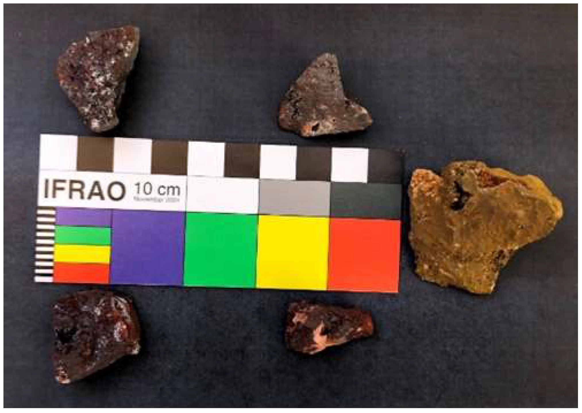
The details of the fires have been described [47] but are summarized here. In the laboratory, samples were heated in a thermostat-controlled Fisher Isotemp Muffle Furnace. Outdoors, the fires (60–80 cm in diameter) were made of well-seasoned wood in a simple, flat hearth [25,26,41,42]. The diameters of the fuel were from 4 to 12 cm, and the lengths were from 40 to 80 cm. Except for Fire 1, the temperatures of the samples were measured every 15 min in triplicate using an Etekcity Lasergrip 800 IR Thermometer; accurate measurements required being within 50 cm of the sample. Fire temperature was controlled by the amount of wood and the rate of stoking. In this configuration, the fires were slightly reducing in the flames, and they were strongly reducing in the coals [47]. Figure 1 illustrates a fire with samples at its center. Figure 2 shows an unheated sample of natural goethite (on the right) and four lumps that had been heated in the fire. A color card from the International Federation of Rock Art Organizations is included as a reference and scale.

3. Results

Eight XRD lines of hematite were selected for study (see Table 1) using the following criteria: sufficient intensity to measure widths accurately and precisely, avoidance of interfering lines, and comparability to measurements reported in the literature. The peak location of hematite lines had a standard deviation of ±0.003°.
As examples of our analysis, the XRD pattern for natural goethite powder heated at 450 °C is illustrated in Figure S1, and the data are listed in a tabular form in Table S1. Similarly, the XRD pattern for natural goethite powder heated at 900 °C is illustrated in Figure S2, and the data are listed in a tabular form in Table S2.
For optimum precision and accuracy in the XRD measurements, we found it necessary to dry samples at 150 °C for two hours immediately before loading them into the diffractometer. All reported measurements are from one-hour scans. For the furnace-heated samples, the reported FWHMs are the average of two or three replicates. In this way, the standard deviation on replicate peak widths was ±0.007°. (Exposure to ambient air for more than an hour allowed humidity to artificially increase peak widths by up to 0.05° and increased the standard deviation on peak widths by up to a factor of four).
To establish the accuracy, resolution, and precision of the instrument independent of the complications of iron oxides, we measured the locations and widths of four quartz lines in 42 ochre samples. The average width of all four lines was 0.135°, comparable to literature values and indicating the excellent resolution of our Rigaku diffractometer. The standard deviation of the location of the quartz lines was ±0.004° and of the width was ±0.02°, indicating excellent precision. The widths of quartz peaks were independent of ambient humidity and the temperature at which samples were heated.
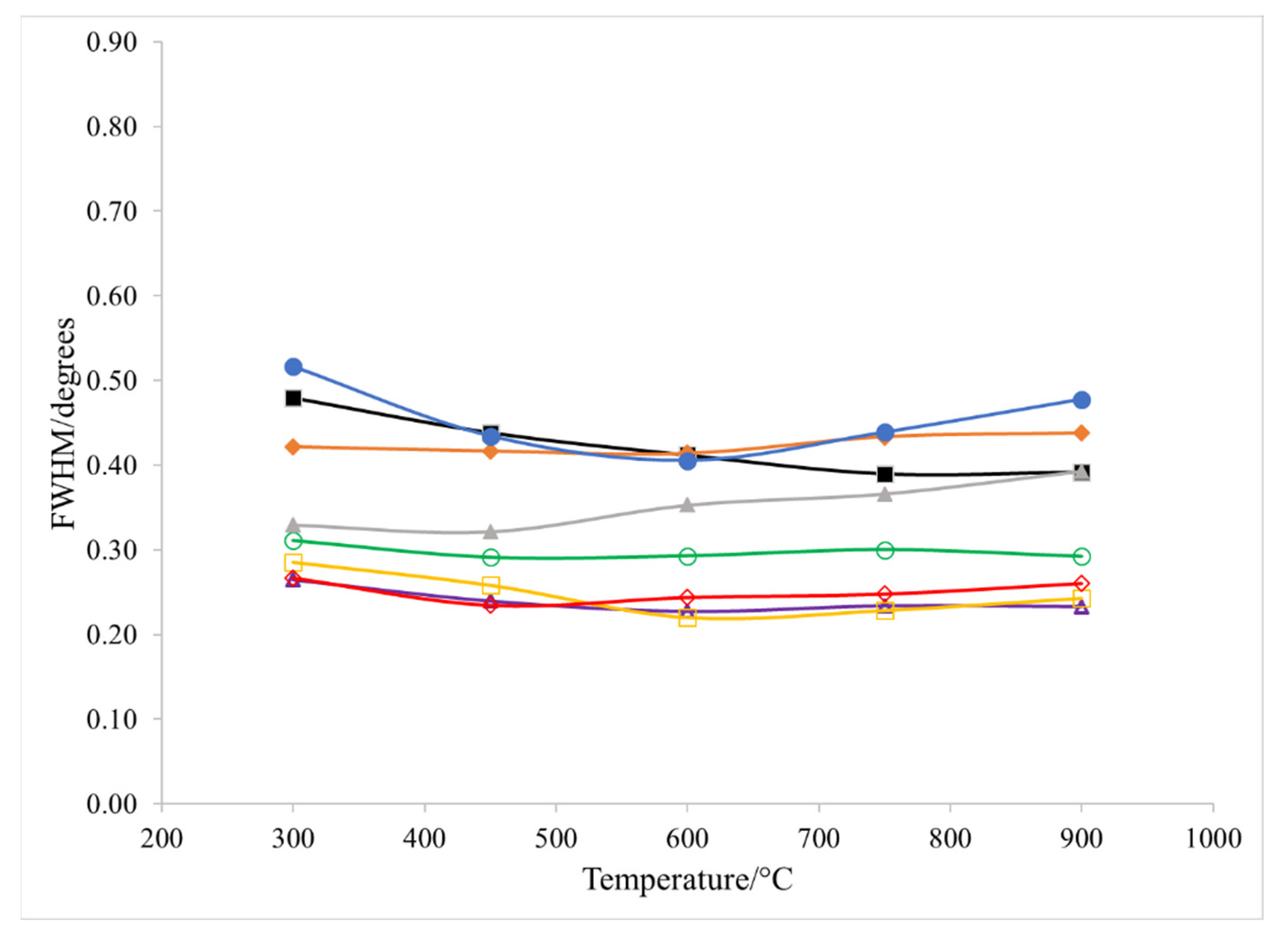
We converted natural goethite into hematite in a furnace at 300, 450, 600, 750, and 900 °C and then measured the peak widths for the eight selected lines. These widths (FWHM) are plotted vs. temperature in Figure 3. All the lines at all temperatures are wider than expected for well-crystallized hematite, and the FWHM values show little or no dependence on temperature. In fact, to our surprise, the widths of only two of the lines (012 and 024) demonstrated a narrowing trend from 300 to 600 °C.
These results for natural goethite are dramatically different from the results from heating synthetic goethite [14,28,32]. In the studies of synthetic goethite, the FWHM vs. T plots of synthetic goethite show a steady decrease in peak width from 300 to 600 °C over a range from about 1.0 to 0.3° for four of the lines (012, 104, 214, 204), lines, which correspond to those predominantly resulting from the lattice of iron ions [28,32]. (For easy comparison, Figure 3 in our study is at approximately the same scale as the corresponding figure in [28].) In contrast, well-crystallized hematite shows widths of 0.13 to 0.15° for these lines [14,32].
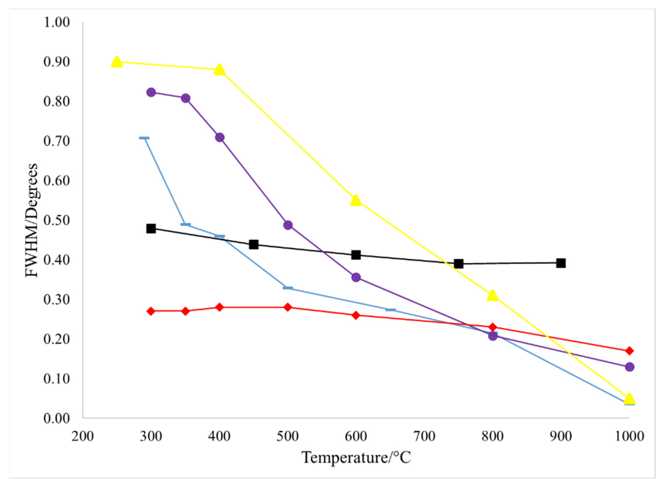
In Figure 4, we display the dependence on temperature for the width of line 012 for three natural goethites: our sample (black square), that of Löffler and Mader (red diamond) [32], and the sample used in Cavallo et al. (yellow triangle) [33]. Two synthetic goethites are also plotted: that of Löffler and Mader (purple circle) [32] and Pomiès et al. (blue dash) [28]. The comparison of results from five different goethites clearly illustrates two points: 1. the three natural goethites are highly variable (purple, red, and yellow lines), ranging from a dramatic dependence of FWHM on temperature to almost no dependence, and 2. the two synthetic samples (black, blue) are similar to each other in that both show a strong dependence of FWHM on temperature.
For a semi-quantitative comparison of the dependence of temperature for all the four selectively broadened lines, we calculated approximate slopes of the plots of FWHM vs. T from 300 to 600 °C where the relationship is quasilinear for the five heated ochres (see Table 2). We achieved this by minimizing the deviation in the y-axis between the points and a straight line. These slopes indicate that the trends illustrated in Figure 4 for line 012 are repeated for 104, 024, and 214. The dramatic differences documented in Figure 3 and Figure 4 and in Table 2 indicate that synthetic goethite makes a poor model for understanding the XRD of natural goethites.
Chemically, all five goethites are FeO(OH), so these results are initially surprising. However, iron oxide formation is notoriously complex [31] and very dependent on the conditions under which they are formed. Crystallinity [47,50], crystal size [51], crystal morphology [39,51,52,53], and impurities [31] all significantly affect the physical properties of goethite, so the variability in XRD line widths demonstrated here is consistent with these observations.
To extend our findings to Stone Age ochres, we replicated the heating of natural goethite in wood fires. We compared the XRD peak widths of natural goethite heated in a furnace to the widths of samples heated in a wood fire. To our knowledge, these are the first XRD results reported for fire-heated goethite.
In our first effort (Fire 1), we heated two samples of the homogenized, powder goethite (described in Section 2) in a campfire for one hour (samples 10-NFireP and 11-NFireP). The XRD lines of goethite had completely disappeared and been replaced by the expected lines for hematite, indicating complete conversion. The widths of those lines are recorded in Table 3. We then compared these widths to those of goethite heated in a furnace (illustrated in Figure 3) to determine the apparent temperature of the fire. The results varied wildly, and different lines led to grossly different predictions of temperatures, including some that are too high for a campfire or too low to have led to complete conversion to hematite in an hour. The difference between furnace-heated and fire-heated samples may be due to the varying temperatures achieved in a fire (see below for more on fire temperatures) or to the complex chemical environment of a fire. In either case, these data suggest that XRD line width is not a promising strategy for determining the temperature at which ochres were heated, especially for goethite that shows little dependence of FWHM on temperature, like the goethite here.
As we had not measured the temperature of Fire 1, we followed this initial effort by recording the XRD patterns for samples in four additional fires. The sample names and fire temperatures are listed in Table 4. The first two, 14-NFireP and 20-NFireP, were from the large batch of the homogenized natural goethite powder. Similar to the results of the powders in Table 3, the width of the lines of the XRD lines did not serve as an accurate proxy for determining the temperature at which the samples were heated. In fact, most of the lines were outside the range of the widths recorded in furnace-heated samples (Table 5).
We then took two new lumps of goethite from the same box of goethite purchased from Wards, 27-NFireL and 28-NFireL, and heated them as lumps directly in a fire (as opposed to heating ground and sieved powders). After heating, we brushed the ash from their surfaces and ground and sieved them. As previously reported, this produced pigments of virtually the same color as those produced by heating in a furnace [47]. We recorded the XRD, and again, the widths did not accurately predict the temperature of the fire (Table 5). Half of the lines were outside the range of the widths recorded in furnace-heated samples.
To measure the variability in three different lumps of goethite taken from the same source of natural goethite independent of any effect of fire, we took two more new lumps from the same box of goethite purchased from Wards, and we heated them in the furnace at 600 °C (samples 36-NFurnL and 37-NFurnL). Although ground via the same procedure and heated for the same amount of time in the same furnace, these samples had XRD line widths very different from those of the first sample. The widths are listed in Table 6. Differences ranged from +0.22° (the 214 line in 36-NFurnL) to −0.21° (the 104 line in 37-NFurnL). These differences are much larger than can be explained by the uncertainty of the measurement, which is better than ±0.01° (see Section 2). This demonstrates that hematite made by heating different pieces of goethite from a single source may have very different line widths due to the natural variability within the source.

4. Discussion

Taken together, these results demonstrate that the selective broadening of XRD lines of heated natural goethite is much less dependent on temperature than for the lines of heated synthetic goethite, questioning the use of synthetic goethite as a model. Furthermore, there is dramatic variation among different sources of natural goethite, probably resulting from differences in crystal size, crystallinity, impurities, and accessory minerals [24,31,32,39,48,52]. To use selective broadening at all requires the measurement of the broadening in the same source of natural goethite as the artists used [33], but, due to the high variability in a single geological source, even this strategy is risky. Furthermore, heating in wood fires yields quite different degrees of broadening than heating in laboratory furnaces. Instead of relying on XRD, a combination of careful archaeology, sourcing of local goethite, and a battery of analytical methods, as exemplified by research groups in Europe, is necessary for firm conclusions [5,7,17,33,34,35,53,54].
The mineralogy of iron oxides is very complex, partly because so many varieties are possible (in addition to goethite and hematite, there are magnetite, maghemite, lepidocrocite, ferrihydrite, etc.) [31]. As emphasized in the literature, the reproducible synthesis of iron oxides requires careful control of pH, reagent concentrations, reagent purity, temperature, convection, and even the material of the container (plastic versus glass), again because of the complexity of their chemistry, e.g., [31,32,50]. In contrast with the clean, controlled environment in laboratories, the formation of natural goethites occurs over geological time scales in complex and uncontrolled environments. We hypothesize that the complex and changing environments in which natural goethites form may disrupt the crystalline structure sufficiently to affect ion rearrangement during the goethite-to-hematite transition. Rietveld’s refinement of X-ray diffractograms may offer a strategy to test this hypothesis in the future.
In particular, the crystallinity of goethite—crystal size and morphology [14,32,35,37,50,51], water content [50], cationic impurities (e.g., Al, Ca, K, etc.) [37,39,52], surface-area-to-volume ratio [32,51,52], crystalline twinning [32,50], accessory minerals (especially other iron oxides) [14,32,37,39,51], crystal aggregation [51,52], and crystal strain [31]—all affect the physical properties of goethite, including the goethite-to-hematite transformation. Given these variables, the differences between natural and synthetic goethite are not surprising. As Goss writes, “Goethites in the natural environment have different grain sizes, shapes, varying degrees of crystallinity and excess water…”, and “a continuous range of goethites exist… Direct comparisons between natural and synthetic goethites cannot be made” [50] (p. 449). Similarly, variations between one natural source of goethite and another are not surprising. In fact, the complexity of natural environments suggests that even goethite from a single geological source will exhibit variation from one subsample to another, consistent with the variability from one lump to another exhibited in Table 6.
Although the interpretation of the XRD of iron oxides is so fraught with complications, several other strategies have proven effective in identifying whether hematite ochres found in archaeological contexts are natural or result from the anthropogenic heating of goethite. As reviewed in the Introduction section, a provenance study (the characterization of the total mineral content in ochres combined with geological prospecting) is an excellent strategy. For example, red ochre found in the Tagliente shelter contained a mix of hematite, quartz, and calcite in proportions that did not exist in any local sources. However, goethite, quartz, and calcite were found in a local source. In combination with careful archaeology and other analyses, the evidence is very persuasive that the ochre was intentionally heated [37]. In another case, the complex mineral composition of hematite ochre found in a Magdalenian burial in El Mirón Cave corresponded closely to a nearby source, demonstrating that the hematite did not result from heated goethite [40].
Another strategy that may prove helpful is the determination of pore size using transmission and scanning electron microscopies [5,32,33,34,35], even possibly to estimate the temperature at which the goethite was heated. However, more work is needed. In some studies of pore size, as for the broadening of XRD peaks, synthetic goethite was used as a model [14,29]. In contrast, the studies comparing laboratory-heated natural goethite to archaeological ochres have yielded more promising results [5,33,39].

5. Conclusions

The XRD line widths of hematite produced by heating natural goethites are very different from those of heated synthetic goethite. Within natural goethites, line widths are highly variable between sources and even within a single source. Heating goethite in a fire complicates selective line broadening compared with heating in a furnace, probably because of the variability in temperatures achieved in an open, wood fire. Thus, the XRD results from heating synthetic goethite in a furnace are shown to be an inappropriate model for understanding natural goethites heated in a wood fire. Whether the TEM and SEM studies of pore size and crystal morphology of heated synthetic goethite are appropriate as models for naturally heated goethite remains a question for future work.

Supplementary Materials

The following supporting information can be downloaded at: https://www.mdpi.com/article/10.3390/colorants1030022/s1, Figure S1: XRD pattern of natural goethite powder heated at 450° in oven for 1 h; Figure S2. XRD pattern of natural goethite powder heated at 900° in oven for 1 h; Table S1: XRD data in tabular form of natural goethite powder heated at 450° in oven for 1 h; Table S2: XRD data in tabular form of natural goethite powder heated at 900° in oven for 1 h.

Author Contributions

Conceptualization, S.R.V. and W.J.B.; methodology, J.C.P., W.J.B. and S.R.V.; formal analysis, J.C.P., J.M.N., S.E.S., S.P.S., S.R.V., Z.L. and W.J.B.; investigation, J.C.P., J.M.N., S.E.S., S.P.S., S.R.V., Z.L. and W.J.B.; writing—original draft preparation, W.J.B.; writing—review and editing, W.J.B.; visualization, J.C.P., J.M.N., S.E.S., S.P.S., S.R.V., Z.L. and W.J.B.; supervision, W.J.B.; project administration, W.J.B.; funding acquisition, W.J.B. All authors have read and agreed to the published version of the manuscript.

Funding

This research received no external funding.

Institutional Review Board Statement

Not applicable.

Informed Consent Statement

Not applicable.

Data Availability Statement

The data presented in this study are available in this article.

Acknowledgments

We thank D. Brooks McKinney and Ilene Nicholas for helpful conversations. We thank Zhengrui Wei for assistance with the design and making of the Graphical Abstract. We thank the anonymous reviewers for their encouragement to expand portions of the manuscript.

Conflicts of Interest

The authors declare no conflict of interest.

References

  1. Barham, L.S. Systematic pigment use in the Middle Pleistocene of South-Central Africa. Curr. Anthr. 2002, 43, 181–190. [Google Scholar] [CrossRef]
  2. Wadley, L. Putting ochre to the test: Replication studies of adhesives that may have been used for hafting tools in the Middle Stone Age. J. Hum. Evol. 2005, 49, 587–601. [Google Scholar] [CrossRef] [PubMed]
  3. Rifkin, R. Assessing the efficacy of red ochre as a prehistoric hide tanning ingredient. J. Afr. Archaeol. 2011, 9, 131–158. [Google Scholar] [CrossRef]
  4. Rifkin, R. Processing ochre in the Middle Stone Age: Testing the inference of prehistoric behaviours from actualistically derived experimental data. J. Anthropol. Archaeol. 2012, 31, 174–195. [Google Scholar] [CrossRef]
  5. Cavallo, G.; Fontana, F.; Gialanella, S.; Gonzato, F.; Riccardi, M.P.; Zorzin, R.; Peresani, M. Heat Treatment of Mineral Pigment During the Upper Palaeolithic in North-East Italy. Archaeometry 2018, 60, 1045–1061. [Google Scholar] [CrossRef]
  6. Dayet, L.; Le Bourdonnec, F.-X.; Daniel, F.; Porraz, G.; Texier, P.-J. Ochre provenance and procurement strategies during the middle stone age at Diepkloof Rock Shelter, South Africa. Archaeometry 2016, 58, 807–829. [Google Scholar] [CrossRef]
  7. Gialanella, S.; Belli, R.; Dalmeri, G.; Lonardelli, I.; Mattarelli, M.; Montagna, M.; Toniutti, L. Artificial or natural origin of hematite-based red pigments in archeological contexts: The case of Riparo Dalmeri (Trento, Italy). Archaeometry 2011, 53, 950–962. [Google Scholar] [CrossRef]
  8. Henshilwood, C.S.; D’Errico, F.; van Niekerk, K.L.; Coquinot, Y.; Jacobs, Z.; Lauritzen, S.-E.; Menu, M.; García-Moreno, R. A 100,000-year-old ochre-processing workshop at Blombos Cave, South Africa. Science 2011, 334, 219–222. [Google Scholar] [CrossRef]
  9. Hodgskiss, T. Identifying grinding, scoring and rubbing use-wear on experimental ochre pieces. J. Archaeol. Sci. 2010, 37, 3344–3358. [Google Scholar] [CrossRef]
  10. Hodgskiss, T.; Wadley, L. How people used ochre at Rose Cottage Cave, South Africa: Sixty thousand years of evidence from the Middle Stone Age. PLoS ONE 2017, 12, e0176317. [Google Scholar] [CrossRef] [Green Version]
  11. MacDonald, B.L.; Fox, W.; Dubreuil, L.; Beddard, J.; Pidruczny, A. Iron oxide geochemistry in the Great Lakes Region (North America): Implications for ochre provenance studies. J. Archaeol. Sci. Rep. 2018, 19, 476–490. [Google Scholar] [CrossRef]
  12. MacDonald, B.L.; Stalla, D.; He, X.; Maschmann, M.R.; Emerson, D.; White, T.A. Reconstructing rock art Fe-oxide pigment pyrotechnology using in situ SEM heating experiments. Microsc. Microanal. 2018, 24, 2124–2125. [Google Scholar] [CrossRef]
  13. MacDonald, B.L.; Chatters, J.C.; Reinhardt, E.G.; Devos, F.; Meacham, S.; Rissolo, D.; Rock, B.; Le Maillot, C.; Stalla, D.; Marino, M.D.; et al. Paleoindian ochre mines in the submerged caves of the Yucatán Peninsula, Quintana Roo, Mexico. Sci. Advant. 2020, 6, eaba1219. [Google Scholar] [CrossRef] [PubMed]
  14. Pomiès, M.; Morin, G.; Vignaud, C. XRD study of the goethite-hematite transformation: Application to the identification of heated prehistoric pigments. Eur. J. Solid State Inorg. Chem. 1998, 35, 9–25. [Google Scholar] [CrossRef]
  15. Pradeau, J.-V.; Salomon, H.; Bon, F.; Mensan, R.; Lejay, M.; Regert, M. Les matières colorantes sur le site aurignacien de plein air de Régismont-le-Haut (Poilhes, Hérault). Bull. De La Soc. Préhistorique Fr. 2014, 111, 631–658. [Google Scholar] [CrossRef]
  16. Rosso, D.E.; Martí, P.; D’Errico, F. Middle Stone Age Ochre Processing and Behavioural Complexity in the Horn of Africa: Evidence from Porc-Epic Cave, Dire Dawa, Ethiopia. PLoS ONE 2016, 11, e0164793. [Google Scholar] [CrossRef]
  17. Salomon, H.; Goemaere, E.; Billard, C.; Dreesen, R.; Bosquet, D.; Hamon, C.; Jadin, I. Analyse critique du protocole de caracterisation des hematites oolithiques mis en place dans le cadre du projet collectif de recherché sur L’origine des hematites oolithiques durant la prehistoire recent entre I’Eifel (DE) et la Normandie (FR). Anthropol. Et Praehist. 2016, 125, 225–246. [Google Scholar]
  18. Velliky, E.C.; Porr, M.; Conard, N.J. Ochre and pigment use at Hohle Fels cave: Results of the first systematic review of ochre and ochre-related artefacts from the Upper Palaeolithic in Germany. PLoS ONE 2018, 13, e0209874. [Google Scholar] [CrossRef]
  19. Villa, P.; Pollarolo, L.; Degano, I.; Birolo, L.; Pasero, M.; Biagioni, C.; Douka, K.; Vinciguerra, R.; Lucejko, J.J.; Wadley, L. A milk and ochre paint mixture used 49,000 years ago at Sibudu, South Africa. PLoS ONE 2015, 10, e0131273. [Google Scholar] [CrossRef]
  20. Wadley, L. Cemented ash as a receptacle or work surface for ochre powder production at Sibudu, South Africa, 58,000 years ago. J. Archaeol. Sci. 2010, 37, 2397–2406. [Google Scholar] [CrossRef]
  21. Watts, I. The pigments from Pinnacle Point Cave 13B, Western Cape, South Africa. J. Hum. Evol. 2010, 59, 392–411. [Google Scholar] [CrossRef] [PubMed]
  22. Wojcieszak, M.; Hodgskiss, T.; Colomban, P.; Wadley, L. Finding chemical and physical evidence of heat treatment of ochre by using non-destructive methods: A preliminary study. In The Exploitation of Raw Materials in Prehistory: Sourcing, Processing and Distribution; Pereira, T., Terradas, X., Bicho, N., Eds.; Cambridge Scholars Publishing: Newcastle upon Tyne, UK, 2017; pp. 587–600. [Google Scholar]
  23. Wojcieszak, M.; Wadley, L. A Raman micro-spectroscopy study of 77,000 to 71,000 year old ochre processing tools from Sibudu, KwaZulu-Natal, South Africa. Herit. Sci. 2019, 7, 24. [Google Scholar] [CrossRef]
  24. Helwig, K. A note on burnt yellow earth pigments: Documentary sources and scientific analysis. Stud. Conserv. 1997, 42, 181–188. [Google Scholar]
  25. Bentsen, S.E. Controlling the heat: An experimental approach to Middle Stone Age pyrotechnology. South Afr. Archaeol. Bull. 2011, 68, 137–145. [Google Scholar]
  26. Wadley, L. Post-depositional heating may cause over-representation of red-coloured ochre in Stone Age sites. South Afr. Archaeol. Bull. 2009, 64, 166–171. [Google Scholar]
  27. Onoratini, G.; Perinet, G. Données minéralogiques sur les colorants rouges préhistoriques de Provence: Démonstration que certains d’ entre eux ont été obtenus par calcination de goethite. C. R. Acad. Sc. Paris 1985, 301, 119–124. [Google Scholar]
  28. Pomiès, M.P.; Menu, M.; Vignaud, C. Red Paleolithic pigments: Natural hematite or heated goethite? Archaeometry 1999, 41, 275–285. [Google Scholar] [CrossRef]
  29. Pomiès, M.P.; Menu, M.; Vignaud, C. TEM observations of goethite dehydration: Application to archaeological samples. J. Eur. Ceram. Soc. 1999, 19, 1605–1614. [Google Scholar]
  30. Pomiès, M.P.; Barbaza, M.; Menu, M.; Vignaud, C. Preparation des pigments rouges prehistoriques par chauffage. L’Anthroplogie 1999, 103, 503–518. [Google Scholar]
  31. Schwertmann, U.; Cornell, R.M. Iron Oxides in the Laboratory, Preparation and Characterization; VCH: Weinheim, Germany, 1991. [Google Scholar]
  32. Löffler, L.; Mader, W. Anisotropic X-ray broadening and twin formation in hematite derived from natural and synthetic goethite. J. Eur. Ceram. Soc. 2006, 26, 131–139. [Google Scholar] [CrossRef]
  33. Cavallo, G.; Ischia, G.; Zorzin, R.; Gialanella, S. Experimental analysis on natural heated goethite from ponte di Veja (Mt Lessini, NE Italy). J. Archaeol. Sci. Rep. 2021, 36, 102871. [Google Scholar] [CrossRef]
  34. Salomon, H.; Vignaud, C.; Coquinot, Y.; Beck LStringer, C.; Strivay, D.; D’Errico, F. Selection and heating of colouring materials in the Mousterian level of Es-Skhul (c. 100, 000 years BP, Mount Carmel, Israel). Archaeometry 2012, 54, 698–722. [Google Scholar] [CrossRef]
  35. Salomon, H.; Vignaud, C.; Lahlil, S.; Menguy, N. Solutrean and Magdalenian ferruginous rocks heat-treatment: Accidental and/or deliberate action? J. Archaeol. Sci. 2015, 55, 100–112. [Google Scholar] [CrossRef]
  36. Cavallo, G.; Riccardi, M.P.; Zorzin, R. Powder diffraction of yellow and red natural earths from Lessini Mountains in NE Italy. Powder Diffr. 2015, 30, 122–129. [Google Scholar] [CrossRef]
  37. Cavallo, G.; Fontana, F.; Gonzato, F.; Guerreschi, A.; Riccardi, M.P.; Sardelli, G.; Zorzin, R. Sourcing and processing of ochre during the late upper Palaeolithic at Tagliente rock-shelter (NE Italy) based on conventional X-ray powder diffraction analysis. Archaeol. Anthr. Sci. 2015, 9, 763–775. [Google Scholar] [CrossRef]
  38. Cavallo, G.; Fontana, F.; Gonzato, F.; Peresani, M.; Riccardi, M.P.; Zorzin, R. Textural, microstructural, and compositional characteristics of Fe-based geomaterials and Upper Paleolithic ochre in the Lessini Mountains. Northeast Italy: Implications for provenance studies. Geoarchaeology 2017, 32, 437–455. [Google Scholar] [CrossRef]
  39. Gialanella, S.; Girardi, F.; Ischia, G.; Lonardelli, I.; Mattarelli, M.; Montagna, M. On the goethite to hematite phase transformation. J. Therm. Anal. 2010, 102, 867–873. [Google Scholar] [CrossRef]
  40. Román, R.S.; Ruiz, M.D.L.; Juan-Juan, J.; Bañón, C.B.; Straus, L.G.; Morales, M.R.G. Sources of the ochres associated with the Lower Magdalenian “Red Lady” human burial and rock art in El Mirón Cave (Cantabria, Spain). J. Archaeol. Sci. Rep. 2019, 23, 265–280. [Google Scholar]
  41. Aldeias, V. Experimental approaches to archaeological fire features and their behavioral relevance. Curr. Anthropol. 2017, 58, S191–S205. [Google Scholar] [CrossRef]
  42. March, R.J.; Lucquin, A.; Joly, D.; Ferreri, J.C.; Muhieddine, M. Processes of formation and alteration of archaeological fire structures: Complexity viewed in the light of experimental approaches. J. Archaeol. Method Theory 2014, 21, 1–45. [Google Scholar] [CrossRef]
  43. Henshilwood, C.S.; Sealy, J.C.; Yates, R.; Cruz-Uribe, K.; Goldberg, P.; Grine, F.E.; Klein, R.G.; Poggenpoel, C.; van Niekerk, K.; Watts, I. Blombos Cave, Southern Cape, South Africa: Preliminary report on the 1992–1999 excavations of the Middle Stone Age levels. J. Archaeol. Sci. 2001, 28, 421–448. [Google Scholar] [CrossRef]
  44. Henshilwood, C.S.; D’Errico, F.; Van Niekerk, K.L.; Dayet, L.; Queffelec, A.; Pollarolo, L. An abstract drawing from the 73,000-year-old levels at Blombos Cave, South Africa. Nature 2018, 562, 115–118. [Google Scholar] [CrossRef]
  45. Wadley, L. Ochre crayons or waste products? Replications compared with MSA ‘crayons’ from Sibudu Cave, South Africa. Farming 2005, 3, 1–12. [Google Scholar] [CrossRef]
  46. Lorblanchet, M.; Labeau, M.; Vernet, J.L.; Fitte, P.; Valladas, H.; Cachier, H.; Arnold, M. Palaeolithic pigments in the Quercy, France. Rock Art Res. 1990, 7, 4–12. [Google Scholar]
  47. Lin, Z.; Natoli, J.M.; Picuri, J.C.; Shaw, S.E.; Bowyer, W.J. Replication of the conversion of goethite to hematite to make pigments in both furnace and campfire. J. Archaeol. Sci. Rep. 2021, 39, 103134. [Google Scholar] [CrossRef]
  48. Elias, M.; Chartier, C.; Prévot, G.; Garay, H.; Vignaud, C. The colour of ochres explained by their composition. Mater. Sci. Eng. B 2006, 127, 70–80. [Google Scholar] [CrossRef]
  49. Garofano, I.; Perez-Rodriguez, J.L.; Robador, M.D.; Duran, A. An innovative combination of non-invasive UV–Visible-FORS, XRD and XRF techniques to study Roman wall paintings from Seville, Spain. J. Cult. Herit. 2016, 22, 1028–1039. [Google Scholar] [CrossRef]
  50. Goss, C.J. The kinetics and reaction mechanism of the goethite to hematite transformation. Miner. Mag. 1987, 51, 437–451. [Google Scholar] [CrossRef]
  51. Lima-de-Faria, J. Dehydration of goethite and diaspore. Z. Für Krist. Bd. 1963, 119, 176–293. [Google Scholar] [CrossRef]
  52. Liu, H.; Chen, T.; Zou, X.; Qing, C.; Frost, R.L. Thermal treatment of natural goethite: Thermal transformation and physical properties. Thermochim. Acta 2013, 568, 115–121. [Google Scholar] [CrossRef]
  53. Botticelli, M.; Appolloni, G.; Candeias, A.; Mirão, J.; Maras, A. Synthesis of yellow and red ochre: A preliminary multianalytical, micro-invasive investigation on recipes and recognition criteria. Eur. Phys. J. Plus 2021, 136, 1–19. [Google Scholar] [CrossRef]
  54. Garilli, V.; Vita, G.; La Parola, V.; Vraca, M.P.; Giarrusso, R.; Rosina, P.; Bonfiglio, L.; Sineo, L. First evidence of Pleistocene ochre production from bacteriogenic iron oxides. A case study of the Upper Palaeolithic site at the San Teodoro Cave (Sicily, Italy). J. Archaeol. Sci. 2020, 123, 105221. [Google Scholar] [CrossRef]
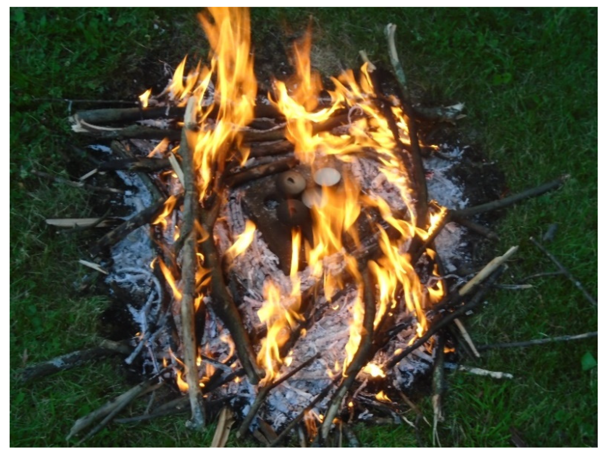
Figure 1. Wood fire for heating ochre samples.
Colorants 01 00022 g001
Figure 2. Four lumps of ochre heated in a wood fire before grinding (top and bottom rows, Munsell color 2.5YR 3/4-6). Lump of unheated goethite (right, Munsell color 10YR 5/6).
Colorants 01 00022 g002
Figure 3. Line widths for eight selected lines of hematite prepared by heating natural goethite from Hibbing, MN, USA, vs. the temperature at which the sample was heated: 012 black, solid square; 104 orange, solid diamond; 110 purple, open triangle; 113 yellow, open square; 024 blue, solid circle; 116 green, open circle; 214 grey, solid triangle; 300 red, open diamond. Each point is the average of two or three replicates.
Colorants 01 00022 g003
Figure 4. Dependence of the width of line 012 on temperature for three natural and two synthetic goethites: natural goethite, this study, black square; natural, Löffler and Mader [32], red diamond; synthetic, Löffler and Mader [32], purple circle; synthetic, Pomiès et al. [28], blue dash; natural, Cavallo et al. [33], yellow triangle.
Colorants 01 00022 g004
Table 1. Miller indices (h k l), atom spacing (d), and location (2θ) of eight hematite lines in heated ochres.
h k ld/Å2θ/°
0 1 23.70523.96
1 0 42.71332.96
1 1 02.52835.42
1 1 32.21540.66
0 2 41.84649.31
1 1 61.69953.88
2 1 41.48962.26
3 0 01.45663.82
Table 2. Slopes of width vs. temperature for four selectively broadened XRD lines (FWHM vs. T (°/°C)·(−1 × 104)) for five hematite samples generated from heated goethite from 300 to 600 °C.
h k lNatural aNatural bNatural cSynthetic dSynthetic e
0 1 22.20.52121711
1 0 40.260.071620.11
0 2 43.70.428.4189.2
2 1 4−0.78−1.26.62214
average1.3−0.05111911
a This study; b [32] (Figure 2); c [33] (Figure 2); d [32] (Figure 4); e [28] (Figure 2).
Table 3. Widths (FWHM) of four XRD lines for two powder samples heated in a fire in crucibles and the apparent fire temperatures as estimated from the width using plot in Figure 3.
10-NFireP 11-NFireP
h k lFWHM/°Temp./°CFWHM/°Temp./°C
0 1 20.406000.41600
1 0 40.32<3000.40>900
0 2 40.36>9000.49350
2 1 40.51<3000.47<300
Table 4. Heating procedure for six samples of natural goethite.
Sample HeatTime/hTemp/°CPowder or Lump
14-NFirePFire 22300–600Powder
20-NFirePFire 32250–600Powder
27-NFireLFire 42300–500Lump
28-NFireLFire 51.5600–750Lump
36-NFurnLFurnace1600Lump
37-NFurnLFurnace1600Lump
Table 5. Temperatures in °C predicted by four XRD lines for samples heated in wood fires.
h k l14-NFireP20-NFireP27-NFireL28-NFireL
0 1 2500350350>900
1 0 4>900>900750>900
0 2 4NP>900350500
2 1 4<300<300<300<300
Table 6. Line widths in degrees measured for three different subsamples of the same natural goethite after heating at 600 °C for one hour in a furnace.
h k l5-NFurnP36-NFurnL37-NFurnL
0 1 20.4120.3710.398
1 0 40.4140.4650.204
0 2 40.4050.4250.494
2 1 40.3530.5730.430
Publisher’s Note: MDPI stays neutral with regard to jurisdictional claims in published maps and institutional affiliations.