Article

Ammonium-Induced Synthesis of Highly Fluorescent Hydroxyapatite Nanoparticles with Excellent Aqueous Colloidal Stability for Secure Information Storage

1
Hubei Provincial Key Laboratory of Green Materials for Light Industry, Hubei University of Technology, Wuhan 430068, China
2
Beijing Institute of Aerospace Testing Technology, Beijing Key Laboratory of Research and Application for Aerospace Green Propellants, Beijing 100074, China
*
Author to whom correspondence should be addressed.
Coatings 2019, 9(5), 289; https://doi.org/10.3390/coatings9050289
Submission received: 28 February 2019 / Revised: 21 April 2019 / Accepted: 23 April 2019 / Published: 27 April 2019

Abstract

:
In this paper, uniform hydroxyapatite (HA) nanoparticles, with excellent aqueous colloidal stability and high fluorescence, have been successfully synthesized via a citrate-assisted hydrothermal method. The effect of the molar ratio of ammonium phosphate in phosphate (RAMP) and hydrothermal time on the resultant products was characterized in terms of crystalline structure, morphology, colloidal stability, and fluorescence behavior. When the RAMP is 50% and the hydrothermal time is 4 h, the product consists of a pure hexagonal HA phase and a uniform rod-like morphology, with 120- to 150-nm length and approximately 20-nm diameter. The corresponding dispersion is colloidally stable, and transparent for at least one week, and has an intense bright blue emission (centered at 440 nm, 11.6-ns lifetime, and 73.80% quantum efficiency) when excited by 340-nm UV light. Although prolonging the hydrothermal time and increasing the RAMP had no appreciable effect on the aqueous colloidal stability of HA nanoparticles, the fluorescence intensity was enhanced. The cause of HA fluorescence are more biased towards carbon dots (which are mainly polymer clusters and/or molecular fluorophores constituents) trapped in the hydroxyapatite crystal structure. Owing to these properties, a highly fluorescent HA colloidal dispersion could find applications in secure information storage.

1. Introduction

Hydroxyapatite [HA, Ca10(PO4)6(OH)2] is the most stable calcium phosphate phase in physiological conditions because of its excellent biocompatibility and biological activity as well as strong ion-exchange capacity [1,2]. It is also the model compound used to denote the mineral component of bone and dentin. The flexible crystal structure and modifiable surface of hydroxyapatite crystals offers many possible applications (Table 1).
Thus, HA is commonly used for diverse purposes such as bone tissue engineering [14,15], heavy metal ion absorption [16,17], fluorescent materials [12,18], protein separation [19,20], drug carriers [21,22,23], and so on.
Hydroxyapatite is intrinsically non-fluorescent [2]. A typical method for imparting fluorescence to HA is to dope rare earth elements into hydroxyapatite crystals, including Eu3+, Tb3+, Gd3+, and so on [4,18,23,24]. An alternative route is through surface modification of the particles with quantum dots or fluorescent dyes [25,26]. Although great efforts have been made in such methods, challenges remain. For example:
  • Synthesizing highly fluorescent hydroxyapatite nanoparticles without sacrificing colloidal stability;
  • Rendering the synthesized fluorescent hydroxyapatite more bio-compatible, such as by using neither quantum dots nor fluorescent dyes;
  • Rendering synthesis facile and not using expensive rare earth elements.
Recently, Lin et al. reported a method for synthesizing luminescent strontium hydroxyapatite, using a citrate-assisted hydrothermal method [27]. The carbon dioxide anion radical impurities in the strontium hydroxyapatite crystal lattice, produced by the decomposition of citrate during hydrothermal treatment, were considered to be essential to the fluorescence. Wang et al. [28] reported the effect of strontium doping on the fluorescence of hydroxyapatite microspheres, and found that the maximum intensity of photoluminescence (PL) is at the molar ratio of 30% strontium. Kim et al. [29] investigated the effect of the molar ratio of citrate to calcium on the fluorescence of hydroxyapatite microspheres, and found that the maximum intensity of photoluminescence is at a molar ratio of 2:1. Fan et al. [30], through a series of comparative experiments on the synthesis of fluorescent HA nanoparticles, proposed another explanation: N-rich carbon dots are the main cause of fluorescence. The use of high-resolution transmission electron microscopy (HR–TEM) on the sample, which indicated that carbon dots are distributed on the surface of HA nanoparticles, strongly supports the proposed mechanism.
Our previous studies have found that by balancing the molar ratio of sodium citrate to calcium, hydrothermal time, and temperature, rod-shaped hydroxyapatite nanoparticles with high crystallinity and excellent colloidal stability can be prepared [11,12]. On this basis, Eu3+-doped hydroxyapatite nanocrystals can be used in inkjet printing for secure information storage [4]. Loo et al. described that the size of the HA nanoparticles increases with increasing synthesis time and synthesis temperature [31]. Kuśnieruk et al. also observed the diameter of HA nanoparticles influenced by the reaction time, pressure and temperature [32].
In the present work, highly fluorescent HA nanoparticles with stable colloidal stability were prepared by a citrate-assisted hydrothermal method. The influence of the molar ratio of ammonium phosphate in phosphate (RAMP) and hydrothermal time on the phase structure, morphology, fluorescence, and colloidal stability of the resultant compounds was systematically studied. Combining our results and those in the scientific literature, the factors imparting florescence and colloidal stability are discussed. In addition, three different compounds containing primary amine groups have been introduced into our fluorescent HA to confirm the proposed mechanism. Particles synthesized under optimal experimental conditions were applied to secure information storage.

2. Materials and Methods

2.1. Materials

Calcium chloride anhydrous (CaCl2, >96.0%), sodium phosphate tribasic dodecahydrate (Na3PO4·H2O, >98.0%), ammonium phosphate tribasic ((NH4)3PO4·3H2O, >98.0%), sodium citrate tribasic dehydrate (C6H5Na3O7·2H2O, >99.0%), arginine (C6H5Na3O7·2H2O, >98.0%), urea (CH4N2O, >99.0%), ethylenediamine (C2H8N2, >99.0%), and absolute ethanol (C2H6O, >99.7%) were purchased from Aladdin Industrial Corporation, Shanghai, China. All chemicals were used as received, without further purification. Deionized water was used throughout our experiments.

2.2. Methods

2.2.1. Preparation of Hydroxyapatite Nanoparticles

Hydroxyapatite nanoparticles were prepared by using a hydrothermal method. In a typical experiment, a sodium citrate (C6H5Na3O7·2H2O) solution (0.01 mol, 10 g water) was added, with continuous stirring, to an aqueous solution of CaCl2 (0.01 mol, 10 g water) over 10 min. Then, an aqueous phosphate solution (Na3PO4⋅12H2O, 0.006−x mol; (NH4)3PO4⋅3H2O, x mol; 15 g water) was added to the mixture with vigorous stirring over 15 min. Next, the mixed solution was transferred, as obtained, to a Teflon-lined stainless steel autoclave with a 50 mL capacity. The solution in the autoclave underwent hydrothermal treatment at 180 °C for 0.5–4 h. After the hydrothermal treatment, the autoclave was allowed to cool down rapidly with water, and the resulting product was purified using a three-cycle process with deionized water and ethanol. Finally, the purified product was re-dispersed in deionized water to form an aqueous dispersion by magnetic stirring (MS-H280-Pro, DLAB Scientific Co., Ltd., Beijing, China) with 600 RPM for 10 min. The pH of the dispersion was adjusted to pH 9.5 by the addition of 0.1 M NaOH. A portion of the product was dried at 80 °C for 12 h to obtain powder for future characterization. For the sake of simplicity, RAMP represents the molar ratio of ammonium phosphate to total phosphate, which one adds in a specific synthesis.

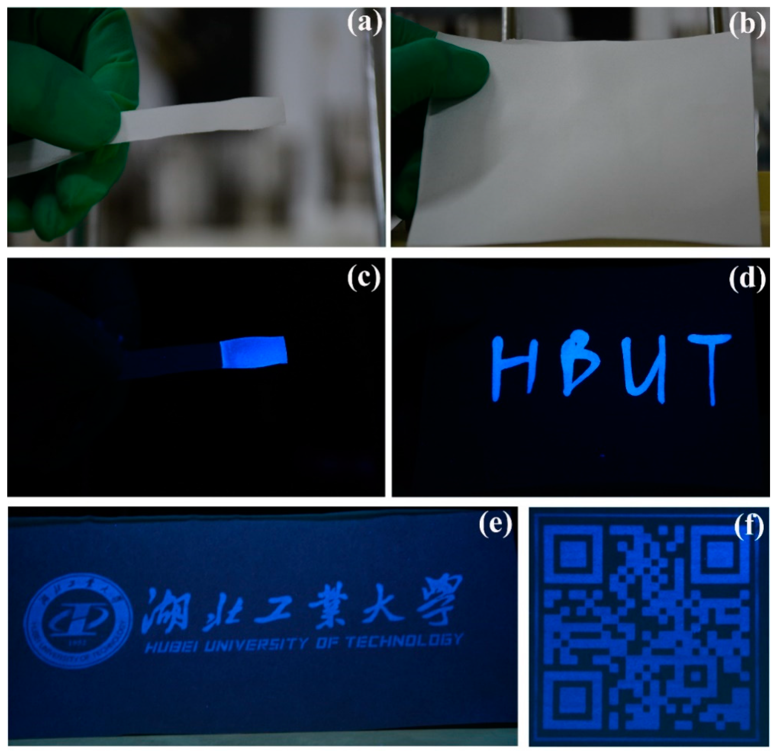
2.2.2. Secure Information Storage Based on Fluorescent Hydroxyapatite Colloidal Dispersion

A fluorescent hydroxyapatite colloidal dispersion with a fixed concentration (5 wt %) acted as an ink. Varied methods for recorded information storage, including dipping, writing with a pen, and printing with an ink-jet printer (Deskjet Ink Advantage 2060 HP, Beijing, China), on commercial paper with no background fluorescence, were conducted. A photograph of the paper was taken using a camera (Nikon 7100D, Tokyo, Japan) under natural or UV light (365 nm) to evaluate the security performance.

2.3. Characterization

The dried powders were characterized using X-ray diffraction (XRD) with an X’Pert PRO X-ray diffractometer (PANalytical B.V., Almelo, the Netherlands). Analyses were performed using a Cu Kα radiation (1.5406 A) source at 60 kV and 60 mA, from 20° to 80°, with a scan rate of 0.5°/min. The main peaks at (002) and (310) were generally used to calculate to investigate the average crystallite size of HA, according to the Debye-Scherrer Formula (1):
D = κλ/(βcosθ)
where D is the average crystal size; κ is a constant related to crystal habit and it is set as 0.9; λ is the wavelength of the Cu Kα radiation (1.5406 Å); β is full width at half maximum (FWHM) of the XRD peak; θ is the Bragg angle.
The morphology of samples was inspected with a field-emission scanning electron microscope (FE–SEM, SU8010, Hitachi, Tokyo, Japan). The morphology of samples was inspected with a FEI Tecnai G2-20 transmission electron microscope (TEM, Hillsboro, OR, USA), using an accelerating voltage of 200 kV. Zeta potential and particle size distribution measurements were performed using a nano-ZS90 Zetasizer (Malvern Instruments Ltd., Malvern, UK). The pH and particle concentration were 9.5 wt % and 1 wt %, respectively. No additional electrolytes were added to each sample for the measurement of zeta potential and particle size distribution. The excitation and emission spectra were measured with a fluorescence spectrophotometer (PerkinElmer LS-55, Waltham, MA, USA). The samples were prepared at 0.1 wt % concentration. Spectra were recorded with a photomultiplier voltage of 120 V, a scan speed of 2400 nm/min, and excitation and emission slit widths of 5 nm. Excitation spectra were recorded at 440-nm emission wavelength, and emission spectra were recorded at 340-nm excitation wavelength. The fluorescence lifetime and quantum yield were recorded with a steady-state/transient fluorescence spectrometer (FLS980, Edinburgh Instruments, Livingston FC, UK). The samples were prepared at 0.1 wt % concentration. X-ray photoelectron spectra (XPS) were recorded with a multifunctional imaging electron spectrometer (Thermo ESCALAB 250XI, ThermoFisher Scientific, Waltham, MA, USA). Fourier-transform infrared spectra (FTIR) were measured with an FTIR spectrometer (NICOLET iS50, Thermo Nicolet Corporation, Livingston Madison, WI, USA). Elemental analysis was conducted with an elemental analyzer (Elementar vario EL cube), using a combustion method to estimate the carbon, nitrogen, and hydrogen content of the products.

3. Results

3.1. Effect of RAMP and Hydrothermal Time on the Physicochemical Properties of the Product

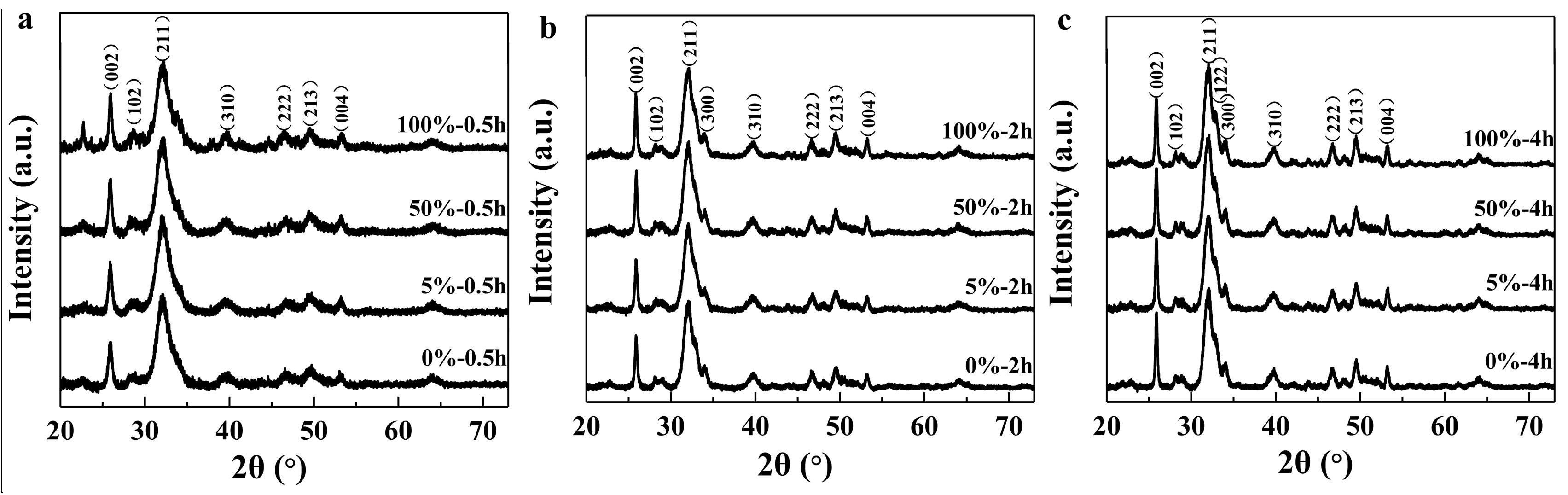
3.1.1. XRD Patterns Analysis

The crystal structure and phase composition of our compounds were determined by X-ray diffraction. Figure 1 shows XRD patterns of samples prepared with different RAMP and hydrothermal times. The diffraction peaks of all patterns can be indexed as a pure hexagonal HA phase without an impurity phase, which is consistent with the standard data for hydroxyapatite (JCPDS files, PDF No. 86-740) and the scientific literature [33,34,35]. This indicates that changes in ammonium phosphate content and hydrothermal time do not induce production of pure hydroxyapatite.
As the reaction temperature increased from 0.5 to 4 h at the same RAMP, the 122 and 300 peaks of the samples gradually appeared and strengthened. This indicates that the crystallinity and crystal integrity of the particles gradually increased. No obvious change was observed from XRD results when the samples were synthesized with different ammonium phosphate content at the same hydrothermal time. This suggests that ammonium phosphate and sodium phosphate do not differ in terms of their effects on hydroxyapatite crystallization.
The average size of crystal domains along the c-axis (D[002]) and the a–b plane (orthogonal to the c-axis, D[310]), calculated by the Scherrer’s equation using the width at half height for the respective reflections 002 and non-overlapped 310, are shown in Table 2. D[002] strongly increased with hydrothermal time from 14.8 to 33.3 nm. D[310] increased with hydrothermal time and RAMP from 8.1 nm to 19.9 nm. The D[002]/D[310] ratio of all samples was above 1, suggesting that the particles preferentially grew along the c-axis and rod-like shape of particles was formed.

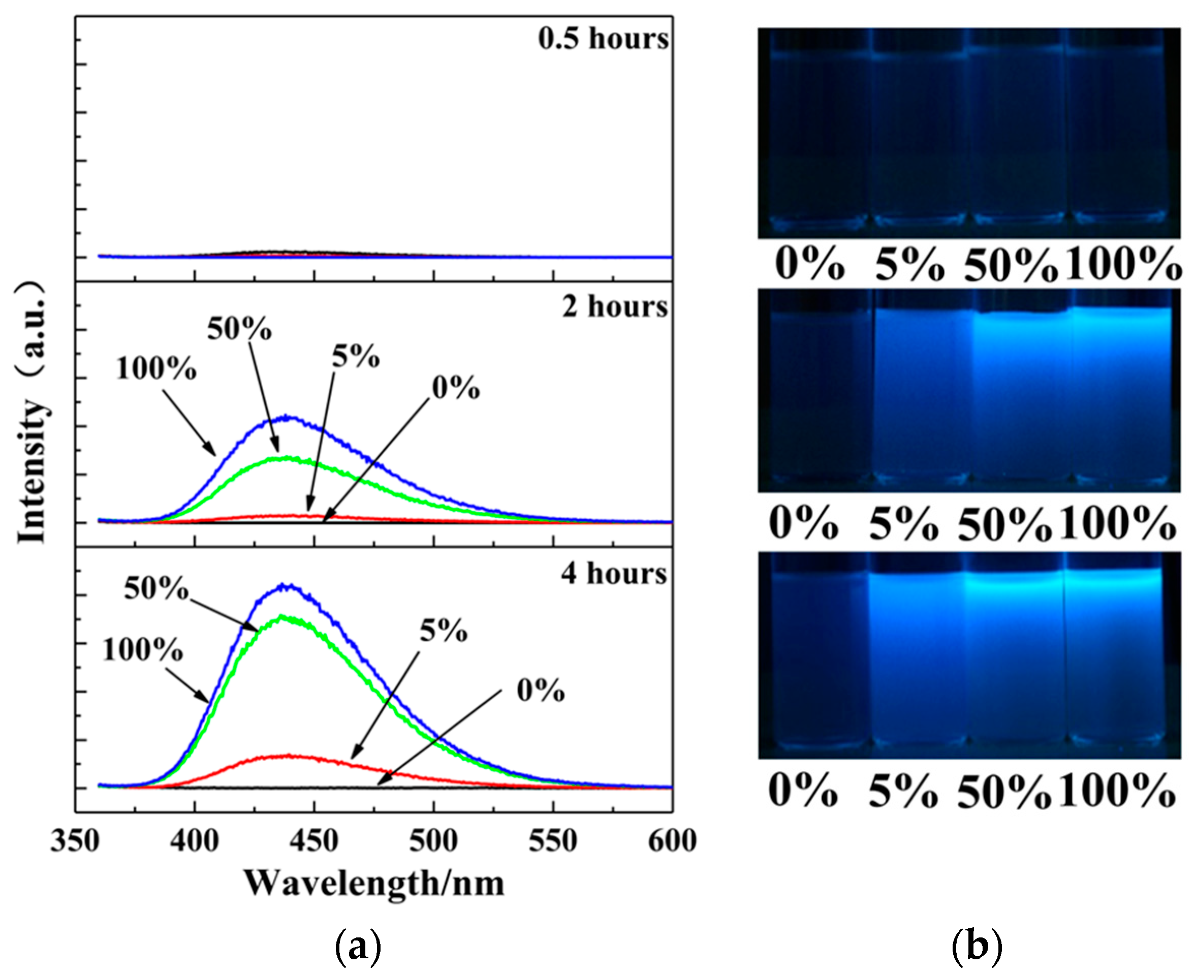
3.1.2. Fluorescence Behavior Analysis

Figure 2 depicts the photoluminescence emission spectrum of samples synthesized with different RAMP and hydrothermal times, excited with 340-nm UV light. When the hydrothermal time was less than 0.5 h, no fluorescence was observed, no matter how the RAMP was changed. When the hydrothermal time was 2 h, the fluorescence intensity of samples increased, and the RAMP increased from 5% to 100%. The maximum fluorescence peak of the samples was approximately 440 nm, which essentially did not change as the RAMP changed. The sample synthesized with sodium phosphate as the phosphate source was not fluorescent. When the hydrothermal time increased to 4 h, the trends were very similar with those of the samples synthesized for 2 h, yet the fluorescence intensity was higher. The sample synthesized with sodium phosphate as the phosphate source for 4 h still did not have fluorescence. The observation of the corresponding dispersions of samples under a 365-nm UV lamp validated the results of the fluorescence spectrum.
From the fluorescence data, one can see that for a specific hydrothermal temperature (180 °C), there is a critical hydrothermal time for generating fluorescence. In the studied hydrothermal time range, the fluorescence of the samples increased in accordance with increasing hydrothermal time. More importantly, the introduction of ammonium phosphate was critical for the fluorescence of HA. At the same hydrothermal time, samples synthesized with a high RAMP exhibited a stronger fluorescence intensity compared to those synthesized with a low RAMP.
To further understand the fluorescence behavior, samples synthesized with a RAMP of 5%, 50%, and 100% were selected to evaluate their fluorescence lifetime (Figure S1; see supporting information) and absolute quantum yield (QY). Table 3 summarizes the results.
The fluorescence lifetime of the selected samples was in the vicinity of 10 ns, indicating that the emitted light was fluorescent. Although the fluorescence intensity was the strongest at 100% RAMP in the fluorescence spectrum, the QY of the fluorescence was not the highest. The sample with 50% RAMP was stronger than that of 100% RAMP, indicating that the quantum efficiency depends on the RAMP. By adjusting the RAMP, one can obtain a very high fluorescence quantum efficiency.

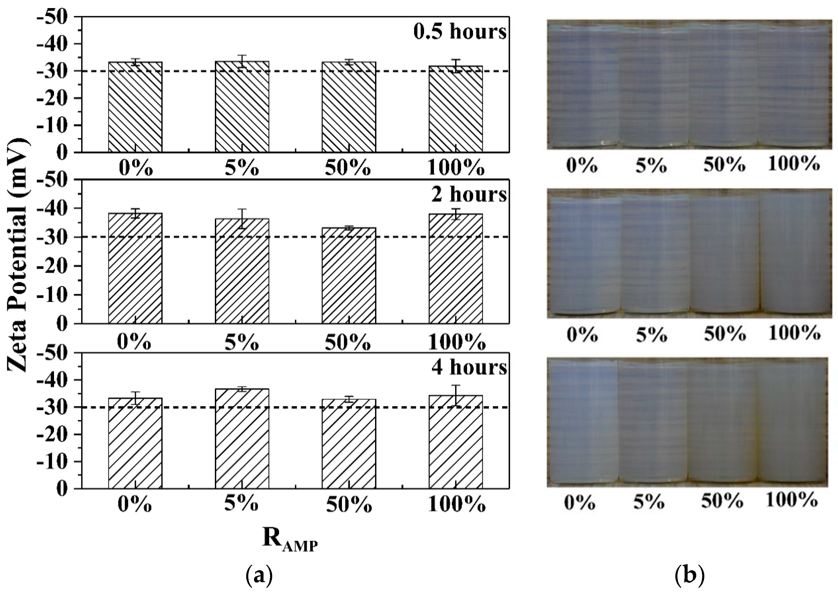
3.1.3. Colloidal Stability Analysis

The colloidal stability of aqueous dispersions synthesized with different RAMP and hydrothermal times were evaluated by zeta potential measurements and long-term observations (Figure 3). The pH and particle concentration of the dispersions was 9.5 wt % and 1 wt %, respectively. Figure 3 shows that the absolute zeta potential values of all the samples were greater than 30 mV, indicative of strong electrostatic repulsive forces between colloidal particles in the aqueous dispersion. Moreover, neither precipitation nor creaming phenomena were observed from dispersions after standing for one week. The dispersions remained homogeneous and transparent. As the RAMP increased, the colorless dispersion gradually turned light yellow.
Thus, the colloidal dispersion was stable over the studied hydrothermal times and RAMP values, without any obvious changes in the colloidal stability. However, the dispersion color deepened with increased hydrothermal time and increasing RAMP, indicating that there may be other chemical reactions in addition to the formation of hydroxyapatite during hydrothermal treatment. One can conclude that this reaction is directly related to the fluorescence behavior.

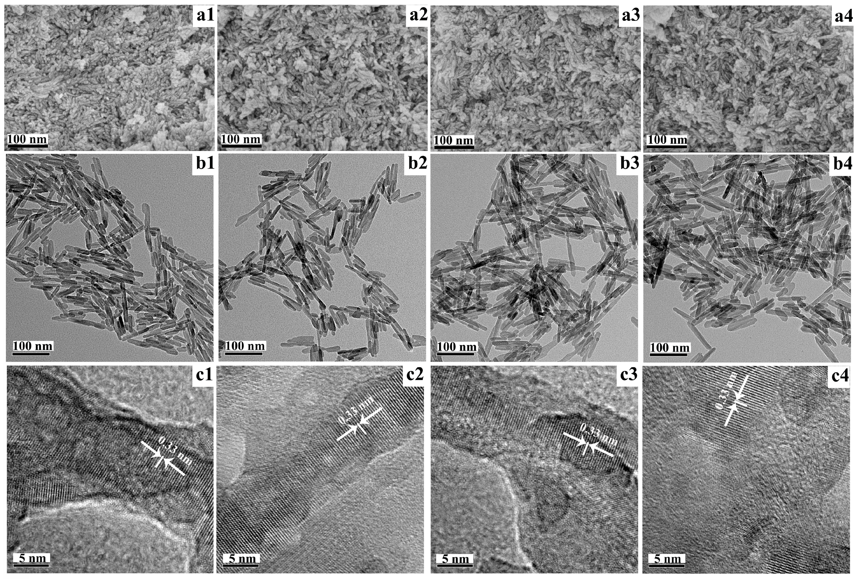
3.1.4. Morphology Analysis

Figure 4 shows the nanoparticle morphology as a function of RAMP at 180 °C for 4 h. The FE–SEM images (Figure 4(a1–a4)) show that the products consisted of relatively uniform rod-shaped particles, with the average size ranging from approximately 100 nm to 250 nm. More detailed information on particle morphology can be obtained from the TEM images (Figure 4(b1–b4)). The average length and diameter of the rod-shaped particles was 120 nm and 10 nm, respectively. In accordance with increasing RAMP, there was no appreciable change in particle morphology or size.
HR–TEM images (Figure 4(c1–c4)) indicate distinct lattice fringes. The distance between these adjacent lattices was approximately 3.3 Å, which is associated with the (002) plane of the hexagonal HA, indicating growth along the (002) lattice plane of the c-axis. A distribution of carbon particles on the surface of the hydroxyapatite nanoparticles was not apparent, which may be related to the shorter hydrothermal time in this study [30,36].

3.1.5. XPS Analysis

Table 4 shows data from the XPS wide scan spectrum (Figure S2a; see supporting information), high-resolution C 1s spectrum (Figure S2b) [37,38], and high-resolution N 1s spectrum (Figure S2c) [37] of nanoparticles synthesized with an RAMP of 5%. Compared with the XPS spectrum of samples synthesized with an RAMP from 5% to 100% (Figure S2d–i), there was a clear enhancement of the intensity of the characteristic peak of the C–N bond. This suggests that over the course of hydrothermal treatment, ammonium ions not only act as cations, but also undergo dehydration condensation reaction with citrate ions.

3.1.6. Elemental Analysis

As shown in Table S1 (see supporting information), the nitrogen content of the product increased from 0.00% to 0.145%, as the RAMP increased from 0% to 100%. The hydrogen content of the reaction product decreased from 0.7255% to 0.503%, as the RAMP increased from 0% to 100%. Combined with the formation of new chemical bonds (C–N, N–H, and C=N) as per the XPS results, one can infer that the nitrogen stays on the surface of the hydroxyapatite nanoparticles by dehydration of ammonium ions and citrate ions, and hydrogen is gradually removed in the form of water.

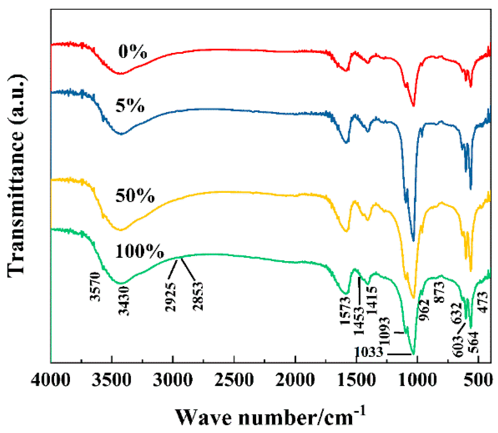
3.1.7. FTIR Analysis

To further understand the dehydration reactions during HA hydrothermal synthesis, an FTIR spectrum was employed to investigate the changes of samples synthesized with different RAMP values (Figure 5). Table 5 summarizes the results.
The C–N chemical band (1404 cm−1) was difficult to identify from the FTIR spectra. This may be attributable to the low number of C–N bonds generated by the dehydration reaction, and the fact that these bonds are easily masked by the absorption peak of the carbonate [30].

4. Discussion

A series of physicochemical properties of the product in accordance with changes in RAMP and hydrothermal time have been investigated. Introducing ammonium ions has no appreciable effect on the crystallization, morphology, or colloidal stability of hydroxyapatite. Nevertheless, it impacts the fluorescence properties. In addition, increasing hydrothermal time (in the time range of our study) can greatly improve crystallization of the product, and has no appreciable effect on the colloidal stability. Nevertheless, there is a critical hydrothermal time limit for the fluorescence properties, which also decisively impacts the fluorescence properties. In other words, in addition to the citrate reported in previous work, ammonium ions and an appropriate hydrothermal time are critical for maximal fluorescent properties. At the same time, we noticed that the change in grain size did not significantly affect the fluorescence performance of the sample.
For the colloidal stability of the particles, we believe that the contribution is mainly due to the following two aspects [33,35,42,43]. The first aspect is based on the formation of hydroxyapatite and consists of two parts: (1) Calcium ion complexation with citrate ions, followed by phosphate ion addition to form amorphous calcium phosphate; and (2) amorphous calcium phosphate transforms into hydroxyapatite along with hydrothermal treatment. Thus, the particle shape and size of the hydroxyapatite are relatively uniform, and the size of the particles is also relatively small. The second aspect is a large amount of citrate ions are adsorbed on the surface of the hydroxyapatite particles after completion of crystallization. In a weak alkaline environment, a good electrostatic repulsion force can be maintained between the particles. These two aspects make the particles with excellent colloidal stability in aqueous phase.
For the fluorescence of the particles, the PL mechanism is not completely clear. It is informative to first compare literature results of HA fluorescence (prepared via the hydrothermal method) with our results (Table S2; see supporting information). One can see that there are some common features:
  • All involve ammonium and citrate ions;
  • The maximum excitation and emission wavelengths are 340 and 440 nm, respectively;
  • All require hydrothermal treatment.
Table S2 also shows that our research affords the highest fluorescence efficiency and quantum yield. This may be attributable to the shorter hydrothermal time of our method [36].
Citrate or ammonium ions often are used as raw materials for synthesizing fluorescent carbon nanodots. Consequently, Table S3 (see supporting information) compares our findings with examples of fluorescent carbon dot synthesis in the literature.
Both the hydrothermal treatment and the resulting fluorescence characteristics are similar to those of our hydroxyapatite research (Table S3). Ammonium ion is also necessary. The fluorescence properties herein should be more biased toward the luminescence mechanism of fluorescent carbon dots [30]. The current results were more biased towards carbon dots (which are mainly polymer clusters and/or molecular fluorophores constituents) trapped in the hydroxyapatite crystal structure. The luminescent center may be one of the fluorescent molecular state, the amorphous carbon dot and the crystalline carbon dot depending on the hydrothermal conditions.
To confirm the aforementioned mechanism, three different nitrogen-containing compounds were chosen, in which the molecular structure contains rich –NH2 groups, to evaluate the effectiveness of fluorescent hydroxyapatite particle synthesis (Table 6). The synthetic steps were essentially the same, except that phosphate was used in sodium phosphate, and the nitrogen-containing compound was added in a 20% molar ratio with respect to sodium citrate. The hydrothermal time was 2 h. All of the samples prepared with different –NH2 sources were pure HA, with excellent colloidal stability and high fluorescence (Figure S3; see supporting information).
The excellent fluorescent properties and aqueous colloidal stability of the resultant nanoparticles render them ideal candidates for storing confidential information, anti-counterfeit printing, and so on [44,45,46,47]. To verify this proposition, inks made from dispersions of hydroxyapatite nanorods were used to dipping, writing, and inkjet printing. A commercially available paper with no background UV fluorescence, upon which the hydroxyapatite nanorods adhered well, was chosen as the printing paper. A sample with an RAMP of 50% and 4-h hydrothermal time were used to prepare a transparent dispersion (particle concentration of 50 mg/mL) as the ink for infiltration, writing, and printing. The commercial paper (no background fluorescence) was infiltrated with the dispersion. As shown in Figure 6a,c, the infiltrated filter paper had no significant imprint under natural light, yet showed bright blue light under 365-nm ultraviolet light. The English letters “HBUT” were written on a paper without background fluorescence (Figure 6b,d). The written paper had no obvious emission under natural light, but showed a bright blue pattern under 365-nm UV light. When Chinese or English characters (Figure 6e) and a QR code (Figure 6f) were printed using the ink containing the hydroxyapatite particle dispersion, blue words could be easily observed under a portable 365-nm UV lamp. More importantly, the information displayed under UV light could not be identified by an unaided eye under natural light. It was not obvious how one could read the printed information, other than fluorescence-aided imaging. Consequently, inks such as these are very suitable for long-term storage of confidential information and for anti-counterfeiting measures.

5. Conclusions

In summary, uniform HA nanoparticles, with excellent aqueous colloidal stability and high fluorescence, have been synthesized via a citrate-assisted hydrothermal method. Introduction of ammonium ions has no appreciable effect on the crystallization, morphology, or colloidal stability of hydroxyapatite. Nevertheless, it is critical to the fluorescence properties. An increased hydrothermal time (in the time range of our study) considerably improved crystallization of the product, and had no appreciable effect on the colloidal stability. Nevertheless, there was a critical hydrothermal time limit for the fluorescence properties. In other words, in addition to the citrate reported in previous work, ammonium ions and an appropriate hydrothermal time are critical for optimum fluorescence properties. For the colloidal stability of the particles, the transformation of amorphous calcium phosphate to hydroxyapatite and surface adsorption of citrate are two important factors for the good colloidal stability of the particles. The cause of HA fluorescence were more biased towards carbon dots (which are mainly polymer clusters and/or molecular fluorophores constituents) trapped in the hydroxyapatite crystal structure. The luminescent center may be one of the fluorescent molecular state, the amorphous carbon dot and the crystalline carbon dot depending on the hydrothermal conditions. Owing to these properties, a highly fluorescent HA colloidal dispersion was applied toward secure information storage.

Supplementary Materials

The following are available online at https://www.mdpi.com/2079-6412/9/5/289/s1, Table S1: Elemental analysis of samples prepared with varied RAMP values for 4-h hydrothermal time; Table S2: Recently reported cases of self-motivated fluorescent hydroxyapatite; Table S3: Recently reported cases of citrate-derived fluorescent carbon nanodots; Figure S1: Lifetime (τ) of fluorescent hydroxyapatite synthesized at RAMP values of (a) 5%; (b) 50%; and (c) 100%; Figure S2: XPS survey spectrum, and C 1s and N 1s high-resolution XPS spectra, of samples with RAMP values of 5% (a–c), 50% (d–f), and 100% (g–i); Figure S3: Synthesizing fluorescent hydroxyapatite using other compounds containing an NH2 group. References [48,49,50,51,52,53,54,55,56,57,58,59,60,61,62,63] are cited in the supplementary materials.

Author Contributions

Conceptualization, J.T.; Investigation, C.C.; Y.F.; and J.W.; Data Curation, C.C.; Y.F.; and J.W.; Formal Analysis, C.C.; Y.L., and J.T; Funding Acquisition, J.T.; Methodology, C.C.; Y.L.; and J.T.; Supervision, J.T.; Writing—Original Draft, C.C., K.T., and J.T.; Writing—Review & Editing, K.T. and J.T.

Funding

This work was financially supported by the Natural Science Foundation of Hubei Province (No. 2018CFB710) and the National Training Program of Innovation and Entrepreneurship for Undergraduates (201710500010; 201810500092). This paper was also supported by the Opening Fund (No. 201611B06) of Hubei Provincial Key Laboratory of Green Materials for Light Industry, Hubei University of Technology.

Conflicts of Interest

The authors declare no conflict of interest.

References

  1. Wang, L.; Nancollas, G.H. Calcium orthophosphates: Crystallization and dissolution. Chem. Rev. 2008, 108, 4628–4669. [Google Scholar] [CrossRef]
  2. Qi, C.; Lin, J.; Fu, L.H.; Huang, P. Calcium-based biomaterials for diagnosis, treatment, and theranostics. Chem. Soc. Rev. 2018, 47, 357–403. [Google Scholar] [CrossRef]
  3. Iannotti, V.; Adamiano, A.; Ausanio, G.; Lanotte, L.; Aquilanti, G.; Coey, J.M.; Lantieri, M.; Spina, G.; Fittipaldi, M.; Margaris, G.; Trohidou, K.; et al. Fe-doping-induced magnetism in nano-hydroxyapatites. Inorg. Chem. 2017, 56, 4446–4458. [Google Scholar] [CrossRef]
  4. Chen, X.; Jin, X.; Tan, J.; Li, W.; Chen, M.; Yao, L.; Yang, H. Large-scale synthesis of water-soluble luminescent hydroxyapatite nanorods for security printing. J. Colloid Interface Sci. 2016, 468, 300–306. [Google Scholar] [CrossRef] [PubMed]
  5. Huang, Y.; Zhang, X.; Mao, H.; Li, T.; Zhao, R.; Yan, Y.; Pang, X. Osteoblastic cell responses and antibacterial efficacy of Cu/Zn co-substituted hydroxyapatite coatings on pure titanium using electrodeposition method. RSC Adv. 2015, 5, 17076–17086. [Google Scholar] [CrossRef]
  6. Samani, S.; Hossainalipour, S.M.; Tamizifar, M.; Rezaie, H.R. In vitro antibacterial evaluation of sol-gel-derived Zn-, Ag-, and (Zn + Ag)-doped hydroxyapatite coatings against methicillin-resistant Staphylococcus aureus. J. Biomed. Mater. Research. Part A 2013, 101, 222–230. [Google Scholar] [CrossRef] [PubMed]
  7. Ciobanu, C.S.; Massuyeau, F.; Constantin, L.V.; Predoi, D. Structural and physical properties of antibacterial Ag-doped nano-hydroxyapatite synthesized at 100 degrees C. Nanoscale Res. Lett. 2011, 6, 613. [Google Scholar] [CrossRef]
  8. Schumacher, M.; Henss, A.; Rohnke, M.; Gelinsky, M. A novel and easy-to-prepare strontium(II) modified calcium phosphate bone cement with enhanced mechanical properties. Acta Biomater. 2013, 9, 7536–7544. [Google Scholar] [CrossRef] [PubMed]
  9. Geng, Z.; Cui, Z.; Li, Z.; Zhu, S.; Liang, Y.; Lu, W.W.; Yang, X. Synthesis, characterization and the formation mechanism of magnesium- and strontium-substituted hydroxyapatite. J. Mater. Chem. B 2015, 3, 3738–3746. [Google Scholar] [CrossRef]
  10. Li, C.; Zhao, L.; Han, J.; Wang, R.; Xiong, C.; Xie, X. Synthesis of citrate-stabilized hydrocolloids of hydroxyapatite through a novel two-stage method: A possible aggregates-breakdown mechanism of colloid formation. J. Colloid Interface Sci. 2011, 360, 341–349. [Google Scholar] [CrossRef]
  11. Jin, X.; Chen, X.; Cheng, Y.; Wang, L.; Hu, B.; Tan, J. Effects of hydrothermal temperature and time on hydrothermal synthesis of colloidal hydroxyapatite nanorods in the presence of sodium citrate. J. Colloid Interface Sci. 2015, 450, 151–158. [Google Scholar] [CrossRef] [PubMed]
  12. Jin, X.; Zhuang, J.; Zhang, Z.; Guo, H.; Tan, J. Hydrothermal synthesis of hydroxyapatite nanorods in the presence of sodium citrate and its aqueous colloidal stability evaluation in neutral pH. J. Colloid Interface Sci. 2015, 443, 125–130. [Google Scholar] [CrossRef]
  13. Ridi, F.; Meazzini, I.; Castroflorio, B.; Bonini, M.; Berti, D.; Baglioni, P. Functional calcium phosphate composites in nanomedicine. Adv. Colloid Interface Sci. 2017, 244, 281–295. [Google Scholar] [CrossRef] [PubMed]
  14. Teotia, A.K.; Raina, D.B.; Singh, C.; Sinha, N.; Isaksson, H.; Tagil, M.; Lidgren, L.; Kumar, A. Nano-hydroxyapatite bone substitute functionalized with bone active molecules for enhanced cranial bone regeneration. ACS Appl. Mater. Interfaces 2017, 9, 6816–6828. [Google Scholar] [CrossRef] [PubMed]
  15. Hu, Y.; Gu, X.; Yang, Y.; Huang, J.; Hu, M.; Chen, W.; Tong, Z.; Wang, C. Facile fabrication of poly(l-lactic acid)-grafted hydroxyapatite/poly(lactic-co-glycolic acid) scaffolds by Pickering high internal phase emulsion templates. ACS Appl. Mater. Interfaces 2014, 6, 17166–17175. [Google Scholar] [CrossRef]
  16. Fernando, M.S.; de Silva, R.M.; de Silva, K.M.N. Synthesis, characterization, and application of nano hydroxyapatite and nanocomposite of hydroxyapatite with granular activated carbon for the removal of Pb2+ from aqueous solutions. Appl. Surf. Sci. 2015, 351, 95–103. [Google Scholar] [CrossRef]
  17. Googerdchian, F.; Moheb, A.; Emadi, R.; Asgari, M. Optimization of Pb(II) ions adsorption on nanohydroxyapatite adsorbents by applying Taguchi method. J. Hazard. Mater. 2018, 349, 186–194. [Google Scholar] [CrossRef]
  18. Victor, S.P.; Gayathri Devi, M.G.; Paul, W.; Vijayan, V.M.; Muthu, J.; Sharma, C.P. Europium doped calcium deficient hydroxyapatite as theranostic nanoplatforms: Effect of structure and aspect ratio. ACS Biomater. Sci. Eng. 2017, 3, 3588–3595. [Google Scholar] [CrossRef]
  19. Sun, Y.; Li, Y.; Xu, J.; Huang, L.; Qiu, T.; Zhong, S. Interconnectivity of macroporous molecularly imprinted polymers fabricated by hydroxyapatite-stabilized Pickering high internal phase emulsions-hydrogels for the selective recognition of protein. Colloids Surf. Biointerfaces 2017, 155, 142–149. [Google Scholar] [CrossRef]
  20. Li, P.; Li, L.; Zhao, Y.; Sun, L.; Zhang, Y. Selective binding and magnetic separation of histidine-tagged proteins using Fe3O4/Cu-apatite nanoparticles. J. Inorg. Biochem. 2016, 156, 49–54. [Google Scholar] [CrossRef]
  21. Das, P.; Jana, N.R. Length-Controlled Synthesis of Calcium Phosphate Nanorod and Nanowire and Application in Intracellular Protein Delivery. ACS Appl. Mater. Interfaces 2016, 8, 8710–8720. [Google Scholar] [CrossRef]
  22. Heng, C.; Zheng, X.; Liu, M.; Xu, D.; Huang, H.; Deng, F.; Hui, J.; Zhang, X.; Wei, Y. Fabrication of luminescent hydroxyapatite nanorods through surface-initiated RAFT polymerization: Characterization, biological imaging and drug delivery applications. Appl. Surf. Sci. 2016, 386, 269–275. [Google Scholar] [CrossRef]
  23. Zheng, X.; Liu, M.; Hui, J.; Fan, D.; Ma, H.; Zhang, X.; Wang, Y.; Wei, Y. Ln(3+)-doped hydroxyapatite nanocrystals: Controllable synthesis and cell imaging. Phys. Chem. Chem. Phys. Pccp 2015, 17, 20301–20307. [Google Scholar] [CrossRef]
  24. Hui, J.; Wang, X. Luminescent, colloidal, F-substituted, hydroxyapatite nanocrystals. Chemistry 2011, 17, 6926–6930. [Google Scholar] [CrossRef]
  25. Guo, Y.; Shi, D.; Lian, J.; Dong, Z.; Wang, W.; Cho, H.; Liu, G.; Wang, L.; Ewing, R.C. Quantum dot conjugated hydroxylapatite nanoparticles for in vivo imaging. Nanotechnology 2008, 19, 175102. [Google Scholar] [CrossRef]
  26. Liu, H.; Chen, F.; Xi, P.; Chen, B.; Huang, L.; Cheng, J.; Shao, C.; Wang, J.; Bai, D.; Zeng, Z. Biocompatible Fluorescent Hydroxyapatite: Synthesis and Live Cell Imaging Applications. J. Phys. Chem. C 2011, 115, 18538–18544. [Google Scholar] [CrossRef]
  27. Zhang, C.; Li, C.; Huang, S.; Hou, Z.; Cheng, Z.; Yang, P.; Peng, C.; Lin, J. Self-activated luminescent and mesoporous strontium hydroxyapatite nanorods for drug delivery. Biomaterials 2010, 31, 3374–3383. [Google Scholar] [CrossRef]
  28. Jiang, F.; Wang, D.P.; Ye, S.; Zhao, X. Strontium-substituted, luminescent and mesoporous hydroxyapatite microspheres for sustained drug release. J. Mater. Sci. Mater. Med. 2014, 25, 391–400. [Google Scholar] [CrossRef]
  29. Singh, R.K.; Kim, T.-H.; Patel, K.D.; Kim, J.-J.; Kim, H.-W. Development of biocompatible apatite nanorod-based drug-delivery system with in situ fluorescence imaging capacity. J. Mater. Chem. B 2014, 2, 2039. [Google Scholar] [CrossRef]
  30. Jiang, D.; Zhao, H.; Yang, Y.; Zhu, Y.; Chen, X.; Sun, J.; Yu, K.; Fan, H.; Zhang, X. Investigation of luminescent mechanism: N-rich carbon dots as luminescence centers in fluorescent hydroxyapatite prepared using a typical hydrothermal process. J. Mater. Chem. B 2017, 5, 3749–3757. [Google Scholar] [CrossRef]
  31. Loo, S.C.; Siew, Y.E.; Ho, S.; Boey, F.Y.; Ma, J. Synthesis and hydrothermal treatment of nanostructured hydroxyapatite of controllable sizes. J. Mater. Sci. Mater. Med. 2008, 19, 1389–1397. [Google Scholar] [CrossRef]
  32. Kusnieruk, S.; Wojnarowicz, J.; Chodara, A.; Chudoba, T.; Gierlotka, S.; Lojkowski, W. Influence of hydrothermal synthesis parameters on the properties of hydroxyapatite nanoparticles. Beilstein J. Nanotechnol. 2016, 7, 1586–1601. [Google Scholar] [CrossRef]
  33. Delgado-Lopez, J.M.; Frison, R.; Cervellino, A.; Gomez-Morales, J.; Guagliardi, A.; Masciocchi, N. Crystal size, morphology, and growth mechanism in bio-inspired apatite nanocrystals. Adv. Funct. Mater. 2014, 24, 1090–1099. [Google Scholar] [CrossRef]
  34. Santos, C.; Almeida, M.M.; Costa, M.E. Morphological evolution of hydroxyapatite particles in the presence of different citrate:Calcium ratios. Cryst. Growth Des. 2015, 15, 4417–4426. [Google Scholar] [CrossRef]
  35. Ibsen, C.J.S.; Chernyshov, D.; Birkedal, H. Apatite formation from amorphous calcium phosphate and mixed amorphous calcium phosphate/amorphous calcium carbonate. Chem.-Eur. J. 2016, 22, 12347–12357. [Google Scholar] [CrossRef]
  36. Zhu, S.; Zhao, X.; Song, Y.; Lu, S.; Yang, B. Beyond bottom-up carbon nanodots: Citric-acid derived organic molecules. Nano Today 2016, 11, 128–132. [Google Scholar] [CrossRef]
  37. Liu, S.; Tian, J.; Wang, L.; Zhang, Y.; Qin, X.; Luo, Y.; Asiri, A.M.; Al-Youbi, A.O.; Sun, X. Hydrothermal treatment of grass: A low-cost, green route to nitrogen-doped, carbon-rich, photoluminescent polymer nanodots as an effective fluorescent sensing platform for label-free detection of Cu(II) ions. Adv. Mater. 2012, 24, 2037–2041. [Google Scholar] [CrossRef]
  38. Zhao, Y.; Shi, L.; Fang, J.; Feng, X. Bio-nanoplatforms based on carbon dots conjugating with F-substituted nano-hydroxyapatite for cellular imaging. Nanoscale 2015, 7, 20033–20041. [Google Scholar] [CrossRef]
  39. Morgan, H.; Wilson, R.M.; Elliott, J.C.; Dowker, S.E.P.; Anderson, P. Preparation and characterisation of monoclinic hydroxyapatite and its precipitated carbonate apatite intermediate. Biomaterials 2000, 21, 617–627. [Google Scholar] [CrossRef]
  40. Max, J.-J.; Chapados, C. Infrared spectroscopy of aqueous carboxylic acids: Comparison between different acids and their salts. J. Phys. Chem. A 2004, 108, 3324–3337. [Google Scholar] [CrossRef]
  41. Zhu, S.; Meng, Q.; Wang, L.; Zhang, J.; Song, Y.; Jin, H.; Zhang, K.; Sun, H.; Wang, H.; Yang, B. Highly photoluminescent carbon dots for multicolor patterning, sensors, and bioimaging. Angew. Chem. 2013, 52, 3953–3957. [Google Scholar] [CrossRef]
  42. Delgado-Lopez, J.M.; Iafisco, M.; Rodriguez, I.; Tampieri, A.; Prat, M.; Gomez-Morales, J. Crystallization of bioinspired citrate-functionalized nanoapatite with tailored carbonate content. Acta Biomater. 2012, 8, 3491–3499. [Google Scholar] [CrossRef] [PubMed]
  43. Manuel Delgado-López, J.; Bertolotti, F.; Lyngsø, J.; Skov Pedersen, J.; Cervellino, A.; Masciocchi, N.; Guagliardi, A. The synergic role of collagen and citrate in stabilizing amorphous calcium phosphate precursors with platy morphology. Acta Biomater. 2017, 49, 555–562. [Google Scholar] [CrossRef]
  44. Lu, Y.; Zhao, J.; Zhang, R.; Liu, Y.; Liu, D.; Goldys, E.M.; Yang, X.; Xi, P.; Sunna, A.; Lu, J.; Shi, Y.; et al. Tunable lifetime multiplexing using luminescent nanocrystals. Nat. Photonics 2013, 8, 32–36. [Google Scholar] [CrossRef]
  45. Deng, C.; Yang, Z.; Zheng, Z.; Liu, N.; Ling, J. Photoluminescent nanoparticles in water with tunable emission for coating and ink-jet printing. J. Mater. Chem. C 2015, 3, 3666–3675. [Google Scholar] [CrossRef]
  46. Han, T.; Yuan, Y.; Liang, X.; Zhang, Y.; Xiong, C.; Dong, L. Colloidal stable quantum dots modified by dual functional group polymers for inkjet printing. J. Mater. Chem. C 2017, 5, 4629–4635. [Google Scholar] [CrossRef]
  47. Yang, R.L.; Zhu, Y.J.; Chen, F.F.; Dong, L.Y.; Xiong, Z.C. Luminescent, fire-resistant, and water-proof ultralong hydroxyapatite nanowire-based paper for multimode anticounterfeiting applications. ACS Appl. Mater. Interfaces 2017, 9, 25455–25464. [Google Scholar] [CrossRef]
  48. Zhang, C.; Yang, J.; Quan, Z.; Yang, P.; Li, C.; Hou, Z.; Lin, J. Hydroxyapatite nano- and microcrystals with multiform morphologies: Controllable synthesis and luminescence properties. Cryst. Growth Des. 2009, 9, 2725–2733. [Google Scholar] [CrossRef]
  49. Zhang, C.; Cheng, Z.; Yang, P.; Xu, Z.; Peng, C.; Li, G.; Lin, J. Architectures of strontium hydroxyapatite microspheres: Solvothermal synthesis and luminescence properties. Langmuir 2009, 25, 13591–13598. [Google Scholar] [CrossRef] [PubMed]
  50. Singh, R.K.; Kim, T.H.; Patel, K.D.; Mahapatra, C.; Dashnyam, K.; Kang, M.S.; Kim, H.W. Novel hybrid nanorod carriers of fluorescent hydroxyapatite shelled with mesoporous silica effective for drug delivery and cell imaging. J. Am. Ceram. Soc. 2014, 97, 3071–3076. [Google Scholar] [CrossRef]
  51. Singh, R.K.; Kim, T.H.; Mahapatra, C.; Patel, K.D.; Kim, H.W. Preparation of self-activated fluorescence mesoporous silica hollow nanoellipsoids for theranostics. Langmuir 2015, 31, 11344–11352. [Google Scholar] [CrossRef]
  52. Wang, C.; Liu, D.D.; Zhang, C.M.; Sun, J.D.; Feng, W.P.; Liang, X.J.; Wang, S.X.; Zhang, J.C. Defect-related luminescent hydroxyapatite-enhanced osteogenic differentiation of bone mesenchymal stem cells via an atp-induced camp/pka pathway. ACS Appl. Mater. Interfaces 2016, 8, 11262–11271. [Google Scholar] [CrossRef]
  53. Qu, D.; Zheng, M.; Du, P.; Zhou, Y.; Zhang, L.; Li, D.; Tan, H.; Zhao, Z.; Xie, Z.; Sun, Z. Highly luminescent s, n co-doped graphene quantum dots with broad visible absorption bands for visible light photocatalysts. Nanoscale 2013, 5, 12272–12277. [Google Scholar] [CrossRef] [PubMed]
  54. Yang, Z.; Xu, M.; Liu, Y.; He, F.; Gao, F.; Su, Y.; Wei, H.; Zhang, Y. Nitrogen-doped, carbon-rich, highly photoluminescent carbon dots from ammonium citrate. Nanoscale 2014, 6, 1890–1895. [Google Scholar] [CrossRef] [PubMed]
  55. Wu, Z.L.; Gao, M.X.; Wang, T.T.; Wan, X.Y.; Zheng, L.L.; Huang, C.Z. A general quantitative pH sensor developed with dicyandiamide n-doped high quantum yield graphene quantum dots. Nanoscale 2014, 6, 3868–3874. [Google Scholar] [CrossRef]
  56. Ju, J.; Zhang, R.Z.; He, S.J.; Chen, W. Nitrogen-doped graphene quantum dots-based fluorescent probe for the sensitive turn-on detection of glutathione and its cellular imaging. RSC Adv. 2014, 4, 52583–52589. [Google Scholar] [CrossRef]
  57. Yang, L.; Jiang, W.H.; Qiu, L.P.; Jiang, X.W.; Zuo, D.Y.; Wang, D.K.; Yang, L. One pot synthesis of highly luminescent polyethylene glycol anchored carbon dots functionalized with a nuclear localization signal peptide for cell nucleus imaging. Nanoscale 2015, 7, 6104–6113. [Google Scholar] [CrossRef]
  58. Yang, H.; Long, Y.W.; Li, H.X.; Pan, S.; Liu, H.; Yang, J.D.; Hu, X.L. Carbon dots synthesized by hydrothermal process via sodium citrate and nh4hco3 for sensitive detection of temperature and sunset yellow. J. Colloid Interface Sci. 2018, 516, 192–201. [Google Scholar] [CrossRef] [PubMed]
  59. Wang, D.Y.; Khan, W.U.; Tang, Z.B.; Wang, Y.H. Applicability evaluation of bright green-emitting carbon dots in the solid state for white light-emitting diodes. Chem. Asian J. 2018, 13, 292–298. [Google Scholar] [CrossRef]
  60. Ren, G.; Meng, Y.; Zhang, Q.; Tang, M.; Zhu, B.; Chai, F.; Wang, C.; Su, Z. Nitrogen-doped carbon dots for the detection of mercury ions in living cells and visualization of latent fingerprints. New J. Chem. 2018, 42, 6824–6830. [Google Scholar] [CrossRef]
  61. Liu, Y.; Zhou, Q.; Yuan, Y.; Wu, Y. Hydrothermal synthesis of fluorescent carbon dots from sodium citrate and polyacrylamide and their highly selective detection of lead and pyrophosphate. Carbon 2017, 115, 550–560. [Google Scholar] [CrossRef]
  62. Xu, Q.; Li, B.; Ye, Y.; Cai, W.; Li, W.; Yang, C.; Chen, Y.; Xu, M.; Li, N.; Zheng, X.; et al. Synthesis, mechanical investigation, and application of nitrogen and phosphorus co-doped carbon dots with a high photoluminescent quantum yield. Nano Res. 2018, 11, 3691–3701. [Google Scholar] [CrossRef]
  63. Lu, S.; Wu, D.; Li, G.; Lv, Z.; Chen, Z.; Chen, L.; Chen, G.; Xia, L.; You, J.; Wu, Y. Carbon dots-based ratiometric nanosensor for highly sensitive and selective detection of mercury(ii) ions and glutathione. RSC Adv. 2016, 6, 103169–103177. [Google Scholar] [CrossRef]
Figure 1. X-ray diffraction patterns of samples synthesized with different RAMP values and hydrothermal times: (a) 0.5 h; (b) 2 h; (c) 4 h.
Coatings 09 00289 g001
Figure 2. Emission spectra (a) of hydroxyapatite dispersions with different RAMP values and hydrothermal times excited at 340-nm UV light. Photographs (b) of corresponding aqueous dispersions of hydroxyapatite nanoparticles excited using a 365-nm UV lamp.
Coatings 09 00289 g002
Figure 3. (a) Zeta potential diagram of samples synthesized with different hydrothermal times and RAMP values. (b) corresponding photos of samples standing for one week under natural light.
Coatings 09 00289 g003
Figure 4. SEM, TEM, and high-resolution transmission electron microscopy (HR–TEM) images of samples synthesized at different RAMP values, with 4 h of hydrothermal treatment. 0% (a1,b1,c1), 5% (a2,b2,c2), 50% (a3,b3,c3), and 100% (a4,b4,c4).
Coatings 09 00289 g004
Figure 5. FTIR spectra of HA nanorods synthesized with different RAMP values at 180 °C for 4 h.
Coatings 09 00289 g005
Figure 6. Photograph of paper infiltrated with fluorescent hydroxyapatite dispersion under natural light (a) and 365-nm UV light (c); Photograph of paper under natural light (b) and 365-nm UV light (d) after the pattern was written using the dispersion; Chinese characters (e) and QR code (f) obtained using ink containing hydroxyapatite dispersion under 365-nm UV light.
Coatings 09 00289 g006
Table 1. Selected applications of hydroxyapatite crystals.
Improved Property or ApplicationsReferences
Rendered magnetic by introducing iron ions[3]
Rendered fluorescent by introducing rare earth elements[4]
Catalytic and antibacterial properties imparted by introducing silver, copper, or zinc ions[5,6,7]
Enhanced biological activity and mechanical performance by introducing cesium ions[8,9]
Stable colloidal stability due to surface modification of citrate[10,11,12]
Utility in drug delivery and other applications due to molecular (drug) adsorption[13]
Good blending with polymers to form nanocomposites due to surface modification by surfactants[14]
Table 2. Average size of crystal domains along the [002] and [310] directions of hydroxyapatite (HA) under different experimental conditions, calculated applying the Scherrer’s equation.
SamplesD[002] (nm)D[310] (nm)D[002]/D[310]
100%-0.5 h19.58.42.3
50%-0.5 h17.75.63.2
50%-0.5 h23.46.93.4
0%-0.5 h14.84.13.6
100%-2 h28.417.01.7
50%-2 h27.312.62.2
5%-2 h24.919.91.3
0%-2 h25.88.73.0
100%-4 h31.114.02.2
50%-4 h30.713.32.3
5%-4 h32.08.14.0
0%-4 h33.38.24.1
Table 3. RAMP, fluorescence lifetime, and absolute quantum yield of HA nanoparticles.
RAMP (%)Fluorescence Lifetime (ns)Absolute Quantum Yield (%)
57.455347.20
507.908373.80
1008.740265.95
Table 4. Summary of X-ray photoelectron spectra (XPS) wide spectrum, high-resolution C 1s spectrum, and high-resolution N 1s spectrum data.
XPS Wide Spectrum aHigh-Resolution C 1s Spectrum aHigh-Resolution N 1s Spectrum a
Peak (eV)Element ConfirmedPeak (eV)Bond ConfirmedPeak (eV)Bond Confirmed
285.0C 1s284.6C–C/C–H399.8C–N
399.0N 1s286.0C–N401.9N–H
532.0O 1s288.0C=O
437.8Ca 2s
346.6Ca 2p
189.0P 2s
130.6P 2p
a Nanoparticles synthesized with an RAMP of 5%.
Table 5. IR peaks and the corresponding functional groups.
Peak (cm−1)Functional Group Confirmed
3570, 632OH stretching modes and vibrations
3430 (broad)OH vibration of absorbed water
473, 565, 605, 962, 1033, 1093PO43− absorption [12]
1415, 1454CO32− replacing PO43− [39]
2925, 2854 (weak peaks)–CH2 groups
1573 (comparatively strong)COO and/or N–H, possibly derived from the citrate chelating ligands or carbon dots within the HA particles [40,41].
Table 6. Synthesizing fluorescent hydroxyapatite using other compounds containing an NH2 group.
Source of NitrogenD
(nm)
PDIZeta Potential
(mV)
EX
(nm)
EM
(nm)
XRD
Arginine85.0 (±38.3)0.124−36.0 (±1.2)340420Pure HA
Urea101.1 (±40.1)0.117−34.2 (±0.6)340427Pure HA
Ethylenediamine107.5 (±46.5)0.112−33.8 (±0.9)340443Pure HA
Notes: “D” is hydrodynamic diameter; “PDI” is polydispersity index; “EX” is excitation wavelength; and “EM” is emission wavelength.

