Article

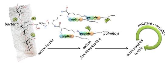
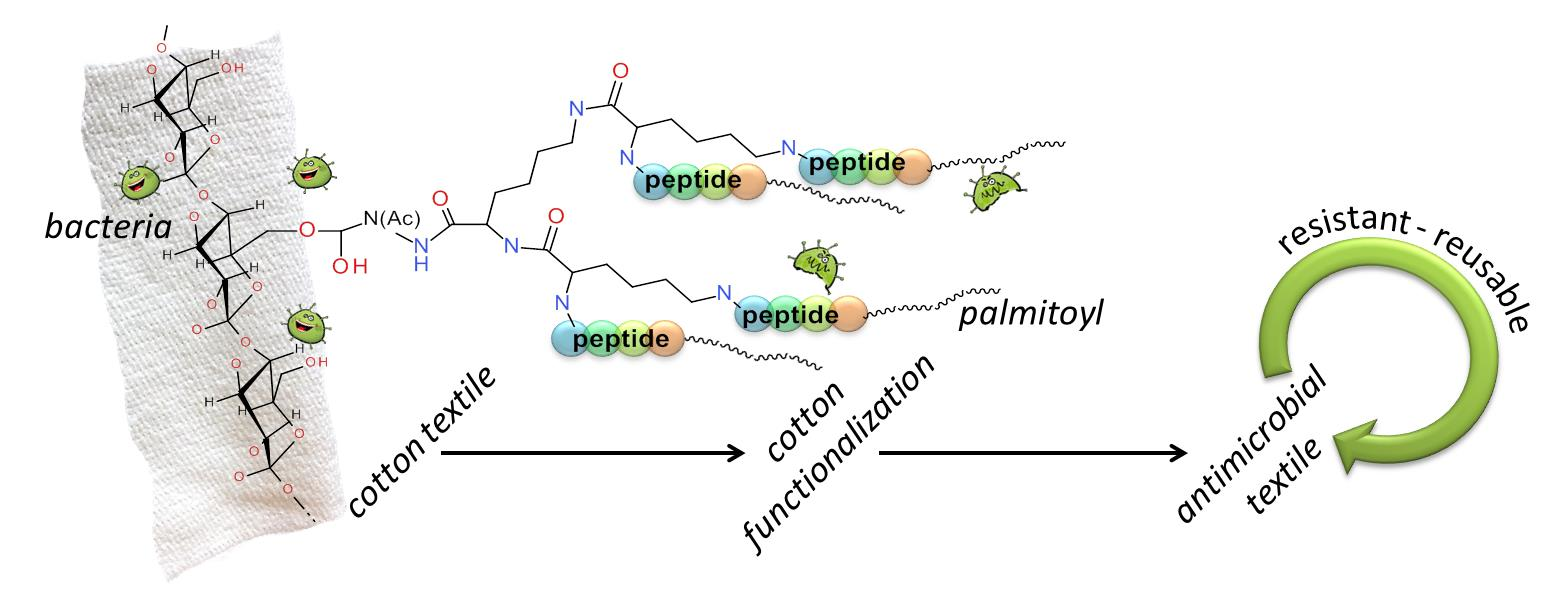
Covalent Graft of Lipopeptides and Peptide Dendrimers to Cellulose Fibers

1
Department of Chemistry, University of Padova, 35131 Padova, Italy
2
ICB CNR, Padova Unit, Department of Chemistry, University of Padova, 35131 Padova, Italy
3
Faculty of Medicine, “Lucian Blaga” University of Sibiu, 550024 Sibiu, Romania
4
Faculty of Engineering, “Lucian Blaga” University of Sibiu, 550024 Sibiu, Romania
5
Department of Agricultural Sciences and Food Engineering, “Lucian Blaga” University of Sibiu, 550024 Sibiu, Romania
*
Author to whom correspondence should be addressed.
Coatings 2019, 9(10), 606; https://doi.org/10.3390/coatings9100606
Submission received: 26 August 2019 / Revised: 16 September 2019 / Accepted: 19 September 2019 / Published: 24 September 2019
(This article belongs to the Special Issue Surface Modification of Cellulose Fibres)

Abstract

:
Introduction: Bacterial proliferation in health environments may lead to the development of specific pathologies, but can be highly dangerous under particular conditions, such as during chemotherapy. To limit the spread of infections, it is helpful to use gauzes and clothing containing antibacterial agents. As cotton tissues are widespread in health care environments, in this contribution we report the preparation of cellulose fibers characterized by the covalent attachment of lipopeptides as possible antimicrobial agents. Aim: To covalently link peptides to cotton samples and characterize them. Peptides are expected to preserve the features of the fabrics even after repeated washing and use. Peptides are well tolerated by the human body and do not induce resistance in bacteria. Materials and Methods: A commercially available cotton tissue (specific weight of 150 g/m2, 30 Tex yarn fineness, fabric density of 270/230 threads/10 cm in the warp and weft) was washed with alkali and bleached and died. A piece of this tissue was accurately weighed, washed with methanol (MeOH) and N,N-dimethylformamide (DMF), and air-dried. Upon incubation with epibromohydrin, followed by treatment with Fmoc-NH-CH2CH2-NH2 and Fmoc removal, the peptides were synthesized by incorporating one amino acid at a time, beginning with the formation of an amide bond with the free NH2 of 1,2–diaminoethane. We also linked to the fibers a few peptide dendrimers, because the mechanism of action of these peptides often requires the formation of clusters. We prepared and characterized seven peptide-cotton samples. Results: The new peptide-cotton conjugates were characterized by means of FT-IR spectroscopy and X-ray Photoelectron Spectroscopy (XPS). This latter technique allows for discriminating among different amino acids and thus different peptide-cotton samples. Some samples maintain a pretty good whiteness degree even after peptide functionalization. Interestingly, these samples also display encouraging activities against a Gram positive strain. Conclusions: Potentially antimicrobial lipopeptides can be covalently linked to cotton fabrics, step-by-step. It is also possible to build on the cotton Lys-based dendrimers. XPS is a useful technique to discriminate among different types of nitrogen. Two samples displaying some antibacterial potency did also preserve their whiteness index.

Graphical Abstract

1. Introduction

Most textiles are characterized by large surfaces that favor microorganism growth under specific moisture and temperature conditions. This feature is particularly harmful in health environments. The bacterial proliferation can promote specific allergies and pathologies and represent a serious concern for patients with low immune defenses.
To impart antimicrobial characteristics to textiles, different approaches were studied and applied. An easy and common solution encompasses absorption and trapping of an antimicrobial agent in the tissue texture or, on occasion, its grafting onto the surface of polymeric fibers [1,2,3,4,5,6,7,8,9]. Common antimicrobial agents used so far include quaternary ammonium compounds, trichlosan, diclofenac, metal salts, silver compounds, or even natural polymers [1,10,11,12,13,14,15]. Inorganic nanoparticles were also exploited to impart antimicrobial activity [7,8,16,17]. However, inorganic salts and nanoparticles rarely give a durable antimicrobial effect to textiles as they are rapidly released during washing and usage.
The lack of a covalent bond with the fabric is the main cause of this drawback. As a consequence, textile deterioration, odor appearance, and color degradation may occur. Occasionally, the antimicrobial agents released may cause allergic responses and potential health risks. Besides being effective against microorganisms, antimicrobial textiles must be non-toxic to the users and should hamper the development of resistant bacteria. They should also be safe for the environment. To meet these issues, many efforts have been devoted to creation of textiles based on natural products, not prone to release metal particles during their production or use [3,18,19].
In this context, we recently started exploring methods to covalently link antimicrobial peptides to a cotton textile. Peptides are expensive molecules, but they have a few advantages over other types of molecules: they are made of natural moieties (α-amino acids), they are biodegradable, and they have negligible toxicity when exploited for external uses.
Cotton was chosen for its widespread use in textiles and in health care environments. In addition, the cellulose fibers can be easily modified to allow covalent peptide attachment or even peptide synthesis directly on its surface. Peptides can be simply adsorbed on cotton through electrostatic interactions, but without covalent bonding they are easily lost [20,21].
Covalent grafting of molecules on cotton fibers has been achieved in different ways [14,22,23,24]. A widely exploited strategy foresees the formation of ester bonds with the naturally occurring hydroxyl group of cellulose [25,26,27,28,29]. However, esters are not very stable at acidic or basic pH or in the presence of hydrolytic enzymes. Therefore, also in this contribution we carried out the covalent attachment of peptides to cellulose through an ether bond [30,31], by exploiting and improving a previous literature report [32]. The free hydroxyl groups of cellulose are directly functionalized, avoiding any oxidation or reduction reaction.
We chose to functionalize our cotton samples with antimicrobial peptides (AMPs) [33,34,35,36] as they are effective against bacteria and some of them also against viruses, fungi, and cancer cells [37]. Many AMPs are helical and amphipathic [36,38,39,40,41,42,43], bearing cationic side chains able to interact with the negatively charged phospholipid membranes. A peculiar class of helical AMPs is represented by lipopeptides that are non-ribosomally produced in bacteria, yeasts, and fungi. The peptide backbone is composed by 6–10 amino acids, linked to an aliphatic acid at the N-terminal end. It was shown that the lipophilic tail is essential for the antimicrobial activity as it elongates these short peptides [44]. Conjugation of aliphatic chains to the N-terminus of short AMPs gives the peptides the capability to perturb a phospholipid bilayer and, in particular, to be selective [44,45,46,47]. This result is rather surprising as some members of this family are composed of as few as four amino acid residues. Nevertheless, these peptides permeate and disrupt membranes with the same potency of many longer AMPs [48,49].
In view of potential applications for the peptide-cotton conjugates, and therefore of a large-scale preparation, in this work we focused on these short lipopeptides as their synthesis is easier and less expensive. We built the peptides directly onto the cotton surface. After derivatization of the cellulose hydroxyl groups, cotton was used as the substrate for the ‘solid phase’ synthesis of peptides (SPPS) [30,41,50]. According to the same procedure, we also built Lys-based dendrimers [51,52,53] of the same peptides as their mechanism of membrane disruption requires the formation of clusters. In these constructs, tree-like peptides protrude from a central core linked to the cotton fiber, thus increasing the local peptide concentration. This approach allows to compensate for the absence of mobility of the peptides, not free to migrate and to form clusters as in solution.
Our new materials were analyzed by means of X-ray Photoelectron Spectroscopy (XPS), a technique that proved to be very informative for these peptide-cotton conjugates. Indeed, it was possible to spot on the fibers and distinguish among Lys, His, and Arg. The influence of the peptide anchoring on the whiteness of the cotton was also examined. Encouraging results were obtained also from a preliminary antibacterial screening. The following flowchart summarizes the work performed (Figure 1).

2. Materials and Methods

2.1. Chemicals and Materials

Fmoc and Boc (Fmoc—fluorenyl-9-methoxycarbonyl, Boc—tert-butyloxycarbonyl) protected amino acids were Bachem products (Bubendorf, Switzerland). Arg(NO2) was purchased from Iris Biotech GmbH (Marktredwitz, Germany) and protected with Fmoc group at the NH2 following a standard protocol [54]. All other chemicals and reagents for the peptide synthesis were purchased from Sigma-Aldrich (St. Louis, MO, USA).
The cotton used in our experiments is a commercially available tissue, ready for garment manufacture. It has a specific weight of 150 g/m2, 30 Tex yarn fineness, a fabric density of 270/230 threads/10 cm in the warp and weft. The fabric was prepared by a process encompassing alkali treatment, bleaching and mercerization, followed by conditioning under standard atmosphere for 24 h.

2.2. Cotton Functionalization

The peptide-cotton samples were prepared according to a protocol previously set up in our laboratory [22,30]. Briefly, for each sample, a piece of cotton fabric of about 2 cm2 was accurately weighed, washed with methanol (MeOH), N,N-dimethylformamide (DMF), MeOH and finally air-dried. The cotton piece (about 0.3 g) was incubated for 4 h with epibromohydrin (1 mL) and a 60% HClO4 solution in DMF (0.1 mL). Then, the sample was washed with DMF, MeOH, and air-dried. This brominated cotton fiber was then soaked into a solution of Fmoc-NH-CH2CH2-NH2 (5 mmol) and triethylamine (5 mmol) in DMF (2 mL) for 12 h. Next, the functionalized cotton-samples were treated for 2 h with a 20% solution of Ac2O in DMF (10 mL) in the presence of triethylamine (1 mL). Fmoc removal was achieved by treatment with a 20% piperidine solution in DMF. The samples were washed with DMF and MeOH and air-dried.

2.3. Peptide Synthesis on the Functionalized Cotton

Amine-functionalized cotton samples of 0.3 g were dunked in DMF. For samples 1, 2, and 3′ (Table 1) the peptides were built step by step by adding the required Fmoc amino acid according to a previously described protocol [30]. For each coupling, we used DMF solutions containing 1.5 equivalents (with respect to the amine functions on the cotton) of Fmoc-Ala-OH, Fmoc-D-Ala-OH, Fmoc-Lys(Boc)-OH, Fmoc-His(Boc)-OH, Fmoc Arg(NO2) OH and palmitic acid (Palm-OH) and of the activation reagents [2-(1H)-7-aza-1,2,3-benzotriazolyl]-1,1,3,3-tetramethyluronium hexafluorophosphate (HATU) and 7-aza-1-hydroxy-1,2,3-benzotriazole (HOAt). At every coupling step 4.5 equivalents of diisopropylethylamine (DIPEA) were also employed. The coupling reactions proceeded for 3 h. Fmoc removal was carried out by treatment with a 20% piperidine solution in DMF (2 × 15 min). Removal of the NO2 protecting group of Arg, to give compound 3, was achieved by catalytic hydrogenation in MeOH in the presence of 10% Pd/C.
For dendrimers 1a, 2a, and 3a, Fmoc-Lys(Fmoc)-OH (1.5 eq.) was first linked to the functionalized cotton by activation with HATU/HOAT (1.5 eq. each) and DIPEA (4.5 eq.) in DMF. After Fmoc removal, a double amount of Fmoc-Lys(Fmoc)-OH (3.0 eq.) was used for a second coupling. Finally, after Fmoc deprotection four amino groups per peptide chain were available. Therefore, the peptide elongation continued on the cotton as described for 1, but by using quadruple quantities of each reagent.
Coupling completeness and Fmoc deprotection were checked at each reaction step by performing the Kaiser test [55]. To this aim, a thread of cotton was washed with DMF, MeOH, and acetone, dried and treated with the three monitor solutions of the Kaiser test, while heating at 100 °C for 5 min. For yield evaluation, the Fmoc removal solution was fully recovered and its UV absorption at 301 nm was recorded by means of a Shimadzu model UV-2501 spectrophotometer (Shimadzu, Kyoto, Japan). At this wavelength it is possible to quantify the amount of dibenzofulvene released upon Fmoc removal.

2.4. Characterization of the Peptide-Cotton Samples by X-Ray Photoelectron Spectroscopy (XPS)

High-resolution XPS spectra for unmodified cotton and for the peptide-functionalized cotton fibers were measured in order to estimate the nitrogen percentage in the samples. We used a Perkin-Elmer Φ 5600ci instrument operating with Al-Kα radiation (1486.6 eV) at 350 W. The working pressure was <5 × 10⁻⁸ Pa (~10−11 torr). The calibration was based on the binding energy (BE) of the Au 4f7/2 line at 83.9 eV with respect to the Fermi level. The standard deviation for the BE values was 0.15 eV. The reported BEs were corrected for charge effects, assigning the BE value of 284.6 eV to the C 1s line of carbon. Survey scans were obtained in the 0–1350 eV range (pass energy 187.5 eV, 1.0 eV/step, 25 ms/step). Detailed scans (29.35 eV pass energy, 0.1 eV/step, 50–150 ms/step) were recorded for the O 1s, C 1s, N 1s regions. The atomic composition, after a Shirley background subtraction, was evaluated using sensitivity factors supplied by Perkin-Elmer. Peak assignment was carried out according to literature data.

2.5. Evaluation of the Whiteness of the Functionalized Cotton Samples

The whiteness of the peptide-functionalized cotton samples was checked using the Datacolor 110 LAV reflection spectrophotometer at standard illuminant/observer D65-10, according to CIE method of ASTM E313 [56] that defines the whiteness and yellowness indices. Whiteness is defined as a measure of how closely a surface matches the properties of a perfect reflecting diffuser, i.e., an ideal reflecting surface that neither absorbs nor transmits light, but reflects it at equal intensities in all directions. The yellowness index, obtained from spectrophotometric data, describes the change in color of a sample from clear or white to yellow. Color studies were quantified using the CIELAB system. The results are expressed as Berger, CIE, and yellowness index in accordance with the illuminant/observer D65/10°.

2.6. Antimicrobial Activity

The antimicrobial effect of the peptide-functionalized cotton fibers was investigated against standard bacterial strains of Gram-negative (Escherichia coli ATCC 25922) and Gram-positive (Staphylococcus aureus ATCC 25923) bacteria. A small piece of functionalized cotton was placed in a test tube containing the bacterial inoculum suspended in physiological solution, diluted 1:200 in the Mueller–Hinton liquid culture medium. An untreated cotton sample was used as control. All cotton samples were subjected to sterilization at 120 °C for 15 min before starting the tests. The antibacterial activity was evaluated by measuring the bacterial cell concentration in suspension after incubation at 37 °C over a 6-day period, using the McFarland standards and the Densimat photometer (BioMérieux, Craponne, France). Tests were performed after 1, 3, and 6 days of incubation. The results are expressed as cell density to approximate their concentration in suspension (CFU/mL). The initial inoculum concentration was 0.5 McFarland, corresponding to 1–2 × 10⁸ CFU/mL. The decrease of turbidity indicates an antimicrobial effect [16].

3. Results and Discussion

3.1. Peptide Design

In this study we focused on the peptide sequence Pal-X-Ala-D-Ala-X [44,57] and applied three variations to the cationic residues X, namely Lys, His, and Arg (Table 1). Cationic residues are fundamental to impart antimicrobial properties even to small peptides [24,48]. Lys, Arg, and His are all protonated under physiological conditions. Thus, their positive charge enables the peptides to bind to the negatively charged bacteria membranes, inducing cell death. On the other hand, Arg, Lys, and His are characterized, respectively, by decreasing pKa values that allow to finely tune the AMP-membrane interaction and, possibly, the selectivity towards different bacterial membranes. Moreover, the Arg guanidinium group enables the formation of multiple H-bonds, thus promoting a better binding to the membranes. Moreover, Arg-rich peptides are able to perturb positively charged membranes as well, as the cationic character of Arg is maintained in both acidic and alkaline environments [38,58,59].
During the synthesis we chose to have the nitro (NO2) moiety to protect the Arg side-chain. This preference came from the finding that the nitro group often increases the ability of bioactive molecules to interact with bacterial membranes [60]. Therefore, we had the possibility to test the antimicrobial activity of both conjugate 3 and 3′.
In general, peptides made of all-L amino acids are highly hemolytic, loose their activity in serum, and are fully cleaved by proteolytic enzymes. On the contrary, peptide diastereoisomers containing D residues are non-hemolytic and maintain full activity in serum [61]. This finding inspired the insertion of a D-Ala residue at position 3 of all our peptides.
The palmitoyl chain (C16) at the N-terminus of the hydrophilic peptide generates a hydrophobic region. Among the different lipid chains found in natural or artificial peptides, C16 is one of the most effective in promoting the interaction of short peptides with membranes. Very likely, the hydrophobic interactions of nonpolar, amino acid side-chains and the core of the lipid bilayers is reinforced when a lipid chain is linked to the peptide.
Finally, a behavior frequently observed in the AMPs action is cooperativity, i.e., a minimum threshold concentration is required to boost the membrane perturbing activity. This phenomenon is usually associated with the formation of clusters responsible for membrane disruption, although through different mechanisms (e.g., pores, rafts, micelles). As our peptides are covalently bound to the cotton fibers, they cannot freely move and thus form clusters. Therefore, they may be inactive if peptide aggregates are necessary to perturb the membranes. We then decided to increase the local peptide concentration on the textile surface to allow possible interactions between closely located peptide chains. To this aim, we synthesized dendrimeric peptides on the cotton fabrics, by exploiting a Lys residue with both amino groups protected by Fmoc. After a one-step Fmoc deprotection, a second coupling with a bis-Fmoc protected Lys originated a tetradentate core on which four peptide chains could be grown step-by step.

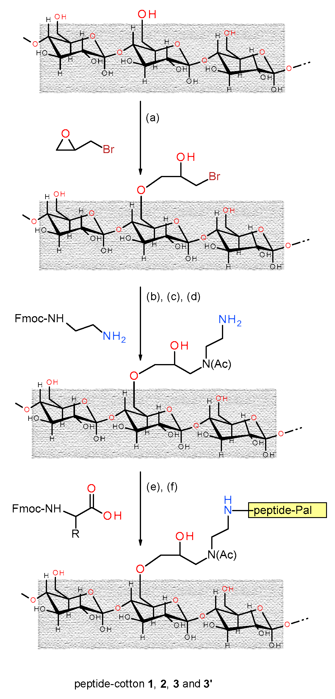
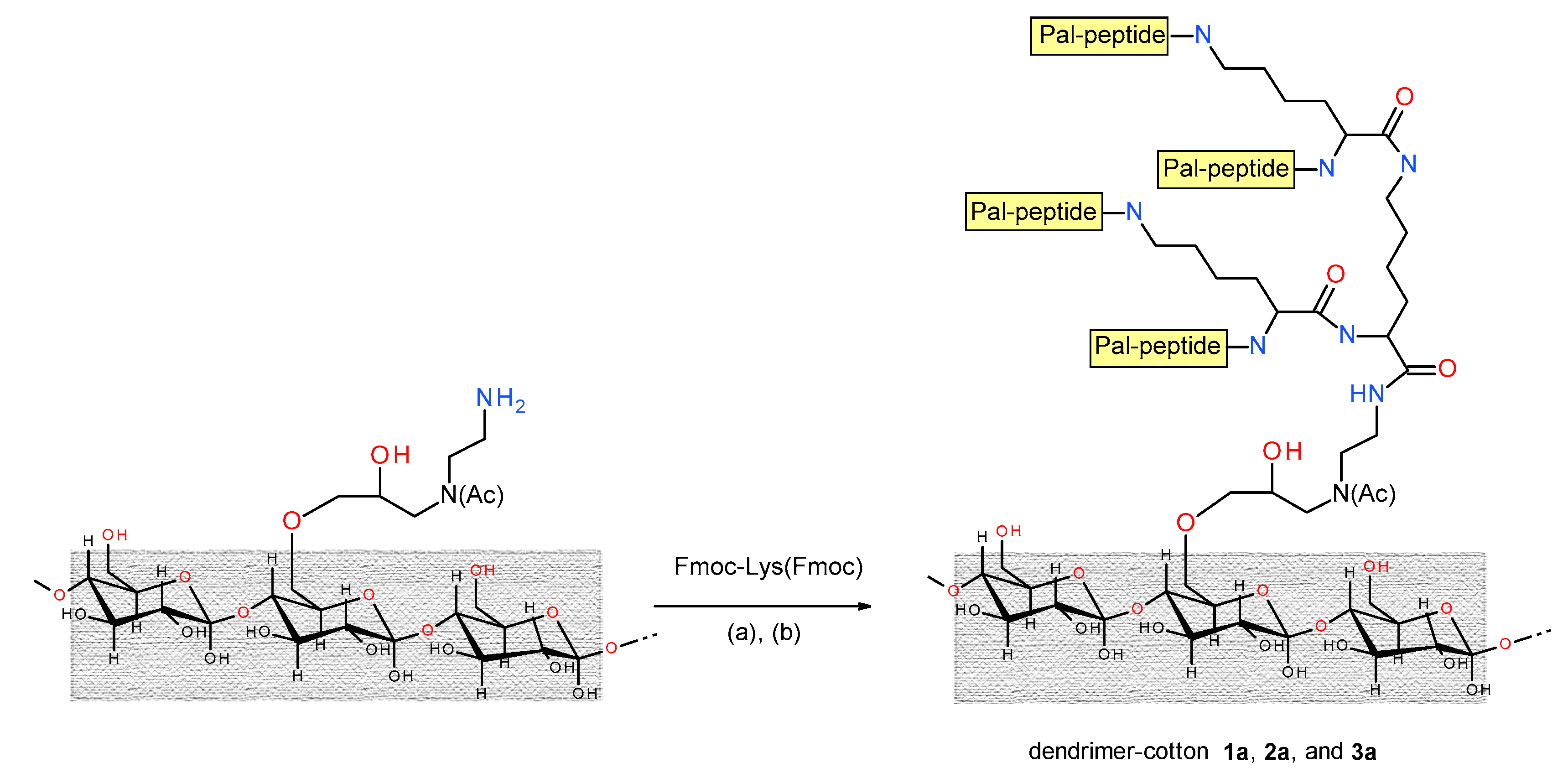
3.2. Synthesis of the Peptide-Cotton Samples

The linear peptides and the Lys-based dendrimers (Table 1) were built directly on the cotton. To allow an efficient peptide anchorage, the cotton surface was appropriately modified according to a protocol already described (Scheme 1) [30,32]. First, the solvent exposed, hydroxy groups of cellulose open the epoxide of epibromohydrin in the presence of HClO4. A stable ether bond cotton forms and a brominated alkane is introduced at the same time. Then, bromine is substituted upon reaction with Fmoc-NH-CH2CH2-NH2. Finally, the acetylation step blocks the newly formed secondary amine in order to avoid the growth of an undesired peptide chains. These synthetic steps allow for introducing a spacer and functionalizing the cotton with an amino group. The peptide is then built on this modified cotton surface according to standard solid-phase peptide synthesis (SPPS) procedures (see Section 2.3). Analogously, the Lys-based dendrimers are built on the NH2-functionalized cotton, as described in Scheme 2. By this procedure, amino acids and peptides are bound to cotton through a very stable amide bond. The alternative, direct anchorage of the peptides to the cotton OH groups generates an easy-to-hydrolyze ester bond. Therefore, the peptide chains would be easily lost on wearing, because of the presence of hydrolytic enzymes leaching from the skin, and upon garment sterilization.
For an effective amide bond formation, Fmoc-amino acids and palmitic acid activation was carried out with the HATU/HOAT reagent combination. Longer reaction times (3 h) were used, with respect to the classical SPPS, to allow for an efficient diffusion of the reagents inside the fibers. Indeed, we recently showed that cotton functionalization does occur also inside the fibers, not only on their external surface [31]. In general, an excess of 1.5 equivalent of acylating reagents, with respect to the amine function on the cotton, was sufficient to achieve efficient couplings. Obviously, in the case of the dendrimers the excess was increased by two or four times, to take into account the multiple number of amino groups developing on the Lys core. Indeed, we performed two consecutive couplings (in single and double quantities, respectively) with the doubly N-protected derivative Fmoc-Lys(Fmoc)-OH, in order to exploit both the α- and ε-amino groups of Lys to grow peptide chains. The side-chain protecting groups (Boc or NO2) were removed as the last step.

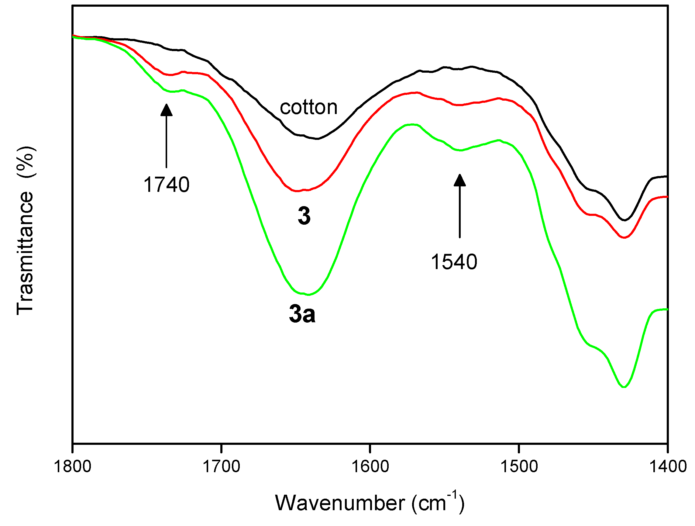
3.3. Peptide Loading Determination and FT-IR Absorption Analysis
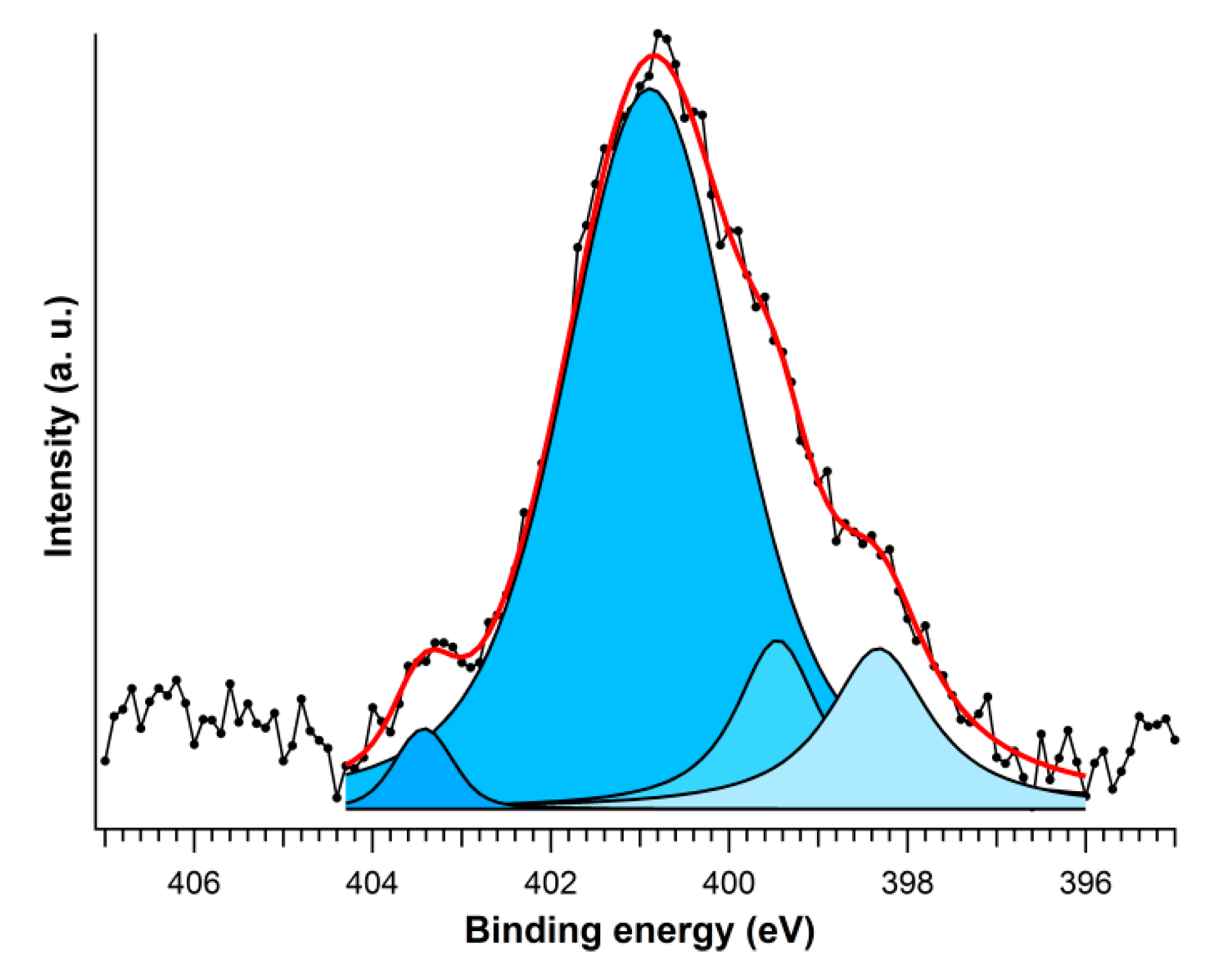
The loading values of the peptide bound to the cotton fibers are reported in Table 1. For 1, 2, 3, and 3′ we observe values ranging from to 0.60 to 0.76 mmol/gcotton. For the dendrimers, we need to take into account the number of peptide units. The value recorded for dendrimer 2a is slightly lower than expected (2.12 instead of 2.52). We tentatively attribute this result to a diminished coupling yield caused, in turn, by the close proximity of four peptide chains that contain the bulky His(Boc) residue.
Additionally, the FT-IR absorption spectroscopy is helpful to assess the anchoring of the peptides to the cotton surface. Informative bands are listed in Table 1. We compared the FT-IR spectrum of pure cotton (fibers on KBr pellet) with those of the cotton-peptide samples 1, 2, and 3 and of the cotton-dendrimer samples 1a, 2a, and 3a. As a representative example, Figure 2 reports the FT-IR spectra of pure cotton and of samples 3 and 3a in the 1800–1500 cm−1 region. The presence of the peptide is revealed by the amide I band (C=O stretching) near 1650 cm−1 and the amide II band (C–N stretching and N–H bending) at about 1540 cm−1 [62]. These bands, although relatively weak and broad, are clearly observed for 3 and 3a, but they are weaker (amide I) or completely missing (amide II) in the pure cotton, thus implying that the peptides are definitely linked to the fibers.
As expected, the intensities of the amide I and amide II absorptions are higher for the cotton functionalized with the dendrimer (3a). The absorption band at 1740 cm−1, not observed for the pure cotton, is likewise due to the carbonyl stretching of esters that may form, on reactive alcoholic functions, in the acetylation step.

3.4. XPS Analysis

XPS analysis on our samples proved to be very useful for the identification and quantification of nitrogen. Indeed, the nitrogen atoms should be missing in a cellulose matrix, but are expected to be present in the cotton functionalized with peptides. The results of the quantitative XPS analyses are presented in Table 2. As expected, nitrogen is virtually absent in the non-functionalized cotton (0.4%). At variance, in the peptide-functionalized samples, the percentage of nitrogen increases from 1.6% for the sample containing only the amino linker, -N(Ac)-CH2CH2-NH2, to 4%–5% for 1, 2, and 3, and to 6.3% for 3′, containing two NO2 protecting groups on the Arg residues. For the dendrimers, this percentage increases further (7%–9%).
Interestingly, the XPS analysis allows also for studying the N 1s core levels. The N 1s region, centered in the binding energy interval 405–395 eV, is not influenced by the structure of the cotton matrix. Therefore, by analyzing this region we could discriminate among samples 13′ and, in particular, we could identify the presence of a particular peptide on the basis of the different types of bonds (hybridization) in which the nitrogen atoms are involved. To this aim, we performed a deconvolution analysis of the spectra of 1, 2 (Figure 3), and 3′ according to literature data [63]. The values calculated and the assignment to a specific nitrogen type are summarized in Table 3.
In all samples we observe the typical band of the amide bond (400.6–400.9 eV). At about 401.5 eV we find the amine nitrogen, attributable for instance to the side chain of Lys in sample 1. It was then possible to assign the side chains for the three different cationic amino acids (Lys, His, and Arg), as detailed in Table 3. It is worth noting that we can even discriminate between the two nitrogen atoms of the His imidazole. They exhibit N 1s BE values at 398.3 and 399.5 eV, in agreement with previously reported data [63]. The values for sample 3′ reflect the guanidinium chemical environment of Arg. Therefore, we believe that XPS spectroscopy may become a valuable tool to investigate peptide-containing solid samples, not easily accessible by other techniques.

3.5. Color Change and Antimicrobial Activity

The whiteness and yellowness indexes are quality parameters used to evaluate the change in whiteness in fabrics of any composition after different types of treatments [15,64]. Functionalization of cotton, in particular with antimicrobial agents, could modify the color properties of the textile and affect also further coloring, if required. We then performed an analysis of the color changes upon cotton functionalization on selected peptide-cotton samples. The results are shown in Table 4.
The variations observed for the functionalized textiles, as compared to an untreated sample (cotton), indicate for 1 and 3 an increased yellow degree, whereas 2 and 2a are little affected. These latter maintain also a pretty good whiteness degree. This finding is very promising for possible applications, as samples 2 and 2a, which also showed antibacterial properties, can be further dyed without major changes of the intended color. Indeed, a preliminary screening against two standard bacterial strains of medical interest (Staphylococcus aureus and Escherichia coli) revealed an antibacterial effect for 2 on both strains. Some antistaphylococcal activity was also observed for samples 2a and 3′, whereas all the other peptide-cotton samples did not exhibit significant antibacterial effects. Interestingly, both samples 2 and 2a contain His, whereas the other peptides bear Arg of Lys. Thus, this preliminary antibacterial analysis suggests that His is the cationic residue of choice for these peptide-cellulose conjugates. Some activity was observed also when the guanidinium group of Arg (sample 3′) was nitro-protected, thus resulting in a diminished basic strength. This feature, in turn, offers a hint to explain the antibacterial behavior observed. The pKa of the side chains of His, Lys, and Arg are, respectively, 6.1, 10.5, and 12.5. An excess of basic strength appears to be detrimental. Indeed, when the pKa of the guanidinium group of Arg is lowered by introducing the nitro group some activity is recovered.
Finally, it is worth noting that all cotton samples were thoroughly washed with DMF and MeOH and subjected to sterilization at 120 °C for 15 min before starting the antibacterial tests (see Section 2.6). As the antimicrobial activity was observed only for cotton samples functionalized with peptides (not for the cotton sample used as reference), we conclude that the peptides are tightly bound to the cellulose by virtue of the connecting covalent bonds. In addition, after the first antibacterial test the most active sample 2 was again sterilized and submitted to a second antimicrobial test. Only a slight decrease in activity was observed, thus indicating that this approach may lead to the development of re-usable antibacterial garments.

4. Conclusions

Short, potentially antimicrobial lipopeptides were successfully linked to cotton fabrics. The peptides were directly built step-by-step on the appropriately functionalized cotton samples. To increase the peptide concentration on the cotton surface, expected to favor the antibacterial activity, we also functionalized the cotton with Lys-based dendrimers. FT-IR measurements confirmed the presence of the peptides on all functionalized samples and quantification was achieved by a UV method. XPS analysis allowed us to detect different types of nitrogen atoms and thus to discriminate among different peptide-cotton samples. Indeed, upon deconvolution of the N1s spectra it was possible to detect the three cationic amino acids present in the investigated peptides.
The functionalized cotton samples were also investigated for the degree of color alteration caused by peptide binding. Interestingly, it was found that two samples displaying some antibacterial potency did also preserve their whiteness index.
To summarize, our strategy offers two main advantages: (i) the covalently bound, antibacterial peptides are not lost upon wearing or sterilization procedures; (ii) they act by changing the membrane permeability with a “physical action”, without altering their nature, thus they are ready to act again after, for instance, a sterilization treatment.

Author Contributions

A.O. synthesized the peptide-cotton conjugates; P.D. performed the XPS measurements and analyses; B.B. contributed to and supervised the spectroscopic analyses; G.H. and S.O. designed and carried out the antibacterial tests; D.C. performed the test on color stability. C.P., S.O. and F.F. conceived the original idea, supervised the peptide design and wrote the manuscript. All authors, according to their expertise, discussed the results and offered hints for the final editing of the manuscript.

Funding

This project was supported by the University of Padova through a grant to CP (PRAT N° UniPD CPDA150532/15) and by the Romanian and Italian authorities that sponsored the Exchange Project RO13MO6/M00301 on the development of antibacterial textiles. Partial support was given also by the Italian Ministry of Education, Universities and Research through grant PRIN 20157WW5EH.

Acknowledgments

The authors wish to thank Dr. Renato Schiesari for the FT-IR measurements.

Conflicts of Interest

The authors declare no conflict of interest. The funders had no role in the design of the study; in the collection, analyses, or interpretation of data; in the writing of the manuscript, or in the decision to publish the results.

References

  1. Gao, Y.; Cranston, R. Recent advances in antimicrobial treatments of textiles. Text. Res. J. 2008, 78, 60–72. [Google Scholar] [CrossRef]
  2. Morais, D.S.; Guedes, R.M.; Lopes, M.A. Antimicrobial approaches for textiles: From research to market. Materials 2016, 9, 498. [Google Scholar] [CrossRef] [PubMed]
  3. Tawiah, B.; Badoe, W.; Fu, S.H. Advances in the development of antimicrobial agents for textiles: The quest for natural products. Review. Fibres Text. East Eur. 2016, 24, 136–149. [Google Scholar] [CrossRef]
  4. Wang, W.Y.; Hui, P.C.L.; Kan, C.W. Functionalized textile based therapy for the treatment of atopic dermatitis. Coatings 2017, 7, 82. [Google Scholar] [CrossRef]
  5. Zhou, C.E.; Kan, C.W.; Matinlinna, J.P.; Tsoi, J.K.H. Regenerable antibacterial cotton fabric by plasma treatment with dimethylhydantoin: Antibacterial activity against S. aureus. Coatings 2017, 7, 11. [Google Scholar] [CrossRef]
  6. Cardoso, V.; Rittmeyer, T.; Correa, R.J.; Brêda, G.C.; Almeida, R.V.; Simões, G.; de França, B.M.; de Azevedo, P.N.; Forero, J.S.B. Photoactive cotton fabric: Synthesis, characterization and antibacterial evaluation of anthraquinone-based dyes linked to cellulose. Dye. Pigment. 2019, 161, 16–23. [Google Scholar] [CrossRef]
  7. Marković, D.; Deeks, C.; Nunney, T.; Radovanović, Ž.; Radoičić, M.; Šaponjić, Z.; Radetić, M. Antibacterial activity of Cu-based nanoparticles synthesized on the cotton fabrics modified with polycarboxylic acids. Carbohyd. Polym. 2018, 200, 173–182. [Google Scholar] [CrossRef]
  8. Yaghoubidoust, F.; Salimi, E. A simple method for the preparation of antibacterial cotton fabrics by coating graphene oxide nanosheets. Fiber Polym. 2019, 20, 1155–1160. [Google Scholar] [CrossRef]
  9. Stular, D.; Sobak, M.; Mihelcic, M.; Sest, E.; Ilic, I.G.; Jerman, I.; Simoncic, B.; Tomsic, B. Proactive release of antimicrobial essential oil from a “smart” cotton fabric. Coatings 2019, 9, 242. [Google Scholar] [CrossRef]
  10. Gugliuzza, A.; Drioli, E. A review on membrane engineering for innovation in wearable fabrics and protective textiles. J. Membr. Sci. 2013, 446, 350–375. [Google Scholar] [CrossRef]
  11. Kenawy, E.-R.; Worley, S.D.; Broughton, R. The chemistry and applications of antimicrobial polymers: A state-of-the-art review. Biomacromolecules 2007, 8, 1359–1384. [Google Scholar] [CrossRef] [PubMed]
  12. Monteiro, D.R.; Gorup, L.F.; Takamiya, A.S.; Ruvollo-Filho, A.C.; de Camargo, E.R.; Barbosa, D.B. The growing importance of materials that prevent microbial adhesion: Antimicrobial effect of medical devices containing silver. Int. J. Antimicrob. Agents 2009, 34, 103–110. [Google Scholar] [CrossRef] [PubMed]
  13. Stobie, N.; Duffy, B.; Hinder, S.J.; McHale, P.; McCormack, D.E. Silver doped perfluoropolyether-urethane coatings: Antibacterial activity and surface analysis. Colloid Surf. B 2009, 72, 62–67. [Google Scholar] [CrossRef] [PubMed] [Green Version]
  14. Cassano, R.; Trombino, S.; Ferrarelli, T.; Barone, E.; Arena, V.; Mancuso, C.; Picci, N. Synthesis, characterization, and anti-inflammatory activity of diclofenac-bound cotton fibers. Biomacromolecules 2010, 11, 1716–1720. [Google Scholar] [CrossRef] [PubMed]
  15. Dhiman, G.; Chakraborty, J.N. Antimicrobial performance of cotton finished with triclosan, silver and chitosan. Fash. Text. 2015, 2, 13. [Google Scholar] [CrossRef]
  16. El-Nahhal, I.M.; Zourab, S.M.; Kodeh, F.S.; Selmane, M.; Genois, I.; Babonneau, F. Nanostructured copper oxide-cotton fibers: Synthesis, characterization, and applications. Int. Nano Lett. 2012, 2, 14. [Google Scholar] [CrossRef]
  17. El-Nahhal, I.M.; Zourab, S.M.; Kodeh, F.S.; Elmanama, A.A.; Selmane, M.; Genois, I.; Babonneau, F. Nano-structured zinc oxide-cotton fibers: Synthesis, characterization and applications. J. Mater. Sci. Mater. Electron. 2013, 24, 3970–3975. [Google Scholar] [CrossRef]
  18. Ayyoob, M.; Khurshid, M.F.; Asad, M.; Shah, S.N.H. Assessment of eco-friendly natural antimicrobial textile finish extracted from aloe vera and neem plants. Fibres Text. East Eur. 2015, 23, 120–123. [Google Scholar] [CrossRef]
  19. Shahid-ul-Islam; Mohammad, F. Natural colorants in the presence of anchors so-called mordants as promising coloring and antimicrobial agents for textile materials. Acs Sustain. Chem. Eng. 2015, 3, 2361–2375. [Google Scholar] [CrossRef]
  20. Gomes, A.P.; Mano, J.F.; Queiroz, J.A.; Gouveia, I.C. Layer-by-layer deposition of antimicrobial polymers on cellulosic fibers: A new strategy to develop bioactive textiles. Polym. Adv. Technol. 2013, 24, 1005–1010. [Google Scholar] [CrossRef]
  21. Gomes, A.P.; Mano, J.F.; Queiroz, J.A.; Gouveia, I.C. Incorporation of antimicrobial peptides on functionalized cotton gauzes for medical applications. Carbohydr. Polym. 2015, 127, 451–461. [Google Scholar] [CrossRef] [PubMed] [Green Version]
  22. He, X.-M.; Chen, X.; Yuan, B.-F.; Feng, Y.-Q. Graft modification of cotton with phosphate group and its application to the enrichment of phosphopeptides. J. Chromatogr. A 2017, 1484, 49–57. [Google Scholar] [CrossRef] [PubMed]
  23. Hsu, B.B.; Klibanov, A.M. Light-activated covalent coating of cotton with bactericidal hydrophobic polycations. Biomacromolecules 2011, 12, 6–9. [Google Scholar] [CrossRef] [PubMed]
  24. Ringot, C.; Sol, V.; Barrière, M.; Saad, N.; Bressollier, P.; Granet, R.; Couleaud, P.; Frochot, C.; Krausz, P. Triazinyl porphyrin-based photoactive cotton fabrics: Preparation, characterization, and antibacterial activity. Biomacromolecules 2011, 12, 1716–1723. [Google Scholar] [CrossRef] [PubMed]
  25. Muresan, E.I.; Balan, G.; Popescu, V. Durable hydrophobic treatment of cotton fabrics with glycidyl stearate. Ind. Eng. Chem. Res. 2013, 52, 6270–6276. [Google Scholar] [CrossRef]
  26. Edwards, J.V.; Fontenot, K.R.; Haldane, D.; Prevost, N.T.; Condon, B.D.; Grimm, C. Human neutrophil elastase peptide sensors conjugated to cellulosic and nanocellulosic materials: Part I, synthesis and characterization of fluorescent analogs. Cellulose 2016, 23, 1283–1295. [Google Scholar] [CrossRef]
  27. Edwards, J.V.; Mao, N.; Russell, S.; Carus, E.; Condon, B.; Hinchliffe, D.; Gary, L.; Graves, E.; Bopp, A.; Wang, Y. Fluid handling and fabric handle profiles of hydroentangled greige cotton and spunbond polypropylene nonwoven topsheets. Proc. Inst. Mech. Eng. Part L J. Mater. Des. Appl. 2016, 230, 847–859. [Google Scholar] [CrossRef]
  28. Muresan, E.I.; Piroi, C.; Creanga, D.; Stelea, L.; Oprica, L.; Sandu, I. Glycidyl esters used for multifunctional finishing of textile materials. Rev. Chim. Buchar. 2016, 5, 871–875. [Google Scholar]
  29. Edwards, J.V.; Fontenot, K.R.; Prevost, N.T.; Pircher, N.; Liebner, F.; Condon, B.D. Preparation, characterization and activity of a peptide-cellulosic aerogel protease sensor from cotton. Sensors 2016, 16, 1789. [Google Scholar] [CrossRef] [PubMed]
  30. Orlandin, A.; Formaggio, F.; Toffoletti, A.; Peggion, C. Cotton functionalized with peptides: Characterization and synthetic methods. J. Pept. Sci. 2014, 20, 547–553. [Google Scholar] [CrossRef]
  31. Dzuba, S.A.; Uvarov, M.N.; Utkin, D.E.; Formaggio, F.; Bedon, A.; Orlandin, A.; Peggion, C. The power of EPR techniques in investigating functionalization and penetration into fibers of cotton-bound antimicrobial peptides. Appl. Magn. Reson. 2017, 48, 943–953. [Google Scholar] [CrossRef]
  32. Nakamura, M.; Iwasaki, T.; Tokino, S.; Asaoka, A.; Yamakawa, M.; Ishibashi, J. Development of a bioactive fiber with immobilized synthetic peptides designed from the active site of a beetle defensin. Biomacromolecules 2011, 12, 1540–1545. [Google Scholar] [CrossRef]
  33. Zasloff, M. Antimicrobial peptides of multicellular organisms. Nature 2002, 415, 389–395. [Google Scholar] [CrossRef]
  34. Matsuzaki, K. Control of cell selectivity of antimicrobial peptides. Biochim. Biophys. Acta (BBA)-Biomembr. 2009, 1788, 1687–1692. [Google Scholar] [CrossRef] [Green Version]
  35. Haney, E.F.; Hunter, H.N.; Matsuzaki, K.; Vogel, H.J. Solution NMR studies of amphibian antimicrobial peptides: Linking structure to function? Biochim. Biophys. Acta (BBA)-Biomembr. 2009, 1788, 1639–1655. [Google Scholar] [CrossRef] [Green Version]
  36. Koehbach, J.; Craik, D.J. The vast structural diversity of antimicrobial peptides. Trends Pharmacol. Sci. 2019, 7, 517–528. [Google Scholar] [CrossRef]
  37. Gabernet, G.; Müller, A.T.; Hiss, J.A.; Schneider, G. Membranolytic anticancer peptides. Med. Chem. Commun. 2016, 7, 2232–2245. [Google Scholar] [CrossRef]
  38. Dalzini, A.; Bergamini, C.; Biondi, B.; De Zotti, M.; Panighel, G.; Fato, R.; Peggion, C.; Bortolus, M.; Maniero, A.L. The rational search for selective anticancer derivatives of the peptide trichogin GA IV: A multi-technique biophysical approach. Sci. Rep. 2016, 6, 24000. [Google Scholar] [CrossRef]
  39. De Zotti, M.; Biondi, B.; Peggion, C.; Formaggio, F.; Park, Y.; Hahm, K.-S.; Toniolo, C. Trichogin GA IV: A versatile template for the synthesis of novel peptaibiotics. Org. Biomol. Chem. 2012, 10, 1285–1299. [Google Scholar] [CrossRef]
  40. Formaggio, F.; Peggion, C.; Crisma, M.; Toniolo, C. Short-chain analogues of the lipopeptaibol antibiotic trichogin GA IV: Conformational analysis and membrane modifying properties. J. Chem. Soc. Perkin Trans. 2001, 2, 1372–1377. [Google Scholar] [CrossRef]
  41. Hilpert, K.; Volkmer-Engert, R.; Walter, T.; Hancock, R.E. High-throughput generation of small antibacterial peptides with improved activity. Nat. Biotechnol. 2005, 23, 1008–1012. [Google Scholar] [CrossRef]
  42. Oancea, S.; Hilma, G.; Peggion, C.; Formaggio, F.; Toniolo, C. Main-chain length control of conformation, membrane activity, and antibiotic properties of lipopeptaibol sequential analogues. Chem. Biodivers. 2008, 5, 681–692. [Google Scholar] [CrossRef]
  43. Giangaspero, A.; Sandri, L.; Tossi, A. Amphipathic, α-helical antimicrobial peptides. Eur. J. Biochem. 2001, 268, 5589–5600. [Google Scholar] [CrossRef]
  44. Makovitzki, A.; Avrahami, D.; Shai, Y. Ultrashort antibacterial and antifungal lipopeptides. Proc. Natl. Acad. Sci. USA 2006, 103, 15997–16002. [Google Scholar] [CrossRef] [Green Version]
  45. Makovitzki, A.; Baram, J.; Shai, Y. Antimicrobial lipopolypeptides composed of palmitoyl di- and tricationic peptides: In vitro and in vivo activities, self-assembly to nanostructures, and a plausible mode of action. Biochemistry 2008, 47, 10630–10636. [Google Scholar] [CrossRef]
  46. Young, A.W.; Liu, Z.; Zhou, C.; Totsingan, F.; Jiwrajka, N.; Shi, Z.; Kallenbach, N.R. Structure and antimicrobial properties of multivalent short peptides. Med. Chem. Commun. 2011, 2, 308–314. [Google Scholar] [CrossRef]
  47. Findlay, B.; Zhanel, G.G.; Schweizer, F. Investigating the antimicrobial peptide ‘window of activity’ using cationic lipopeptides with hydrocarbon and fluorinated tails. Int. J. Antimicrob. Agents 2012, 40, 36–42. [Google Scholar] [CrossRef]
  48. Opitakorn, A.; Rauytanapanit, M.; Waditee-Sirisattha, R.; Praneenararat, T. Non-leaching antibacterial cotton fabrics based on lipidated peptides. RSC Adv. 2017, 7, 34267–34275. [Google Scholar] [CrossRef] [Green Version]
  49. Sato, H.; Feix, J.B. Peptide–membrane interactions and mechanisms of membrane destruction by amphipathic α-helical antimicrobial peptides. Biochim. Biophys. Acta (BBA)-Biomembr. 2006, 1758, 1245–1256. [Google Scholar] [CrossRef]
  50. Lebl, M. Solid-phase synthesis on planar supports. Biopolym. (Pept. Sci.) 1998, 47, 397–404. [Google Scholar] [CrossRef]
  51. Tam, J.P. Synthetic peptide vaccine design: Synthesis and properties of a high-density multiple antigenic peptide system. Proc. Natl. Acad. Sci. USA 1988, 85, 5409–5413. [Google Scholar] [CrossRef]
  52. Tam, J.P.; Lu, Y.-A.; Yang, J.-L. Antimicrobial dendrimeric peptides. Eur. J. Biochem. 2002, 269, 923–932. [Google Scholar] [CrossRef]
  53. Kowalczyk, W.; Monsó, M.; de la Torre, B.G.; Andreu, D. Synthesis of multiple antigenic peptides (MAPs)—Strategies and limitations. J. Pept. Sci. 2011, 17, 247–251. [Google Scholar] [CrossRef]
  54. Raillard, S.P.; Mann, A.D.; Baer, T.A. An efficient procedure for the preparation of Fmoc-amino acids. Org. Prep. Proced. Int. 1998, 30, 183–186. [Google Scholar] [CrossRef]
  55. Kaiser, E.; Colescott, R.L.; Bossinger, C.D.; Cook, P.I. Color test for detection of free terminal amino groups in the solid-phase synthesis of peptides. Anal. Biochem. 1970, 34, 595–598. [Google Scholar] [CrossRef]
  56. ASTM E313-00 Standard Practice for Calculating Yellowness and Whiteness Indices from Instrumentally Measured Color Coordinates; ASTM International: West Conshohocken, PA, USA, 2000.
  57. Vallon-Eberhard, A.; Makovitzki, A.; Beauvais, A.; Latge, J.P.; Jung, S.; Shai, Y. Efficient clearance of Aspergillus fumigatus in murine lungs by an ultrashort antimicrobial lipopeptide, palmitoyl-Lys-Ala-DAla-Lys. Antimicrob. Agents Chemother. 2008, 52, 3118–3126. [Google Scholar] [CrossRef]
  58. Strøm, M.B.; Rekdal, Ø.; Svendsen, J.S. Antimicrobial activity of short arginine- and tryptophan-rich peptides. J. Pept. Sci. 2002, 8, 431–437. [Google Scholar] [CrossRef]
  59. Oh, D.; Shirazi, A.N.; Northup, K.; Sullivan, B.; Tiwari, R.K.; Bisoffi, M.; Parang, K. Enhanced cellular uptake of short polyarginine peptides through fatty acylation and cyclization. Mol. Pharmaceut. 2014, 11, 2845–2854. [Google Scholar] [CrossRef]
  60. Gholivand, K.; Dorosti, N. Novel ammonium phosphinates containing peptide moiety: Synthesis, structure, and in vitro antimicrobial activity. Chem. Pap. 2012, 66, 765–771. [Google Scholar] [CrossRef]
  61. Papo, N.; Oren, Z.; Pag, U.; Sahl, H.-G.; Shai, Y. The Consequence of sequence alteration of an amphipathic α-helical antimicrobial peptide and its diastereomers. J. Biol. Chem. 2002, 277, 33913–33921. [Google Scholar] [CrossRef]
  62. Doh, S.; Lee, J.; Lim, D.; Im, J. Manufacturing and analyses of wet-laid nonwoven consisting of carboxymethyl cellulose fibers. Fibers Polym. 2013, 14, 2176–2184. [Google Scholar] [CrossRef]
  63. Stevens, J.S.; de Luca, A.C.; Pelendritis, M.; Terenghi, G.; Downes, S.; Schroeder, S.L.M. Quantitative analysis of complex amino acids and RGD peptides by X-ray photoelectron spectroscopy (XPS). Surf. Interface Anal. 2013, 45, 1238–1246. [Google Scholar] [CrossRef] [Green Version]
  64. Palamutcu, S.; Keskin, R.; Sengul, M.; Devrent, N.; Ikis, Y.; Hascelik, B. Physical properties of antibacterial treated cotton fabrics and effect of laundry cycle. Ann. Univ. Oradea Fascicle Text. Leatherwork 2014, 15, 79–82. [Google Scholar]
Figure 1. Flowchart summarizing the steps performed on the cotton surface.
Coatings 09 00606 g001
Scheme 1. Synthesis of peptide-cotton samples 1, 2, 3 and 3′: (a) 60% HClO4 in N,N-dimethylformamide (DMF); (b) triethylamine in DMF; (c) Ac2O, triethylamine in DMF; (d) 20% piperidine in DMF; (e) step-by-step solid-phase peptide synthesis (SPPS): Fmoc-AA-OH, HATU/HOAt, diisopropylethylamine (DIPEA) in DMF; then, piperidine in DMF; (f) last coupling: palmitic acid, HATU/HOAt, DIPEA in DMF.
Coatings 09 00606 sch001
Scheme 2. Synthesis of the dendrimer-cotton samples 1a, 2a, and 3a starting from NH2-functionalized cotton: (a) HATU/HOAt and DIPEA in DMF, then piperidine in DMF, twice; (b) step-by-step SPPS: Fmoc-AA-OH, HATU/HOAt, DIPEA in DMF; then, piperidine in DMF; last coupling: palmitic acid, HATU/HOAt, DIPEA in DMF.
Coatings 09 00606 sch002
Figure 2. FT-IR spectra (KBr disc) of cotton (black) and sample 3 (red) and 3a (green).
Coatings 09 00606 g002
Figure 3. Deconvolution analysis of the XPS spectrum of the His-containing sample 3.
Coatings 09 00606 g003
Table 1. Amino acid composition of the peptide-cotton samples, peptide loading values and relevant IR absorption signals.
SamplePeptide Loading (mmol/g)IR (KBr) (cm−1)
1Pal-Lys-Ala-D-Ala-Lys-NH-CH2-CH2-N(Ac)-cotton0.761653, 1543
2Pal-His-Ala-D-Ala-His-NH-CH2-CH2-N(Ac)-cotton0.631643, 1540
3Pal-Arg-Ala-D-Ala-Arg-NH-CH2-CH2-N(Ac)-cotton0.60 11674, 1539, 1256
3′Pal-Arg(NO2)-Ala-D-Ala-Arg(NO2)-NH-CH2-CH2-N(Ac)-cotton0.601674, 1539, 1256
1a[(Pal-Lys-Ala-D-Ala-Lys)4-Lys2]-Lys-NH-CH2-CH2-N(Ac)-cotton2.851734, 1654, 1532
2a[(Pal-His-Ala-D-Ala-His)4-Lys2]-Lys- NH-CH2-CH2-N(Ac)-cotton2.121737, 1657, 1531
3a[(Pal-Arg-Ala-D-Ala-Arg)4-Lys2]-Lys-NH-CH2-CH2-N(Ac)-cotton2.431733, 1653, 1534, 1249
1 The loading value of sample 3 is considered to be the same as its precursor 3′.
Table 2. Atomic concentration of C, N, and O detected by X-ray Photoelectron Spectroscopy (XPS) on our peptide-cotton samples.
Sample%C%N%O
Cotton78.00.421.6
Cotton-N(Ac)-CH2-CH2-NH259.81.738.5
1Pal-Lys-Ala-D-Ala-Lys-NH-CH2-CH2-N(Ac)-Cotton68.14.727.2
2Pal-His-Ala-D-Ala-His-NH-CH2-CH2-N(Ac)-Cotton71.44.923.7
3Pal-Arg-Ala-D-Ala-Arg-NH-CH2-CH2-N(Ac)-Cotton69.24.826.0
3′Pal-Arg(NO2)-Ala-D-Ala-Arg(NO2)-NH-CH2-CH2-N(Ac)-cotton72.66.321.1
1a[(Pal-Lys-Ala-D-Ala-Lys)4-Lys2]-Lys-NH-CH2-CH2-N(Ac)-cotton70.38.920.8
2a[(Pal-His-Ala-D-Ala-His)4-Lys2]-Lys-NH-CH2-CH2-N(Ac)-cotton71.27.322.5
3a[(Pal-Arg-Ala-D-Ala-Arg)4-Lys2]-Lys-NH-CH2-CH2-N(Ac)-cotton70.08.121.9
Table 3. XPS spectra and data of 1, 2, and 3′ containing Lys, His, and Arg, respectively, in the 405–395 eV range.
SampleSide ChainNitrogen SpeciesBinding Energy (eV)Area (%)
1 Coatings 09 00606 i001Amide400.628.6
Amine401.534.1
Alkyl ammonium401.837.3
2 Coatings 09 00606 i002Imino imidazole398.311.0
Amino imidazole399.59.2
Amide400.977.7
Protonated amide403.42.1
3′ Coatings 09 00606 i003Imino guanidinium398.43.0
Amino guanidinium399.824.2
Amide400.943.2
Amine401.725.3
Nitro402.84.3
Table 4. Effects of the peptide functionalization on the whiteness degree of selected samples.
SampleBerger WhitenessCIE Whiteness IndexYellowness Index
Cotton60.8462.457.59
149.6750.011.28
250.2951.59.87
325.3526.219.54
2a57.9657.378.16

