Article

Photocatalytic Properties of ZnO/WO3 Coatings Formed by Plasma Electrolytic Oxidation of a Zinc Substrate in a Tungsten-Containing Electrolyte

1
Faculty of Physics, University of Belgrade, Studentski trg 12-16, 11000 Belgrade, Serbia
2
Faculty of Forestry, University of Belgrade, Kneza Višeslava 1, 11000 Belgrade, Serbia
3
Department of Atomic Physics, INS Vinča—National Institute of the Republic of Serbia, University of Belgrade, Mike Petrovića Alasa 12-14, 11000 Belgrade, Serbia
4
IChTM-Department of Catalysis and Chemical Engineering, University of Belgrade, Njegoševa 12, 11000 Belgrade, Serbia
*
Author to whom correspondence should be addressed.
Coatings 2025, 15(6), 657; https://doi.org/10.3390/coatings15060657
Submission received: 17 April 2025 / Revised: 25 May 2025 / Accepted: 28 May 2025 / Published: 29 May 2025

Abstract

:
ZnO/WO3 coatings were synthesized by the plasma electrolytic oxidation of zinc in an alkaline phosphate electrolyte (supporting electrolyte, SE) with the addition of WO3 particles or tungstosilicic acid (WSiA, H4SiW12O40) at concentrations of up to 1.0 g/L. These coatings were intended for the decomposition of methyl orange (MO) through photocatalysis. The morphology, chemical composition, crystal structure and absorption properties of the coatings were investigated using scanning electron microscopy, energy dispersive X-ray spectroscopy, wavelength-dispersive X-ray spectroscopy, X-ray diffraction, photoelectron spectroscopy and diffuse reflectance spectroscopy. Under artificial sunlight, the PA of the coatings was investigated using MO decomposition. The photocatalytic activity (PA) of the ZnO/WO3 coatings was higher than that of the ZnO obtained in SE. The decrease in the recombination rate of photo-generated electron/hole pairs due to the coupling of ZnO and WO3 is related to the increased PA. The PA for ZnO and the most photocatalytically active ZnO/WO3 was around 72% and 96%, respectively, after 8 h of irradiation. A mechanism for MO photo-degradation by the ZnO/WO3 photocatalyst was also proposed.

1. Introduction

Due to its unique combination of physical, chemical and electronic properties, ZnO is an important material that is very universal and usable in a variety of applications [1,2,3,4,5,6,7,8]. ZnO in its various forms is a material distinguished by a wide direct energy band gap (~3.37 eV), a considerable exciton binding energy (~60 meV), elevated photosensitivity and photostability, a high melting point (1975 °C), a remarkable thermal and chemical stability, a significant piezoelectric coefficient and electrochemical coupling coefficient, a high electron mobility and transfer efficiency and high optical transmittance in the visible light spectrum [1,2,3,4,5,6,7,8,9].
One of the most common applications of ZnO is in the photocatalytic degradation of various pollutants [10,11,12]. The two main obstacles to the practical use of ZnO in photocatalytic reactions are its wide energy gap, which restricts the use of ZnO to the ultraviolet region, and the fast recombination of electron/hole pairs that occurs after the absorption of photons when ZnO is exposed to radiation with an energy equal to or greater than its band gap [13,14]. Various techniques have been employed to reduce the electron/hole recombination rate and to change the energy structure of ZnO, i.e., to extend the light absorption range from the ultraviolet to the visible region and thus increase the photocatalytic efficiency of ZnO.
Coupling ZnO with semiconductors that have a narrower band gap than ZnO and suitable band edge positions, such as WO3 [15], CuO [16], SnO2 [17], Bi2O3 [18], CdS [19], CeO2 [20], etc., can improve the photocatalytic efficiency of ZnO for two reasons: (i) photosensitization with visible light is achieved with semiconductors with a narrow band gap; (ii) charge carrier transfer between the two semiconductors is enabled by the positions of the band levels of the coupled semiconductors, which also reduce the recombination of photo-generated electron/hole pairs.
WO3 is one of the most promising semiconductor candidates for coupling with ZnO because of its high oxidation potential; good electron transfer performance; chemical, photochemical and thermal stability; and its narrow band gap (2.4–2.8 eV), which enables the utilization of visible light [21]. Since the edges of the conduction and valence band of WO3 are lower than those of ZnO, electrons generated by light can migrate from the conduction band of ZnO to the conduction band of WO3, while holes generated by light can migrate in the reverse direction. Consequently, the recombination rates of electrons and holes decrease in both semiconductors, increasing the overall photocatalytic activity of ZnO/WO3 compared to its constituents [15].
Several works have investigated the photocatalytic activity of ZnO/WO3 composites prepared by different methods, including hydrothermal [22,23], precipitation [24,25] and green biosynthesis [26], among others. To prepare ZnO/WO3 photocatalysts, we employed the plasma electrolytic oxidation (PEO) of zinc in tungsten-containing electrolytes (an alkaline electrolyte containing WO3 particles or H4SiW12O40 in different concentrations). PEO is a high-voltage electrochemical method distinguished by the existence of micro-discharges over the coating surface obtained on certain metals (Al, Ti, Mg, Zr, Zn, Hf, Ta and Nb) or their alloys when immersed in a suitable electrolyte and subjected to a voltage higher than the dielectric breakdown [27,28]. At the micro-discharge sites, the high temperature and high pressure trigger thermal oxidation and plasmachemical and electrochemical processes. Consequently, the substrate material is ejected from the micro-discharge channels, quickly solidifying and crystallizing as a result of melting and reacting with the much cooler electrolyte. The microstructure of these coatings is porous and primarily composed of substrate oxide.
Recently, PEO has proven to be a useful method for the production of coatings for the photocatalytic degradation of organic dyes [29,30], mainly on titanium [31], but also on aluminum [32], magnesium alloys [33], zinc [34], zirconium [35], tantalum [36], niobium [37] and brass [38]. The photocatalytic performance of ZnO/WO3 coatings on zinc was evaluated using methyl orange (MO) as a model for an organic dye. The complex chemical structure of MO makes it a common dye pollutant that is difficult to degrade [39]. MO is widely used in the food, paper, leather and textile industries. Its toxicity, carcinogenic properties, genetic mutagenicity and limited biodegradability make it a serious health risk to humans and animals if not removed from wastewater.
The objective of this study was to investigate whether ZnO/WO3 coatings on zinc substrates can be produced with PEO and used to degrade organic pollutants. Most studies have focused on powdered ZnO/WO3 photocatalysts [22,23,24,25,26], but these are unsuitable for real applications. It is common for powdered photocatalysts to aggregate, especially when their concentration is higher. Due to this aggregation, they have a lower effective surface area, resulting in lower light absorption and fewer reactive sites. This also leads to mass transfer limitations that reduce reaction rates and overall efficiency by hindering the effective diffusion of contaminants to and from the catalyst surface. It is also difficult, costly and time-consuming to extract powdered photocatalysts from treated wastewater. Furthermore, photocatalysts in powder form, especially nanoparticles, pose a health risk primarily through inhalation, leading to oxidative stress, inflammation and potential carcinogenicity in the lungs and other organs. ZnO/WO3 photocatalysts supported on a stable substrate are harmless to health and only need to be removed from the treated wastewater after use, require no recovery effort and are suitable for practical applications. The short processing time of PEO (a few minutes) in environmentally friendly alkaline electrolytes is its main advantage in the synthesis of ZnO/WO3 coatings on zinc substrates for photocatalysis.

2. Materials and Methods
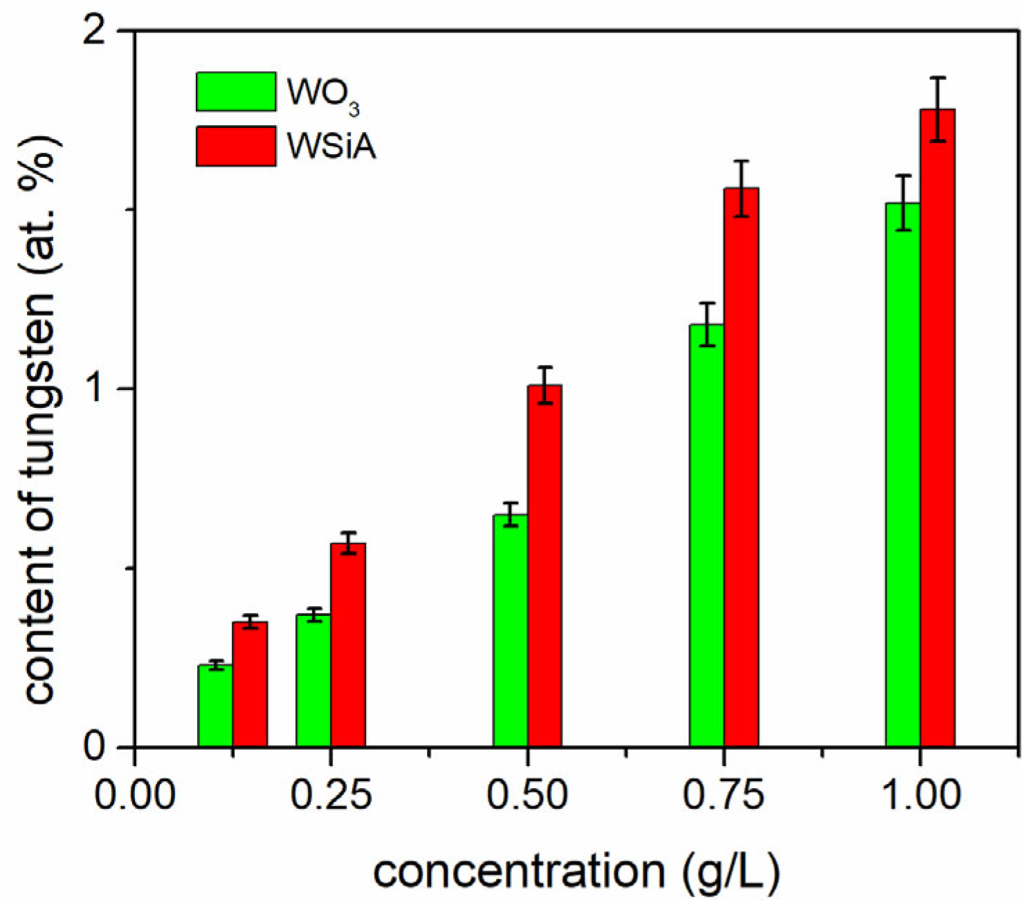
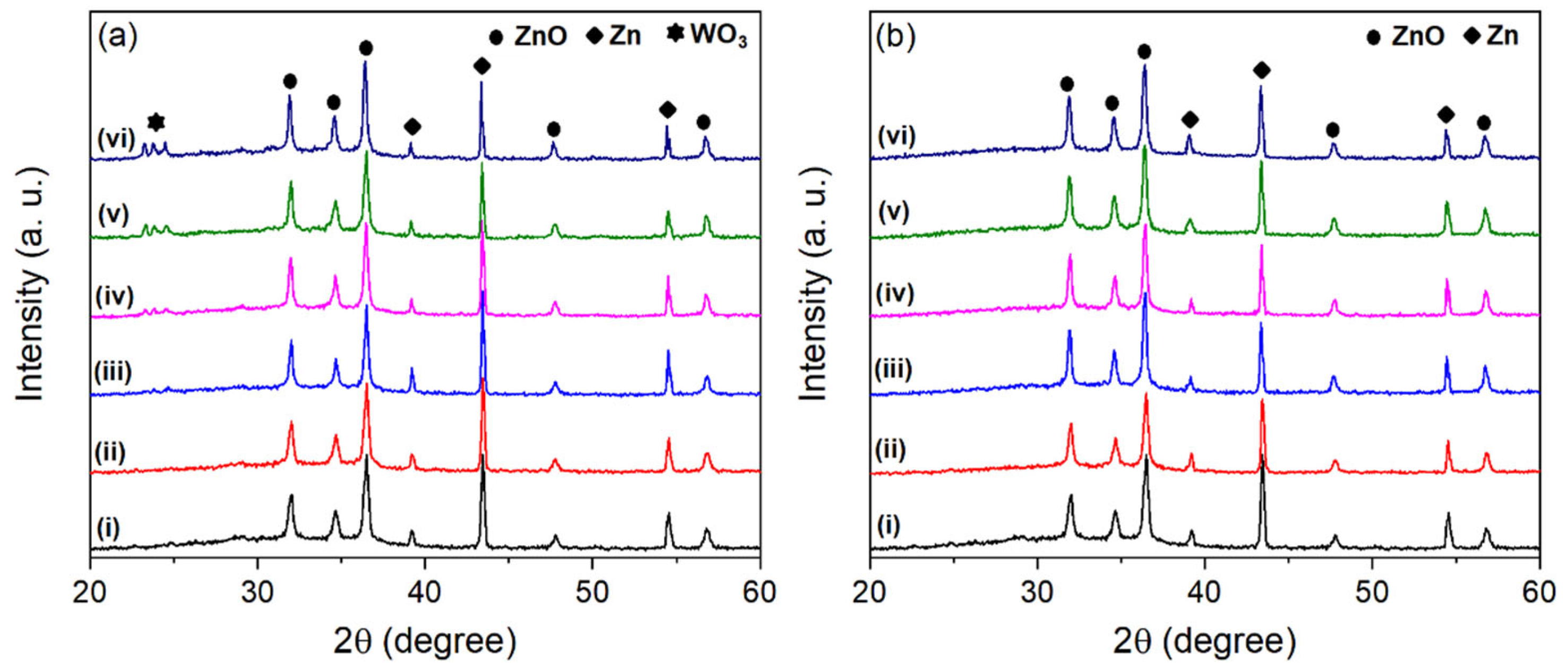
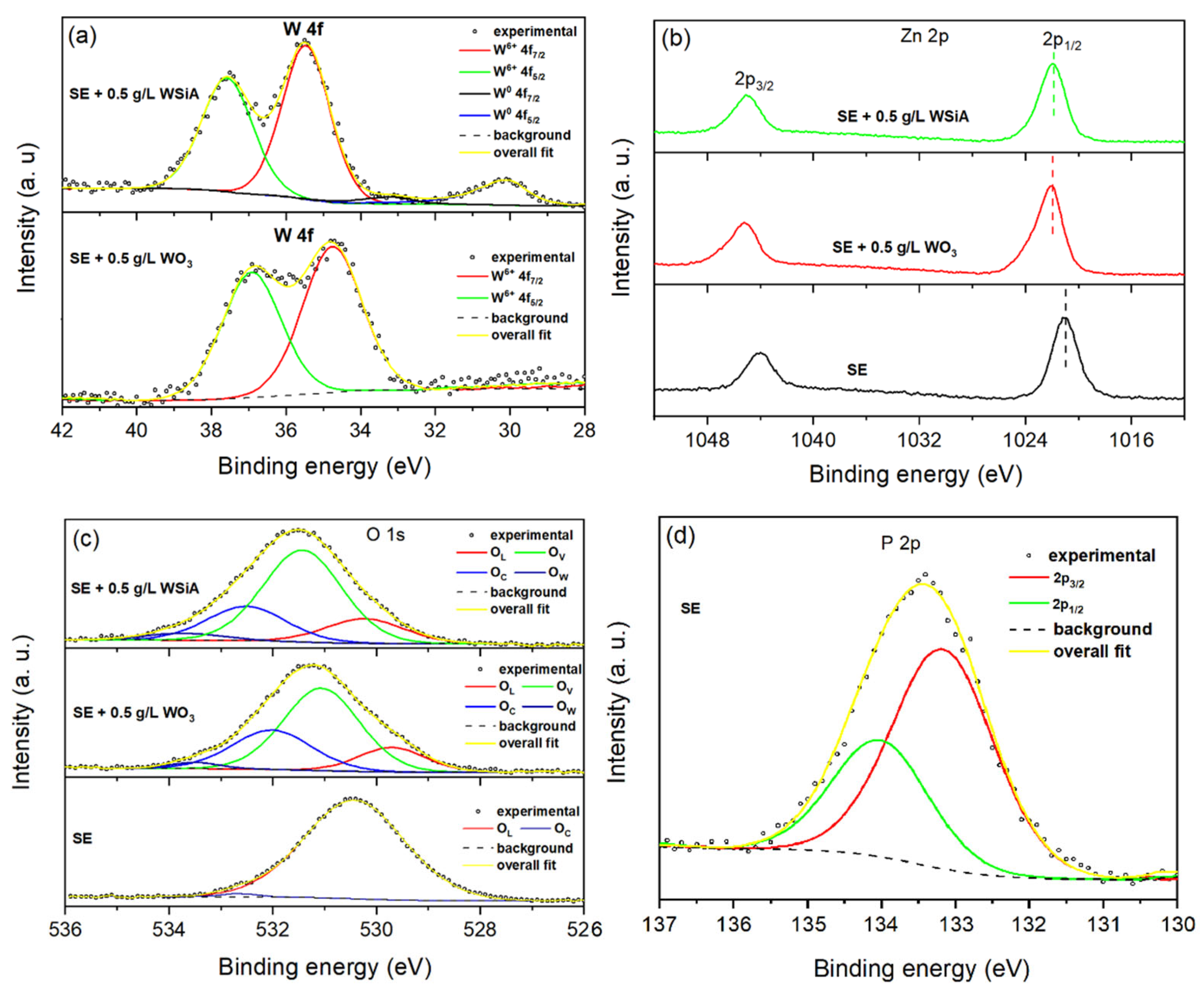
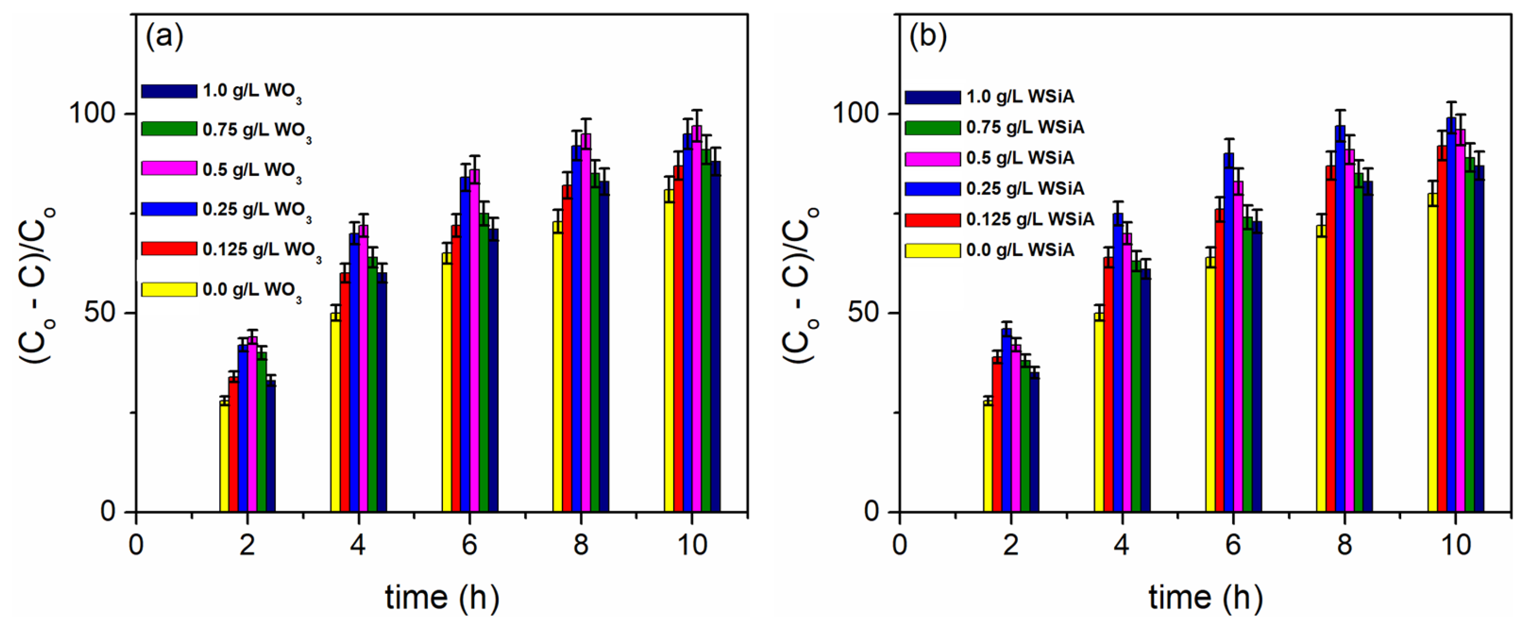
The test material was 0.25 mm thick zinc foil (Alfa Aesar, Haverhill, MA, USA, 99.98% pure). Before PEO, the foil was cut into 2.5 cm × 1.0 cm samples, cleaned using acetone in an ultrasonic bath and covered with insulating resin so that a 1.5 cm × 1.0 cm area was accessible for the electrolyte. In a double-walled glass electrolytic cell with water cooling, zinc samples were used as anodes, which were surrounded by a tubular cathode made of stainless steel. PEO was performed for 2 min at a constant current density of 300 mA/cm2 in an aqueous solution that contained 10 g/L Na3PO4·12H2O + 2 g/L KOH (supporting electrolyte—SE) with the addition of WO3 particles (Sigma-Aldrich, St. Louis, MO, USA, ≤100 nm particle size) or H4SiW12O40 (WSiA—12-tungstosilicic acid, Sigma-Aldrich, St. Louis, MO, USA, purity 99.9%) in concentrations of 0.125 g/L, 0.25 g/L, 0.5 g/L, 0.75 g/L and 1.0 g/L. Under working conditions, the pH value of the SE was 12.4. A magnetic stirrer was used to mix the electrolyte to ensure a temperature of (20 ± 0.5) °C and to have a uniform particle distribution. After the PEO procedure, the samples were washed with distilled water to avoid the accumulation of electrolyte components and then dried for three minutes under a stream of hot air.
A scanning electron microscope (SEM, JEOL 840A, Tokyo, Japan) was used to study the morphology and thickness of the coatings. Chemical composition of the coatings was investigated by utilizing energy dispersive X-ray spectroscopy (EDS, Oxford INCA, Abingdon, UK) and wavelength-dispersive X-ray fluorescence (XRF, Shimadzu XRF-1800, Tokyo, Japan). The phase composition of the coatings was determined using X-ray diffraction (XRD, Rigaku Ultima IV, Tokyo, Japan). In addition, the surface of the coating was analyzed using X-ray photoelectron spectroscopy (XPS, SPECS System, Berlin, Germany). UV/Vis diffuse reflectance spectroscopy (DRS, Shimadzu UV-3600, Tokyo, Japan) was used to analyze the optical absorption properties of the coatings.
A description of the experimental setup and the methodology of the photocatalytic measurement can be found in Ref. [35]. Water was used to cool the double-walled photocatalytic reactor. Before starting the photocatalytic test, the samples were submerged in 10 cm3 MO solution, positioned on a stainless steel holder 5 mm above the reactor’s base, and maintained in darkness for one hour with continuous stirring to achieve adsorption-desorption equilibrium. Once equilibrium was reached, the initial absorbance of the MO solution was measured using a UV/Vis spectrometer (Thermo Electron Nicolet, Evolution 500, Cambridge, UK). To start the photocatalytic reaction, the solution was exposed to a 300 W light source (OSRAM ULTRA-VITALUX UV-A, Munich, Germany) positioned 25 cm above the surface of the solution, corresponding to a power intensity of about 12 W/m2. To monitor the degradation of MO, absorbance values were measured every two hours for a total of ten hours. At 464 nm, a linear relationship between concentration and absorbance was demonstrated by converting absorbance to MO concentration using a standard curve.

3. Results

Figure 1a,b shows the top-view and cross-sectional morphologies of the coatings obtained in SE, as well as those obtained in SE with the addition of 1.0 g/L WO3 particles and 1.0 g/L WSiA, while Figure 1c shows the corresponding EDS spectra of the coatings in Figure 1a. The escape of gases during PEO leaves porous structures, including micro-discharge channels and craters where the micro-discharges have taken place, and the quick cooling of the molten material leads to the formation of solidified areas [35]. The morphology and thickness of the coatings obtained are not considerably altered by the addition of WO3 particles or WSiA to SE. The coatings have a thickness of about (40 ± 4) μm. The coating obtained in SE consists mainly of Zn, O and P (Figure 1c(i)). After the addition of WO3 particles or WSiA in SE, W or W and Si can be detected on the obtained coatings.
The content of W in the coatings is low and approaches the detection limit of our EDS system, particularly when small amounts of WO3 particles or WSiA are added to SE. To monitor the W content, we performed a wavelength-dispersive XRF measurement. The average content of W in the coatings formed on five different samples is shown in Figure 2. The content of W is low and rises as the concentration of WO3 particles and WSiA in the SE increases.
The XRD patterns of the coatings are presented in Figure 3. In the XRD pattern of the pure ZnO coating obtained in SE, several reflections at 2θ = 31.9°, 34.5°, 36.4°, 47.6° and 56.6° can be seen. These reflections correspond to the crystallographic planes (100), (002), (101), (102) and (110), respectively, of the hexagonal wurtzite crystal structure of ZnO (JCPDS Card No. 36–1451). The XRD patterns also show diffraction peaks for the Zn substrate. This is the result of the reflection of X-rays from the substrate when they penetrate the porous structure.
The XRD patterns of the coatings obtained in SE with the added WO3 particles (Figure 3a) show ZnO diffraction peaks as well as small distinct diffraction peaks at about 2θ = 23.2°, 23.7° and 24.5° corresponding to the (020), (020) and (200) planes of the triclinic phase of WO3, respectively (PDF Card No. 1010618), confirming the incorporation of WO3 particles in ZnO coatings.
During PEO, the particles can be incorporated as reactive, partially reactive or inert into the coatings. The two most significant variables that influence the type of incorporation are the melting point and the particle size [40]. Particles such as WO3, which have a high melting point, are typically incorporated inertly into PEO coatings, regardless of their size [41,42,43]. WO3 particles are negatively charged in SE (alkaline electrolyte) and easily migrate toward the zinc anode in the high electric field generated by an applied positive potential [40,41]. The melting point of WO3 particles is about 1470 °C, which is well below the plasma electron temperature for zinc PEO [44]. In the micro-discharge channels, mixed oxide coatings can be formed by the reaction of molten WO3 particles with components from the electrolyte and the substrate.
In the XRD patterns of the coatings obtained in SE with the addition of WSiA (Figure 3b), no phases corresponding to tungsten species can be recognized. The Keggin anions [SiW12O40]4−, which are anionic electrolyte components, migrate towards the zinc anode during PEO. The decomposition of WSiA (H4SiW12O40 → H2SiO3 + 12WO3 + H2O) [45] is caused by the high temperature at the micro-discharge sites, which should allow for the deposition of solid WO3 together with ZnO on the growing oxide surface.
In order to additionally examine the chemical composition and oxidation states of tungsten, zinc, oxygen and phosphorus, an XPS analysis was performed with a C 1s peak of 284.8 eV as a reference position. Figure 4 shows the high-resolution XPS spectra of the W 4f, Zn 2p, O 1s and P 2p of the obtained coatings. Figure 4a shows the high-resolution W 4f XPS spectra of coatings obtained in SE with the addition of 0.5 g/L WO3 particles or 0.5 g/L WSiA. In the spectrum of the coating obtained in SE + 0.5 g/L WO3 particles, two XPS peaks were detected at about 34.2 eV and 36.3 eV, corresponding to the W 4f7/2 and W 4f5/2 orbitals, respectively. These double energy bands correspond to the W6+ oxidation state and the spin-orbit separation of W 4f7/2 and W 4f5/2 is 2.1 eV, which agrees with the theoretical value for WO3 [46]. The high-resolution W 4f core level spectrum of the coating obtained in SE + 0.5 g/L WSiA shows two groups of doublets: one at binding energies around 35.2 eV and 37.3 eV ascribed to W 4f5/2 and 4f7/2, respectively, corresponding to the W6+ oxidation state, and another at energies around 31.2 eV and 33.2 eV attributed to the W 4f5/2 and W 4f7/2 of metallic tungsten, respectively [47]. However, the low intensities of the XPS peaks of metallic tungsten indicate that the tungsten is mainly WO3.
The high-resolution XPS spectra of Zn 2p from coatings obtained in SE and in SE with the addition of 0.5 g/L WO3 particles or 0.5 g/L WSiA are shown in Figure 4b. Two distinct and well-separated peaks have a spin/orbit splitting of 23.1 eV and can be assigned to Zn 2p1/2 and Zn 2p3/2, respectively, proving that Zn is in the Zn2+ oxidation state [48]. The binding energies of 2p3/2 and Zn 2p1/2 of the ZnO coating obtained in SE are at 1021 eV and 1044.1 eV, respectively, and are 1 eV lower than the binding energies of the Zn 2p1/2 and Zn 2p3/2 of the ZnO/WO3 coatings. The high-energy shift and slight broadening of the Zn 2p line is due to the incorporation of tungsten into the ZnO crystal structure. The W6+ substitution for Zn2+ in the ZnO lattice creates a charge imbalance that is predominantly compensated by the formation of zinc vacancies [49].
Figure 4c shows the high-resolution XPS spectra of O 1s for the coatings obtained in SE and in SE with the addition of 0.5 g/L WO3 particles or 0.5 g/L WSiA. The XPS spectrum of the ZnO coating obtained in SE can be decomposed into two components, where the peaks at 529.91 eV and 532.11 eV can be attributed to lattice oxygen (OL) and chemisorption oxygen (OC), respectively [50]. The O 1s XPS spectra of ZnO/WO3 coatings contain two additional contributions. The O 1s peak at a binding energy level of around 531.4 eV is a frequently observed region for hydroxyl groups and structural defects such as oxygen vacancies on both ZnO and WO3. Since this peak is not present in the XPS spectrum of pure ZnO, it is assumed that the peak around 531.4 eV in the O 1s XPS spectra of WO3-containing ZnO samples probably originates from hydroxyl groups in the WO3 phase (Ov in Figure 4). As the W 4f spectra did not show contributions from W5+ or W4+ in the presence of oxygen, vacancies is unlikely. This aligns with studies showing that WO3 surfaces preferentially adsorb hydroxyl groups and oxygen-containing species compared to ZnO [51]. The other contribution at around 534 eV is ascribed to adsorbed water (Ow).
The high-resolution XPS spectrum of P 2p from the coating obtained in SE can be decomposed into two peaks at a binding energy of 134.1 eV and 133.2 eV, ascribed to 2p1/2 and 2p3/2, respectively, corresponding to the phosphate group (PO4)3− (Figure 4d) [52]. This result indicates the formation of amorphous Zn3(PO4)2, which transforms into crystalline Zn3(PO4)2 over a long PEO time in SE [34].
Figure 5a,b shows the results of a DRS investigation of the absorption characteristics of the obtained coatings. The wide band gap of ZnO is responsible for the broad absorption band of the coatings in the ultraviolet range. ZnO/WO3 coatings do not exhibit a discernible shift in the absorption band toward visible light when compared to ZnO. The following formula can be used to calculate the band gap energy (Eg) of the ZnO and ZnO/WO3 coatings based on the absorption spectra:
αhν = A(Eg)η
where A is the constant, h is the Planck constant, α is the absorption coefficient and ν is the incident photon frequency. The exponent η is determined by the type of electron transition in the semiconductor (η = 1/2 and η = 2 for direct and indirect transitions, respectively). ZnO has a direct band gap, and the linear region of the Tauc plot is extended to the intersection with the axis to determine the band gap values of the coatings (Figure 5c,d). ZnO coating has a calculated Eg of (3.154 ± 0.012) eV, while ZnO/WO3 coatings obtained in the SE by adding 1.0 g/L WO3 particles or 1.0 g/L WSiA have a calculated Eg of (3.138 ± 0.011) eV and (3.134 ± 0.012) eV, respectively. The low concentration of WO3, which is evenly distributed over the entire surface of the ZnO layers, explains the insignificant shifts in the b and gap [53,54]. At low WO3 concentrations, the electronic properties of ZnO/WO3 are dominated by ZnO, which comprises the bulk of the material. The WO3 does not significantly alter the overall band structure. A uniform distribution prevents the formation of large clusters or aggregates of WO3, which could lead to more pronounced localized electronic states that affect the band gap.
Figure 6 shows the PA tests for MO degradation as a function of WO3 particles and WSiA content in the SE. For each concentration of WO3 particles and WSiA in the SE, five photocatalysts were prepared, and the average PA value is shown. Co stands for the initial MO concentration, while C stands for the concentration at time t. The concentration of WO3 particles and WSiA added to the way that SE affects the PA of ZnO/WO3 coatings, which is higher than that of pure ZnO coatings. The highest PA of the ZnO/WO3 coatings was achieved by adding 0.25 g/L WSiA and 0.5 g/L WO3 particles to the SE. Their PA after 8 h of irradiation is about 95%, while the ZnO coating is about 72%.
Because the surface morphology, crystalline structure and absorption properties of all PEO coatings are almost identical, their photocatalytic performance can be ascribed to the varying WO3 contents in the ZnO/WO3 coatings, which is controlled by the WO3 particles and the WSiA concentration in SE.
The conduction (CB) and valence (VB) bands of ZnO are higher than the corresponding bands of WO3 in ZnO/WO3 coatings [55]. This allows for the easy transfer of photo-generated electrons from the CB of ZnO to the CB of WO3. From the VB of WO3 to the VB of ZnO, the holes move concurrently in the opposite direction. The separation effect reduces recombination between the electron/hole pairs and increases the probability of electrons and holes participating in photocatalytic reactions. The hydroxyl ions (OH) and the water molecules (H2O) adsorbed on the ZnO/WO3 coating react with the photo-generated holes and oxidize them to hydroxyl radicals (OH). OH is a strong oxidant that decomposes MO, in particular, into CO2 and H2O. The formation of superoxide ions (O2¯) from the combination of photo-generated electrons and oxygen molecules adsorbed on the ZnO/WO3 coating surface also contributes to the degradation of MO.
Increasing the concentrations of WO3 particles and WSiA in SE up to 0.5 g/L and 0.25 g/L, respectively, leads to increased PA, which is associated with slower recombination of electron/hole pairs. When the concentrations of WO3 particles and WSiA are further increased, PA decreases, suggesting that the WO3 incorporated into the ZnO coatings not only slows down the recombination of electron/hole pairs but also serves as a trapping center for photo-induced electrons. These captured electrons are not readily available for the reduction reactions required for photocatalysis, resulting in a decrease in PA. Basically, the WO3 becomes a sink for electrons instead of just enabling charge separation.
The suitability of ZnO/WO3 coatings obtained by PEO for photocatalysis is largely determined by their chemical and physical stability, which also has an impact on their lifetime and operating costs. Figure 7 shows the PA after five consecutive tests with the most active ZnO/WO3 coatings obtained in SE with the addition of WO3 particles and WSiA. The samples were washed with water after each test, dried and then used for the next photocatalytic test. The obtained ZnO/WO3 coatings are reliable and effective photocatalysts, as evidenced by the exceptional stability of the coatings during the photocatalytic degradation of MO.
Since PEO-formed ZnO/WO3 coatings on zinc eliminate the necessity for costly and time-consuming recycling and reuse of the photocatalyst after wastewater degradation treatment, they are more useful than ZnO/WO3 photocatalysts in powder form.

4. Conclusions

In this study, ZnO/WO3 coatings on zinc substrates were prepared by PEO in an alkaline phosphate electrolyte (supporting electrolyte/SE) to which WO3 particles or WSiA were added in different concentrations. The photocatalytic activity (PA) of these coatings in the degradation of methyl orange was evaluated. The amount of tungsten in the ZnO/WO3 coatings determines their PA. ZnO/WO3 coatings have a higher PA than ZnO coatings obtained in SE because the coupling of ZnO and WO3 reduces the recombination rate of photo-generated electron/hole pairs. The highest PA is found in ZnO/WO3 coatings obtained in SE using 0.25 g/L WSiA or 0.5 g/L WO3 particles.
In order to further improve the photocatalytic performance of PEO-formed ZnO coatings and to extend the activation range to the visible range, efforts should be made in the future to incorporate oxide particles of transition metals (Fe, Cu, Ni, Co, Mn and V) into the ZnO coatings. ZnO photocatalysts on solid supports have advantages over those in particle form, and PEO can easily incorporate particles in the coatings.

Author Contributions

Conceptualization, S.S. and N.R.; methodology, S.S.; validation, S.S., D.P. and N.R.; formal analysis, S.S.; investigation, S.S., D.P. and N.R.; writing—original draft preparation, S.S. and D.P.; writing—review and editing, S.S.; visualization, D.P. and N.R.; supervision, S.S.; project administration, S.S. and N.R.; funding acquisition, S.S. All authors have read and agreed to the published version of the manuscript.

Funding

This research was funded by the Ministry of Science, Innovation, and Technological Development of the Republic of Serbia (Grants 451-03-136/2025-03/200162 and 451-03-136/2025-03/200026) and the Science Fund of the Republic of Serbia, grant number 7309 ZEOCOAT.

Institutional Review Board Statement

Not applicable.

Informed Consent Statement

Not applicable.

Data Availability Statement

The data presented in this study are available on request from the corresponding author.

Conflicts of Interest

The authors declare no conflicts of interest.

References

  1. Ansari, A.A.; Lv, R.; Gai, S.; Parchur, A.K.; Solanki, P.R.; Archana; Ansari, Z.A.; Dhayal, M.; Yang, P.; Nazeeruddin, M.K.; et al. ZnO nanostructures—Future frontiers in photocatalysis, solar cells, sensing, supercapacitor, fingerprint technologies, toxicity, and clinical diagnostics. Coord. Chem. Rev. 2024, 515, 215942. [Google Scholar] [CrossRef]
  2. Sun, Y.; Zhang, W.; Li, Q.; Liu, H.; Wang, X. Preparations and applications of zinc oxide based photocatalytic materials. Adv. Sens. Energy Mater. 2023, 2, 100069. [Google Scholar] [CrossRef]
  3. Monika, P.; Krishna, R.H.; Hussain, Z.; Nandhini, K.; Pandurangi, S.J.; Malek, T.; Kumar, S.G. Antimicrobial hybrid coatings: A review on applications of nano ZnO based materials for biomedical applications. Biomater. Adv. 2025, 172, 214246. [Google Scholar] [CrossRef]
  4. Raha, S.; Ahmaruzzaman, M. ZnO nanostructured materials and their potential applications: Progress, challenges and perspectives. Nanoscale Adv. 2022, 4, 1868–1925. [Google Scholar] [CrossRef]
  5. Gartner, M.; Stroescu, H.; Mitrea, D.; Nicolescu, M. Various applications of ZnO thin films obtained by chemical routes in the last decade. Molecules 2023, 28, 4674. [Google Scholar] [CrossRef]
  6. Bhadwal, N.; Mrad, R.B.; Behdinan, K. Review of zinc oxide piezoelectric nanogenerators: Piezoelectric properties, composite structures and power output. Sensors 2023, 23, 3859. [Google Scholar] [CrossRef]
  7. Chu, L.; Xu, C.; Zeng, W.; Nie, C.; Hu, Y. Fabrication and application of different nanostructured ZnO in ultraviolet photodetectors: A review. IEEE Sens. J. 2022, 22, 7451–7462. [Google Scholar] [CrossRef]
  8. Kaur, P.; Kriti; Rahul; Kaur, S.; Kumar, V.; Kandasami, A.; Singh, D.P. Temperature-dependent characteristics of ZnO phosphors from synchrotron-based vacuum ultraviolet photoluminescence spectroscopy. Eur. Phys. J. Plus 2022, 137, 142. [Google Scholar] [CrossRef]
  9. Sharma, D.K.; Shukla, S.; Sharma, K.K.; Kumar, V. A review on ZnO: Fundamental properties and applications. Mater. Today: Proc. 2022, 49, 3028–3035. [Google Scholar] [CrossRef]
  10. Zheng, A.L.T.; Abdullah, C.A.C.; Chung, E.L.T.; Andou, Y. Recent progress in visible light-doped ZnO photocatalyst for pollution control. Int. J. Environ. Sci. Technol. 2023, 20, 5753–5772. [Google Scholar] [CrossRef]
  11. Aftab, S.; Shabir, T.; Shah, A.; Nisar, J.; Shah, I.; Muhammad, H.; Shah, N.S. Highly efficient visible light active doped ZnO photocatalysts for the treatment of wastewater contaminated with dyes and pathogens of emerging concern. Nanomaterials 2022, 12, 486. [Google Scholar] [CrossRef]
  12. Mutalib, A.A.A.; Jaafar, N.F. ZnO photocatalysts applications in abating the organic pollutant contamination: A mini review. Total Environ. Res. Themes 2022, 3–4, 100013. [Google Scholar] [CrossRef]
  13. Tuama, A.N.; Alzubaidi, L.H.; Jameel, M.H.; Abass, K.H.; bin Mayzan, M.Z.H.; Salman, Z.N. Impact of electron–hole recombination mechanism on the photocatalytic performance of ZnO in water treatment: A review. J. Sol-Gel Sci. Technol. 2024, 110, 792–806. [Google Scholar] [CrossRef]
  14. Hezam, A.; Drmosh, Q.A.; Ponnamma, D.; Bajiri, M.A.; Qamar, M.; Namratha, K.; Zare, M.; Nayan, M.B.; Onaizi, S.A.; Byrappa, K. Strategies to enhance ZnO photocatalyst’s performance for water treatment: A comprehensive review. Chem. Rec. 2022, 22, e202100299. [Google Scholar] [CrossRef]
  15. Xu, Y.; Yan, H.; Chen, T. Application of ZnO/WO3 composite nanofiber photocatalysts in textile wastewater treatment. Separations 2023, 10, 339. [Google Scholar] [CrossRef]
  16. Mubeen, K.; Irshad, A.; Safeen, A.; Aziz, U.; Safeen, K.; Ghani, T.; Khan, K.; Ali, Z.; ul Haq, I.; Shah, A. Band structure tuning of ZnO/CuO composites for enhanced photocatalytic activity. J. Saudi Chem. Soc. 2023, 27, 101639. [Google Scholar] [CrossRef]
  17. Guerram, A.; Laouini, S.E.; Mohammed, H.A.; Hasan, G.G.; Tedjani, M.L.; Alharthi, F.; Menaa, F. Synergistic Performance of ZnO/SnO2 nanocomposites: Synthesis, characterization, and applications in photocatalysis and superoxide radical scavenger. J. Clust. Sci. 2024, 35, 2231–2242. [Google Scholar] [CrossRef]
  18. Shahzad, R.; Muneer, M.; Khalid, R.; Amin, H.M.A. ZnO-Bi2O3 Heterostructured composite for the photocatalytic degradation of orange 16 reactive dye: Synergistic effect of UV irradiation and hydrogen peroxide. Catalysts 2023, 13, 1328. [Google Scholar] [CrossRef]
  19. Nadikatla, S.K.; Chintada, V.B.; Gurugubelli, T.R.; Koutavarapu, R. Review of recent developments in the fabrication of ZnO/CdS heterostructure photocatalysts for degradation of organic pollutants and hydrogen production. Molecules 2023, 28, 4277. [Google Scholar] [CrossRef]
  20. Stojadinović, S.; Radisavljevic, Z.; Petrović, Z.; Radić, N. ZnO/Zn3(PO4)2/CeO2 photocatalysts formed on zinc by plasma electrolytic oxidation. Solid State Sci. 2024, 158, 107748. [Google Scholar] [CrossRef]
  21. Desseigne, M.; Dirany, N.; Chevallier, V.; Arab, M. Shape dependence of photosensitive properties of WO3 oxide for photocatalysis under solar light irradiation. Appl. Surf. Sci. 2019, 483, 313–323. [Google Scholar] [CrossRef]
  22. Xu, Y.; Chen, T. Development of nanostructured based ZnO@WO3 photocatalyst and its photocatalytic and electrochemical properties: Degradation of Rhodamine B. Int. J. Electrochem. Sci. 2023, 18, 100055. [Google Scholar] [CrossRef]
  23. Sangkhanak, S.; Kunthakudee, N.; Hunsom, M.; Ramakul, P.; Serivalsatit, K.; Pruksathorn, K. Highly efficient ZnO/WO3 nanocomposites towards photocatalytic gold recovery from industrial cyanide-based gold plating wastewater. Sci. Rep. 2023, 13, 22752. [Google Scholar] [CrossRef]
  24. Zhang, D.; Liu, Z.; Mou, R. Preparation and characterization of WO3/ZnO composite photocatalyst and its application for degradation of oxytetracycline in aqueous solution. Inorg. Chem. Commun. 2022, 142, 109667. [Google Scholar] [CrossRef]
  25. Aziz, F.; Warsi, A.; Somaily, H.H.; Din, M.I.; Sabeeh, H.; Warsi, M.F.; Shakir, I. Zinc oxide-tungsten oxide (ZnO-WO3) composite for solar light-assisted degradation of organic dyes. Korean J. Chem. Eng. 2023, 40, 1497–1509. [Google Scholar] [CrossRef]
  26. Rania, M.; Keshu; Pandey, S.; Rishabh; Sharma, S.; Shanker, U. Sunlight assisted highly efficient photocatalytic remediation of organic pollutants by green biosynthesized ZnO@ WO3 nanocomposite. J. Photochem. Photobiol. A Chem. 2024, 446, 115160. [Google Scholar] [CrossRef]
  27. Clyne, T.W.; Troughton, S.C. A review of recent work on discharge characteristics during plasma electrolytic oxidation of various metals. Int. Mater. Rev. 2019, 64, 127–162. [Google Scholar] [CrossRef]
  28. Kaseem, M.; Fatimah, S.; Nashrah, N.; Ko, Y.G. Recent progress in surface modification of metals coated by plasma electrolytic oxidation: Principle, structure, and performance. Prog. Mater. Sci. 2021, 117, 100735. [Google Scholar] [CrossRef]
  29. Karbasi, M.; Chaharmahali, E.N.R.; Hosseini, R.; Giannakis, S.; Bahramian, H.; Kaseem, M.; Fattah-alhosseini, A. A review on plasma electrolytic oxidation coatings for organic pollutant degradation: How to prepare them and what to expect of them? J. Environ. Chem. Eng. 2023, 11, 110027. [Google Scholar] [CrossRef]
  30. Saji, V.S. Plasma electrolytic oxidation (PEO) layers grown on metals and alloys as supported photocatalysts. Next Energy 2025, 8, 100259. [Google Scholar] [CrossRef]
  31. Fattah-alhosseini, A.; Chaharmahali, R.; Kaseem, M. Exploring nanoparticle contributions to enhanced photocatalytic activity of PEO coatings on titanium: A review of the recent advancements. Nano-Struct. Nano-Objects 2024, 39, 101273. [Google Scholar] [CrossRef]
  32. Fattah-alhosseini, A.; Chaharmahali, R.; Alizad, S.; Babaei, K.; Stojadinović, S. A review on the revealed improved photocatalytic activity of PEO coatings applied on Al alloys. Nano-Struct. Nano-Objects 2024, 39, 101233. [Google Scholar] [CrossRef]
  33. Fattah-Alhosseini, A.; Stojadinović, S.; Chaharmahali, R.; Gnedenkov, A. An examination of the enhanced photocatalytic performance of PEO coatings applied on Mg alloys: A review. J. Magnes. Alloys 2024, 12, 4422–4435. [Google Scholar] [CrossRef]
  34. Stojadinović, S.; Radić, N. Photocatalytic performance of ZnO and ZnO/Zn3(PO4)2 coatings formed by plasma electrolytic oxidation of zinc. Solid State Sci. 2024, 153, 107578. [Google Scholar] [CrossRef]
  35. Stojadinović, S.; Radić, N.; Perković, M. Highly efficient ZrO2 photocatalysts in the presence of UV radiation synthesized in a very short time by plasma electrolytic oxidation of zirconium. Opt. Mater. 2023, 146, 114608. [Google Scholar] [CrossRef]
  36. Stojadinović, S.; Radić, N.; Vasilić, R. Application of micro-arc discharges during anodization of tantalum for synthesis of photocatalytic active Ta2O5 coatings. Micromachines 2023, 14, 701. [Google Scholar] [CrossRef]
  37. Stojadinović, S.; Radić, N.; Perković, M. Nb2O5 and AlNbO4 coatings formed by plasma electrolytic oxidation of niobium: Synthesis, characterization, and photocatalytic activity. J. Mater. Sci. Mater. Electron. 2024, 35, 410. [Google Scholar] [CrossRef]
  38. Fattah-alhosseinia, A.; Karbasi, M.; Fardosi, A.; Kaseem, M. Optimization of electrolyte composition for enhanced photocatalytic performance of the ceramic coating produced on brass by plasma electrolytic oxidation. Ceram. Int. 2024, 50, 25822–25831. [Google Scholar] [CrossRef]
  39. Hanafi, M.F.; Sapawe, N. Areview on the water problem associate with organic pollutants derived from phenol, methyl orange, and remazol brilliant blue dyes. Mater. Today Proc. 2020, 31, A141–A150. [Google Scholar] [CrossRef]
  40. Lu, X.; Mohedano, M.; Blawert, C.; Matykina, E.; Arrabal, R.; Kainer, K.U.; Zheludkevich, M.L. Plasma electrolytic oxidation coatings with particle additions—A review. Surf. Coat. Technol. 2016, 307, 1165–1182. [Google Scholar] [CrossRef]
  41. Salimi, H.; Fattah-alhosseini, A.; Karbasi, M.; Nikoomanzari, E. Development of WO3-incorporated porous ceramic coating: A key role of WO3 nanoparticle concentration on methylene blue photodegradation upon visible light illumination. Ceram. Int. 2023, 49, 32181–32192. [Google Scholar] [CrossRef]
  42. Zehra, T.; Kaseem, M.; Patil, S.A.; Shrestha, N.K.; Fattah-alhosseini, A.; Kaseem, M. Anionic assisted incorporation of WO3 nanoparticles for enhanced electrochemical properties of AZ31 Mg alloy coated via plasma electrolytic oxidation. J. Alloys Compd. 2022, 916, 165445. [Google Scholar] [CrossRef]
  43. Kaseem, M.; Hussain, T.; Rehman, Z.U.; Ko, Y.G. Stabilization of AZ31 Mg alloy in sea water via dual incorporation of MgO and WO3 during micro-arc oxidation. J. Alloys Compd. 2021, 853, 157036. [Google Scholar] [CrossRef]
  44. Stojadinović, S.; Tadić, N.; Vasilić, R. Formation and characterization of ZnO films on zinc substrate by plasma electrolytic oxidation. Surf. Coat. Technol. 2016, 307, 650–657. [Google Scholar] [CrossRef]
  45. Klisch, M. 12-Tungstosilicic Acid (12-TSA) as a tungsten precursor in alcoholic solution for deposition of xWO3(1−x)SiO2 thin films (x ≤ 0.7) exhibiting electrochromic coloration ability. J. Sol-Gel Sci. Technol. 1998, 12, 21–33. [Google Scholar] [CrossRef]
  46. Bouvard, O.; Krammer, A.; Schüler, A. In situ core-level and valence-band photoelectron spectroscopy of reactively sputtered tungsten oxide films. Surf. Interface Anal. 2016, 48, 660–663. [Google Scholar] [CrossRef]
  47. Wakizaka, M.; Chun, W.-J.; Imaoka, T.; Yamamoto, K. Metallic tungsten nanoparticles that exhibitan electronic state like carbides during the carbothermal reduction of WCl6 by hydrogen. Inorg. Chem. 2020, 59, 15690–15695. [Google Scholar] [CrossRef]
  48. Biesinger, M.C.; Lau, L.W.M.; Gerson, A.R.; Smart, R.S.C. Resolving surface chemical states in XPS analysis of first row transition metals, oxides and hydroxides: Sc, Ti, V, Cu and Zn. Appl. Surf. Sci. 2010, 257, 887. [Google Scholar] [CrossRef]
  49. Vines, F.; Iglesias-Juez, A.; Fernandez-García, M.; Illas, F. Understanding W doping in Wurtzite ZnO. J. Phys. Chem. C 2018, 122, 19082–19089. [Google Scholar] [CrossRef]
  50. Kwoka, M.; Kulis-Kapuscinska, A.; Zappa, D.; Comini, E.; Szuber, J. Novel insight on the local surface properties of ZnO nanowires. Nanotechnology 2020, 31, 465705. [Google Scholar] [CrossRef]
  51. Adhikari, S.; Sarkar, D.; Madras, G. Highly efficient WO3–ZnO mixed oxides for photocatalysis. RSC Adv. 2015, 5, 11895–11904. [Google Scholar] [CrossRef]
  52. Naciri, Y.; Chennah, A.; Jaramillo-Páez, C.; Navío, J.A.; Bakiz, B.; Taoufyq, A.; Ezahri, M.; Villain, S.; Guinneton, F.; Benlhachemi, A. Preparation, characterization and photocatalytic degradation of Rhodamine B dye over a novel Zn3(PO4)2/BiPO4 catalyst. J. Environ. Chem. Eng. 2019, 7, 103075. [Google Scholar] [CrossRef]
  53. Malik, M.S.; Roy, D.; Chun, D.-M.; Abd-Elrahim, A.G. One-step dry coating of hybrid ZnO–WO3 nanosheet photoanodes for photoelectrochemical water splitting with composition-dependent performance. Micromachines 2023, 14, 2189. [Google Scholar] [CrossRef]
  54. Qin, H.; Chen, L.; Yu, X.; Wu, M.; Yan, Z. Preparation and photocatalytic performance of ZnO/WO3/TiO2 composite coatings formed by plasma electrolytic oxidation. J. Mater. Sci. Mater. Electron. 2018, 29, 2060–2071. [Google Scholar] [CrossRef]
  55. Sajjad, A.K.L.; Sajjad, S.; Iqbal, A.; Ryma, N.A. ZnO/WO3 nanostructure as an efficient visible light catalyst. Ceram. Int. 2018, 44, 9364–9371. [Google Scholar] [CrossRef]
Figure 1. (a) Top-view micrographs. (b) Cross-section micrographs. (c) EDS spectra of coatings obtained in (i) SE; (ii) SE + 1.0 g/L WO3; (iii) SE + 1.0 g/L WSiA.
Coatings 15 00657 g001
Figure 2. Tungsten content determined by XRD in coatings obtained in SE with the addition of different concentrations of WO3 particles and WSiA.
Coatings 15 00657 g002
Figure 3. XRD patterns of coatings obtained in SE with addition of (a) WO3 and (b) WSiA in various concentrations: (i) 0 g/L; (ii) 0.125 g/L; (iii) 0.25 g/L; (iv) 0.5 g/L; (v) 0.75 g/L; (vi) 1.0 g/L.
Coatings 15 00657 g003
Figure 4. High-resolution XPS spectra of coatings obtained in SE and SE + 0.5 g/L WO3 particles, as well as SE + 0.5 g/L WSiA: (a) W 4f; (b) Zn 4p; (c) O 1s; (d) P 2p.
Coatings 15 00657 g004
Figure 5. DRS spectra of coatings obtained in SE with addition of (a) WO3 and (b) WSiA; in various concentrations. Tauc plots of coatings obtained in (c) SE and SE + 1.0 g/L WO3; (d) SE and SE + 1.0 g/L WSiA.
Coatings 15 00657 g005
Figure 6. PA of coatings obtained in SE with addition of (a) WO3 and (b) WSiA in various concentrations.
Coatings 15 00657 g006
Figure 7. MO photo-degradation recycling test of coatings obtained in (a) SE + 0.5 g/L WO3; (b) SE + 0.25 g/L WSiA.
Coatings 15 00657 g007
Disclaimer/Publisher’s Note: The statements, opinions and data contained in all publications are solely those of the individual author(s) and contributor(s) and not of MDPI and/or the editor(s). MDPI and/or the editor(s) disclaim responsibility for any injury to people or property resulting from any ideas, methods, instructions or products referred to in the content.

