Article

Ultrathin Biomaterial Coating for Flame-Retardant Medical Paper

1
School of the Emergency Management (School of the Environment and Safety Engineering), Jiangsu University, Zhenjiang 212013, China
2
College of Safety Science and Engineering, Nanjing Tech University, Nanjing 211816, China
3
School of Chemical Engineering, Xuzhou College of Industrial Technology, Xuzhou 221140, China
*
Authors to whom correspondence should be addressed.
Coatings 2025, 15(2), 192; https://doi.org/10.3390/coatings15020192
Submission received: 23 November 2024 / Revised: 19 December 2024 / Accepted: 30 December 2024 / Published: 6 February 2025
(This article belongs to the Special Issue Surface Modification and Strengthening of Bio-Based Materials)

Abstract

:
Paper has the multiple advantages of being breathable, sustainable, environmentally friendly, and non-toxic for medical care. However, the flammability stemming from the raw materials of paper has limited its use in medical heat therapy. In this paper, a composite flame-retardant coating is assembled layer by layer on a medical paper surface using medically safe natural biomaterials with starch and adenosine triphosphate as internal layers, and starch and phytic acid as external layers. With the layer-by-layer assembly using the ultrathin adsorption method, the microscopic morphology and elemental mapping reveal that all the biomaterials are deposited uniformly and have completely capsulated the paper surface fiber. The flame-retardant coating shows less impact on medical paper appearance morphology and mechanical properties in medical usability. The coated medical paper exhibits significant flame-retardant performance, such that the limiting oxygen index increases from 19.70% to 25.40% where both internal and external layers reached 100 layers (BL), and relevant residual charring in the thermogravimetric test increases 17.00 wt% in a nitrogen atmosphere and 18.00 wt% in an air atmosphere at 800 °C. The peak and total heat release rates of 100 BL medical paper reduced by approximately 91.10% and 53.10%, respectively, and the variations in both CO and CO2 production also suggest that flame-retardant coating could effectively inhibit combustion. Benefiting from the intumescent flame-retardant function of different biomaterial combinations and the multilayer design on different thermal response temperatures, the flame retardancy of medical paper significantly improved, and this advancement will make medical heat therapy safer and healthier for patients.

1. Introduction

Paper, formed from plant fibers of natural cellulose, is a commodity printing and packaging material for humans that has been widely used for thousands of years [1,2,3,4,5,6]. Nowadays, paper is still a vital material for medical care, such as pharmaceutical packaging, medical-grade coating, medical dialyzing, and even wound care dressing [7,8]. As a natural substrate, paper is breathable, porous, flexible, inexpensive, biocompatible, and non-hazardous. In traditional oriental medicine and pharmacy, medicinal herbal plaster, with herbal medicines smeared on paper or fiber, has been used to treat various diseases for a long time. The plaster usually needs to be heated with fire to improve the therapeutic effects before or during medical treatment. Medicinal paper is commonly used in indirect fire therapy to treat cold-related illnesses. The medicated paper acts as a heat transfer medium, isolating the skin from fire and enhancing the therapeutic effect of the medicines on the paper. Figure 1 shows a photograph of indirect fire therapy using medicated paper to treat knee diseases. However, the innate flammability properties of paper, which stem from its natural organic materials, have also limited its use in heat-related applications. Thus, improving the flame retardancy of paper is very important for the safety of medical treatment.
Incorporating flame retardants into flammable materials is commonly used to reduce fire risk, including ignitability, heat release, and flame spread. However, while flame retardants are effective for fire safety, not all are safe for humans or the environment. The safety of flame retardants has long been one of the main issues in research on human health and environmental pollution. Halogenated flame retardants are artificial chemicals that contain halogenated elements like fluorine, chlorine, and bromine, which have been added widely to various products for combustion prevention. Evidence shows that these substances are inevitably transformed or released into the environment and living organisms during production and use. That toxicity can affect human health and ecological safety. For human health, adverse effects have been reported to be closely related to cancer, endocrine disruption, and immune system suppression. Various studies have indicated that brominated flame retardants are neurotoxic, endocrine toxic [9], reproductive-toxic [10], and even carcinogenic [11,12,13]. Perfluoroalkyl substances have a significant association with an increased risk of hyperlipidemia, metabolic syndrome, diabetes, and hyperuricemia [14]. Those flame retardants are highly persistent and have a high potential for bioaccumulation, biomagnification, and long-range transport. Therefore, many flame retardants, such as mirex, chlorinated chemicals (polychlorinated biphenyls, pentachlorobenzene), brominated chemicals (polybrominated diphenyl ethers), chlorinated paraffin (short-chain chlorinated paraffin, medium-chain chlorinated paraffin), fluorinated chemicals (perfluorooctane sulfonic acid, perfluorooctanoic acid, perfluorohexane sulfonic acid), continue to be recognized as persistent organic pollutants (POPs) and have been listed in the Stockholm Convention to stop their production. Phosphate chemicals are another primary type of flame retardant. Some of them have also been proven toxic to human health as the pathophysiological determinant of cancer cell growth, whether organic or inorganic. Hence, traditional artificial chemical flame retardants are generally considered unsuitable for medical goods on grounds of long-term patient safety.
Nature-based biomaterials have attracted increasing interest in recent years due to their great potential usages in many sectors, such as biomedicine [15,16,17], construction [18], fuel [19,20], chemicals [21,22], energy [23], and environmental [24]. The biomaterials also perform well as flame retardants for many flammable materials. Fully biomaterial-based intumescent flame retardants prepared from biologically derived phytic acid and guanosine have been used in polybutylene succinate, effectively improving charring ability and flame retardancy [25]. However, if only biomaterials are used as flame retardants, flame retardancy is usually poorly characterized [26]. As a result, biomaterials commonly act as auxiliary agents to synergize primary flame retardants to achieve the flame retardant of combustibles in many studies [27,28].
For biomaterial-based intumescent flame retardants, phytic acid containing six phosphate groups is often used as the acid source [29]. Starch, a biomaterial-based polymer with rich carbon content, is widely found in the root and seed of plants for energy storage [30]. Adenosine triphosphate is an energy-carrying molecule found in the cells of all living things. Adenosine triphosphate is a nucleotide that consists of adenine, ribose, and a chain of three phosphate groups. So, it also could be a natural intumescent flame retardant, similar to DNA, which meets the three fundamental qualifications for intumescent flame retardancy. The phosphate chain of adenosine triphosphate will release phosphoric acid when heated to pyrolysis, which may work as an acid source like phytic acid. Besides the phosphate groups, the ribose and adenine in adenosine triphosphate could serve as the carbon source and blowing agent [31].
The layer-by-layer assembly is a simple and controllable method to create nanoscale thin organized films on different substrates and has garnered significant attention since its inception [32,33,34]. The different driving forces, depending on the physical and chemical properties of the chosen materials on electrostatic interactions, hydrogen bonds, hydrophobic interactions, and covalent bonds, are employed to step by step build the organizable and tunable structured film [35,36]. This method has many advantages in practical and economical for surface coating of paper products [37,38,39]. Due to layer-by-layer assembly, the assembled materials are absorbed only on the surface of the substrates, and the self-assembly coating is super thin, so it gives less impact than the other methods [40,41]. In previous studies, many flame-retardant coatings were layer-by-layer assembled using two different kinds of materials, and there are fewer reports on the composite structural coating which uses more materials [42].
In this paper, starch, adenosine triphosphate, and phytic acid, the common materials used in medical care, are selected to prepare the biomass flame-retardant coating for medical paper. The preparation process utilizes the different electrostatic properties of those biomaterials to self-assemble coating on the medical paper surface via the self-made ultrathin adsorption device. The prepared coating has a multilayered structure with internal and external layers constituted by the biomaterial combinations on different thermal response temperatures. The changes in the macro and micro views of coated medical paper are performed and discussed. Then, a series of combustion tests are conducted to evaluate its flame retardancy. In addition, the related mechanical properties of coated medical paper in practical applications are also assessed. Finally, the flame-retardant mechanism of coated medical paper is discussed in view of the intumescent flame retardant.

2. Materials and Methods

2.1. Materials

All biomaterials were commercial products that could be directly used without further purification. Phytic acid solution (PA, 70%) was purchased from Shandong Yousuo Chemical Technology Co., Ltd., Linyi, China. Adenosine triphosphate (ATP, 98%) was purchased from Hengji Chemical Co., Ltd., Xi’an, China. The purities of starch, including cationic starch and water-soluble starch, were all 99% and were bought, respectively, from Guangdong Hongxin Biotechnology Co., Ltd., Dongguan, China and Yunfeng Starch Co., Ltd., Nantong, China. The paper was provided by Shandong Chenming Paper Holdings Co., Ltd., Shouguang, China. The aqueous solutions of each biomaterial were prepared using deionized water.

2.2. Ultrathin Adsorption Device

The ultrathin adsorption device was self-made and used for each biomaterial adsorbed on the medical paper surface. The device comprises a top-opened transparent body frame, an ultrasonic atomization part, and a high-voltage electrostatic part, as shown in Figure 2. Before coating preparation, the biomaterial aqueous solution fills the bottom of the device and immerses the ultrasonic nebulizer. The electrostatic generator provides a 30 KV static voltage, whose positive pole immerses in the solution, and the negative pole connects to the metal coil situated slightly lower and around the top of the device body. The metal coil is covered with an insulated jacket and has two exposed short needle electrodes that point to the notches on the side top of the device body. The paper is laid on the little holders fixed on the top frame of the device body. During the coating preparation, the immersed ultrasonic nebulizer atomizes the biomaterial solution into the mist that gathers at the bottom of the device. Then, the electrostatic forces drive the mist moving toward the opened top of the device body where the paper lies. Due to the positioning between the top surface of the device body and the metal coil, the mist can be continuously and effectively absorbed on the paper surface [43,44]. Figure 3 gives the generation and movement process of the biomaterial solution mist.

2.3. Preparation Method of the Coating

Both the ATP and the PA solutions, with a 5% mass concentration, were prepared by separately dissolving ATP and PA in deionized water with magnetic stirring. The same method was used to prepare the water-soluble starch solution. The 2 wt% starch solution was made by mixing the cationic starch solution and the water-soluble starch solution with the mass ratio of cationic starch and water-soluble starch 1:19. The preparation process of the starch solution includes two steps, one is the gelatinization of cationic starch, and another is the retrogradation of mixed starch solutions. In the first step, the cationic starch is added to the deionized water, then heated and stirred to ensure complete dissolution. In this step, water molecules penetrate the semi-crystalline region of the cationic starch granules and disrupt the hydrogen bonds to make the orderly structural transition to a disordered state. The loss of crystallinity and molecular organization let cationic starch disperse in the water to form a hydrophilic colloid. In the second step, the water-soluble starch solution is slowly added to the hot gelatinized cationic starch solution with continuous stirring and then cooled naturally. In this step, as thermal motion decreases, the different starch molecules gradually reassociate into an entangled structure by hydrogen bonding.
The suitable-sized square paper samples were surface cleaned using an air stream before the self-assembly. Figure 4 illustrates the preparation process of the self-assembly coating. Each layer in both internal and external layers was prepared with the successive adsorptions of two different solutions mist. The first adsorption used the starch solution for all the layers, and then the second used the ATP solution for the internal layers and the PA solution for the external layers. Every adsorption lasted 10 s, and then the paper sample was naturally dried for the next. All the layers coated the two sides of the paper sample in the same way. All the prepared paper samples had the same layer count of internal and external layers and were marked using the layer count number. For example, the n BL paper sample means the paper sample had n layers internal layers and n layers external layers. The 0 BL paper refers to the original paper, which had no internal and external layers. The 20 BL, 40 BL, 60 BL, 80 BL, and 100 BL paper samples were prepared using this method.
All the materials used for self-assembly coating were natural biomaterials, so it was necessary to disinfect the coated paper before the practical medical applications. Nowadays, many methods are used to disinfect medical materials. However, some are not suitable for the coated paper because the water or steam used in disinfection methods will damage the coating via the dissolution of the water-soluble materials. For the coated paper, ultraviolet irradiation and ethylene oxide disinfection methods can used to disinfect it. After the disinfection, the coated paper should be sealed and stored as usual as other medical materials until medical use.

2.4. Combustion Tests and Characterization

In combustion tests, the horizontal burning test referred to as the UL 94 method was carried out using a CZF-3 horizontal and vertical combustion tester (HBT, Nanjing Jiangning District Analytical Instrument Co., Ltd., Nanjing, China). The JF-3 oxygen index tester measured the limiting oxygen index required for combustion (LOI, Nanjing Jiangning District Analytical Instrument Co., Ltd., Nanjing, China). The combustion behavior of paper samples was performed by the iConemini cone calorimeter, where the irradiative heat flux was 30 KW/m2 (CONE, Fire Testing Technology Ltd., West Sussex, UK).
In characterization, the Fourier transform infrared spectroscopy of the paper samples was measured using the Nicolet IS5 Fourier transform infrared spectrometer under the wave range of 4000 to 500 cm−1 (FTIR, Thermo Fisher Scientific Inc., Waltham, MA, USA). The JSM-7800F scanning electron microscope was used to image the morphology of the paper samples and their combustion residues under vacuum-sprayed gold conditions, and equipped energy-dispersive X-ray spectroscopy was used for elemental analysis (SEM-EDS, Japan Electron Optics Laboratory Co., Ltd., Tokyo, Japan). The thermogravimetric variation of the paper samples, in the range of 35–800 °C with 10 °C/min under nitrogen and air atmospheres, were measured by the TG 209F3 analyzer (TGA, NETZSCH Scientific Instruments Trading Ltd., Selb, Germany). X-ray photoelectron spectroscopy, provided by the ESCALAB QXi X-ray Photoelectron Spectrometer, was used for surface elemental analysis of paper sample combustion residues (XPS, Thermo Fisher Scientific Co., Ltd., Waltham, MA, USA).
Furthermore, the mechanical properties of paper samples were also investigated based on the consideration of practical medical applications. The tensile index, tear index, and folding endurance of paper samples were tested by the YC-KZ-W2 tensile strength tester, the YC-SL-B tear strength tester, and the YC-NZ-MIT MIT folding endurance tester following the instructions of ISO standards (TS, TR, and MIT, Shandong Yicheng Equipment Co., Ltd., Jinan, China).

3. Results and Discussion

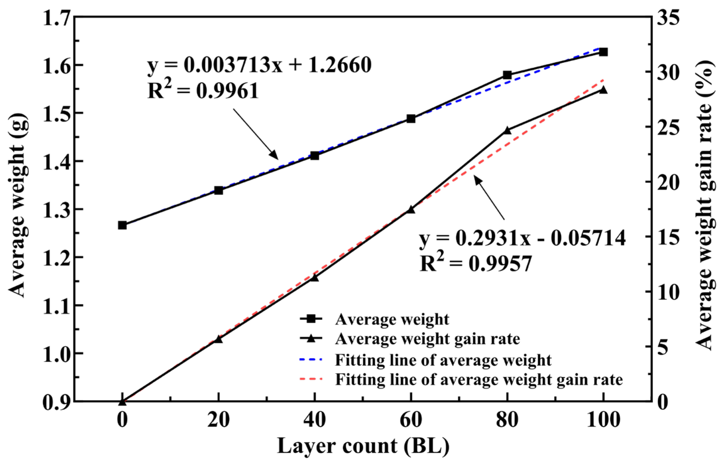
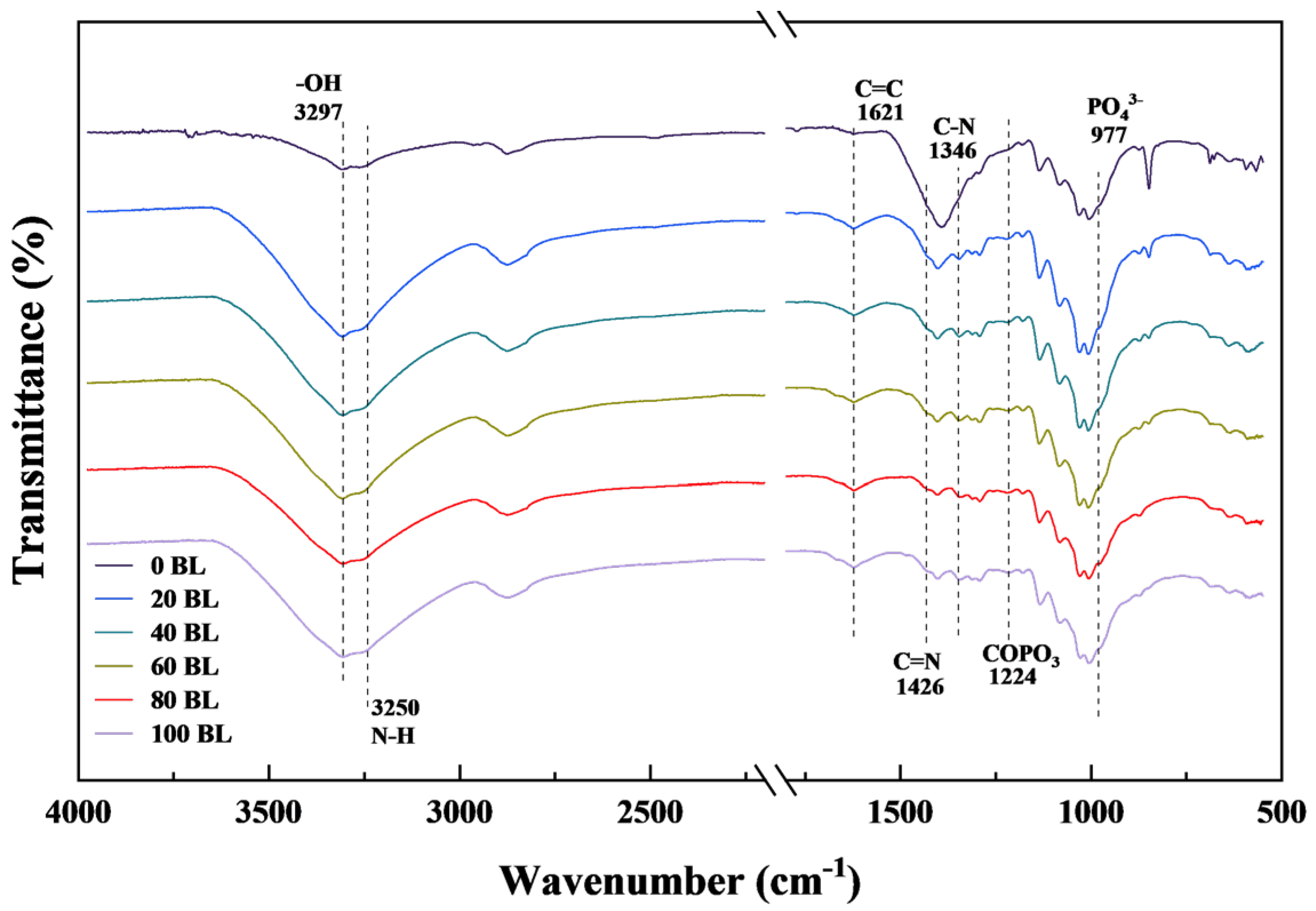
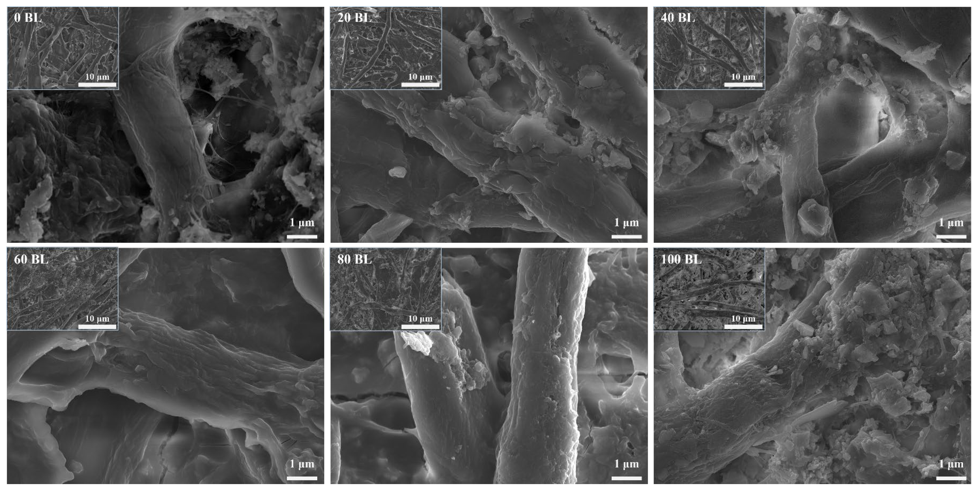
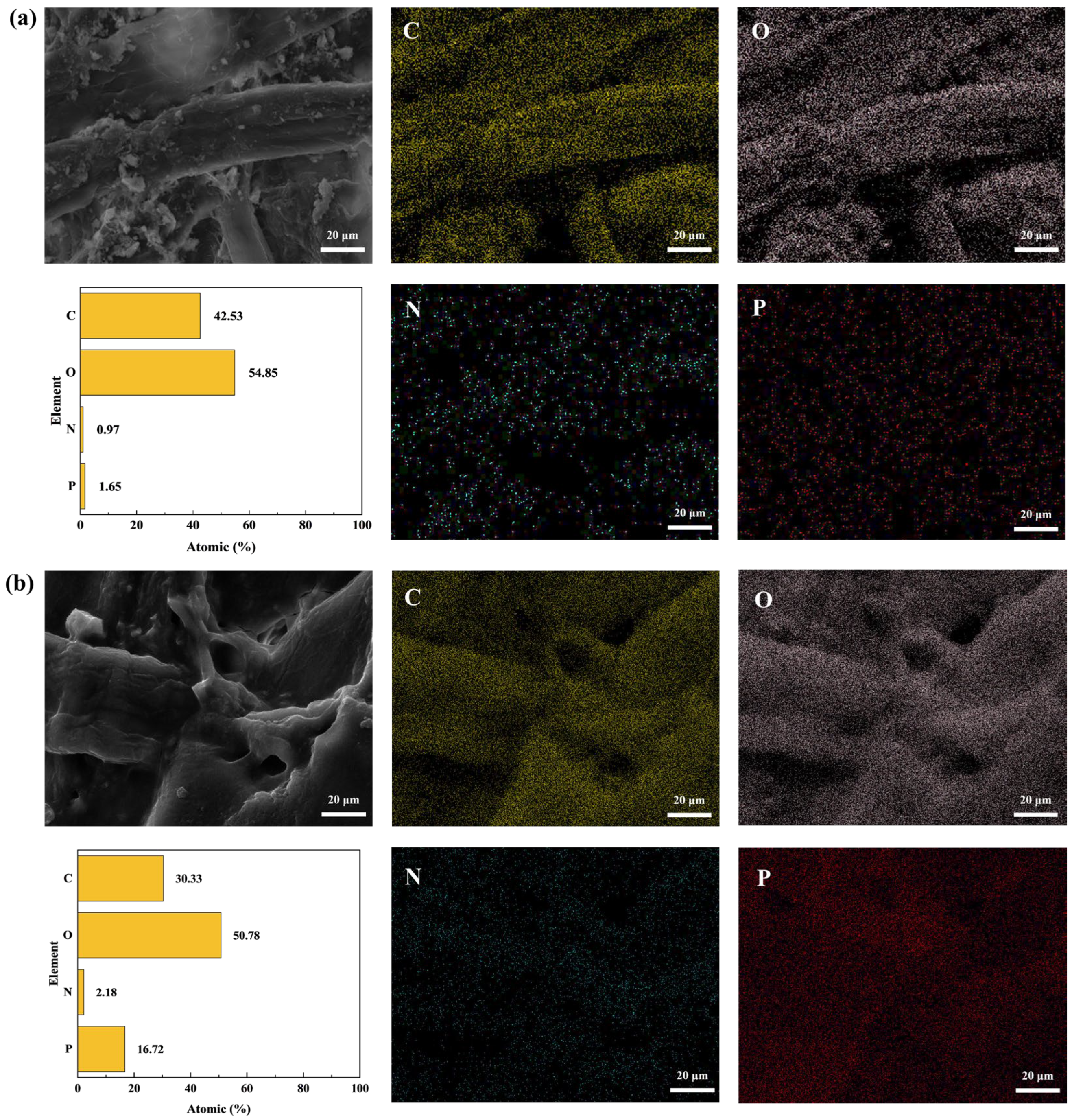
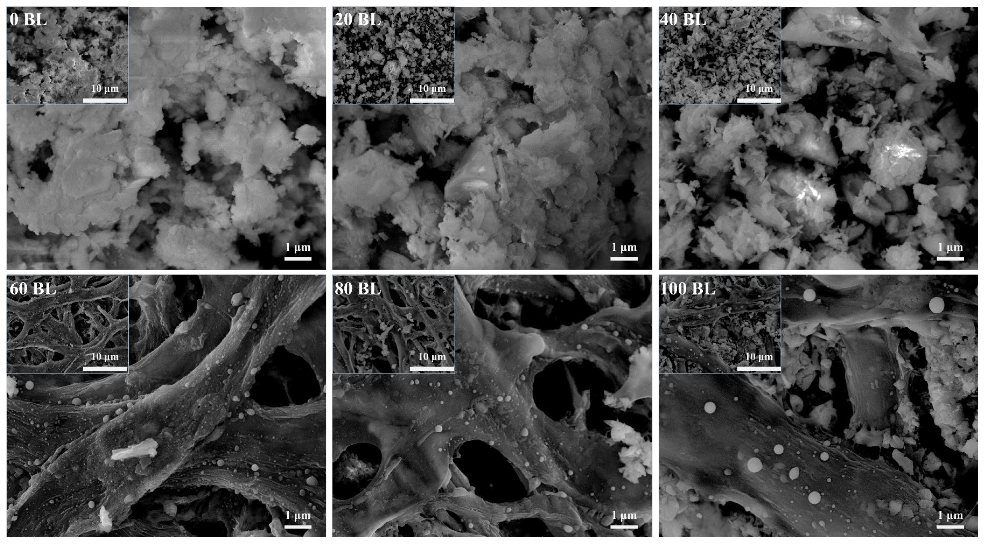
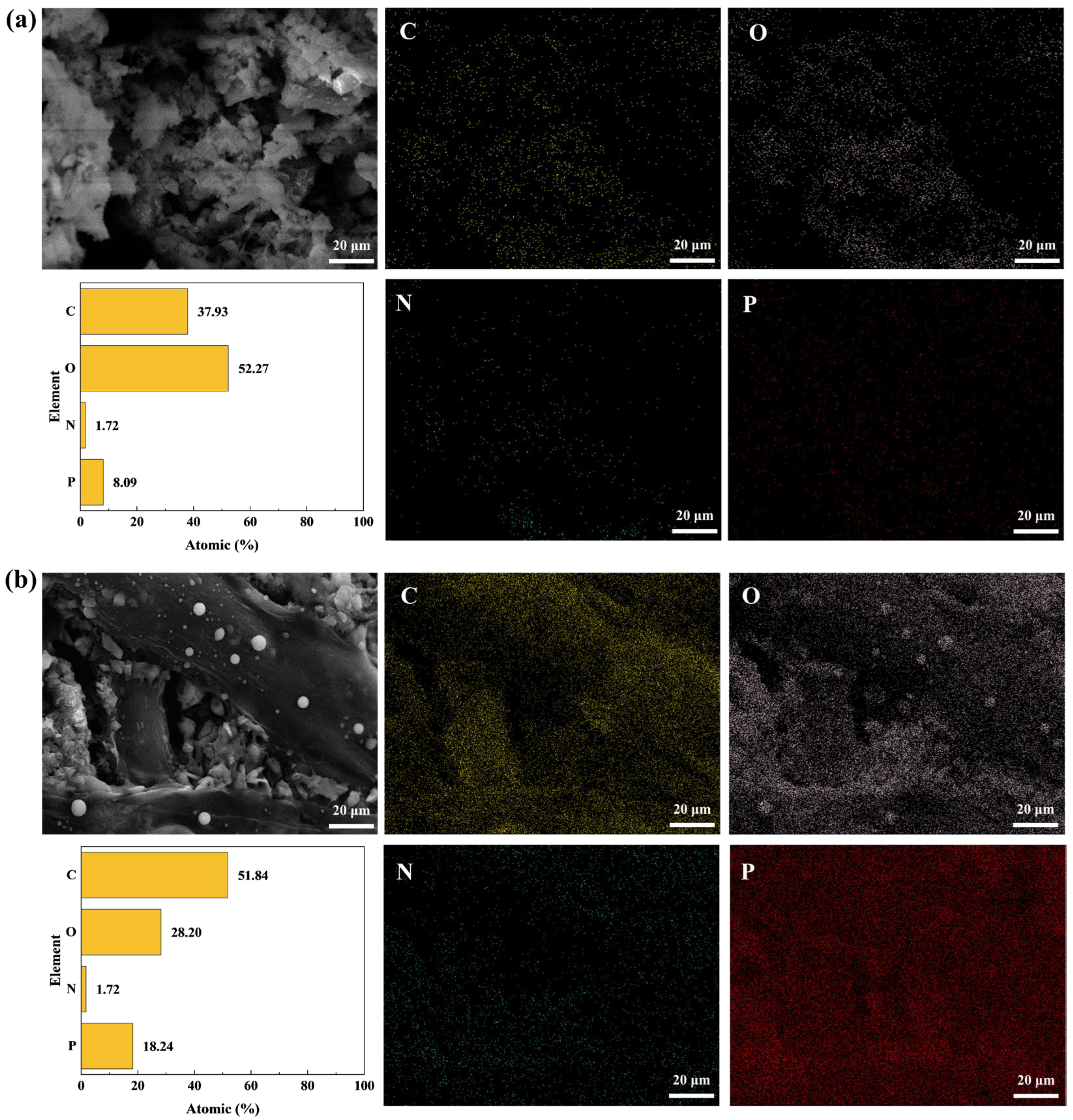
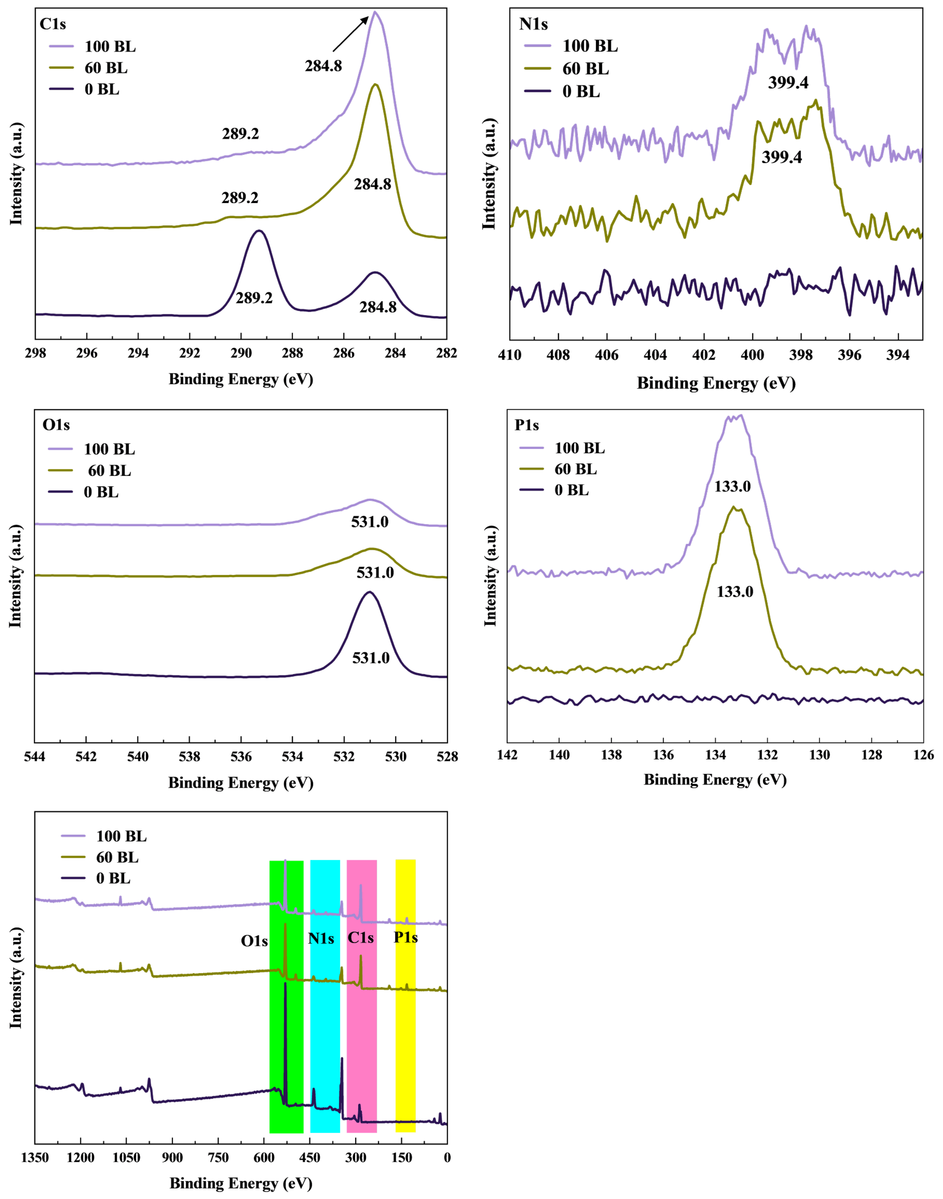
3.1. Characterization of the Self-Assembly Coating

In previous studies, many methods have been used to prepare the self-assembly coating for different materials. Some are popular and widely used, like immersion, painting, and spraying. All these methods will bring lots of water to the materials if they use aqueous solutions for self-assembly. Yet it is not suitable for paper in many applications. The hydrogen bond association provides the connection of natural cellulose fiber, which will easily be disrupted by the water because the oxygen–hydrogen bond of water forms hydrogen bonds with cellulose fiber. Figure 5 shows the photographs of the original paper and paper that had been treated with different methods for self-assembly coating, including spraying, painting, immersion, and ultrathin adsorption methods. It is easy to see that the paper was seriously deformed using the spray, painting, and immersion methods. In contrast, the ultrathin adsorption method treats paper exhibited less change than the original paper. This is thanks to the superfine solution mist and the short adsorption time of the ultrathin adsorption method, which only brings a little water to the paper.
The coated paper of 20 BL, 40 BL, 60 BL, 80 BL, and 100 BL also reflects the advantages of the ultrathin adsorption method on low-impact paper morphology. Figure 6 gives the top- and side-view photographs of the coated paper. The photographs show that the coated paper became slightly transparent in some areas as the coated layers reached 80 BL. With the coated layers increasing, more and more biomaterials not only covered the paper surface but also entered the paper through the gaps in the cellulose fiber. Those materials on or in the paper crystallize after natural drying, and the paper produces visual changes. From the macro view, the coated paper exhibited fewer changes in surface topography, except for the lesser amount of warping on the paper edge. All of these express that the ultrathin adsorption method is a good way to prepare the self-assembly coating and keep the paper surface clean and flat.
The weight change of the coated paper is the key indicator reflecting the thickness of the specific flame-retardant coating, just like the paper grammage for paper. For the coating prepared by the ultrathin adsorption method, Figure 7 gives the average weight and its gain rate of the paper samples. Both the average weight and its gain rate show good linear growth, and the R-squared of each fitting line is over 0.995, performing more linearly than other methods that have been reported before. This reveals that the ultrathin adsorption method has better stability and controllability. The slope of the average weight gain rate fitting line is 0.2931, which is much lower than the other methods: only about one-fifth of the spray method and one-twentieth of the immersion method. So, the coating prepared by the ultrathin adsorption method is much thinner than the other commonly used methods. And that is why this method is named with ultrathin.
The infrared spectroscopy, measured by FTIR, provides the molecular structure information about the self-assembly coating of paper samples. The result is shown in Figure 8. Compared to the original paper, the coated paper had an enhanced telescopic vibration peak of the hydroxyl group that appeared at ~3297 cm−1, primarily attributed to the starch solution. The water-soluble starch and cationic starch contained a lot of hydroxyl groups, which could hydrogen bond together during the mixed solution retrogradation [45]. So, the starch solution adsorbed on the paper surface directly improves the hydroxyl group contained in the self-assembly coating. When the coated paper catches fire, those hydroxyl groups may react to generate H2O and suppress the combustion. The absorption peaks appeared at ~1346 cm−1 by the C–N stretching vibration of the quaternary ammonium group, indicating cationic starch had assembled in the coating [46]. The characteristic absorption bands of N–H, C=C, and C=N in ATP appeared at ~3250 cm−1, ~1621 cm−1, and ~1426 cm−1, respectively [47]. The ~1224 cm−1 and ~977 cm−1 corresponded to COPO3 and PO43− absorption bands of PA [48]. The FTIR results indicate that all biomaterials were successfully assembled and coated on the paper surface.
The microscopic morphology of the paper samples, including the original paper and the coated paper, was imaged by SEM and is shown in Figure 9. The original paper surface and cellulose fiber exhibited more smoothness than the coated paper. Following the assembled layers increasing, the paper surface adsorbed more and more flame retardants, and the cellulose fiber coarsened to make the paper surface rough. These reveal the biomaterials adhered well to the cellulose fiber, where the coating assembled as expected. Figure 9 also shows that fiber gaps in the paper samples gradually filled with flame retardants as the layers increased. However, this phenomenon performed less well than other methods due to the fewer flame retardants adsorbed in the coating preparation process. The flame retardants that covered the cellulose fiber and filled in the fiber gaps formed a structured flame-retardant coating that could protect the coated fiber and isolate the inside of the paper from fire. According to the intumescent flame-retardant function, the coating will become thicker and expand more coverage when heated by fire.
The coating elemental mapping about the C, O, N, and P elements of the original paper and 100 BL paper is shown in Figure 10. According to the comparison between elemental mappings, the content of the P element in the original paper was much less than 100 BL paper, due to ATP and PA in the coating having lots of phosphates. As previous studies reported, the phosphate group is conducive to flame retardancy and plays a vital role in intumescent flame retardancy. For the N element, which was mainly contained in the cationic starch and ATP, it slightly increased in 100 BL paper. With the increase in the P and N elements, the contents of the C and O elements showed a decreasing performance on related percentages. All the elemental mappings show that flame retardants on the coated paper were densely and evenly distributed, reflecting that the coating assembled tightly.

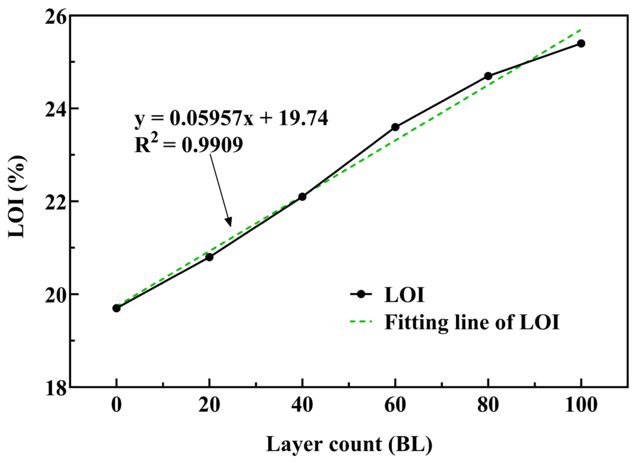
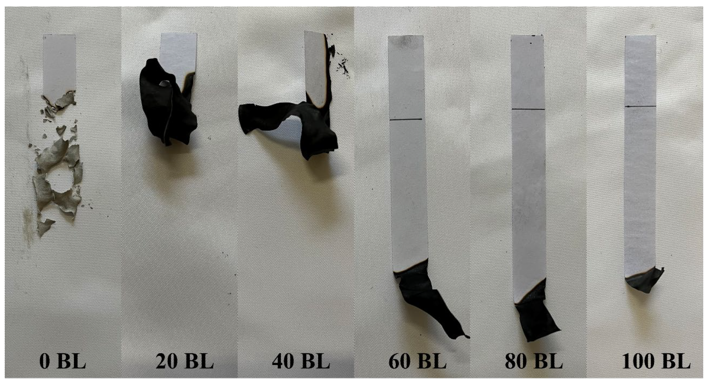
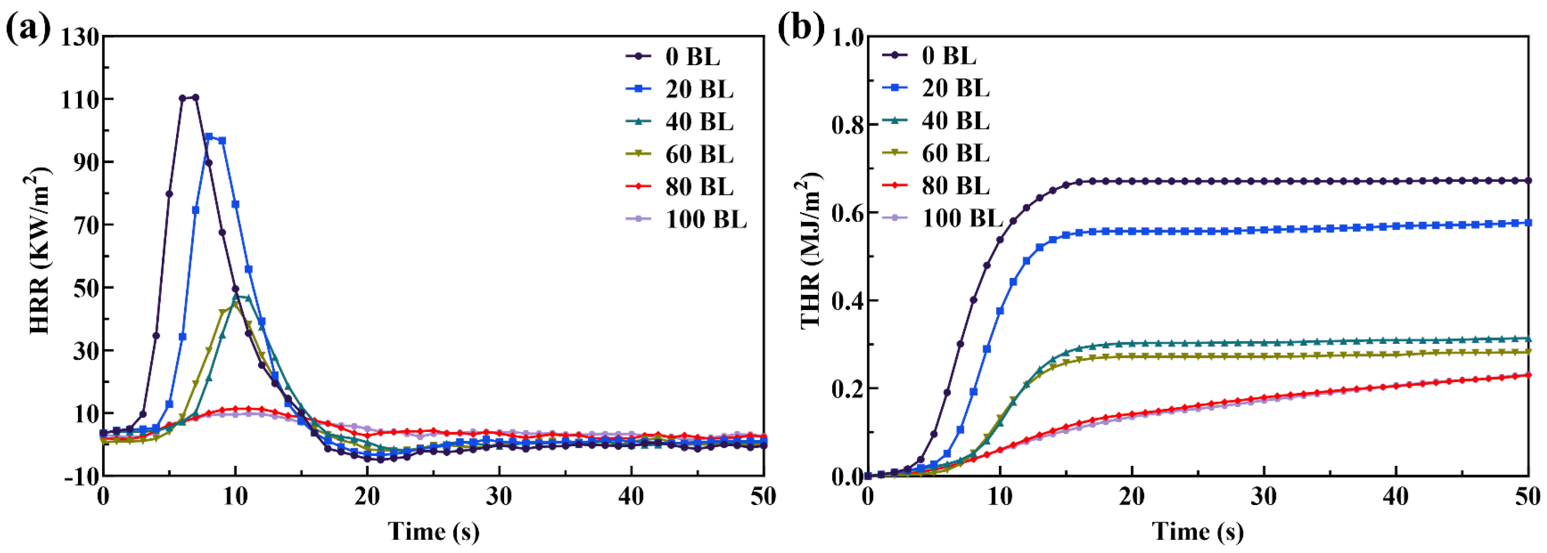
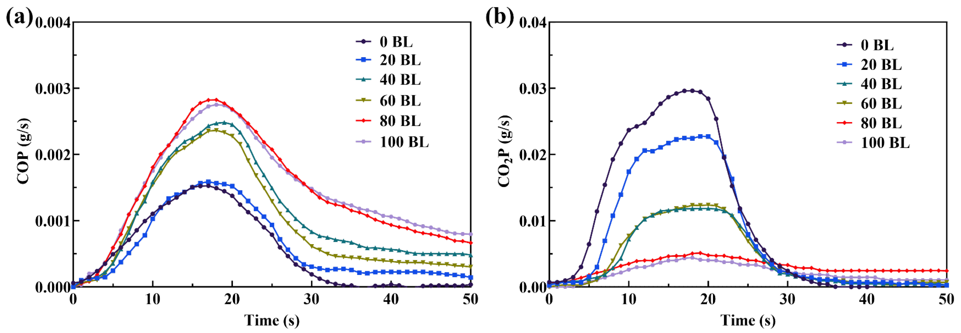
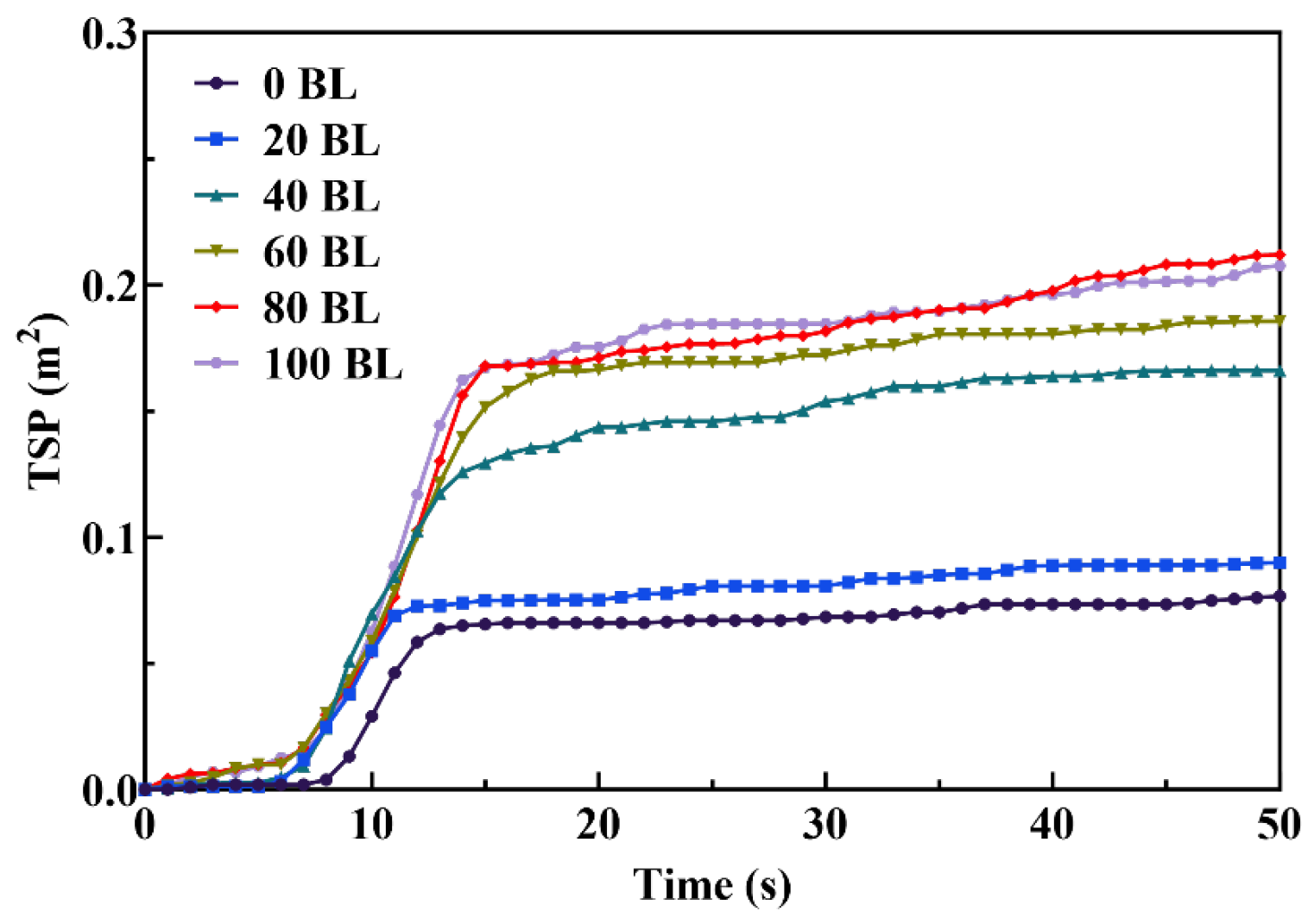
3.2. Combustion Behaviors of the Paper Samples

The LOI is the minimum oxygen concentration for the flaming combustion of the paper samples, directly reflecting their flammability. The LOI of paper samples was tested and is shown in Figure 11. As the assembled layers increase, the LOI shows a good linear increasing trend, reaching 25.40% when the layer count was 100 BL. The LOI of 40 BL paper was 22.10%, which is over the oxygen concentration of the air. The linear increasing trend of the LOI fitting line suggests that the flame-retardant effect of the coating is controllable. Due to the flame retardants having a uniform distribution on the surface of all paper samples treated with the ultrathin adsorption method, the flame retardants in each layer will provide a unanimous action on the flame-retardant function. As a result, the flammability of the paper samples can be adjusted by the layer count of the self-assembly coating. The slope of the fitting line shows that the increasing trend of the LOI was a little less than that of paper prepared by other methods, because of the fewer flame retardants adsorbed on the paper surface when using the ultrathin adsorption method.
The flammability of the paper samples was further evaluated by HBT. The burning rate and self-extinguishing performance of the paper samples were obtained and shown in Table 1. During the HBT, the 0 BL, 20 BL, and 40 BL paper samples could not self-extinguish in the test, while the 60 BL, 80 BL, and 100 BL paper samples were self-extinguished. The burning rate of the paper samples tended to decrease as the assembled layers increased, which indicates that the flame-retardant effect of coating was gradually enhanced with the assembled layers increase.
The burnt length of the paper samples varied with different layer count numbers after HBT, as shown in Figure 12. The original paper burnt leaves only residual ash, which had an incomplete structure and showed light grey. This is different from the coated paper. With the assembled layers increasing, the burnt length of the coated paper became shorter, which reflects the coating has a good effect on flame retardancy. At the same time, all the coated paper produced the structurally stable residual after HBT, which was colored black. These morphology performances indicate that the self-assembly coating uniformly covered the paper surface and could work effectively.
The CONE test was carried out to perform the combustion behavior of the paper samples, and the results are shown in Figure 13. The heat release rate (HRR) and total heat release rate (THR) show the difference in the combustion behavior of the paper samples. The peak HRR declined with the assembled layers increased. Compared between the original paper and the coated paper, the peak HRR of 100 BL paper reduced from 110.45 kW/m2 to 9.87 kW/m2, only 8.94% of the original paper. The significant reduction in the peak HRR suggested the coating had suppressed the exothermic combustion reaction of paper during combustion. Due to this effectiveness, the THR also shows a high reduction from the original paper to the coated paper. So, the reduced performance on both HRR and THR indicated the flame-retardant coating made the combustion of paper incomplete. According to the theory of intumescent flame retardancy, the coating is carbonized when the paper surface is exposed to the fire and forms the protection layer that isolates the paper from the fire and heat. This protection layer will become more sturdy and effective while the assembled layers increase.
The mainly gaseous combustion products of the paper samples, including carbon monoxide and carbon dioxide, show the opposite trend in the production rate as the assembled layers increase. Figure 14 exhibits their variations during the paper samples CONE test. With the assembled layers increasing, the peak value of the carbon monoxide production rate (COP) of the paper samples was increased, while the related peak value of the carbon dioxide production rate (CO2P) was decreased. For the peak value of COP, the 0 BL and 20 BL paper had similar values of about 0.0015 g/s, where the values of 40 BL to 100 BL paper were higher and ranged from 0.0024 g/s to 0.0028 g/s. At the same time, 0 BL paper had the highest peak value of CO2P, approximately 0.030 g/s, whereas 100 BL paper had the lowest one, approximately 0.004 g/s, only 13.33% of 0 BL paper. According to the combustion theory, these variations reflect the increase in the assembled layers led to the complete combustion of the paper samples transited to incomplete combustion, and then continuing to intensify. The intense incomplete combustion will make the coating, which is made of flammable biomaterials, intumesced and carbonized to produce the dense carbon layer on the paper surface and protect the paper from the fire. The total smoke production (TSP) of the paper samples, as shown in Figure 15, also increased with the assembled layers, similar to COP. The TSP and the main smoke-producing stage of 100 BL paper were nearly more than double those of 0 BL paper. The more smoke produced by the coated paper than the original paper was mainly from the incomplete combustion of the coating. The incomplete combustion of the coating will enhance its intumesced and carbonized processes to form a better carbon layer. And that is why the coated paper with more assembled layers showed a better flame-retardant performance.

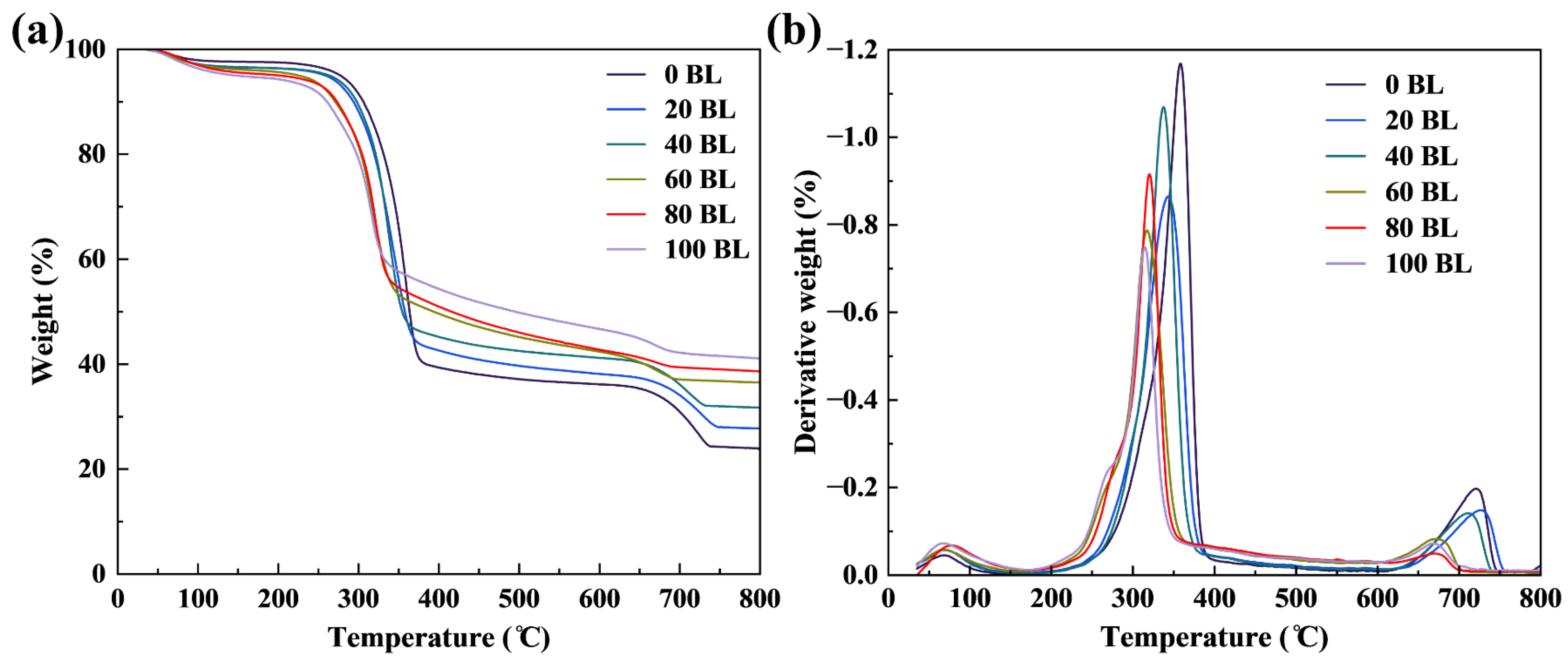
3.3. Thermal Behaviors of the Paper Samples

The thermogravimetric variations of the paper samples were performed using TGA under nitrogen and air atmospheres. Figure 16 and Figure 17 present TGA results and their derivatives (DTG) under different atmospheres, with the corresponding key data recorded in Table 2 and Table 3. In TGA, the 5% weight-loss temperature is regarded as the initial decomposition temperature and marked T5%. The Tmax is the corresponding temperature of the maximum weight loss rate. TGA of the paper samples under the nitrogen atmosphere is a one-step thermal decomposition process, whereas TGA under the air atmosphere is a two-step thermal decomposition process. So, TGA under the nitrogen atmosphere has one Tmax value, marked Tmax. TGA under the air atmosphere has two Tmax values, marked Tmax1 and Tmax2. The variation of T5%, both under nitrogen and air atmospheres, indicated the thermal decomposition of the coated paper started earlier than the original paper. That reflects that the coating will react before the paper combusts as the temperature increases. As the number of assembled layers increases, this phenomenon becomes more significant. T5% of 100 BL paper is approximately 67.87% that of 0 BL paper under the nitrogen atmosphere, which is only 50.85% under the air atmosphere. Tmax and Tmax1 of the coated paper are also lower than the original paper, which may be affected by the coating with the low thermal decomposition temperature. As the assembled layers increase, more and more flame retardants are absorbed on the paper surface, and this affection will become more apparent. Hence, Tmax and Tmax1 of the paper samples continue to reduce as the number of assembled layers increases. The acid sources, PA and ATP, used in the coating have low thermal decomposition temperatures that let the coating respond earlier to form carbon layers and provide fire protection to the paper [49,50]. The variations of DTG show the maximum weight loss rate of the original paper is higher than the coated paper, which also reflects the protection effect of the coating. The variation of TGA under the air atmosphere shows a two-step thermal decomposition process [51]. The first step is the depolymerizing and dehydrating processes of the paper, which produces volatile products and aliphatic char. The second step is the thermal oxidating process of the first step products, which produces aromatic char and volatile products. As the assembled layers increase, the Tmax2 exhibits a contrary tendency to Tmax and Tmax1. That may be caused by the protection effect of the carbon layer, which is produced in the first step. Due to this protection effect, the weight of the coated paper residues significantly improved at the end of TGA. The weight of 100 BL paper residues is 1.71 times and 2.38 times that of 0 BL paper under nitrogen and air atmospheres, respectively. According to TGA, the residues of the paper samples under the nitrogen atmosphere are mainly char residues, so the extra residue mass of the coated paper can be regarded as the weight of the carbonized coating. Therefore, the extra residue mass can be used to express the capacity of coating carbonizing. For intumescent flame retardants, the greater capacity of coating carbonizing may bring a better flame retardant effect. Correspondingly, the residues of the paper samples, produced under the air atmosphere, are mainly ashes, so the extra residue mass is from the incombustible component of the coating. This extra residue mass reflects the non-carbon content of the carbon layers. Furthermore, the effective carbon content of the coating could be calculated by the extra residue mass under the nitrogen atmosphere minus that under the air atmosphere.
The microscopic morphology and elemental mapping of the burnt residues after HBT, including the original paper and the coated paper, have been imaged by SEM-EDS, and are shown in Figure 18 and Figure 19, respectively. The burnt residues of the original paper were much looser than the coated paper, and the fibrous structure of most paper fiber had been disintegrated during the combustion process of HBT. The coated paper outperformed the original paper on the fibrous structure, and most paper fiber contours remained intact when the assembled layers exceeded 60 BL. That is just like the macro performance of the paper. The original paper burnt residues are fragmentable, whereas the coated paper burnt residues are more intact and could bear a little external force. Over the observation from the photographs of 20 BL paper to 100 BL paper, there could find the carbon layers covered on the paper fiber surface gradually became clear. The carbon layers, produced by the coating carbonizing, provide the solid-phase flame retardant effect that will starve the fuel source of the combustion. For 60 BL to 100 BL paper, many different-sized bubbles could be observed on the surface of paper fiber residues. Those bubbles may be the solidified foams produced by the inert gases boiling over the carbon layer surface. The inert gases, the key factor for carbon layers intumescing, may mainly be generated by the reaction between acid and carbon sources or the thermal decomposition of intumescent flame retardants. Moreover, the inert gases also provide the gas-phase flame retardant effect that will dilute the oxygen and fuel concentrations and suppress the combustion.
Compared with the elemental mappings of burnt residues between 0 BL and 100 BL paper, every elemental mapping has enhanced image performance on the coated paper. The residual percentage content of the C element has significantly increased 13.91%, whereas the O element has decreased 24.07%. That directly indicates the carbon layers covered on the coated paper surface and inhibited the oxidation of the combustion. Although the N element has the same percentage content value in both 0 BL and 100 BL paper burnt residues, the image performance of 100 BL paper is much stronger, revealing many N element content materials are not burnt out during combustion. The P element has improved 10.15% from 100 BL to 0 BL paper, inferring phosphoric acid produced by PA and ATP left in the carbon layers and enhanced their carbonization [52]. The elemental mappings between the paper samples and their burnt residues also reflect the changes before and after flame retardant application. For 0 BL paper, the percentage content of every element changed less between 0 BL paper and its burnt residue. The loss of the C element is due to the combustion that made it oxidate into gaseous carbon oxidation products. For 100 BL paper, the C element percentage content was greatly improved, indicating the coating bulk carbonizes into solid char residues in combustion to form the carbon layers. Meanwhile, the oxidation of the coating in combustion had been inhibited, where the O element percentage content was reduced. The N element percentage content decreased because the N element content materials produced inert gases during combustion.
Furthermore, XPS was utilized to provide more information about the elemental composition of the paper sample burnt residues, and the results are shown in Figure 20 and Table 4. According to changes among 0 BL, 60 BL, and 100 BL paper on the C, N, O, and P elements, the coating directly participated in the combustion process and constructed the new surface of the paper. The peaks were observed at 289.2 eV and 284.8 eV in C1s, which indicated the presence of C=O–C and C–C bonds, respectively [53]. The C=O–C reflects that the original paper produced carbonates on the burnt residue surface during the combustion. With the assembled layers increasing, the peak at 284.8 eV was enhanced for the coated paper, whereas the peak at 289.2 eV disappeared. That reveals the coating has successfully carbonized on the paper surface, leaving the carbon layers to block the paper oxidized. This is also reflected in the changes of the peak at 531.0 eV in O1s, which indicated the oxides have reduced on the burnt residue surface of coated paper than the original paper. The atomic percentage of C1s shows the coated paper has nearly doubled the C element content on the burnt residue surface than the original paper, whereas the O1s has reduced by about a quarter. The peak observed from the coated paper at 399.4 eV in N1s indicates the presence of the N element in the burnt residue surface [54]. Compared with the original paper, this does not appear in its N1s. Therefore, it can be confirmed the peak is from the partial combustion residues of ATP and cationic starch. As previous studies report, one of the main functions of the nitrogen-containing materials in intumescent flame retardants is working as blowing agents to produce inert gases foaming and intumescing the carbon layers. The peak at 133.0 eV in P1s, which only appeared in the coated paper, is the P element from burnt residues of PA and ATP in the coating [55]. During the combustion, PA and ATP could release phosphoric acid, carbonizing the coating to form the carbon layers.

3.4. Mechanical Properties of the Paper Samples

In addition to flame-retardant, the mechanical properties are another vital characteristic of the paper used in medical care. The tensile index (TS), tear index (TR), and folding endurance (MIT) of the paper samples were tested, and the results are shown in Table 5. With the assembled layers increasing, the TS and TR of the paper samples exhibited varying degrees of increase, where MIT was observed to decrease. Compared with the original paper, TS and TR of the 100 BL paper were respectively improved by 41.08% and 30.00%, whereas MIT was reduced by 68.75%. The mechanical properties of the coated paper continue to meet the basic requirements for medical care in the simulation of practical medical applications. The different variations in mechanical properties of the coated paper reflect that the coating and its preparation methods have different influences on the mechanical properties of paper. For the ultrathin adsorption method, one enormous advantage of it over others is that leaves much less water on the paper surface during its preparation process. So, less water left on the paper surface had less disruptive effects on the hydrogen bonds of the paper fiber and better protected the existing fiber connection. In addition, the flame retardant absorbed on the paper surface will act like the glue that binds the cellulose fiber tightly. Finally, the coated paper performed better on TS and TR than the original paper. However, the shortcomings of the coating also need to be noticed. The flame retardants adsorbed on the paper surface and dried to form a hard shell that covered the cellulose fiber and its gaps. The covered shell provides fire protection to the paper, but it also cures the cellulose fiber on the paper surface and restrains its flexible connection. Although the ultrathin adsorption method absorbs a little flame retardant on the paper surface, the formed coating will still adversely impact the MIT performance of the coated paper.

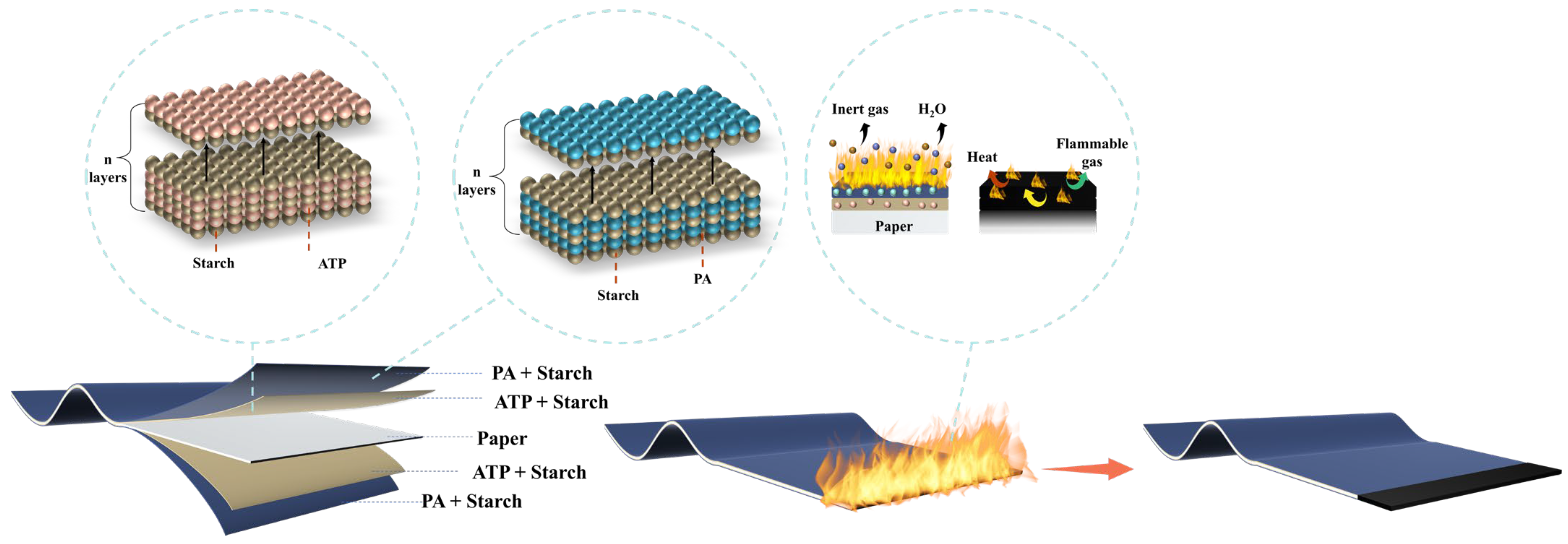
3.5. Flame-Retardant Mechanism

According to the theory of intumescent flame retardancy, the necessary carbon source, acid source, and blowing agent combine to constitute the fundamental components of intumescent flame retardants [56]. Figure 21 shows the flame-retardant mechanism of the coated paper. In this paper, starch with rich carbon content is the main component used in both internal and external layers, and it mainly serves as the carbon source of the flame-retardant coating. ATP and PA both have phosphate groups that will work as the acid source to produce phosphoric acid when they disintegrate at high temperatures. These acids will dehydrate starch in the coating to create the carbon layer. The gas produced by the blowing agent further makes the carbon layer foam and intumesce. The intumescent carbon layer covering the paper surface has a structure just like the sponge, which will block the combustion heat transfer from the fire to the paper and cut off the fuel mass transfer path of the combustion. The structural design of internal and external layers provides dual fire protection for the paper. Due to the different thermal decomposition temperatures of PA and ATP, the external layers will act earlier than the internal layers. Compared with the previous studies, the successive formation of the different carbon layers by the external and internal layers will provide a stronger dual-carbonized-layer structure to the coating when exposed to fire and enhance its flame-retardant effect. Furthermore, the different response temperatures of the internal and external layers also provide a continuous stepped response on the flame-retardant for the coated paper, which lets the coating still be usable after being exposed to fire with only external layers carbonized.

4. Conclusions

In this paper, the ultrathin biomaterial coating has been prepared using the ultrathin adsorption method to improve the medical paper flame retardancy. The coating has internal and external layers, where the internal layers are layer-by-layer assembled by starch and adenosine triphosphate via electrostatic interactions, and the external layers are assembled in the same way by starch and phytic acid. With the different thermal decomposition temperatures of PA and ATP, the dual-layer structural design offered the successive carbonized capacity to the coating, which will produce stronger carbon layers when exposed to fire. On the benefits of less water brought to the paper, the ultrathin adsorption method makes less change on the paper than other methods. The FTIR and SEM-EDS results show that all biomaterials were successfully assembled on the paper surface, and the cellulose fiber became coarsened to make the paper surface rough. Due to the ultrathin biomaterial coating, the flame retardancy of the coated paper has significantly improved. The LOI of 100 BL paper was 25.40% and 1.29 times that of the original paper. This was also reflected in the CONE test, where both peak HRR and THR were reduced for the coated paper. The changes of the COP and CO2P reveal the complete combustion of the paper samples transited to incomplete combustion by the coating. Due to the low thermal decomposition temperatures of PA and ATP, the coating responds earlier to form carbon layers and provides fire protection to the paper. As a result, the coated paper exhibited a higher maximum weight loss rate than the original paper and left more residues at the end of TGA. The morphology and element analyses indicated that coating provides the solid-phase and gas-phase flame retardant effects on paper combustion and forms the carbon layers covering the paper surface. Furthermore, although the mechanical properties of coated paper exhibited varying impacts due to the coating, they still met the basic requirements for medical care in the simulation of practical medical applications.

Author Contributions

Conceptualization, Z.S.; methodology, Z.S.; software, X.L.; validation, X.L. and J.L.; formal analysis, X.L.; investigation, Z.S.; resources, J.L.; data curation, X.L.; writing—original draft preparation, X.L.; writing—review and editing, Z.S. and C.Y.; visualization, X.L.; supervision, X.X.; project administration, X.P.; funding acquisition, Z.S. All authors have read and agreed to the published version of the manuscript.

Funding

This research was funded by the Fundamental Research Funds for the Central Universities, the Research Fund for Advanced Talents of Jiangsu University (No. 15JDG147), and the Research Project of the School of Emergency Management of Jiangsu University (No. JG-04-13, JG-03-20), the Life and Health Soft Project of Huai’an Natural Science (No. 2023KX0088).

Institutional Review Board Statement

Not applicable.

Informed Consent Statement

Not applicable.

Data Availability Statement

The data presented in this study are available on request from the corresponding author.

Acknowledgments

Thanks to Fei Xu and Qiaosheng Hu of Lianshui People’s Hospital for their technical support. Thanks to Shandong Yicheng Equipment Co., Ltd. for providing the mechanical properties tests of paper samples.

Conflicts of Interest

The authors declare no conflicts of interest.

References

  1. Abdelkader, A.; Moos, C.; Pelloux, A.; Pfeiffer, M.; Alter, C.; Kolling, S.; Keck, C.M. Tablets Made from Paper—An Industrially Feasible Approach. Pharmaceuticals 2022, 15, 1188. [Google Scholar] [CrossRef] [PubMed]
  2. Abdelkader, A.; Nallbati, L.; Keck, C.M. Improving the Bioactivity of Norfloxacin with Tablets Made from Paper. Pharmaceutics 2023, 15, 375. [Google Scholar] [CrossRef] [PubMed]
  3. Abdelkader, A.; Preis, E.; Keck, C.M. SmartFilm Tablets for Improved Oral Delivery of Poorly Soluble Drugs. Pharmaceutics 2022, 14, 1918. [Google Scholar] [CrossRef] [PubMed]
  4. Eckert, R.W.; Wiemann, S.; Keck, C.M. Improved Dermal and Transdermal Delivery of Curcumin with SmartFilms and Nanocrystals. Molecules 2021, 26, 1633. [Google Scholar] [CrossRef] [PubMed]
  5. Ornik, J.; Heidrich, L.; Schesny, R.; Castro-Camus, E.; Keck, C.M.; Koch, M. Non-destructive crystallinity assessment of indomethacin in tablets made from smartFilms® using terahertz time-domain spectroscopy. Sci. Rep. 2022, 12, 1–7. [Google Scholar] [CrossRef]
  6. Stumpf, F.; Keck, C.M. Tablets made from paper. Int. J. Pharm. 2018, 548, 812–819. [Google Scholar] [CrossRef] [PubMed]
  7. Rauscher, M.; Rauscher, A.; Hu, L.Y.; Schlitt, H.J.; Krauß, S.; Illg, C.; Wolfertstetter, P.R.; Hofmann, A.; Knorr, C.; Denzinger, M. Influence of Accumulation of Humidity under Wound Dressings and Effects on Transepidermal Water Loss (TEWL) and Skin Hydration. Appl. Sci. 2024, 14, 7739. [Google Scholar] [CrossRef]
  8. Shin, Y.S.; Kim, H.J.; Moon, N.; Ahn, Y.H.; Kim, K. The effects of uncoated paper on skin moisture and transepidermal water loss in bedridden patients. J. Clin. Nurs. 2012, 21, 2469–2476. [Google Scholar] [CrossRef]
  9. Lam, A.K.-Y. Update on Adrenal Tumours in 2017 World Health Organization (WHO) of Endocrine Tumours. Endocr. Pathol. 2017, 28, 213–227. [Google Scholar] [CrossRef] [PubMed]
  10. Hales, B.F.; Robaire, B. Effects of brominated and organophosphate ester flame retardants on male reproduction. Andrology 2020, 8, 915–923. [Google Scholar] [CrossRef]
  11. Mughal, B.B.; Demeneix, B.A. Flame retardants and increased risk of thyroid cancer. Nat. Rev. Endocrinol. 2017, 13, 627–628. [Google Scholar] [CrossRef]
  12. Shen, C.; Zhang, K.; Shi, J.; Yang, J.; Wang, Y.; Li, Z.; Dai, H.; Yang, W. Association between brominated flame retardants and risk of endocrine-related cancer: A systematic review and meta-analysis. Toxicol. Lett. 2024, 394, 11–22. [Google Scholar] [CrossRef]
  13. Terrell, M.L.; Rosenblatt, K.; Wirth, J.; Cameron, L.L.; Marcus, M. Breast cancer among women in Michigan following exposure to brominated flame retardants: Table 1. Occup. Environ. Med. 2016, 73, 564–567. [Google Scholar] [CrossRef]
  14. You, L.; Kou, J.; Wang, M.; Ji, G.; Li, X.; Su, C.; Zheng, F.; Zhang, M.; Wang, Y.; Chen, T.; et al. An exposome atlas of serum reveals the risk of chronic diseases in the Chinese population. Nat. Commun. 2024, 15, 1–18. [Google Scholar] [CrossRef]
  15. Biswal, T.; BadJena, S.K.; Pradhan, D. Sustainable biomaterials and their applications: A short review. Mater. Today Proc. 2020, 30, 274–282. [Google Scholar] [CrossRef]
  16. Joyce, K.; Fabra, G.T.; Bozkurt, Y.; Pandit, A. Correction to: Bioactive potential of natural biomaterials: Identification, retention and assessment of biological properties. Signal Transduct. Target. Ther. 2021, 6, 175. [Google Scholar] [CrossRef]
  17. Troy, E.; Tilbury, M.A.; Power, A.M.; Wall, J.G. Nature-Based Biomaterials and Their Application in Biomedicine. Polymers 2021, 13, 3321. [Google Scholar] [CrossRef] [PubMed]
  18. Yadav, M.; Agarwal, M. Biobased building materials for sustainable future: An overview. Mater. Today Proc. 2021, 43, 2895–2902. [Google Scholar] [CrossRef]
  19. Liu, C.-M.; Wu, S.-Y. From biomass waste to biofuels and biomaterial building blocks. Renew. Energy 2016, 96, 1056–1062. [Google Scholar] [CrossRef]
  20. Popa, V.I. Biomass for fuels and biomaterials. In Biomass as Renewable Raw Material to Obtain Bioproducts of High-Tech Value; Popa, V.I., Volf, I., Eds.; Elsevier: Amsterdam, The Netherlands, 2018; pp. 1–37. [Google Scholar]
  21. Cho, E.J.; Trinh, L.T.P.; Song, Y.; Lee, Y.G.; Bae, H.-J. Bioconversion of biomass waste into high value chemicals. Bioresour. Technol. 2019, 298, 122386. [Google Scholar] [CrossRef] [PubMed]
  22. Enawgaw, H.; Tesfaye, T.; Yilma, K.T.; Limeneh, D.Y. Multiple Utilization Ways of Corn By-Products for Biomaterial Production with Bio-Refinery Concept; a Review. Mater. Circ. Econ. 2023, 5, 1–14. [Google Scholar] [CrossRef]
  23. Xu, X.; Zhou, Q.; Yu, D. The future of hydrogen energy: Bio-hydrogen production technology. Int. J. Hydrogen Energy 2022, 47, 33677–33698. [Google Scholar] [CrossRef]
  24. Leong, Y.K.; Chang, J.S. Chapter 6—Bioprocessing for production and applications of bioplastics from algae. In Biomass, Biofuels, and Biochemicals; Elsevier: Amsterdam, The Netherlands, 2022; pp. 105–132. [Google Scholar]
  25. Chen, S.; Wu, F.; Hu, Y.; Lin, S.; Yu, C.; Zhu, F.; Wang, X. A fully bio-based intumescent flame retardant for poly(butylene succinate). Mater. Chem. Phys. 2020, 252, 123222. [Google Scholar] [CrossRef]
  26. Zhang, S.; Jin, X.; Gu, X.; Chen, C.; Li, H.; Zhang, Z.; Sun, J. The preparation of fully bio-based flame retardant poly(lactic acid) composites containing casein. J. Appl. Polym. Sci. 2018, 135, 46599. [Google Scholar] [CrossRef]
  27. Xu, F.; Ma, W.; Wang, W.; Wang, H.; An, S.; Zhu, Z.; Wang, R. Fully bio-based intumescent flame retardant hybrid: A green strategy towards reducing fire hazard and improving degradation of polylactic acid. Int. J. Biol. Macromol. 2024, 269, 131985. [Google Scholar] [CrossRef]
  28. Jin, X.; Cui, S.; Sun, S.; Gu, X.; Li, H.; Liu, X.; Tang, W.; Sun, J.; Bourbigot, S.; Zhang, S. The preparation of a bio-polyelectrolytes based core-shell structure and its application in flame retardant polylactic acid composites. Compos. Part A Appl. Sci. Manuf. 2019, 124, 105485. [Google Scholar] [CrossRef]
  29. Cheng, X.-W.; Guan, J.-P.; Yang, X.-H.; Tang, R.-C. Improvement of flame retardancy of silk fabric by bio-based phytic acid, nano-TiO2, and polycarboxylic acid. Prog. Org. Coatings 2017, 112, 18–26. [Google Scholar] [CrossRef]
  30. Sonnier, R.; Taguet, A.; Ferry, L.; Lopez-Cuesta, J.-M. Biobased Flame Retardants. In Towards Bio-based Flame Retardant Polymers; Springer: Cham, Switzerland, 2018; pp. 33–72. [Google Scholar]
  31. Alongi, J.; Cuttica, F.; Di Blasio, A.; Carosio, F.; Malucelli, G. Intumescent features of nucleic acids and proteins. Thermochim. Acta 2014, 591, 31–39. [Google Scholar] [CrossRef]
  32. Decher, G. Fuzzy Nanoassemblies: Toward Layered Polymeric Multicomposites. Science 1997, 277, 1232–1237. [Google Scholar] [CrossRef]
  33. Decher, G.; Eckle, M.; Schmitt, J.; Struth, B. Layer-by-layer assembled multicomposite films. Curr. Opin. Colloid Interface Sci. 1998, 3, 32–39. [Google Scholar] [CrossRef]
  34. Yang, N.; Zhou, X.; Yu, D.; Jiao, S.; Han, X.; Zhang, S.; Yin, H.; Mao, H. Pesticide residues identification by impedance time-sequence spectrum of enzyme inhibition on multilayer paper-based microfluidic chip. J. Food Process. Eng. 2020, 43, e13544. [Google Scholar] [CrossRef]
  35. Decher, G.; Hong, J.-D. Buildup of ultrathin multilayer films by a self-assembly process, 1 consecutive adsorption of anionic and cationic bipolar amphiphiles on charged surfaces. Makromol. Chem. Macromol. Symp. 1991, 46, 321–327. [Google Scholar] [CrossRef]
  36. Lvov, Y.; Decher, G.; Moehwald, H. Assembly, structural characterization, and thermal behavior of layer-by-layer deposited ultrathin films of poly(vinyl sulfate) and poly(allylamine). Langmuir 1993, 9, 481–486. [Google Scholar] [CrossRef]
  37. Liu, L.; Yao, M.; Zhang, H.; Zhang, Y.; Feng, J.; Fang, Z.; Song, P. Aqueous Self-Assembly of Bio-Based Flame Retardants for Fire-Retardant, Smoke-Suppressive, and Toughened Polylactic Acid. ACS Sustain. Chem. Eng. 2022, 10, 16313–16323. [Google Scholar] [CrossRef]
  38. Zheng, X.-T.; Dong, Y.-Q.; Liu, X.-D.; Xu, Y.-L.; Jian, R.-K. Fully bio-based flame-retardant cotton fabrics via layer-by-layer self assembly of laccase and phytic acid. J. Clean. Prod. 2022, 350, 131525. [Google Scholar] [CrossRef]
  39. Zhang, C.; Li, C.; Aliakbarlu, J.; Cui, H.; Lin, L. Typical application of electrostatic layer-by-layer self-assembly technology in food safety assurance. Trends Food Sci. Technol. 2022, 129, 88–97. [Google Scholar] [CrossRef]
  40. Li, Y.; Wang, X.; Sun, J. Layer-by-layer assembly for rapid fabrication of thick polymeric films. Chem. Soc. Rev. 2012, 41, 5998–6009. [Google Scholar] [CrossRef]
  41. Qiu, X.; Li, Z.; Li, X.; Zhang, Z. Flame retardant coatings prepared using layer by layer assembly: A review. Chem. Eng. J. 2018, 334, 108–122. [Google Scholar] [CrossRef]
  42. De Villiers, M.M.; Otto, D.P.; Strydom, S.J.; Lvov, Y.M. Introduction to nanocoatings produced by layer-by-layer (LbL) self-assembly. Adv. Drug Deliv. Rev. 2011, 63, 701–715. [Google Scholar] [CrossRef]
  43. Stewart-Clark, S.S.; Lvov, Y.M.; Mills, D.K. Ultrasonic nebulization-assisted layer-by-layer assembly for spray coating of multilayered, multicomponent, bioactive nanostructures. J. Coatings Technol. Res. 2010, 8, 275–281. [Google Scholar] [CrossRef]
  44. Wang, L.; Luo, Z.; Yan, J.; Ban, Z.; Yang, M.; Qi, M.; Xu, Y.; Wang, F.; Li, L. Ultrasonic nebulization-assisted layer-by-layer assembly based on carboxymethyl chitosan: An emerging alternative for promoting phenylpropanoid metabolism. Ultrason. Sonochemistry 2020, 68, 105184. [Google Scholar] [CrossRef]
  45. Zhang, L.; Li, X.; Janaswamy, S.; Chen, L.; Chi, C. Further insights into the evolution of starch assembly during retrogradation using SAXS. Int. J. Biol. Macromol. 2020, 154, 521–527. [Google Scholar] [CrossRef]
  46. Pal, S.; Mal, D.; Singh, R. Cationic starch: An effective flocculating agent. Carbohydr. Polym. 2005, 59, 417–423. [Google Scholar] [CrossRef]
  47. Yin, S.-J.; Chen, G.-Y.; Zhang, C.-Y.; Wang, J.-L.; Yang, F.-Q. Zeolitic imidazolate frameworks as light-responsive oxidase-like mimics for the determination of adenosine triphosphate and discrimination of phenolic pollutants. Microchim. Acta 2022, 190, 1–10. [Google Scholar] [CrossRef] [PubMed]
  48. He, Z.; Honeycutt, C.W.; Zhang, T.; Bertsch, P.M. Preparation and FT–IR Characterization of Metal Phytate Compounds. J. Environ. Qual. 2006, 35, 1319–1328. [Google Scholar] [CrossRef]
  49. Alongi, J.; Carletto, R.A.; Di Blasio, A.; Cuttica, F.; Carosio, F.; Bosco, F.; Malucelli, G. Intrinsic intumescent-like flame retardant properties of DNA-treated cotton fabrics. Carbohydr. Polym. 2013, 96, 296–304. [Google Scholar] [CrossRef]
  50. Zhang, T.; Yan, H.; Shen, L.; Fang, Z.; Zhang, X.; Wang, J.; Zhang, B. Chitosan/Phytic Acid Polyelectrolyte Complex: A Green and Renewable Intumescent Flame Retardant System for Ethylene–Vinyl Acetate Copolymer. Ind. Eng. Chem. Res. 2014, 53, 19199–19207. [Google Scholar] [CrossRef]
  51. Pan, Y.; Liu, L.; Zhao, H. Recyclable flame retardant paper made from layer-by-layer assembly of zinc coordinated multi-layered coatings. Cellulose 2018, 25, 5309–5321. [Google Scholar] [CrossRef]
  52. Lu, Y.; Lu, Y.; Yang, Y.; Liu, Y.; Ding, D.; Chen, Y.; Zhang, G. Synthesis and application of a novel high durable cotton flame retardant rich in P N covalent bonds and ammonium phosphate groups. Chem. Eng. J. 2022, 454, 140422. [Google Scholar] [CrossRef]
  53. Johansson, L.; Campbell, J.M.; Rojas, O.J. Cellulose as the in situ reference for organic XPS. Why? Because it works. Surf. Interface Anal. 2020, 52, 1134–1138. [Google Scholar] [CrossRef]
  54. Bertóti, I. Characterization of nitride coatings by XPS. Surf. Coatings Technol. 2002, 151–152, 194–203. [Google Scholar] [CrossRef]
  55. Sherwood, P.M.A. Introduction to Studies of Phosphorus-Oxygen Compounds by XPS. Surf. Sci. Spectra 2002, 9, 62–66. [Google Scholar] [CrossRef]
  56. Carosio, F.; Laufer, G.; Alongi, J.; Camino, G.; Grunlan, J.C. Layer-by-layer assembly of silica-based flame retardant thin film on PET fabric. Polym. Degrad. Stab. 2011, 96, 745–750. [Google Scholar] [CrossRef]
Figure 1. Photograph of indirect fire therapy using medicated paper to treat knee diseases.
Coatings 15 00192 g001
Figure 2. Schematic diagram of the ultrathin adsorption device.
Coatings 15 00192 g002
Figure 3. Photographs of the mist generation and movement process.
Coatings 15 00192 g003
Figure 4. Preparation process of the self-assembly coating.
Coatings 15 00192 g004
Figure 5. Photographs of the (a) original paper and 20 BL paper samples prepared using (b) spray, (c) painting, (d) immersion, and (e) ultrathin adsorption methods.
Coatings 15 00192 g005
Figure 6. Top- and side-view photographs of the paper samples.
Coatings 15 00192 g006
Figure 7. Weight changes of the paper samples.
Coatings 15 00192 g007
Figure 8. FTIR spectra of the paper samples.
Coatings 15 00192 g008
Figure 9. Microscopic morphology of the paper samples.
Coatings 15 00192 g009
Figure 10. Elemental mapping of (a) 0 BL and (b) 100 BL papers.
Coatings 15 00192 g010
Figure 11. LOI of the paper samples.
Coatings 15 00192 g011
Figure 12. Photographs of the paper samples after HBT.
Coatings 15 00192 g012
Figure 13. (a) HRR and (b) THR of the paper samples.
Coatings 15 00192 g013
Figure 14. (a) COP and (b) CO2P of the paper samples.
Coatings 15 00192 g014
Figure 15. TSP of the paper samples.
Coatings 15 00192 g015
Figure 16. (a) TGA and (b) DTG of the paper samples under the nitrogen atmosphere.
Coatings 15 00192 g016
Figure 17. (a) TGA and (b) DTG of the paper samples under the air atmosphere.
Coatings 15 00192 g017
Figure 18. Microscopic morphology of the burnt residues of paper samples.
Coatings 15 00192 g018
Figure 19. Elemental mapping of the burnt residues of (a) 0 BL and (b) 100 BL papers.
Coatings 15 00192 g019
Figure 20. The XPS results of the burnt residues of 0 BL, 60 BL, and 100 BL papers.
Coatings 15 00192 g020
Figure 21. Flame-retardant mechanism of the coated paper.
Coatings 15 00192 g021
Table 1. Burning rate and self-extinguishing performance of the paper samples.
Paper SampleBurning Rate (mm/min)Self-Extinguishing
0 BL340 ± 4No
20 BL315 ± 1No
40 BL240 ± 2No
60 BL230 ± 2Yes
80 BL220 ± 2Yes
100 BL130 ± 3Yes
Table 2. Key data of TGA under the nitrogen atmosphere.
SampleT5% (°C)Tmax (°C)Residues at 800 °C (wt%)
0 BL27735824
20 BL25934228
40 BL26433932
60 BL22531937
80 BL20231939
100 BL18831441
Table 3. Key data of TGA under the air atmosphere.
SampleT5% (°C)Tmax1 (°C)Tmax2 (°C)Residues at 800 °C (wt%)
0 BL23433244013
20 BL22331646421
40 BL19832547021
60 BL16931747625
80 BL10131348429
100 BL11931148631
Table 4. Atomic percentage of the burnt residues of 0 BL, 60 BL, and 100 BL papers.
ElementAtomic Percentage (%)
0 BL60 BL100 BL
C1s25.2050.2553.55
O1s45.2832.8831.03
N1s-3.113.31
P1s-5.165.24
Table 5. Mechanical properties of the paper samples.
SampleTensile Index/N·m·g−1Tear Index
/mN·m2·g−1
Folding Endurance
/Times
0 BL27.530.6016
20 BL28.230.6114
40 BL29.250.6311
60 BL32.220.6410
80 BL35.050.696
100 BL38.840.785
Disclaimer/Publisher’s Note: The statements, opinions and data contained in all publications are solely those of the individual author(s) and contributor(s) and not of MDPI and/or the editor(s). MDPI and/or the editor(s) disclaim responsibility for any injury to people or property resulting from any ideas, methods, instructions or products referred to in the content.

