Article

Biological Response and Antimicrobial Behaviour of Sputtered TiO2/Cu Coatings Deposited on Ti6Al4V Alloy

by
Maria P. Nikolova
1,*,
Yordan Handzhiyski
2,
Tanya V. Dimitrova
2,
Andreana Andreeva
3,
Stefan Valkov
4,5,
Maria Ormanova
4,5 and
Margarita D. Apostolova
2
1
Department of Material Science and Technology, University of Ruse “Angel Kanchev”, 8 Studentska Street, 7017 Ruse, Bulgaria
2
Roumen Tsanev Institute of Molecular Biology, Bulgarian Academy of Science, Academician Georgi Bonchev Street, Block 21, 1113 Sofia, Bulgaria
3
Faculty of Physics, Sofia University “St. Kliment Ohridski”, 15 Tsar Osvoboditel Boulevard, 1504 Sofia, Bulgaria
4
Academician Emil Djakov Institute of Electronics—Bulgarian Academy of Science, 72 Tzarigradsko Chaussee, 1784 Sofia, Bulgaria
5
Department of Mathematics, Informatics and Natural Sciences, Technical University of Gabrovo, 4 Hadzi Dimitar Street, 5300 Gabrovo, Bulgaria
*
Author to whom correspondence should be addressed.
Coatings 2025, 15(11), 1341; https://doi.org/10.3390/coatings15111341
Submission received: 5 October 2025 / Revised: 13 November 2025 / Accepted: 17 November 2025 / Published: 18 November 2025

Abstract

Nanostructured TiO2/Cu coatings were deposited on Ti6Al4V alloy by a two-step glow-discharge sputtering process and evaluated for their structural, electrochemical, and biological properties. Dual-acid etching produced microroughened substrates before TiO2 layer deposition, followed by surface Cu sputtering with varied deposition times. Characterisation by AFM, OM, SEM/EDS, and XRD confirmed the formation of TiO2 with Cu/Cu2O-containing hybrid coatings with good adhesion to the substrate. Increasing Cu deposition enhanced surface hydrophobicity and copper ion release. EIS measurements proved that the coatings retained stable protective behaviour in simulated body fluid (SBF). Antibacterial tests against Escherichia coli showed up to 98% improved efficacy compared to bare Ti6Al4V, confirming the strong antimicrobial role of copper. However, MG63 osteoblast-like cells exhibited reduced viability even after pre-immersion in PBS, suggesting that cytotoxicity was associated not only with excess Cu ion release but also with direct interaction between cells and surface Cu nanostructures. Overall, the results indicate that TiO2/Cu coatings provide excellent antimicrobial activity, good protection and strong adhesion, but their limited biocompatibility highlights the need for fine-tuned copper incorporation in future biomedical implant applications.

Graphical Abstract

1. Introduction

Despite their high strength, good toughness, low density, and elastic modulus similar to that of natural bone, titanium alloys still suffer from postoperative complications such as implant-related infections and poor bone integration [1]. An implant’s performance life is influenced by the surrounding tissue integration and antibacterial properties at the hosting tissue-implant interface [2]. The biological response of cells to an implanted material is determined largely by the chemical and physical properties of its surface, as well as topographical features at micro- and nanoscales. Hierarchical micro- and nano-modifications have been shown not only to stimulate osteogenic differentiation in osteoblasts [3] but also to significantly enhance osseointegration in vivo [4]. Based on their geometrical features, nanostructures can be classified as zero-, one-, two-, and three-dimensional [5]. Considering both the complexity of fabrication and clinical feasibility, zero-dimensional nanostructures (e.g., nanoparticles, clusters, etc.) often seem to offer the optimal compromise between effective biological cues and manufacturability [6]. Therefore, surface modifications of titanium alloys that include such nanostructures hold great promise for improving implant performance in a clinical setting.
Many studies have shown that surface titanium dioxide (TiO2) layers, especially with different fractions of anatase and rutile phases, are more conducive to bone binding than bare titanium implants [7,8,9]. TiO2 has drawn a lot of interest in the biomedical industry due to its superior corrosion resistance and biocompatibility [10]. Nonetheless, TiO2 on its own lacks bactericidal activity under normal ambient conditions without exposure to ultraviolet or visible light [11]. To overcome this limitation, the incorporation of essential trace elements with chemical stability and heat resilience, such as copper (Cu), has become a widely used strategy to impart broad-spectrum antibacterial properties. Moreover, Cu promotes the creation of new blood vessels, participates in enzyme-based processes for bone metabolism, and enhances the healing of early skin wounds [12,13]. At the same time, the release of copper ions allows them to pass through microbial membranes, a mechanism thought to be responsible for their bactericidal efficacy [14]. When combined with other materials, CuO is thermodynamically more stable and less reactive than Cu2O, which benefits the coating formulation, even though it has been claimed that Cu2O nanoparticles have more antibacterial activity than CuO due to their lower oxidation state [15]. However, excessive release of Cu ions can lead to cytotoxicity, which remains a major concern for the development of Cu-containing coatings. For instance, TiO2 coatings doped with 3.0 g/L copper nanoparticles (NPs) exhibited clear cytotoxicity, while 0.3 g/L Cu encouraged osteoblast adhesion and proliferation [7]. On the other hand, the coating synthesised with 3.0 g/L Cu was non-toxic to endothelial cells and promoted cell adhesion and proliferation. Therefore, to encourage implant ingrowth and preserve the antibacterial properties, a special sustained-release platform must be established that can maintain copper ions in proper amounts to minimise negative effects on surrounding tissue.
The surface of titanium implants can be altered using a variety of techniques, including loading with biocompatible coatings, such as titanium plasma spray [16]; sandblasting with large grit and acid-etching (SLA) [17]; anodic oxidation [18], micro-arc oxidation (MAO) [19]; and physical and chemical vapour deposition [20]. Among these techniques, physical vapour deposition (PVD) processes enable the preparation of element-containing and zero-dimensional nanostructured coatings with precise control over composition, thickness, adherence, and microstructure [21]. Furthermore, co-sputtering allows for precise control of component concentrations to build multicomponent composite layers [22]. For instance, magnetron co-sputtered Ti-Cu coatings with varying element ratios (Ti81-Cu19, Ti49-Cu51, Ti29-Cu71, and Ti14-Cu86) were synthesised by Mahmoudi-Qashqay et al. [23]. It was found that the binary amorphous Ti14-Cu86 layers with the highest percentage of Cu exhibited the best bactericidal rate against S. aureus and E. coli, and that the nanoscale surfaces became rougher and less wettable as the concentration of Cu increased. Liang and Li [24] decorated TiO2 nanorods with sputtered metallic copper coatings and post-annealed the as-synthesised layers at temperatures ranging from 200 to 400 °C. With the increase in temperature, the crystalline Cu2O turned into co-existing Cu2O-CuO phases, and at 400 °C, only TiO2-CuO composite structure dominated. Both Refs. [25,26] used magnetron sputtering to construct TiO2/Cu/TiO2 multilayers with different thicknesses of Cu interlayer. Nevertheless, none of them exhibited copper or copper-containing oxide XRD peaks because the thick TiO2 top layer screened the Cu metallic interlayer. By using alternating magnetron sputtering, Zhao et al. [27] created NPs-Cu/TiO2 composite coatings with varying sputtering times and Cu layer counts. They found that Cu nanoparticles scattered in an amorphous TiO2 matrix grow in size and partially agglomerate with longer Cu deposition times. However, due to Cu’s oxidative nature and diffusion into TiO2, Cu nanoparticle-embedded TiO2 coatings have not received enough research attention [28]. In contrast, Deibolt [29] stated that Ag cannot reduce Ti4+ due to the low heat of production of the most stable Ag oxide and the weak Ag–TiO2 compound interactions. Furthermore, there are not many studies that focus on using copper on TiO2 layers to provide antibacterial properties and a proper biological response of implants by PVD treatment.
For that reason, in the present work, we attempt to build upon these advances by constructing a superficially deposited copper-containing layer on TiO2 coating using a two-step glow-discharge sputtering process—first depositing TiO2 in a pure oxygen atmosphere, then coating its surface with Cu in an argon atmosphere. Following detailed characterisation of structure, composition, and copper ion release, we examine the antibacterial properties of the TiO2-Cu coatings against Escherichia coli, a pathogen relevant to many implant-associated infections. Simultaneously, we explore the biological activity of the coatings using MG-63 osteoblast cells. Through this approach, we aim to strike a balance between effective antibacterial performance and osteoblastic cell compatibility, potentially offering improved multifunctional surfaces for titanium implants.

2. Materials and Methods

2.1. Sample Preparation

The experiment employed the Ti6Al4V (Gr 5) alloy, which is composed of 6.23 weight percent Al, 4.18 weight percent V, 0.12 weight percent Fe, 0.17 weight percent O, 0.014 weight percent H, and Ti. After being laser-cut, samples measuring 14 × 14 × 2 mm underwent dual acid etching. All specimens were etched sequentially using 48% HF and 37% HCl (PanReac AppliChem) acids. The samples were first etched with HF for three minutes at room temperature, followed by HCl etching for two hours at 60 °C. Concentrated hydrofluoric and hydrochloric acids were used in the etching process without additional dilution to effectively remove the native oxide layer and achieve a reproducible surface morphology suitable for subsequent coating deposition. After being purified once in 96% ethanol for 3 min in an ultrasonic bath, all samples underwent further rinsing three times with deionised water (dH2O), each for 3 min, also in the ultrasonic bath.
Π-shaped sputtering cathodes in a cubic vacuum chamber with water-cooled walls were utilised for the glow discharge deposition. The base pressure in the sputtering chamber was reduced to 6 × 10⁻² Pa. First, a pure Ti cathode was employed with a 1200 V target voltage (3 A current). During the initial stage, a Ti sublayer was deposited by glow discharge sputtering in pure argon (6 Pa, 30 min) to improve the adhesion of the subsequent TiO2 coating. After that, TiO2 coating was deposited for 240 min in a pure O2 environment with an operating pressure of 8 Pa (deposition rate of approximately 7 nm/min). These technological parameters ensured stable plasma conditions and complete oxidation of the sputtered species and were optimised to achieve a uniform, adherent TiO2 layer on the metallic substrates. To cool the substrates and remove any remaining oxygen from the chamber, 45 min were allowed to pass after the TiO2 was deposited. Following that, copper was sputtered using a pure Cu target in a pure argon atmosphere with a target voltage of 1200 V (3 A current) and a working pressure of 6 Pa. To obtain varying amounts of nanostructured copper on the surface of the TiO2 layer, the deposition times were equal to 6, 15, and 30 s. From now on, the 6–, 15–, and 30–second copper-coated samples will be referred to by the abbreviations TiO2-Cu-6, TiO2-Cu-15, and TiO2-Cu-30. During the procedures, pure Ti foils were also coated for cross-section observations. Additionally, samples with Cu layers deposited for 1500 s under the same conditions as previously mentioned were created to examine the crystal structure of the Cu coating.

2.2. Characterisation

Using a scanning electron microscope (SEM, LYRA I XMU, Tescan, Brno, Czech Republic) fitted with an energy-dispersive spectrometer (EDS, Quantax 200, Bruker, Billerica, MA, USA), the characteristics and chemistry of the implant surface were investigated. For the optical microscopy, an Axiolab 5 (Carl Zeiss, Oberkochen, Germany) metallographic microscope equipped with a digital Axiocam 208 colour camera was used. Ni-filtered CuKα radiation (λ = 0.154178 nm) in grazing incidence and symmetrical Bragg–Brentano modes was used for X-ray diffraction (XRD, Philips PW1050 (Amsterdam, The Netherlands) and URD 6 (Seiferd&Co, Radevormwald, Germany), respectively). A 2θ range of 20–80° and steps of 0.1° lasting for 6 s were used to study the phase composition. The grazing incidence XRD was performed with the incident beam angle of 2° relative to the surface.
The MFP-3D Classic AFM (Asylum Research, Oxford Instruments, Santa Barbara, CA, USA) was used to measure the topographic images. Using a standard tapping mode cantilever that is appropriate for measuring coatings, the measurements were made in AC Air Topography mode (tapping mode). The region that was scanned was 3 × 3 µm.
Coating adherence up to a normal load of 30 N was assessed using a CSEM-Macroscratch tester (CSEM, Neuchâtel, Switzerland) fitted with an optical microscope and a standard Rockwell-C diamond indenter (cone with a 200 µm tip radius and a 120° apex angle). The studies were carried out outdoors and at room temperature.
Using the sessile drop method, the static contact angle was measured. Three drops (5 µL) of simulated body fluid (SBF) were successively applied to the surface of each sample after they had been washed with ethanol. Tris hydroxymethyl-aminomethane ((CH2OH)3CNH2) and 1N HCl were added to distilled water at pH 7.4 after NaCl, NaHCO3, KCl, K2HPO4·3H2O, MgCl2·6H2O, CaCl2, and Na2SO4 were successively dissolved in it. This was performed at 37 °C in accordance with [30]. Nearly the same amount of ions was present in the solution as in the plasma of human blood. Three droplets on each surface were photographed repeatedly for 300 s at room temperature at consistent intervals. Advancement of contact angles (±standard deviation values) was obtained at the end of the sequence. We used Autodesk AutoCAD 2018 software to measure the contact angle values from pictures.
In the SBF solution, electrochemical experiments were performed with an Interface 1010E potentiostat/galvanostat (Gamry Instruments, Warminster, PA, USA). A three-electrode cell was used for the measurements: a platinum counter electrode, a saturated calomel reference electrode, and a 0.8 cm2 exposed area working electrode. To establish a balanced open circuit potential, the samples were submerged in SBF at 37 °C for 3300 s. The EIS test was performed at an open-circuit potential with a sinusoidal wave potential of 5 mV amplitude and a frequency range of 10−2 Hz to 105 Hz following the OCP measurement. At 37 °C, working electrodes were submerged in 10 mL SBF. A water bath with high temperature stability (±0.5 °C) was used to submerge the electrochemical cell. For each sample group, EIS measurements were taken three times, using a different specimen and solution each time.
The amount of copper released from the TiO2-Cu coatings was determined by immersion tests in 10 mL SBF. The solution was kept in polypropylene tubes with each coated sample at 37 °C. The concentration of copper ions in each sample solution at 1, 2, 3, 5 and 7 days of immersion was recorded spectrophotometrically (HI83300, Hanna Instruments, Sat Nusfalau, Romania). Three samples of each group were measured to determine the statistical significance between the groups. The data were shown as mean ± standard deviation.

2.3. Cytotoxicity Evaluation

Human osteosarcoma cells (MG63, CRL-1427) were used to test the effects of sample surface alteration on cell viability. The cells were maintained at 37 °C in a humidified CO2 atmosphere in High Glucose Dulbecco’s Modified Eagle Medium (DMEM, Gibco, Life Technologies Limited, Paisley, UK) with 10% foetal bovine serum (FBS, Lonza, Basel, Switzerland), 100 units/mL penicillin, and 100 µg/mL streptomycin. Using 4,6-diamidino-2-phenylindole staining (DAPI, Roshe Diagnostics GmbH, Mannheim, Germany), they were regularly examined for mycoplasma contamination and were determined to be clear of it. In 6-well plates, the cells were planted on the sample surface with a density of 6.5 × 10⁴ cells/cm2 in full DMEM medium. 3 mL of growth media was added three hours later, and the mixture was incubated for an additional twenty-four hours. Three separate technical tests were conducted in duplicate. MTT tests were used to measure the cell growth [10]. In summary, MG63 cells were cultured for 24 h in 3 mL of medium and then incubated for a further 3 h at 37 °C after 300 µL of MTT solution (5 mg/mL) was added to each well. After removing the cell media, 300 µL of 100% anhydrous isopropanol was applied per surface to dissolve the MTT’s formazan product. After the formazan was fully extracted, the samples were taken out, and a DTX880 spectrophotometer (Beckman Coulter Inc., Wals, Austria) was used to determine the optical density of the resulting solutions at 550 nm.

2.4. Antibacterial Evaluation

The plate counting approach demonstrated the coatings’ antimicrobial qualities. From the National Bank for Industrial Microorganisms and Cell Cultures (Sofia, Bulgaria), the Escherichia coli (E. coli) K12 AB1157 (F-thr-1 leu-6 proA2 his-4 argE3 thi-1 lacY1 galK2 ara-14 xyl-5 mtl-1 tsx-33 rspL31 supE44) strain was acquired. Five mL of sterile LB medium containing 1% protein hydrolysate, 0.5% yeast extract, and 0.5% NaCl adjusted to pH 7.4 were injected with a single E. coli colony. Cells were grown at 37 °C for the entire night. 100 µL of these cells was taken in 10 mL of fresh, sterile LB media the next day. The cells were cultivated to an optical density of 0.6 at 37 °C. After that, 1 mL of cells was centrifuged at 2500 rpm for 5 min. The bacterial pellet was dissolved in 1 mL of sterile PBS, and the supernatant was disposed of. The coated and etched Ti6Al4V alloy was exposed to 100 µL of bacterial suspension, which was then cultured for 24 h at 37 °C. Following incubation, 10 µL aliquots were extracted and diluted 100,000 times. On LB agar plates, 100 µL of the dilution was seeded. The bacterial colonies were photographed and counted following a 24 h incubation period at 37 °C. Formula (1) was used to obtain the inhibition percentage:
R = (B − A)/B × 100%
where A and B are the colony numbers on the test and control samples, respectively.

2.5. Statistical Analysis

Analysis of variance (ANOVA) and Tukey’s post-hock test were used to assess the results. Results that differed at the p < 0.05 level were deemed statistically significant. The PASW 18.0 statistical software package (IBM) for Windows was used to do the statistical analysis.

3. Results and Discussions

Various studies have focused on achieving the best combination of surface features after modification by different approaches, considering their biocompatibility and antimicrobial activity. In this study, we have adopted a dual acid etching technique to obtain the initial surface roughness of the alloy and deposit TiO2/Cu coating over it. After coating with TiO2 (Figure 1A,B), typical multi-crystalline coatings with uniform distribution on the micro-rough surface can be seen. Large numbers of rough oxide nanocrystals with irregular shapes appeared at higher magnification (Figure 1C). Based on cross-sectioned sample micrographs, the average coating thickness was determined to be about 2 µm for the oxide and the underlying Ti layer (Figure 1C).
The distribution of copper deposited on the TiO2 layer was examined by optical microscopy and is shown in Figure 2. The micrographs reveal that, unlike the thicker TiO2 coating that uniformly covers the surface, ultrathin red-yellow copper deposits on high-aspect-ratio surfaces preferentially coat the asperity tops, while the valleys remain shadowed and uncovered. This behaviour results from geometric shadowing combined with limited adatom mobility and island growth at low thicknesses. Very thin coatings typically follow the island (Volmer–Weber) growth mode, in which nuclei form at favourable sites such as peaks and gradually coalesce as thickness increases [31]. Because the substrate was not heated and copper deposition in argon was rapid [32], adatoms had insufficient mobility to diffuse from peaks into valleys, favouring local nucleation at the initial landing sites [33]. Furthermore, sharp peaks receive a higher local flux and act as energetically favourable nucleation centres.
Micrographs overlapped with EDS maps of copper in samples coated with varying concentrations of Cu are displayed in Figure 3. The findings confirm the optical microscope observations that copper deposits tend to cover preferentially peaks (hilltops) rather than deeper valleys on the TiO2-coated surface. It should be mentioned that, in contrast to bigger copper nanoparticles, smaller ones could not be distinguished or defined using the EDS resolution. However, with time increase, the formation of larger metallic copper clusters is seen. EDS was used to determine the coatings’ relative titanium and dopant copper concentrations, and the acquired results are depicted in Table 1. Because of the high sputtering rate of Cu in Ar [32], the surface concentration of Cu reached about 1.06, 1.37, and 2.3 at% after 6, 15 and 30 s deposition, respectively (Table 1). This increase in Cu atomic concentration with deposition time is consistent with the expected accumulation of Cu atoms on the coated surface. Moreover, as the copper deposition time increased from 6 s to 30 s, the Ti/Cu atomic ratio decreased from 35.76 to 14.32, indicating progressively thicker Cu deposits on the TiO2 surface. EDS was unable to provide an accurate measurement of the coatings’ oxygen content.
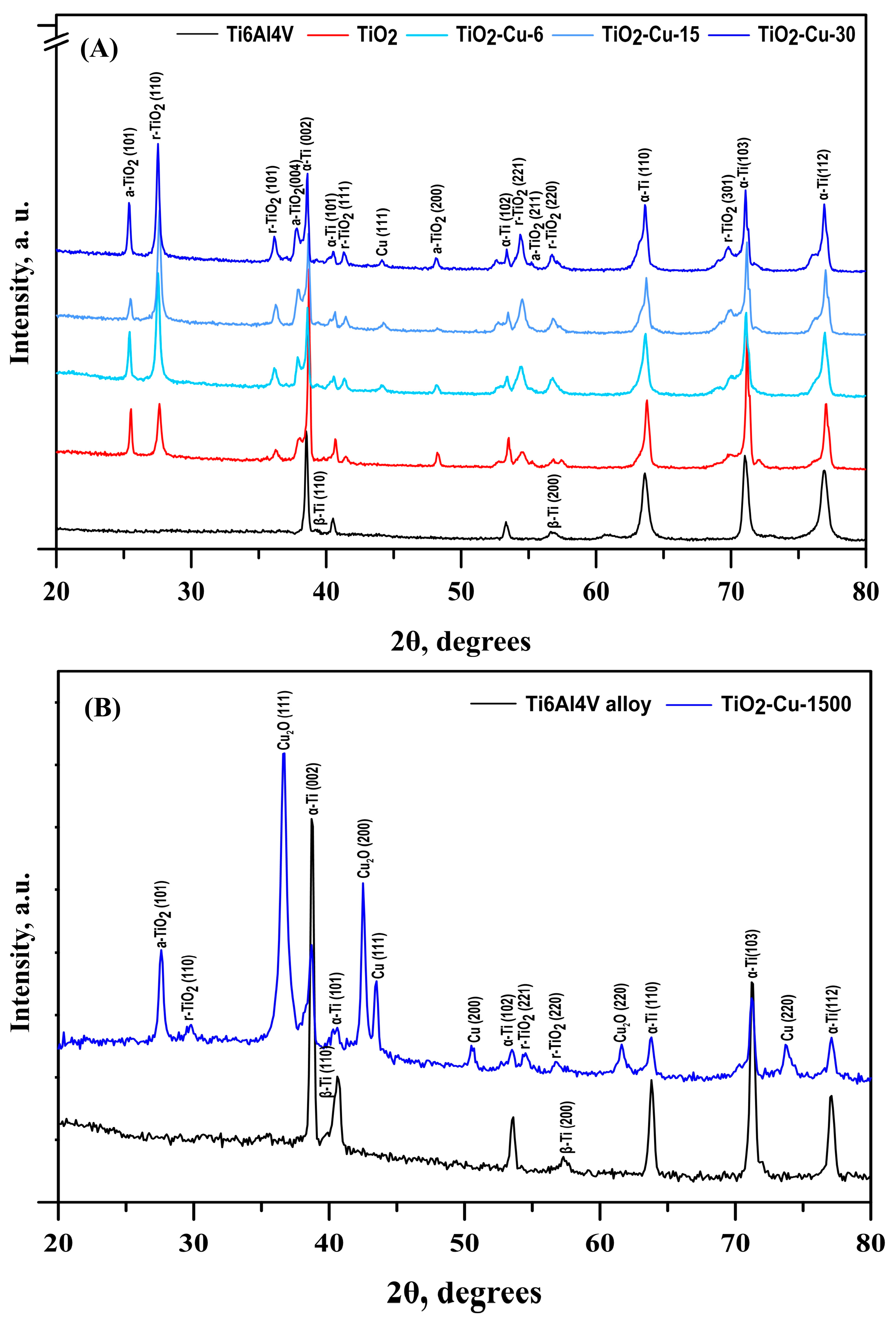
The grazing-incidence XRD (GIXRD) patterns presented in Figure 4A show the phase composition and structural evolution of the coatings with increasing copper deposition time. Except for α- (JCPDS No. 01-1198) and β-Ti (JCPDS No. 09-0098) peaks from the pristine substrate, the coatings exhibited maxima for anatase (JCPDS No. 21-1272) and rutile (JCPDS No. 21-1276) TiO2 phases. Typical diffraction maxima of rutile-phase crystal planes are visible in the XRD pattern. The patterns exhibited six diffraction peaks that correspond to the crystallographic planes (1 1 0), (1 0 1), (1 1 1), (2 2 1), (2 2 0), and (3 0 1), respectively. Four further reflections are associated with the anatase phase’s planes (1 0 1), (0 0 4), (2 0 0), and (2 1 1). According to some authors, the simultaneous presence of both phases in the coating is the most effective way to enhance the biological and physicochemical properties of the implant’s surfaces [34]. Furthermore, small reflections around 2θ ≈ 43.3°, which can be assigned to the (1 1 1) copper plane, indicate the presence of metallic copper nanostructures on the TiO2 surface. Since Cu has low loading on the fine TiO2 dispersion surface, no extra reflections can be seen. In contrast to the pristine TiO2 coating, a certain effect of an increase in the rutile reflection intensities from the surface after copper deposition was also observed. This phenomenon may be ascribed to the alteration of redox balance and the defective structure of TiO2. Numerous metals, including Cr, Co, and Fe, that were deposited on the surface of TiO2 reacted with the oxygen in TiO2 to reduce TiO2 and oxidise metals [29]. Similarly, when Cu is deposited on TiO2, the redox exchange can create oxygen vacancies (V0) and Ti3+ defect sites, modifying the TiO2 lattice. Oxygen vacancies and Ti3+ facilitate lattice rearrangement, which may lower the activation barrier for the anatase-to-rutile transformation and favour rutile transformation.
Since copper oxide phases were not observed in the grazing-incidence diffraction patterns of the coatings—likely due to their small thickness and/or peak overlap—a similar copper deposition on TiO2 coatings was carried out for a longer sputtering time of 1500 s (Figure 4B). In addition, (1 1 1), (2 0 0), and (2 2 0) Cu peaks (JCPDS No 04-0836) with a face-centred cubic crystal lattice, distant (1 1 0), (1 1 1), (2 0 0) and (2 2 0) diffraction peaks of Cu2O phase (JCPDS No 77-0199) were formed. It follows that the deposited Cu layers were oxidised to Cu2O, as indicated by the Bragg–Brentano XRD results. This oxidation likely occurred due to the interaction of the Cu layer with the underlying TiO2 and/or in situ bulk oxidation of residual oxygen contamination in the sputtering chamber. Similar copper phases were reported by Potočnik et al. [14] for superficially electrochemically deposited Cu on e-beam glancing angle deposited TiO2, as well as Carvalho et al. [28] for DC sputtered Cu on TiO2 coatings. The gradual increase in the relative intensity and a decrease in the full width at half maximum (FWHM) of the Cu diffraction peaks suggest the formation of thicker and more continuous Cu layers, resulting from the coalescence and growth of Cu crystallites during prolonged sputtering. Moreover, the bulk phase composition of the coatings was found to be richer in anatase. It follows that the phase composition varies with depth in the coating due to Cu-induced surface reduction or lattice rearrangement.
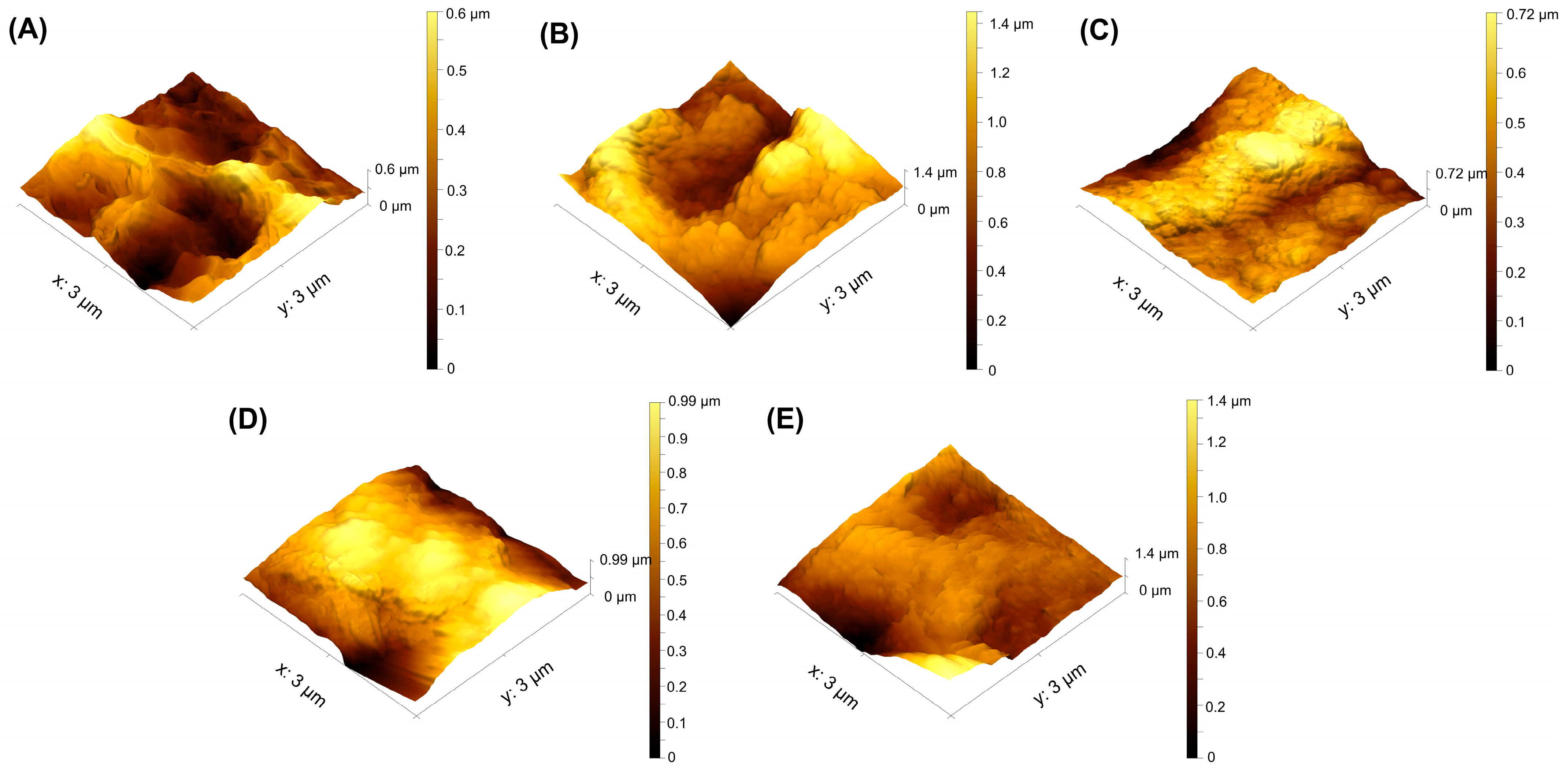
Using AFM imaging, the surface topographic profiles are shown in Figure 5. The coated groups exhibited a granular surface, suggesting homogeneous dispersion of the nanoparticles throughout the substrate surface. The tridimensional profile illustrates the peaks and valleys obtained for each surface. The average surface roughness (Sa) may be calculated from these pictures, and the findings are shown in Table 2. Antibacterial applications may benefit from surfaces with nanoscale roughness, especially when paired with sufficient antibacterial compounds, such as copper. As indicated in Table 2, the TiO2-coated sample showed the highest values of Sa, followed by TiO2/Cu-6. With longer Cu deposition times, Sa values decrease because copper preferentially accumulates on high asperities, rounding and levelling them. This reduces the height difference across the surface and therefore lowers the Sa roughness values, even if valleys are not yet fully coated. A higher Cu concentration results in a stronger tendency for Cu to aggregate, which reduces surface holes and gaps.
The results obtained for the skewness (Ssk), showing the asymmetry in the distribution of surface heights, demonstrate that after coating deposition, the value of this indicator is negative, indicating that the depressions of the profile predominate over the heights compared to the uncoated alloy. With longer Cu deposition time, Ssk becomes less negative. For Ssk > 0, the ridges are more than the valleys of the profile. Such Ssk values are obtained for both the etched alloy and TiO2/Cu-30 samples. This means that, in contrast with the other coated samples, the cell interactions of the etched alloy and TiO2/Cu-30 will be dominated by asperity tops.
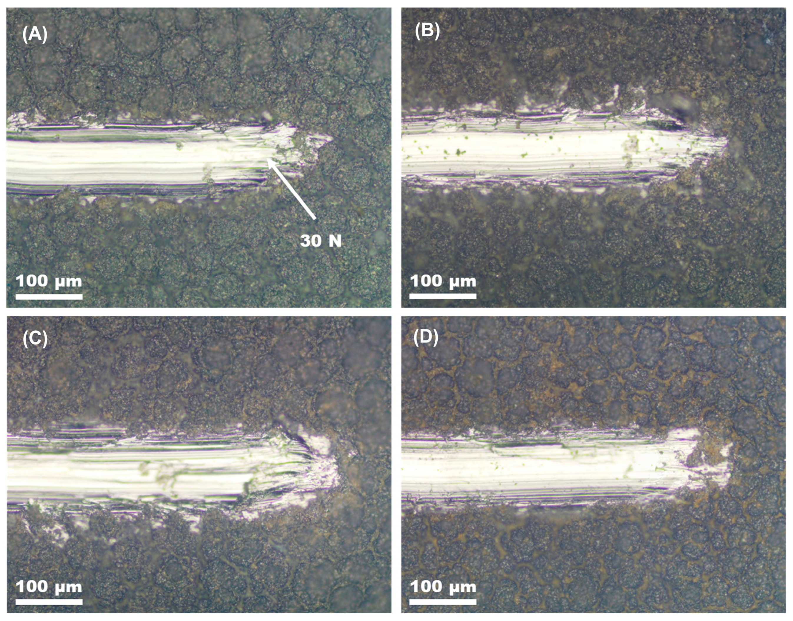
Figure 6 shows the ends of the scratch tracks of all coated specimens before and after copper deposition. The scratch testing of the coatings deposited on the etched alloy revealed that all coatings have excellent adhesion to the substrate. No flaking was seen in the coatings up to a maximal load of 30 N.
The contact angle between the surface and SBF droplets was measured to define the coating’s wettability. The contact angle measurements indicate that all tested surfaces, even after 5 min of contact with SBF solution, remain hydrophobic (Figure 7A). The least hydrophobic is the etched alloy, followed by the TiO2–coated group. The hydrophobicity of the coatings increased with the amount of Cu present on their surface. However, this effect is likely due to the formation of a Cu2O layer on the surface, rather than the presence of metallic Cu itself. The deposition of Cu onto the TiO2 layer can enhance hydrophobicity through the surface oxidation of Cu to Cu2O. A similar tendency was reported by Mahmoudi-Qashqay et al. [23]. With a filled outer d-orbital, Cu has high electron saturation, which minimises dangling bonds and lowers surface energy [35]. Elements with low surface energy enrich the surface, forming Cu–O bonds while showing a weaker affinity for hydrophilic groups such as –OH. Moreover, the increased rutile content near the surface after copper deposition can contribute to higher hydrophobicity since it was found that rutile-rich films become hydrophobic over time, while anatase-rich films remain hydrophilic, indicating rutile is more hydrophobic [36]. As a result, TiO2/Cu coatings exhibit greater hydrophobicity than the pure TiO2 layer. However, as research shows that surface hydrophobicity and surface roughness are closely connected when the contact angle is larger than 90°, all wettability results can be ascribed to the high surface roughness values of the acid-etched substrate and coated samples.
Figure 7B shows the release profiles of Cu from the surface of the Cu-containing sample in SBF over 7 days of immersion. Cu ions can diffuse into the solution by dissolution or reaction with OH ions from the solution [37]. The Cu release profiles of the three samples are similar. The highest absolute release was observed for the TiO2/Cu-30 sample, followed by TiO2-Cu-15 and TiO2-Cu-6. For all samples, the highest Cu release was observed during the first 24 h. Such a burst way of release due to leaching of Cu species from the TiO2 surface may cause cytotoxicity in the short term [38]. Following that, the release proceeded gradually for a minimum of seven days. This release could ensure the implant surface’s ongoing antibacterial activity.
The long-term stability of implants in the organism depends on their interaction with body fluids. To determine the effect of the coatings on the corrosion resistance of Ti6Al4V surfaces in SBF, electrochemical impedance spectroscopy (EIS) tests were conducted. The results are presented in Figure 8 and Table 3. Compared with the uncoated Ti6Al4V substrate, the coated groups exhibited improved electrochemical behaviour. The Bode-impedance curve of the etched alloy (Figure 8A) indicated a thinner and defect-rich oxide layer, whereas the coated specimens showed higher resistance values, reflecting the protective role of the TiO2/Cu layers. As shown in the Bode-phase plots (Figure 8B), the substrate alloy demonstrated a single capacitive response with a phase angle approaching −80° in the medium-low frequency range, confirming its ability to form a passive surface [39]. In contrast, the coated samples exhibited two distinct time constants, corresponding to the response of the coating/SBF interface, which is characteristic of multilayer protective systems. Notably, at both high and low frequencies, all coated surfaces displayed greater phase angles than the uncoated substrate, confirming their enhanced capacitive and barrier properties. Although the maximum phase angle values of all coated groups were somewhat decreased, roughly to −67 ÷ −50 degrees, the coated surfaces function as stable capacitors, providing durable protection in the corrosive SBF environment.
A semi-circle capacitive behaviour and a high impedance value are seen for all coated specimens in the Nyquist plot (Figure 8C). It is commonly recognised that a semicircle’s diameter indicates its corrosion performance, with a bigger diameter often indicating greater corrosion resistance. Figure 8C shows that the greatest arc radius was noted for the TiO2-Cu-15 and TiO2-Cu-30, whereas the etched Ti6Al4V sample had a lower impedance.
Figure 8D shows the electrical equivalent circuit (EEC) model that was used to fit the impedance values. Due to the porous oxide layer, the electrolyte resistance (Rs) is in series with a parallel combination (R1//Q1) for the high- and intermediate-frequency range response that includes the oxide layer resistance (R1) and a constant phase element (Q1). For the low-frequency response measurements, which could be related to the corrosion process, R2 is connected in series with a second R3//Q3 sub-circuit in parallel. The charge transfer resistance, or R3, is inversely proportional to the corrosion rate, and the double-layer capacitance represented by Q3. The Q is an ideal capacitor when n = 1 and an inductor when n = −1 [40].
In contrast to the substrate alloy, the R1 values for all coatings are noticeably lower than the R2 and R3 values, suggesting that the outer rough layer has lower barrier properties. Owing to the high roughness and low SBF wettability, penetration of the solution into the inner coating layers is expected to be less during the initial hours of testing of the copper-containing coatings. It was discovered that the R2 and Q2 parameters for every coated specimen were of the same order of magnitude. This effect occurs because the SBF solution can suppress corrosion by promoting the precipitation of protective compounds within the pores. As the copper deposition time increases, the n2 values rise as well, indicating that the oxide layer is becoming more homogeneous. The charge transfer resistance (R3) also has similar values, except for TiO2-Cu-30, which indicates a higher value. The capacitance of the double layer (Q3) was in the range 2–5.5 × 10⁻⁵ with n values about 0.6 for all coatings. The only alloy with a lower Q3 and n value that matches those of a perfect capacitor (n = 1) is the substrate alloy. Although the outer rough layer exhibited lower resistance, the porous structure allowed precipitation within the pores, further enhancing protection. Increased Cu deposition time promoted oxide coating homogeneity, with TiO2-Cu-30 showing the highest resistance values. Overall, the addition of superficial copper did not compromise the protective effect of TiO2, confirming that sputtered TiO2/Cu coatings provide durable corrosion resistance in physiological conditions.
The bacteria E. coli, which is frequently used to test materials’ antibacterial qualities and is a prevalent pathogen that causes diseases linked to healthcare [41], was used to test the bactericidal activity of TiO2-Cu coatings. The Cu-containing coatings demonstrated extremely effective antibacterial activity against this important coloniser of implants, compared to TiO2-coated and Ti6Al4V alloy. Figure 9 depicts representative images of E. coli colonies on the agar plates after culturing on the surface of each sample for 24 h. It can be seen that a large number of colonies covered the solid agar plates of the etched Ti6Al4V alloy and TiO2-coated samples, indicating that E. coli can normally grow after 24 h of contact with these groups. The lower hydrophobicity could, in turn, promote the adhesion of cells, including bacteria. The rougher pristine TiO2-coated surface shows slightly higher bactericidal activity compared to the uncoated alloy, confirming that the TiO2 material only shows activity when exposed to UV or visible light.
In contrast, a few live bacteria appeared on the agar plates of TiO2-Cu-6 and, in general, no bacteria were found on the agar plates of TiO2-Cu-15 and TiO2-Cu-30 after 24 h of incubation. The percentage of inhibition compared to the TiO2-coated sample was equal to 99.1% for TiO2-Cu-6 and 100% for TiO2-Cu-15 and TiO2-Cu-30 groups. It follows that all copper-containing surfaces generate excellent antibacterial effects. Such an inhibitory effect of copper species decorated on the surface of TiO2 can be related to the release and contact-killing mechanism of copper ions [42]. Cu2+ ions disrupt bacterial membranes by increasing permeability and releasing lipopolysaccharides, proteins, and intracellular molecules [43]. In addition, interactions with sulphur-containing proteins in cell walls can alter their structure and impair key metabolic processes [44]. Cu ions might also impair respiratory chain activity and disturb gene replication of bacteria [37]. Additionally, scientists [45] discovered that interaction with Cu, not ion release, activates the coating’s antibacterial property. A strong difference in contact killing activity was demonstrated between CuO and Cu2O, with a higher antimicrobial effect for Cu2O [46]. According to other research, when Cu (I) oxides came into direct contact with proteins, they absorbed and denatured them more than Cu (II) oxides [47]. By switching between the redox states of Cu+ and Cu2+ ions, Cu may behave as an electron donor or acceptor, which could be the mechanism behind the antibacterial characteristics. After coming into direct contact with the bacteria, these ions are taken up by the outer membrane and react with it in accordance with the element’s electronegative tendencies. Moreover, the hydrophobic surfaces usually effectively inhibit bacterial adhesion, which is consistent with our experimental results. It is extremely encouraging that all TiO2-Cu-6, TiO2-Cu-15 and TiO2-Cu-30 groups achieved an R value greater than 99%, which translates to an almost 99% reduction of the bacterial burden.
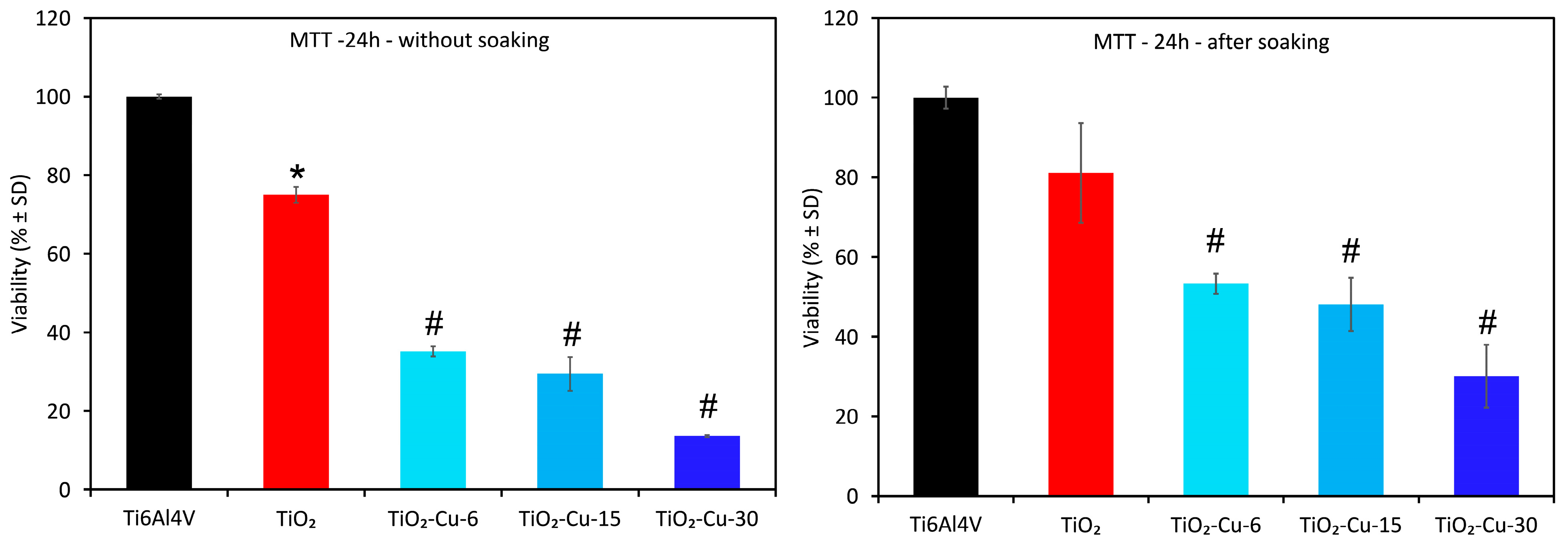
In vitro cell viability tests are important to evaluate the biological performance of the coatings. The cell viability of the cultured MG63 cells for 24 h with all groups is presented in Figure 10. After day 1, all experimental groups reduced cell viability compared to the etched Ti6Al4V alloy, possibly due to unfavourable interactions or excess copper concentration in the culture medium. The cytotoxic effect of Cu-containing surfaces corresponds to the increase in copper content on the surface of the coatings. Since the highest amount of copper was released during the first 24 h, and after that, the release was lower and stable, a second experiment after 24 h of immersion in PBS was conducted. The outcomes demonstrated less damage to the cells’ viability from the Cu-containing surfaces. However, all Cu-containing surfaces were found to be cytotoxic to MG63 after day 1, meaning that the Cu release levels from the surface of the coatings are not within the safe concentration and form for MG63 osteoblastic cell growth. A linear decrease in the number of alive cells was also found with the increase in Cu content in Cu-containing groups. The MTT activity of MG63 cells decreased in the following order: Ti6Al4V > TiO2 > TiO2-Cu-6 > TiO2-Cu-15 > TiO2-Cu-30 coated alloy. When the culture time was increased to 5 days, the cells slightly proliferated, indicating that such Cu-containing coatings were non-conductive to cell growth and had obvious toxicity.
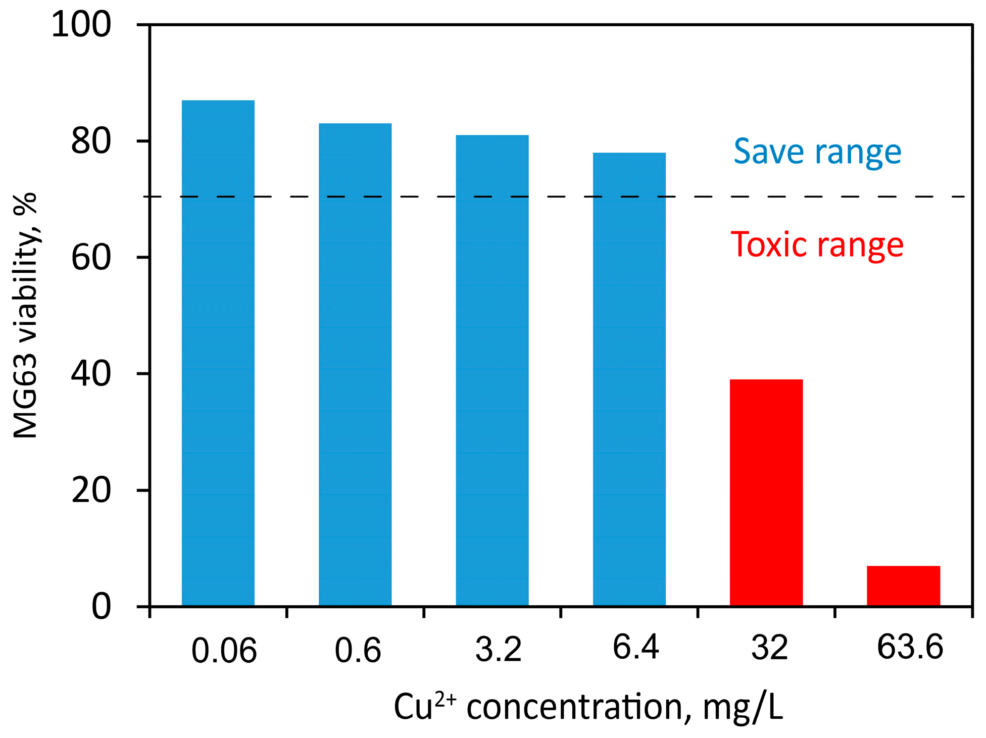
Similarly, Zhang et al. reported a great inhibitory effect of microporous anatase and rutile TiO2 coating doped with an even lower concentration of copper (1.93 wt%) on fibroblasts [42]. On the contrary, anodised Ti–Cu alloy with Cu content of 5 wt% showed a higher proliferation rate and lower apoptosis rate of MC3T3–E1 cells compared to pure Ti samples [48]. The excessive Cu2+ ions are thought to interact non-specifically with macromolecules, thus changing their conformation and increasing clinical oxidative stress, finally leading to the dysfunction of cellular processes and cell apoptosis [42]. It is thus believed that the high content of Cu on the surface alone seriously affects the viability of osteoblast cells by inhibiting spreading and cell viability. Hallab et al. [49] provided in vitro studies about copper ion concentrations that show toxicity or adverse effects to MG63 osteoblastic cells. The results reported in their study are plotted in Figure 11. Taking into account the ISO 10993-5 standard, a reduction in cell viability higher than 30% compared to the control can be considered cytotoxic [50]. This indicates that the Cu ion content released from all deposited coatings is within the cell-safe range for the MG63 cell line.
At the same time, according to the standard [50], samples with 100% cell viability are non-cytotoxic (grade 0). Viability of 80%–100% indicates slight cytotoxicity (grade 1), 50%–80% mild (grade 2), 30%–50% moderate (grade 3), and below 30% severe cytotoxicity (grade 4). Therefore, without initial soaking, TiO2-Cu-6 and TiO2-Cu-15 belong to the moderate (grade 3) cytotoxicity group, while TiO2-Cu-30 demonstrate severe cytotoxicity (grade 4). The examined copper–containing coatings cause obvious toxicity toward osteoblasts even after 24 h of pre-immersion in PBS, which decreases copper ion release. This implies that the cytotoxicity and antimicrobial properties of the examined Cu-containing coatings are dictated not only by excess ions but also by direct contact between cells and Cu species that are not removed during immersion. The preferential deposition of Cu on asperity peaks rather than in valleys means that, even after pre-immersion in PBS to reduce ion release, cells directly encounter Cu-rich sites at the surface. With increasing deposition time, larger copper clusters form, leading to more pronounced direct contact between osteoblasts and metallic oxide Cu species. This correlates with the observed evolution of surface skewness (Ssk). The contact with the superficially located copper species may disrupt cell membranes, alter adhesion, or trigger oxidative stress locally at the interface. Therefore, the observed cytotoxicity cannot be attributed solely to excess Cu ion release but is strongly influenced by the topographical distribution and aggregation of Cu species. These results emphasise that both ion release kinetics and surface geometry, particularly the presence of Cu clusters on protruding features, govern the biological response of TiO2/Cu coatings.
The nano-specific properties of superficially located copper impart greater toxicity and burst release of Cu ions over a short time frame in the biological media. Achieving an optimal implant design requires controlling both Cu release kinetics and surface exposure of Cu nanoparticles to minimise osteoblast toxicity while maintaining antibacterial properties. Therefore, the use of continuous layers could more effectively control Cu release, considering the decrease in the total area of interaction with the biological medium and cells. Moreover, since the metallic bond is weaker than the ionic bond, fewer copper ions will be released if Cu participates in the coatings in its stable oxide form.

4. Conclusions

We have demonstrated that the superficial copper concentration and its form are crucial factors that influence the physicochemical properties of sputtered TiO2/Cu coatings and provides a way to construct desired antimicrobial efficacy. Cu and Cu2O phases preferentially accumulated on asperity peaks, forming nanoscale clusters that reduced surface roughness and altered skewness values. All Cu-containing TiO2 coatings exhibited excellent antibacterial efficiency against E. coli, achieving up to 100% inhibition, which was attributed to both Cu ion release and direct contact-killing mechanisms. Compared to the other groups, TiO2-Cu-6 showed acceptable MG63 cell viability reduction not only due to the chemical composition of the coating but also related to its physical properties, such as a less hydrophobic profile, corrosion resistance and lower Cu release from the surface. The addition of superficial copper did not impair the protective role of the TiO2 layer, but with prolonged sputtering, MG63 cell compatibility was reduced, which was likely due to excessive Cu ion release from thicker surface layers. Hence, Cu form and its release from the surface should be properly selected to be below the cytotoxic level. For that reason, in our next studies, the co-sputtering of Ti and Cu to form complex oxide coatings will be tested for their antibacterial properties and cytocompatibility.

Author Contributions

Conceptualization, M.P.N.; methodology, M.P.N., Y.H. and M.D.A.; software, M.P.N., M.D.A., T.V.D. and S.V.; validation, M.D.A., M.O. and Y.H.; formal analysis, M.P.N., M.D.A., T.V.D. and S.V.; investigation, M.O., Y.H. and A.A.; resources, M.P.N.; data curation, M.D.A., T.V.D. and M.P.N.; writing—original draft preparation, M.P.N.; writing—review and editing, M.P.N. and M.D.A.; visualisation, M.P.N., M.D.A., T.V.D. and S.V.; supervision, M.D.A.; project administration, M.P.N.; funding acquisition, M.P.N. All authors have read and agreed to the published version of the manuscript.

Funding

This research was funded by the Bulgarian National Science Fund, grant number KΠ-06-H67-5 (2022).

Institutional Review Board Statement

Not applicable.

Informed Consent Statement

Not applicable.

Data Availability Statement

The original contributions presented in the study are included in the article; further inquiries can be directed to the corresponding author.

Conflicts of Interest

The authors declare no conflicts of interest.

References

  1. Liu, S.; Zhang, Z.; Zhang, J.; Qin, G.; Zhang, E. Construction of a TiO2/Cu2O multifunctional coating on Ti-Cu alloy and its influence on the cell compatibility and antibacterial properties. Surf. Coat. Technol. 2021, 421, 127438. [Google Scholar] [CrossRef]
  2. Brunello, G.; Elsayed, H.; Biasetto, L.J.M. Bioactive glass and silicate-based ceramic coatings on metallic implants: Open challenge or outdated topic? Materials 2019, 12, 2929. [Google Scholar] [CrossRef]
  3. Wang, C.; Fan, Z.; Han, Y. Formation and osteoblast behavior of HA nano-rod/fiber patterned coatings on tantalum in porous and compact forms. J. Mat. Chem. B 2015, 3, 5442–5454. [Google Scholar] [CrossRef]
  4. Li, G.; Cao, H.; Zhang, W.; Ding, X.; Yang, G.; Qiao, Y.; Liu, X.; Jiang, X. Enhanced osseointegration of hierarchical micro/nano-topographic titanium fabricated by micro-arc oxidation and electrochemical treatment. ACS Appl. Mater. Interf. 2016, 8, 3840–3852. [Google Scholar] [CrossRef]
  5. Nikolova, M.P.; Chavali, M.S. Metal Oxide Nanoparticles as Biomedical Materials. Biomimetics 2020, 5, 27. [Google Scholar] [CrossRef]
  6. Huang, Q.; Liu, X.; Zhang, R.; Yang, X.; Lan, C.; Feng, Q.; Liu, Y. The development of Cu-incorporated micro/nano-topographical bio-ceramic coatings for enhanced osteoblast response. Appl. Surf. Sci. 2019, 465, 575–583. [Google Scholar] [CrossRef]
  7. Zhang, X.; Li, J.; Wang, X.; Wang, Y.; Hang, R.; Huang, X.; Tang, B.; Chu, P.K. Effects of copper nanoparticles in porous TiO2 coatings on bacterial resistance and cytocompatibility of osteoblasts and endothelial cells. Mater. Sci. Eng. C Mater. Biol. Appl. 2018, 82, 110–120. [Google Scholar] [CrossRef]
  8. Nikolova, M.; Ormanova, M.; Nikolova, V.; Apostolova, M. Electrochemical, Tribological and Biocompatible Performance of Electron Beam Modified and Coated Ti6Al4V Alloy. Int. J. Mol. Sci. 2021, 22, 6369. [Google Scholar] [CrossRef] [PubMed]
  9. Zhang, L.; Guo, J.; Yan, T.; Han, Y. Fibroblast responses and antibacterial activity of Cu and Zn co-doped TiO2 for percutaneous implants. Appl. Surf. Sci. 2018, 434, 633–642. [Google Scholar] [CrossRef]
  10. Balamurugan, A.; Kannan, S.; Rajeswari, S. Evaluation of TiO2 coatings obtained using the sol–gel technique on surgical grade type 316L stainless steel in simulated body fluid. Mater. Lett. 2005, 59, 3138–3143. [Google Scholar] [CrossRef]
  11. Fuentes, S.; Tapia, A.; Pozo, P. Synthesis, characterization, and antibacterial activity evaluation of Cu@TiO2 nanocomposites. Mater. Lett. 2021, 296, 129885. [Google Scholar] [CrossRef]
  12. Wu, C.T.; Zhou, Y.H.; Xu, M.C.; Han, P.P.; Chen, L.; Chang, J.; Xiao, Y. Copper-containing mesoporous bioactive glass scaffolds with multifunctional properties of angiogenesis capacity, osteostimulation and antibacterial activity. Biomaterials 2013, 34, 422–433. [Google Scholar] [CrossRef]
  13. Li, J.Y.; Zhai, D.; Lv, F.; Yu, Q.Q.; Ma, H.S.; Yin, J.B.; Yi, Z.F.; Liu, M.Y.; Chang, J.; Wu, C.T. Preparation of copper-containing bioactive glass/eggshell membrane nanocomposites for improving angiogenesis, antibacterial activity and wound healing. Acta Biomater. 2016, 36, 254–266. [Google Scholar] [CrossRef] [PubMed]
  14. Potočnik, J.; Latas, N.; Milošević, M.; Janakiev, T.; Stamenković, T.; Srejić, I. Copper-enhanced TiO2 nanostructured thin films: Structural, chemical, and antibacterial properties. Appl. Surf. Sci. 2025, 708, 163747. [Google Scholar] [CrossRef]
  15. Hosseini, M.; Chin, A.W.H.; Behzadinasab, S.; Poon, L.L.M.; Ducker, W.A. Cupric Oxide Coating That Rapidly Reduces Infection by SARS-CoV-2 via Solids. ACS Appl. Mater. Interfaces 2021, 13, 5919–5928. [Google Scholar] [CrossRef] [PubMed]
  16. Wang, F.; Li, C.; Zhang, S.; Liu, H.J.S. Tantalum coated on titanium dioxide nanotubes by plasma spraying enhances cytocompatibility for dental implants. Surf. Coat. Technol. 2020, 382, 125161. [Google Scholar] [CrossRef]
  17. Kim, S.; Park, C.; Moon, B.-S.; Kim, H.-E.; Jang, T.-S. Enhancement of osseointegration by direct coating of rhBMP-2 on target-ion induced plasma sputtering treated SLA surface for dental application. J. Biomater. Appl. 2017, 31, 807–818. [Google Scholar] [CrossRef]
  18. Wu, B.; Xiong, S.; Guo, Y.; Chen, Y.; Huang, P.; Yang, B. Tooth-colored bioactive titanium alloy prepared with anodic oxidation method for dental implant application. Mater. Lett. 2019, 248, 134–137. [Google Scholar] [CrossRef]
  19. Durdu, S.J.C. Characterization, bioactivity and antibacterial properties of copper-based TiO2 bioceramic coatings fabricated on titanium. Coatings 2018, 9, 1. [Google Scholar] [CrossRef]
  20. Yılmaz, G.E.; Göktürk, I.; Ovezova, M.; Yılmaz, F.; Kılıç, S.; Denizli, A. Antimicrobial Nanomaterials: A Review. Hygiene 2023, 3, 269–290. [Google Scholar] [CrossRef]
  21. Du, H.; Shi, Z.; Hou, Q.; Xia, S.; Yin, P.; Dastan, D.; Cui, H.; Fan, R. Gold sputtering at the interfaces: An easily operated strategy for enhancing the energy storage capability of laminated polymer dielectrics. ACS Appl. Mater. Interfaces 2023, 15, 17103–17112. [Google Scholar] [CrossRef]
  22. Shiravand, M.; Ghobadi, N.; Hatam, E.G. The investigation of structural, surface topography, and optical behaviors of Al-doped ZnO thin films with annealing temperature deposited by RF magnetron sputtering. J. Mater. Sci. Mater. Electron. 2023, 34, 713. [Google Scholar] [CrossRef]
  23. Mahmoudi-Qashqay, S.; Zamani-Meymian, M.R.; Sadati, S.J. Improving antibacterial ability of Ti-Cu thin films with co-sputtering method. Sci. Rep. 2023, 13, 16593. [Google Scholar] [CrossRef] [PubMed]
  24. Liang, Y.-C.; Li, T.-H. Sputtering-Assisted Synthesis of Copper Oxide–Titanium Oxide Nanorods and Their Photoactive Performances. Nanomaterials 2022, 12, 2634. [Google Scholar] [CrossRef] [PubMed]
  25. El-Moula, A.; El-Hossary, F.; Raaif, M.; Thabet, A.; El-Kassem, M. Effect of Cu Metallic Interlayer Thickness on Optoelectronic Properties of TiO2-Based Multilayers Deposited by DC Pulsed Magnetron Sputtering. J. Electron. Mater. 2021, 50, 2699–2709. [Google Scholar] [CrossRef]
  26. Awad, M.; Raaif, M. Optical and electrical performance of transparent conductive TiO2/Cu/TiO2 multilayers prepared by magnetron sputtering. J. Mater. Sci. Mater. Electron. 2018, 29, 2815–2824. [Google Scholar] [CrossRef]
  27. Zhao, Z.; Sun, J.; Zhang, G.; Bai, L. The study of microstructure, optical and photocatalytic properties of nanoparticles (NPs)-Cu/TiO2 films deposited by magnetron sputtering. J. Alloys Compd. 2015, 652, 307–312. [Google Scholar] [CrossRef]
  28. Carvalho, H.W.; Batista, A.P.; Hammer, P.; Ramalho, T.C. Photocatalytic degradation of methylene blue by TiO2–Cu thin films: Theoretical and experimental study. J. Hazard. Mater. 2010, 184, 273–280. [Google Scholar] [CrossRef]
  29. Diebold, U. The surface science of titanium dioxide. Surf. Sci. Rep. 2003, 48, 53–229. [Google Scholar] [CrossRef]
  30. Kokubo, T.; Kushitani, H.; Sakka, S.; Kitsugi, T.; Yamamuro, T. Solutions able to reproduce in vivo surface-structure changes in bioactive glass-ceramic A-W. J. Biomed. Mater. Res. 1990, 24, 721–734. [Google Scholar] [CrossRef]
  31. Venables, J.A. Introduction to Surface and Thin Film Processes; Cambridge University Press: Cambridge, UK, 2000. [Google Scholar] [CrossRef]
  32. Angstrom Science Home Page. Available online: https://www.angstromsciences.com/sputtering-yields (accessed on 30 September 2023).
  33. Ohring, M. Materials Science of Thin Films, 2nd ed.; Academic Press: San Diego, CA, USA, 2001. [Google Scholar] [CrossRef]
  34. Pantaroto, H.N.; Cordeiro, J.M.; Pereira, L.T.; de Almeida, A.B.; Nociti Junior, F.H.; Rangel, E.C.; Azevedo Neto, N.F.; da Silva, J.H.D.; Barão, V.A.R. Sputtered crystalline TiO2 film drives improved surface properties of titanium-based biomedical implants. Mater. Sci. Eng. C 2021, 119, 111638. [Google Scholar] [CrossRef]
  35. Lu, K.; Sun, M.; Jiang, Y.; Wu, X.; Zhao, L.; Xu, J. Insight into the Structural and Performance Correlation of Photocatalytic TiO2/Cu Composite Films Prepared by Magnetron Sputtering Method. Catalysts 2024, 14, 621. [Google Scholar] [CrossRef]
  36. Vrakatseli, V.; Farsari, E.; Mataraset, D. Wetting Properties of Transparent Anatase/Rutile Mixed Phase Glancing Angle Magnetron Sputtered Nano-TiO2 Films. Micromachines 2020, 11, 616. [Google Scholar] [CrossRef] [PubMed]
  37. Zhang, D.; Han, Q.; Yu, K.; Lu, X.; Liu, Y.; Lu, Z.; Wang, Q. Antibacterial activities against porphyromonas gingivalis and biological characteristics of copper-bearing PEO coatings on magnesium. J. Mater. Sci. Technol. 2021, 61, 33–45. [Google Scholar] [CrossRef]
  38. He, X.; Zhang, G.; Zhang, H.; Hang, R.; Huang, X.; Yao, X.; Zhang, X. Cu and Si co-doped microporous TiO2 coating for osseointegration by the coordinated stimulus action. Appl. Surf. Sci. 2020, 503, 144072. [Google Scholar] [CrossRef]
  39. Hwang, I.J.; Choe, H.C.; Brantley, W.A. Electrochemical characteristics of Ti-6Al-4V after plasma electrolytic oxidation in solutions containing Ca, P, and Zn ions. Surf. Coat. Technol. 2017, 320, 458–466. [Google Scholar] [CrossRef]
  40. Halvorsen, I.J.; Pivac, I.; Bezmalinović, D.; Barbir, F.; Zenith, F. Electrochemical low-frequency impedance spectroscopy algorithm for diagnostics of PEM fuel cell degradation. Int. J. Hydrogen Energy 2019, 45, 1325–1334. [Google Scholar] [CrossRef]
  41. Thukkaram, M.; Vaidulych, M.; Kylián, O.; Rigole, P.; Aliakbarshirazi, S.; Asadian, M.; Nikiforov, A.; Biederman, H.; Coenye, T.; Du Laing, G.; et al. Biological Activity and Antimicrobial Property of Cu/a-C:H Nanocomposites and Nanolayered Coatings on Titanium Substrates. Mater. Sci. Eng. C 2021, 119, 111513. [Google Scholar] [CrossRef]
  42. Zhang, L.; Guo, J.; Huang, X.; Zhang, Y.; Han, Y. The dual function of cu-doped TiO2 coatings on titanium for application in percutaneous implants. J. Mater. Chem. B 2016, 4, 3788–3800. [Google Scholar] [CrossRef] [PubMed]
  43. Artunduaga Bonilla, J.J.; Paredes Guerrero, D.J.; Sánchez Suárez, C.I.; Ortiz López, C.C.; Torres Sáez, R.G. In vitro antifungal activity of silver nanoparticles against fluconazole-resistant Candida species. World J. Microbiol. Biotechnol. 2015, 31, 1801–1809. [Google Scholar] [CrossRef] [PubMed]
  44. Haider, A.; Kwak, S.; Gupta, K.C.; Kang, I.-K. Antibacterial activity and cytocompatibility of PLGA/CuO hybrid nanofiber scaffolds prepared by electrospinning. J. Nanomater. 2015, 2015, 832762. [Google Scholar] [CrossRef]
  45. Shimabukuro, M.; Tsutsumi, Y.; Nozaki, K.; Chen, P.; Yamada, R.; Ashida, M.; Doi, H.; Nagai, A.; Hanawa, T. Investigation of antibacterial effect of copper introduced titanium surface by electrochemical treatment against facultative anaerobic bacteria. Dent. Mater. J. 2020, 39, 639–664. [Google Scholar] [CrossRef] [PubMed]
  46. Hans, M.; Erbe, A.; Mathews, S.; Chen, Y.; Solioz, M.; Mücklich, F. Role of copper oxides in contact killing of bacteria. Langmuir 2013, 29, 16160–16166. [Google Scholar] [CrossRef] [PubMed]
  47. Pan, C.; Shen, H.; Liu, G.; Zhang, X.; Liu, X.; Liu, H.; Xu, P.; Chen, W.; Tian, Y.; Deng, H.; et al. CuO/TiO2 Nanobelt with Oxygen Vacancies for Visible-Light-Driven Photocatalytic Bacterial Inactivation. ACS Appl. Nano Mater. 2022, 5, 10980. [Google Scholar] [CrossRef]
  48. Zhang, W.; Zhang, S.; Liu, H.; Ren, L.; Wang, Q.; Zhang, Y. Effects of surface roughening on antibacterial and osteogenic properties of Ti-Cu alloys with different Cu contents. J. Mater. Sci. Technol. 2021, 88, 158–167. [Google Scholar] [CrossRef]
  49. Hallab, N.J.; Vermes, C.; Messina, C.; Roebuck, K.A.; Glant, T.T.; Jacobs, J.J. Concentration- and composition-dependent effects of metal ions on human MG-63 osteoblasts. J. Biomed. Mater. Res. 2002, 60, 420–433. [Google Scholar] [CrossRef]
  50. ISO 10993-5:2009; Biological Evaluation of Medical Devices Part 5: Tests for Invitro Cytotoxicity. International Organization for Standardization/ANSI: Geneva, Switzerland, 2009.
Figure 1. Micrographic images of the TiO2 coating deposited on the surface of the etched Ti6Al4V alloy; (A,B) topography of the oxide coating at different magnifications; (C) cross-section of the layers revealing the thickness of both Ti sublayer and overlying TiO2.
Coatings 15 01341 g001
Figure 2. Representative optical microscope topographic images of (A) TiO2 coating deposited on the surface of the etched Ti6Al4V alloy; (B) TiO2-Cu-30 surface morphology.
Coatings 15 01341 g002
Figure 3. Elemental EDS mapping of Cu-TiO2 coatings demonstrating the cluster structure of the superficially located copper: (A) TiO2-Cu-6, (B) TiO2-Cu-15 and (C) TiO2-Cu-30 surface morphology.
Coatings 15 01341 g003
Figure 4. XRD patterns of the substrate and TiO2 and TiO2-Cu coated samples (A) grazing incidence configuration, (B) symmetrical Bragg–Brentano geometry. a-TiO2 refers to the corresponding peaks of anatase, whereas r-TiO2 refers to those of rutile.
Coatings 15 01341 g004
Figure 5. Representative AFM topography of (A) etched sample; (B) TiO2 coated; (C) TiO2-Cu-6, (D) TiO2/Cu-15 and (E) TiO2-Cu-30.
Coatings 15 01341 g005
Figure 6. Scratch track images on (A) TiO2 coated; (B) TiO2-Cu-6, (C) TiO2/Cu-15 and (D) TiO2-Cu-30.
Coatings 15 01341 g006
Figure 7. Surface contact angle with SBF solution (A) and changes in the cumulative Cu release from the surface at different immersion periods in SBF at 37 °C (B).
Coatings 15 01341 g007
Figure 8. Representative electrochemical curves: (A) Bode-impedance; (B) Bode-phase; (C) Nyquist, of the samples in SBF solution at 37 °C. (D) Equivalent circuit model of the uncoated and coated samples used for modelling impedance data.
Coatings 15 01341 g008
Figure 9. Control and coated samples’ antibacterial activity against E. coli. Results are shown as average ± SD of 5 independent experiments. CFU—colony-forming unit.
Coatings 15 01341 g009
Figure 10. Cell viability after 1 day of culture without and with initial immersion via MTT analysis. Data are expressed as mean ± standard deviation. Statistically significant differences between the groups are indicated by symbols: * p < 0.02; # p < 0.007.
Coatings 15 01341 g010
Figure 11. MG63 cell viability as a function of Cu2+ ion concentration according to [46].
Coatings 15 01341 g011
Table 1. Copper and titanium content and their ratio on the surface of the TiO2-Cu coatings. The mean ± standard deviation is used to express the data.
SampleCu, at%Ti, at%Ti/Cu Ratio
TiO2-Cu-61.06 ± 0.0737.91 ± 0.4535.76
TiO2-Cu-151.37 ± 0.0935.05 ± 0.6225.58
TiO2-Cu-302.29 ± 0.1032.79 ± 1.0314.32
Table 2. Statistical parameters of surface roughness.
SampleSa, nmSsk
Ti6Al4V113.1 ± 17.80.30 ± 0.27
TiO2210.6 ± 79.8−0.31 ± 0.32
TiO2-Cu-6162 ± 49,1−0.28 ± 0.24
TiO2-Cu-15133.2 ± 43.1−0.24 ± 1.06
TiO2-Cu-30125.1 ± 20.20.22 ± 0.5
Table 3. EIS fitting parameters derived from the Bode-phase, Nyquist, and Bode-impedance diagrams.
SpecimenQ1
−1cm−2sn)
n1R1
(Ωcm2)
Q2
−1cm−2sn)
n2R2
(Ωcm2)
Q3
−1cm−2sn)
n3R3
(Ωcm2)
Χ2
Ti6Al4V1.3 × 10−50.91.0 × 1044.2 × 10−40.75.1 × 1033.2 × 10−315.0 × 1051.1 × 10−4
TiO22.8 × 10−60.85.5 × 1021.9 × 10−50.72.1 × 1035.5 × 10−50.61.9 × 1055.1 × 10−4
TiO2-Cu-61.4 × 10−60.89.2 × 1027.9 × 10−60.75.2 × 1034.4 × 10−50.61.6 × 1059.2 × 10−4
TiO2-Cu-152.4 × 10−60.86 × 1021.3 × 10−60.92.1 × 1032.2 × 10−50.64.5 × 1051.7 × 10−4
TiO2-Cu-304.4 × 10−70.84.9 × 1035.7 × 10−70.95.7 × 1031.9 × 10−50.64.5 × 1061.9 × 10−3
Disclaimer/Publisher’s Note: The statements, opinions and data contained in all publications are solely those of the individual author(s) and contributor(s) and not of MDPI and/or the editor(s). MDPI and/or the editor(s) disclaim responsibility for any injury to people or property resulting from any ideas, methods, instructions or products referred to in the content.