Article

Bio-Entities Based on Albumin Nanoparticles and Biomimetic Cell Membranes: Design, Characterization and Biophysical Evaluation

by
Marcela-Elisabeta Barbinta-Patrascu
1,
Sorina Iftimie
1,
Nicoleta Cazacu
1,
Diana Lavinia Stan
1,
Andreea Costas
2,
Adriana Elena Balan
1 and
Claudia Gabriela Chilom
1,*
1
Faculty of Physics, University of Bucharest, 405 Atomistilor Street, P.O. Box MG-11, 077125 Magurele, Romania
2
National Institute of Materials Physics, 077125 Magurele, Romania
*
Author to whom correspondence should be addressed.
Coatings 2023, 13(4), 671; https://doi.org/10.3390/coatings13040671
Submission received: 17 February 2023 / Revised: 18 March 2023 / Accepted: 22 March 2023 / Published: 24 March 2023
(This article belongs to the Special Issue Multilayer Coatings for Nanomaterials: From Synthesis to Applications)

Abstract

:
Protein-based particles are one of the most important research topics in nanomedicine, being used especially as drug delivery systems. From the wide variety of proteins, albumins offer several advantages in biomedical applications due to their special properties. Albumin nanoparticles play an important role as carriers in the drug delivery of chemical and biomolecular drugs, such as anticancer drugs; offer many advantages, such as biocompatibility and biodegradability; and are well-tolerated, without any side effects. In this work, various types of bovine serum albumin nanoparticles (BSA NPs), with or without ascorbic acid or glucose, were prepared via different nanoprecipitation methods. The obtained BSA NPs were characterized by UV–Vis absorption spectroscopy. Their size and morphology were studied by Scanning Electron Microscopy (SEM) and Atomic Force Microscopy (AFM). The stability in time of the developed BSA NPs was spectrally monitored. Three types of bio-entities containing BSA NPs and chlorophyll-labeled artificial cell membranes were “green” developed. The designed biohybrids were characterized by UV–Vis absorption and fluorescence emission spectroscopy, and their three-dimensional topography was investigated by AFM. Both the size and shape of the developed bio-entities were monitored through SEM analysis. These results could be exploited in the development of novel drug carrier systems or as bio-coatings to be used in the biomedical field.

1. Introduction

Serum albumin, the most abundant plasma protein, is used more and more in direct clinical applications, such as maintaining blood homeostasis [1,2], and can be used as a biomarker for kidney and liver diseases [3].
A very current direction in biomedical research is the synthesis of bio-conjugates and assemblies based on albumin that can be used as additives and versatile bio-functional carriers in different applications [4]. The physical–chemical stability and biochemical inertia of the albumin molecule, as well as the fact that it has specific binding sites for numerous endogenous and exogenous therapeutic compounds [5,6,7], recommend this protein to be used as a drug carrier, including the management of some tumor problems [8]. The stability and biochemical inertia of albumin allow for the modification of the surface and the covering of the albumin molecule with various substrates [4,9], resulting in functionalized hybrid structures, used in biomedical applications. The functional characteristics of the albumin molecule depend, to a large extent, on factors such as temperature, pH, various solvents or ligands [10,11].
The literature abounds in information on the development of drug delivery systems, such as dendrimers [12], liposomes [13], nanoparticles [14], carbon nanotubes [15] or quantum dots [16]. Among these, the nanoparticles (NPs) protect the therapeutic agents from degradation and mediate the penetration of the cell membranes by these active principles [17,18]. In order for the drug–cell interaction to be effective, a delivery is required, which ensures a certain concentration of drug for a certain period of time. Formulations such as albumin nanoparticles aim to improve the permeation and retention effect (EPR effect) so that targeted delivery and retention of anticancer compounds in the target tissue is achieved [19]. The size and physicochemical properties of albumin nanoparticles are essential for them to achieve the EPR effect. BSA has more than 40 kDa and a very long half-life (~20 days) in the bloodstream [20]. BSA NPs have sizes in the range of 100–200 nm, and this is considered optimal for achieving EPR.
The advantage of using albumin NPs in clinical applications lies in the properties of albumin, such as biodegradability, very low toxicity and immunogenicity, and also easy preparation in a reproducible manner [21]. As in the case of all NPs, the physicochemical properties, shape, dimensions and degree of coverage greatly influence the degree of drug release at the cellular level.
Methods for the preparation of protein NPs, including serum protein NPs, can be classified into several categories, the most common of which are chemical, physical and self-assembly methods (e.g., dissolution method) [22,23,24]. The chemical method allows the production of protein NPs through the use of a chemical reaction (e.g., emulsion and complex coacervation). The advantages of this method include cost effectiveness, high versatility in surface chemistry, stability and encapsulation efficiency, easy functionalization, size controllability, thermal stability and reduced dispersity. The particles obtained from using the chemical method are larger than those that result from the desolvation method, but it also presents other disadvantages, such as low purity, thermodynamic instability and the use of toxic chemicals and organic solvents. The physical method includes physically aggregating after separating proteins into nanosized particles (e.g., electrospray technique and nano-spray drying methods). Using this method has the following advantages: time efficiency; obtainment of stable particles; control of particle size, shape and morphology; and no use of toxic chemicals. Instead, this method comes with a set of disadvantages, such as a high cost, exposure to radiation and the altered surface chemistry and physicochemical properties of nanoparticles. On the other hand, the self-assembly approach allows the synthesis of nanoparticles by self-agglutinating proteins.
Desolvation, also known as the antisolvent precipitation method, is based on solvent displacement in protein solutions, which leads to protein self-assembly and particle formation (Figure 1). This method is one of the most used and simple approaches for the fabrication of albumin NPs. NPs are obtained when a desolvating agent (usually acetone, alcohols or acetonitrile) is added with a constant rate of dripping to an aqueous solution of protein under constant stirring, using a magnet, to dehydrate the protein, resulting in conformational change from stretched to coil conformation. A very important parameter in this method to obtain favorable size of NPs is the constant maintenance of the flow rate and the volume of the added desolvating agent. The next important step after the formation of NPs, after adding the desolvating agent, consists of increasing their stability. This step is achieved by using a crosslinker agent [21]. The prepared suspension is kept under continuous stirring with the aim to complete crosslinking of all amino acid residues in the protein [25]. As a result of this process, the nanoparticles need to be purified by removing the supernatant to obtain the protein particles. The nanoparticles’ suspension is purified by using three cycles of centrifugation for 10–20 min to remove the unreacted albumins, desolvating agent and the excess crosslinking agent. The disadvantages of this method include the limited availability and high cost of materials, as well as the fact that it can be applied only for proteins that can be minimally affected by the dissolution process. However, it presents very important benefits, such as a simple procedure, high stability and encapsulation efficiency, and the formation of small NPs.
In order to chemically attach the specific functional groups on protein without significantly alteration of their structures and complexes, crosslinking agents are used. These agents are molecules that contain two or more reactive ends which are capable of chemically attaching to specific functional groups on protein [26]. They play an important role in the nanoparticle preparation for the crosslinking and stabilization of derived NPs. One of the most used crosslinkers is glutaraldehyde, but it is well-known that it has undesirable effects on human health. Other crosslinking agents used so far are tannic acid [27], ascorbic acid [28], citric acid [29], sorbitol [30] and glucose [21]. They can influence the bio-decomposability of the drug and its release from the nanocarriers’ system. The study carried out by Amighi et al. (2020) [31] on the effect of different crosslinking agents (e.g., glucose, vitamin C, tannic acid, sorbitol and citric acid) on the preparation of bovine serum albumin nanoparticles (BSA NPs) supports the idea that using glucose as a crosslinking agent can be an appropriate alternative to using toxic glutaraldehyde.
Albumin nanoparticles play an important role as carriers in the drug delivery of chemical and biomolecular drugs, such as anticancer drugs, and offer many advantages, such as biocompatibility, biodegradability and being well-tolerated, without any side effects [32]. Due to the various albumin receptors that are overexpressed in many cancer cells, the conjugation of a targeted ligand to albumin-based NPs could increase specific nanoparticle uptake into cancer cells [33]. The anticancer activity of some drugs can be improved by using drug formulas loaded in albumin nanoparticles. They selectively target and accumulate in cancer cells [32,34]. Moreover, due to the functional residues on the surfaces of the albumin nanoparticles, different fragments can be introduced into them that give them additional functionalities [34]. In the NPs synthesis, human serum albumin (HSA) and bovine serum albumin (BSA) are the most commonly used [33].
BSA is the most abundant plasma protein in bovine blood circulation. BSA has three homologous domains (I-III) and two tryptophan (Trp) residues, i.e., Trp134, which is located on the BSA surface, and Trp213 residue, which is located in a hydrophobic pocket of the protein [35,36]. BSA is the most used protein in drug–protein interaction studies and is a model protein in clinical medicine due to its structural homology to HSA and because it is cheaper and much more stable than HSA [37].
Albumin’s ability to interact with proteins and albumin-binding membrane receptors (e.g., gp60 receptor) is the basis of the process of its internalization by cells [38,39], as well as of its accumulation in tumor cells [40]. For this reason, albumin-based delivery systems are used to deliver drugs to tumor tissues and to accumulate hydrophobic drugs at their site of action [39], without using toxic solvents or surfactants. BSA NPs were used in erythrocyte membranes as drug transporters without the risk of erythrocyte hemolysis [41]. A direction that is increasingly exploited in recent years is the functionalization of liposomal membranes with albumin-based systems in order to use them as carriers of anticancer drugs and active molecules with low water solubility [42].
Peng et al. [43] demonstrated that the albumin corona enhances the biostability of NP-based drug delivery systems, extending the circulation time of NPs. Wei and Ba [44] developed a liposome–curcumin–albumin conjugate with an enhanced bio-performance, e.g., enhanced stability, nontoxicity, long-circulating delivery, low phagocytosis and slow drug release. The albumin component contributes to the enhancement of the biostability of these drug delivery systems. A recent review also points out the role of proteins (albumin, gelatin, silk fibroin, etc.) used as liposome coatings to improve the stability, the long blood retention of the active compounds and a high capacity of controlled targeting [45].
Considering these aspects, the aim of this study was to assess BSA NPs for use as a drug delivery system. In this paper, the properties of BSA, preparation techniques of BSA NPs and their applications as carriers for therapeutic drugs are discussed. For the first time, a bottom-up strategy to build up supramolecular bio-entities through self-assembling albumin nanoparticles and biomimetic membranes labeled with chlorophyll a (Chla) is presented. Liposomes are lipid vesicles consisting of one or more phospholipid bilayers entrapping aqueous media. Their membranes are very similar to biological ones, and this is one of the reasons, along with biocompatibility and nontoxicity, for their use in the biomedical field. Liposomes were successfully used in site-specific targeting in anticancer therapy [46]. In this study, Chla loaded in the biomimetic membranes was used as a spectral biosensor to investigate the biohybrids. Recent studies [47,48] reported the use of Chla–liposomes in the development of nanomaterials with interesting properties that can be exploited in the biomedical field. The presented method to design novel bio-entities consisting of protein particles and bio-inspired lipid membranes could be used as a strategy to develop new protein-based drug carriers.

2. Materials and Methods

Materials. Bovine serum albumin (BSA, 66 kDa) and Phosphate-Buffered Saline (PBS) tablets were purchased from Fisher Scientific (Loughborough, UK). Ascorbic acid (vitamin C, 176.12 Da), glucose (180.15 Da), ethanol (EtOH, 46.07 Da, ≥99.8%) and chloroform (CHCl3, 119.38 g/mol; ≥99.9%) were purchased from Merck Company (Darmstadt, Germany). Soybean lecithin (≥97%) was supplied by Carl Roth (Karlsruhe, Germany). Chlorophyll a (Chla) was extracted in-house from fresh leaves of Spinacia oleracea, as previously described [49].
Preparation of albumin nanoparticles. The desolvation method that we adapted does not involve the use of salts in the preparation of nanoparticles. We chose distilled water precisely to avoid the salting-out and sugaring-out effects. The first step in the development of BSA NPs was the preparation of 12 mL of BSA solution (200 mg/mL) in distilled water. The solution was brought to pH 9 by adding concentrated sodium hydroxide solution (NaOH). Only one-third of this BSA solution was used for the preparation of BSA NPs with acetone, and the other two-thirds were used for the preparation of BSA NPs with ethanol (using vitamin C and glucose as reticulation agents) by employing the desolvation method. The use of glucose offers many advantages, such as low toxicity, long-term stability, biocompatibility and easily assimilation into the body. Santos-Rebelo et al. demonstrated that BSA–glucose formed a very stable complex [50]. On the other hand, sugar ligand molecules such as glucose can bind to drug delivery systems, making them targeted [51]. The property of vitamin C and glucose to crosslink chitosan was used in elaboration of a versatile platform for drug delivery [52].
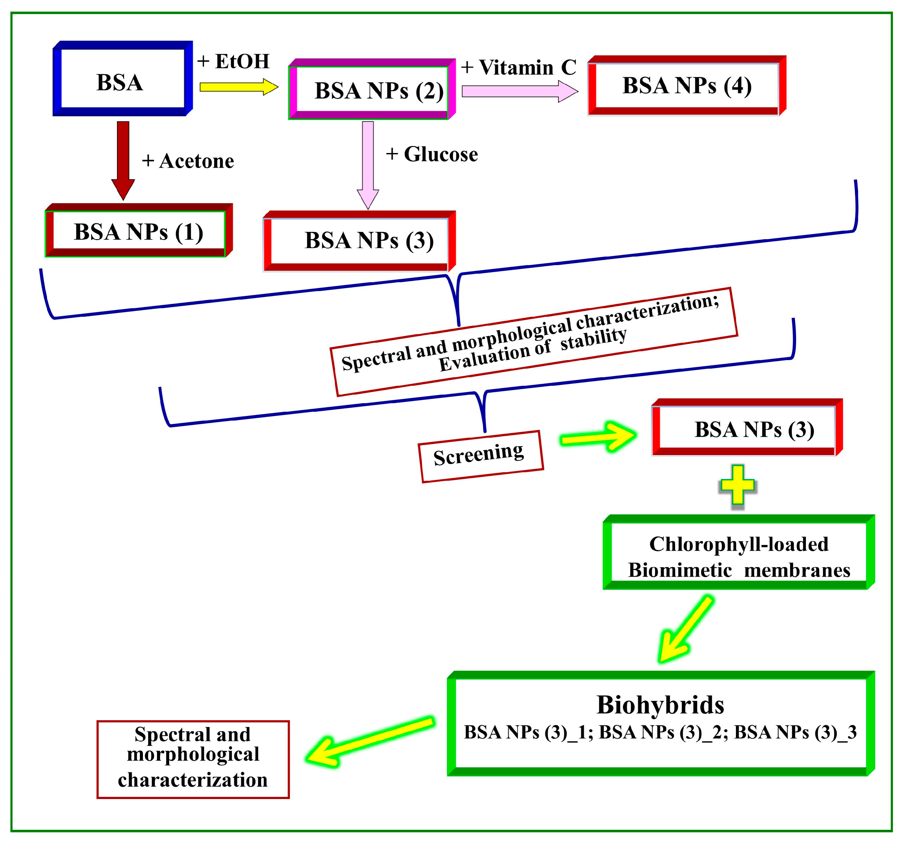
The schematic representation of the preparation of albumin particles by desolvation is displayed in Figure 2. The samples obtained are noted as follows: BSA NPs(1)—NPs produced by the acetone method, without crosslinking agent; BSA NPs(2)—NPs produced by the ethanol method, without crosslinking agent; BSA NPs(3)—NPs produced by the ethanol method, with glucose as crosslinking agent; and BSA NPs(4)—NPs produced by the ethanol method, with vitamin C as crosslinking agent.
Two types of desolvating agents were used: acetone and ethanol. As compared to BSA NPs obtained by acetone (named BSA NPs(1)), BSA NPs obtained by the EtOH method (named BSA NPs(2)) proved to be more stable in time (see Section 3.1); therefore, these particles were further used in the following experiments related to the use of the eco-friendly crosslinkers: ascorbic acid (vitamin C) and glucose. The linking of these crosslinkers to BSA NPs was carried out under continuous stirring (550 rpm, CRS 15X CAPP, Denmark) at room temperature (RT).
Preparation of albumin particles using the acetone method. Over the BSA stock solution, a volume of 8 mL of acetone (as desolvating agent) was added dropwise (1 mL/min) at room temperature (25 °C), under continuous stirring (550 rpm, CRS 15X CAPP, Region Syddanmark, Denmark), until the solution became cloudy. The nanoparticles thus obtained were subjected to centrifugation three times (15,000× g, for 10 min) with a SIGMA 2–16 K centrifuge (SciQuip Ltd., Merrington, Shrewsbury Shropshire SY4 3QJ UK, Newtown, UK). Each centrifugation step was followed by resuspension of the pellet in the same volume of distilled water as the removed supernatant, under ultrasound treatment (ultrasound systems: Hielscher UP 100H, GmbH, 14513 Teltow, Germany; BRANSON 1210, Marshall Scientific, Hampton, NH, USA).
Preparation of albumin particles using ethanol method. To obtain BSA NPs via the desolvation method, using EtOH as the desolvating agent [25], the BSA stock solution was continuously stirred (550 rpm, CRS 15X CAPP, Odense, Denmark) at room temperature (25 °C), and a volume of 8 mL of pure EtOH 96% was added dropwise (1 mL/min), until the suspension became cloudy, acquiring a yellowish color. This suspension was centrifuged three times (10,000× g, for 10 min), and each centrifugation was followed by ultrasonication, alternatively: in an ultrasound bath (BRANSON 1210, Marshall Scientific, Hampton, NH, USA) for 5 min, and then with Ti probe (Hielscher UP 100H, GmbH, 14513 Teltow, Germany) for 5 min. After each centrifugation step, the suspension was resuspended in the same volume of distilled water, with the obtained BSA NPs showing as in Figure 3 after each centrifugation step.
The resulting BSA NPs suspension obtained by the ethanol method was divided into 2 equal parts. To each half of the BSA NPs suspension, a volume of 350 μL of 8% (w/w) crosslinker agent (glucose or vitamin C) aqueous solution was added, under continuous stirring for 12 h at room temperature (RT), resulting in the samples named BSA NPs(3) and BSA NPs(4), respectively [21,31].
Preparation of artificial cell membranes labeled with Chla. Artificial cell membranes loaded with Chla were obtained by the thin-film hydration method, as previously described [53,54]. Briefly, soybean lecithin and Chla were co-dissolved in chloroform, in a mole ratio of 100:1. After solvent evaporation, the obtained thin lipid film was hydrated with a simulating biological fluid phosphate-buffered saline solution (PBS, KH2PO4/Na2HPO4/NaCl, pH 7.4), and then the suspension was vigorously mechanical stirred (VIBRAX stirrer—Milian USA, OHIO 43230 USA, 200 rpm, ~40 min), resulting in multilamellar lipid vesicles (MLVs). The experiments were carried out in the dark, at a working temperature above the critical temperature of the phase transition of lipids (41 °C), resulting in biomimetic membranes in the liquid crystal state.
Bottom-up “green” design of biohybrids. Three types of hybrid bio-entities BSA NPs/liposomes were prepared with the following volumetric ratios of BSA NPs: liposomes of 1:29 ((BSA NPs(3)-1)), 1:9 ((BSA NPs(3)-2)) and 1:2 (v/v) ((BSA NPs(3)-3)), through the self-assembly strategy, using strong stirring (1500 rpm, Vortex Fisher Scientific) for 15 min, with breaks.
Scanning electron microscopy (SEM). The morphology of the fabricated samples was investigated using a Zeiss Gemini SEM 500 field-emission scanning electron microscope (FESEM). The SEM images were acquired at an accelerating voltage of 1.5 kV and 2.5 kV, a working distance of 2 mm and a magnification of 10,000× and 20,000×, Respectively.
Atomic Force microscopy (AFM). Three-dimensional topography and phase-contrast images were obtained by means of SPM-NTegra Prima AFM (NT-MDT), operated in semi-contact mode, using an NSG 01 cantilever (resonance frequency, 87–230 kHz; force constant, 1.45–15.1 N/m). Samples were deposited on freshly cleaved mica and dried at room temperature.
UV–Vis absorption spectroscopy. The UV–Vis absorption spectra were recorded on a double-beam Perkin Elmer Lambda 750 UV–Vis spectrophotometer (Waltham, MA 02451, USA), at room temperature, in the wavelength range of 200–800 nm and at a scan speed of 1 nm/s.
Fluorescence spectroscopy. The fluorescence emission spectra of Chla–liposomes and their biohybrids with BSA NPs were recorded on a Perkin Elmer LS55 spectrophotometer (Waltham, MA 02451, USA) (by excitation at 430 nm). Chla is a natural porphyrin with intense absorption in the blue region (Soret band, 410–430 nm) and in the red region of electromagnetic spectrum. By exciting the Chla samples in blue, at 430 nm, an intense and sharp emission peak can be observed [55]. The scan speed was 500 nm/min, and the slit widths for the excitation and emission monochromators were 7.0 nm and 5.0 nm, respectively.

3. Results and Discussion

After their fabrication, we studied the influence of the process parameters on BSA NPs’ formulation. The BSA NPs were characterized by UV–Vis absorption spectroscopy and SEM in order to investigate the stability and the morphological aspects of the obtained albumin NPs.

3.1. Characterization of Nanoparticles by UV–Vis Absorption Spectroscopy

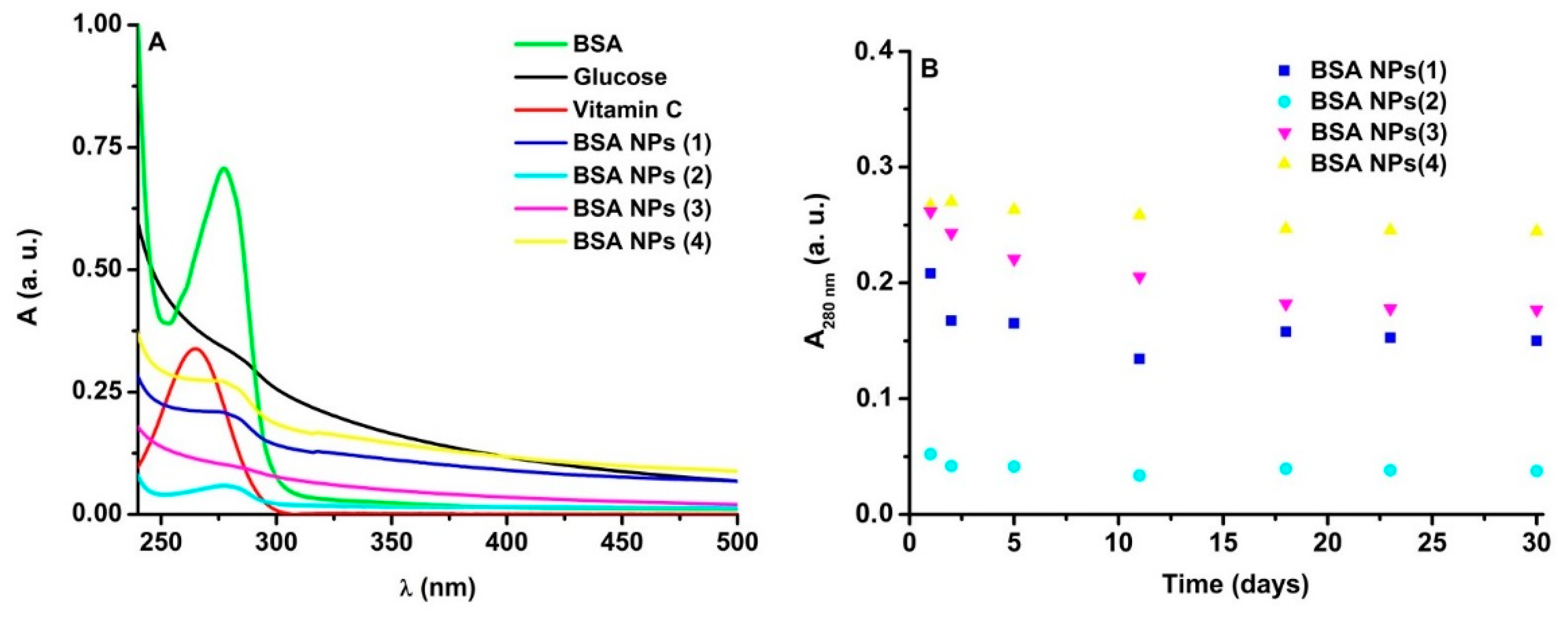
The spectral fingerprint of Trp from the protein (280 nm) was modified in the NPs samples (Figure 4A). The UV–visible spectral profile of BSA NPs is comparable to those reported in the literature for these NPs [56]. The UV–visible spectral profiles of pure BSA, glucose, vitamin C and BSA NPs (without and with crosslinking agents) were found to be significantly different when a spectral scan was evaluated between 200 and 400 nm.
As is known, larger particles scatter light more, so the baselines of the spectra are positioned at a higher height than in the case of the spectra of smaller particles. As observed in Figure 4A, according to the baseline height which is related to light scattering, it is expected that the BSA NPs mean size increases as follows: size (BSA NPs(2)) < size (BSA NPs(3)) < size (BSA NPs(1)) < size (BSA NPs(4)). A similar trend has been obtained by Amighi et al. [31]. These assumptions were further confirmed by SEM analysis. BSA NPs prepared by desolvation were monitored to determine stability over time. The absorbance of the nanoparticles at 280 nm was recorded over 30 days, and the collected data are represented in Figure 4B. After 30 days, the absorbance of BSA NPs samples reached 28% for BSA NPs(1) and BSA NPs(2), 32.4% for BSA NPs(3), and 8.4% for BSA NPs(4). Therefore, in the following experiments on BSA NPs/liposome bio-entities, we chose BSA NPs(3) as the samples of interest. All types of NPs showed similar stability in time, except for the BSA NPs(4).
Liposomes have been proposed and even used in the clinic in various applications, from vaccine-like formulations, drug delivery, treatment of infections and skin conditions, to cancer treatment [57]. The biohybrids obtained and characterized in this study come as a necessity to innovate and streamline the production of liposomes. The presence of biomimetic membranes increases the cellular uptake of BSA formulations. Moreover, this lipid coat improves the loading of hydrophobic drugs.

3.2. Characterization of Albumin Nanoparticles by SEM

The morphological properties of the surface were displayed by SEM analysis in order to confirm the formation of BSA NPs and to observe their shape. The BSA NPs(1) (Figure 5) generated particles that were polydispersed (non-uniformly distributed), with diameters ranging from the nano- and microscale. On the contrary, the ethanol method generated more homogenous populations of protein particles that were spherical in shape and nanosized (BSA NPs(2)) (Figure 5B).
BSA NPs from the samples without crosslinking agent (Figure 5A), as well as from those in which the crosslinking agent was glucose (Figure 5C), and vitamin C, respectively (Figure 5D), showed that the average size of BSA NPs was ≈170 nm for simple nanoparticles, ≈200 nm for the nanoparticles prepared with glucose and larger for those prepared with ascorbic acid. The SEM analysis also revealed that the protein particles obtained by EtOH desolvation presented spherical shapes, and they are smaller in size compared to those from the acetone desolvation method (Figure 5). Similar results were obtained in a previous report [31].
The particle size distribution based on the SEM results was analyzed and emphasized as histograms for the sample with BSA NPs(2) without a reticulation agent, and for the sample with BSA NPs(3) with glucose as a reticulation agent. The particle size distribution for the two samples was evaluated using ImageJ software on the SEM images.
Corroborating the results from the UV–Vis absorption spectroscopy and SEM analysis, in our studies, we further used the BSA NPs(2), BSA NPs(3), andBSA NPs(4) (generated by EtOH method), because this method gives rise to nanosized particles as compared to the acetone desolvation method (BSA NPs(1)). On the other hand, in other experiments leaded by us, we went further in studies with glucose as the reticulating agent since it produced nanoparticles smaller in size as compared to those produced with ascorbic acid. These findings are similar to those obtained by Amighi et al. [31] and Santos-Rebelo et al. [50].

3.3. Characterization of BSA NPs/Liposomes Bio-Entities

Taking into account the obtained results, we will further use BSA NPs(3) (prepared with EtOH method) and linked with glucose, for the development of three types of biohybrids (see Section 2). The resulting BSA NPs/liposomes bio-nano-constructs were characterized by UV–Vis absorption and emission spectroscopy by exploiting the optical properties of Chla embedded in the bio-inspired membranes. Furthermore, the three-dimensional topography of biohybrids was investigated by AFM.

3.3.1. Spectral Characterization of Biohybrids

Chla was successfully used as a spectral marker for the liposome-based materials’ characterization, as reported in our previous studies [47,48,53,54]. The UV–Vis absorption spectra of BSA NPs/liposomes bio-entities (BSA NPs(3)-1, BSA NPs(3)-2 and BSA NPs(3)-3; see Figure 6A) displayed the spectral signatures of Trp at 280 nm, and of Chla at 670 nm and at 410–420 nm. The changes in the baseline position of each UV–Vis absorption spectrum were observed. A correlation with a particle size could be achieved in function of the baseline position. Thus, it could predict that the liposome size is greater than that of biohybrids.
The fluorescence emission spectra (Figure 6B) revealed the characteristic fluorescence emission peak of Chla at 670 nm. A fluorescence quenching occurred in the presence of BSA NPs due to the interaction between amino-acid residues (e.g., Trp and Tyr) in BSA NPs and Chla–liposomes, leading to a reorganization in the artificial lipid bilayers, a fact that was further demonstrated by AFM images, which detected a morphological change of the biomimetic membranes. Similar findings were obtained by Vavilin et al. [58], who observed Chl fluorescence quenching due to the Trp interaction. Another study suggests that pheophytin, a chlorophyll derivative, is able to interact with HSA by means of hydrogen bonds, resulting in a fluorescence quenching [59]. As can be seen from Figure 6, among the three bio-entities, the most suitable one is the BSA NPs(3)-3, since a more accentuated change of the spectral Vis signatures of the natural biosensor, Chla (Figure 6A), and a more pronounced Chla-fluorescence quenching were observed in this case (Figure 6B), indicating a better interaction between the components of this biohybrid. An important role in biohybrid formation can be attributed to the interaction between functional groups of amino acids in BSA NPs and polar head groups of lipid molecules in biomimetic membranes. Thus, the biohybrid BSA NPs(3)-3 will be used further in our studies.
Liposomes have been shown to increase the therapeutic efficacy of drugs such as doxorubicin (DOX) by improving its pharmacokinetic profile, increasing its circulation time and reducing its side effects [60]. As BSA is a commonly used protein-based drug carrier system for drug delivery, including DOX [61], we expect the biohybrids obtained and characterized in this study to be necessity to innovate and streamline the production of liposomes. The presence of biomimetic membranes increases the cellular uptake of BSA formulations. Moreover, this lipid coat improves the loading of hydrophobic drugs.

3.3.2. Morphological Characterization of BSA NPs and Their Bio-Entities

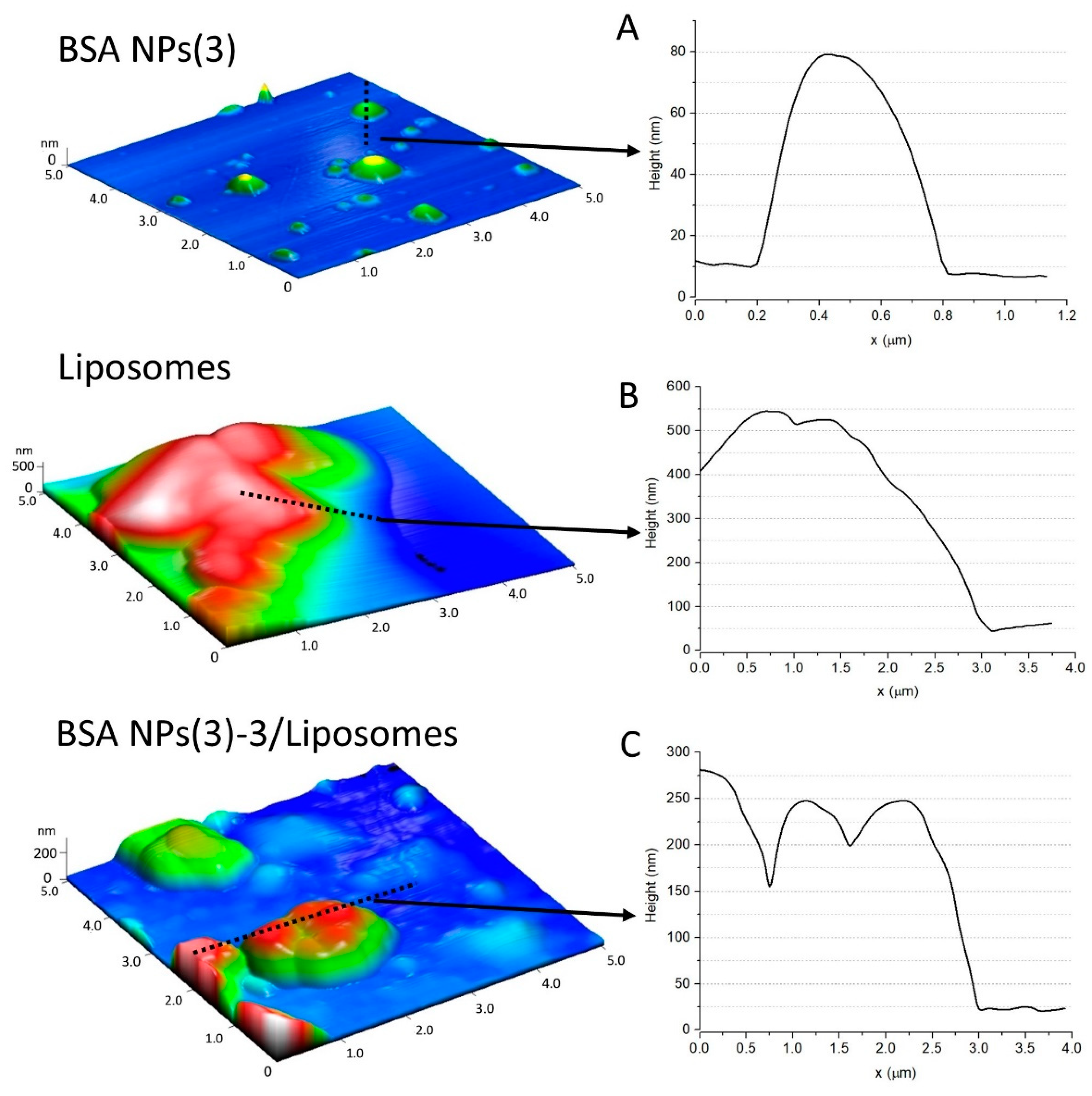
Furthermore, BSA NPs(3) (obtained by EtOH and glucose method) were investigated by AFM revealing the 3D profile (Figure 7). Using the semi-contact mode AFM, the characterization of individual particles and particle groups of BSA NPs (Figure 7A) deposited on a mica substrate was performed. BSA NPs(3) are spherical shaped, with a smooth surface, and their average diameter is ∼70 nm. No visible aggregates of free BSA molecules were evident, indicating that the preparation of BSA NPs(3) via the desolvation process was adequate. The particle size obtained from AFM was smaller than that from the SEM measurements. This may be due to different shrinkage results related to the interaction with the support with BSA NPs during the drying process for the AFM image (on mica substrate) and SEM (on Si substrate). Similar findings have also been reported in the literature [62,63].
To monitor the effect of BSA NPs on liposomes, the morphology of liposomes was first investigated. Among the three types of biohybrids, we chose to investigate the morphology of the sample BSA NPs(3)-3, because we considered that it has an optimal ratio to obtain BSA NPs:Liposomes hybrids, as indicated by fluorescence emission spectra (see Figure 6B).
Figure 7B shows that the spherical liposome was deformed on the mica surface, and a flattened vesicle was obtained. The vertical profile of the image shows that the liposome is 500 nm in height; the lipid vesicle interacts with the mica substrate, leading to a flattened structure, because the diameter of the multilamellar vesicles is greater than 500 nm, going up to 1000 nm [64].
The BSA NPs adsorbed on the surface of liposomes are visualized in Figure 8. The presence of BSA NPs leads to the aggregation of three lipid vesicles, as indicated in the vertical profile image. Presumably, the lipid vesicles are adsorbed on the mica support, and thus the liposome size in the presence of BSA NP dropped to ∼250 nm. Therefore, the multilamellar vesicles (>500 nm) shrink and turn into large unilamellar vesicles (>100 nm). The AFM results are well correlated with the spectral analysis.
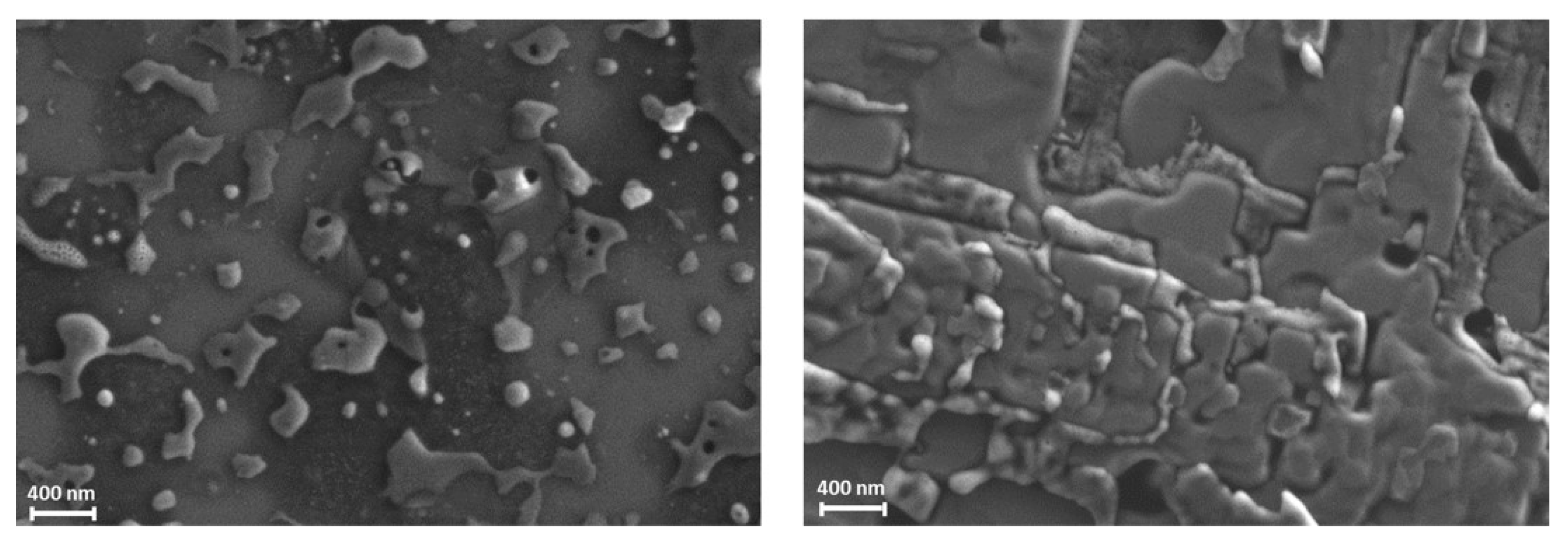
Other morphological aspects of the obtained bio-entities are displayed in the SEM micrographs (Figure 8). Different interactions can be observed between the prepared systems and the substrate on which they were deposited. Thus, the bare liposomes showed a spherical or quasi-spherical shape (Figure 8, left), while aggregated multilayered ordered coatings were observed in the case of biohybrids (Figure 8, right). This behavior is due to the composition of biohybrid particles, as it involves specific interactions at the bio-interfaces between their components, and also between the biohybrids and the substrates. The SEM analysis correlates with the AFM results, where spherical liposomes were deformed on the mica surface and flattened vesicles were obtained. The difference in particle size obtained from the AFM and SEM analyses can be attributed to a combination of factors, including the shape of particles, the nature of the sample environment, the method of data analysis and the type of instrument used.
Nanoparticles with a size greater than 200 nm are typically considered to be in the microscale range, rather than the nanoscale range. This larger size may limit their ability to effectively penetrate certain biological barriers, such as cell membranes or the blood–brain barrier, which could potentially limit their usefulness as drug delivery systems. However, albumin particles with a size >200 nm have been used for biomedical applications. Thus, Karimi et al. [17] highlighted the role of albumin microspheres (1–3 μm diameter) as drug delivery vehicles into solid tumors, as well as the liver and kidney. In addition, the use of glucose and BSA, both with good biocompatibility and biodegradability, can lead to the faster degradation of NPs in biological environments. There are still potential ways in which these larger nanoparticles could be utilized for localized, targeted, or slow-release drug delivery. For example, in localized delivery, NPs could be designed to remain at the site of administration, rather than needing to travel throughout the body. This could be achieved through various methods, such as embedding the nanoparticles in a gel or using them in a patch or implant [65]. In targeted delivery, while larger NPs may have limited ability to passively diffuse into tissues, they may still be able to actively target specific cells or tissues. This could be achieved through various targeting strategies, such as adding a ligand to the nanoparticle surface that binds specifically to a cell surface receptor [66]. In slow-release delivery, larger NPs may have a larger surface area available for drug loading, allowing for a higher drug payload. This could be beneficial for drugs that require a slow and sustained release over time [67]. Overall, while nanoparticles greater than 200 nm may have limitations for drug delivery compared to smaller nanoparticles, there are still potential strategies that could be used to make them effective drug delivery systems.

4. Conclusions

This paper reported, for the first time, a bio-strategy to design albumin-based soft materials; this is a very promising approach, as it ensures that BSA NPs have certain features prior to loading with the drug. A simple preparation method (using ethanol) of BSA NPs with and without vitamin C or glucose as an eco-friendly crosslinking agent was used. The morphological characterization of the prepared protein particles confirmed the formation of BSA NPs with spherical and (quasi)spherical shapes. The stability in time of the obtained BSA NPs was demonstrated by UV–Vis absorption and emission spectroscopy.
The addition of artificial cell membranes to the BSA NPs resulted in the development of bio-entities with modified size and morphological aspects as compared to each individual component. The insight into the formation of these biohybrids was also provided by the spectral signature of Chla on fluorescence emission and UV–Vis absorption spectra.
The biomimetic membranes coated with BSA NPs layer presented nano-scale size and a (quasi)spherical morphology. The lipid coating of albumin nanoparticles could be a useful strategy to build effective drug delivery nanosystems, which can simultaneously perform two important functions: (i) improvement of cellular uptake (due to the lipid coat) and (ii) opsonization avoiding (due to BSA NPs).
The obtained bio-entities constitute, therefore, the building blocks for the development of novel multifunctional drug carriers with applications in the biomedical field. Moreover, the obtained biohybrids could be used as bio-coatings to increase the biocompatibility of various nanocarriers. These studies will be continued to develop new protein nanosystems that will be more stable over time and which could incorporate various therapeutic agents.

Author Contributions

Conceptualization, C.G.C. and M.-E.B.-P.; methodology, M.-E.B.-P. and C.G.C.; investigation, C.G.C., M.-E.B.-P., N.C., D.L.S., A.E.B., A.C. and S.I.; writing—original draft preparation, C.G.C. and M.-E.B.-P.; writing—review and editing, C.G.C., M.-E.B.-P. and N.C.; visualization, C.G.C. and M.-E.B.-P.; supervision, C.G.C. and M.-E.B.-P. All authors have read and agreed to the published version of the manuscript.

Funding

This research was funded by the Romanian Ministry of Research, Innovation and Digitization, CNCS—UEFISCDI—Project No. 582PED/2022, New hybrid protein nanostructures for targeting specific in colon tumor cells (Prot-Col-Target), PN-III-P2-2.1-PED-2021-1323/01.08.2022.

Institutional Review Board Statement

Not applicable.

Informed Consent Statement

Not applicable.

Data Availability Statement

Not applicable.

Acknowledgments

The authors acknowledge to the Romanian Ministry of Research, Innovation and Digitization, CNCS—UEFISCDI, for the financial support—Project No. 582PED⁄2022, New hybrid protein nanostructures for targeting specific in colon tumor cells (Prot-Col-Target), PN-III-P2-2.1-PED-2021-1323/01.08.2022.

Conflicts of Interest

The authors declare no conflict of interest.

References

  1. Hankins, J. The Role of Albumin in Fluid and Electrolyte Balance. J. Infus. Nurs. 2006, 29, 260–265. [Google Scholar] [CrossRef] [Green Version]
  2. Francis, G.L. Albumin and mammalian cell culture: Implications for biotechnology applications. Cytotechnology 2010, 62, 1–16. [Google Scholar] [CrossRef] [PubMed] [Green Version]
  3. Kuntip, N.; Japrung, D.; Pongprayoon, P. How human serum albumin-selective DNA aptamer binds to bovine and canine serum albumins. Biopolymers 2021, 112, e23421. [Google Scholar] [CrossRef] [PubMed]
  4. Tao, C.; Chuah, Y.J.; Xu, C.; Wang, D.-A. Albumin conjugates and assemblies as versatile bio-functional additives and carriers for biomedical applications. J. Mater. Chem. B 2019, 7, 357–367. [Google Scholar] [CrossRef]
  5. Chilom, C.; Barangă, G.; Găzdaru, D.; Popescu, A. Characterisation by fluorescence of human and bovine serum albumins in interaction with eosin Y. J. Optoelectron. Adv. Mater. 2013, 15, 311–316. [Google Scholar]
  6. Chilom, C.G.; Nistorescu, A. A spectroscopic study of the interaction of HSA with tetracaine. IJBB 2016, 53, 206–211. [Google Scholar]
  7. Sandu, N.; Chilom, C.G.; Popescu, A.I. Spectroscopic Insights on the Binding of Rutin to Bovine Serum Albumin. Rom. J. Phys. 2020, 65, 703. [Google Scholar]
  8. Neumann, E.; Frei, E.; Funk, D.; Becker, M.D.; Schrenk, H.-H.; Müller-Ladner, U.; Fiehn, C. Native albumin for targeted drug delivery. Expert Opin. Drug Deliv. 2010, 7, 915–925. [Google Scholar] [CrossRef] [PubMed]
  9. Chilom, C.G.; Bălan, A.; Sandu, N.; Bălăşoiu, M.; Stolyar, S.; Orelovich, O. Exploring the Conformation and Thermal Stability of Human Serum Albumin Corona of Ferrihydrite Nanoparticles. Int. J. Mol. Sci. 2020, 21, 9734. [Google Scholar] [CrossRef] [PubMed]
  10. Chilom, C.; Barangă, G.; Găzdaru, D.; Popescu, A. A spectroscopic approach of pH effect on thermal denaturation of human and bovine serum albumins. J. Optoelectron. Adv. Mater. 2011, 13, 583–587. [Google Scholar]
  11. Baler, K.; Martin, O.A.; Carignano, M.A.; Ameer, G.A.; Vila, J.A.; Szleifer, I. Electrostatic Unfolding and Interactions of Albumin Driven by pH Changes: A Molecular Dynamics Study. J. Phys. Chem. B 2014, 118, 921–930. [Google Scholar] [CrossRef]
  12. Froehlich, E.; Mandeville, J.S.; Jennings, C.J.; Sedaghat-Herati, R.; Tajmir-Riahi, H.A. Dendrimers Bind Human Serum Albumin. J. Phys. Chem. B 2009, 113, 6986–6993. [Google Scholar] [CrossRef]
  13. Taguchi, K.; Okamoto, Y.; Matsumoto, K.; Otagiri, M.; Chuang, V. When Albumin Meets Liposomes: A Feasible Drug Carrier for Biomedical Applications. Pharmaceuticals 2021, 14, 296. [Google Scholar] [CrossRef]
  14. Spada, A.; Emami, J.; Tuszynski, J.A.; Lavasanifar, A. The Uniqueness of Albumin as a Carrier in Nanodrug Delivery. Mol. Pharm. 2021, 18, 1862–1894. [Google Scholar] [CrossRef]
  15. Lu, N.; Sui, Y.; Ding, Y.; Tian, R.; Li, L.; Liu, F. Adsorption of human serum albumin on functionalized single-walled carbon nanotubes reduced cytotoxicity. Chem. Interact. 2018, 295, 64–72. [Google Scholar] [CrossRef] [PubMed]
  16. Wojnarowska-Nowak, R.; Polit, J.; Zięba, A.; Stolyarchuk, I.; Nowak, S.; Romerowicz-Misielak, M.; Sheregii, E. Colloidal quantum dots conjugated with human serum albumin-interactions and bioimaging properties. Opto-Electron. Rev. 2017, 25, 137–147. [Google Scholar] [CrossRef]
  17. Karimi, M.; Bahrami, S.; Ravari, S.B.; Zangabad, P.S.; Mirshekari, H.; Bozorgomid, M.; Shahreza, S.; Sori, M.; Hamblin, M.R. Albumin nanostructures as advanced drug delivery systems. Expert Opin. Drug Deliv. 2016, 13, 1609–1623. [Google Scholar] [CrossRef] [Green Version]
  18. Chilom, C.G.; Zorilă, B.; Bacalum, M.; Bălăşoiu, M.; Yaroslavtsev, R.; Stolyar, S.V.; Tyutyunnicov, S. Ferrihydrite nanopar-ticles interaction with model lipid membranes. Chem. Phys. Lipids 2020, 226, 104851. [Google Scholar] [CrossRef]
  19. Wu, J. The Enhanced Permeability and Retention (EPR) Effect: The Significance of the Concept and Methods to Enhance Its Application. J. Pers. Med. 2021, 11, 771. [Google Scholar] [CrossRef] [PubMed]
  20. Chubarov, A.S. Serum Albumin for Magnetic Nanoparticles Coating. Magnetochemistry 2022, 8, 13. [Google Scholar] [CrossRef]
  21. Niknejad, H.; Mahmoudzadeh, R. Comparison of Different Crosslinking Methods for Preparation of Docetaxel-loaded Albumin Nanoparticles. Iran. J. Pharm. Res. 2015, 14, 385–394. [Google Scholar]
  22. Hong, S.; Choi, D.W.; Kim, H.N.; Park, C.G.; Lee, W.; Park, H.H. Protein-Based Nanoparticles as Drug Delivery Systems. Pharmaceutics 2020, 12, 604. [Google Scholar] [CrossRef] [PubMed]
  23. Bloch, K.; Pardesi, K.; Satriano, C.; Ghosh, S. Bacteriogenic Platinum Nanoparticles for Applicationin Nanomedicine. Front. Chem. 2021, 9, 624344. [Google Scholar] [CrossRef]
  24. Hornok, V. Serum Albumin Nanoparticles: Problems and Prospects. Polymers 2021, 13, 3759. [Google Scholar] [CrossRef] [PubMed]
  25. Jahanban-Esfahlan, A.; Dastmalchi, S.; Davaran, S. A simple improved desolvation method for the rapid preparation of albumin nanoparticles. Int. J. Biol. Macromol. 2016, 91, 703–709. [Google Scholar] [CrossRef]
  26. Chu, F.; Thornton, D.T.; Nguyen, H. Chemical cross-linking in the structural analysis of protein assemblies. Methods 2018, 144, 53–63. [Google Scholar] [CrossRef]
  27. Luo, R.; Lin, M.; Zhang, C.; Shi, J.; Zhang, S.; Chen, Q.; Hu, Y.; Zhang, M.; Zhang, J.; Gao, F. Genipin-crosslinked human serum albumin coating using a tannic acid layer for enhanced oral administration of curcumin in the treatment of ulcerative colitis. Food Chem. 2020, 330, 127241. [Google Scholar] [CrossRef] [PubMed]
  28. Kistriyani, L.; Sulistyawan, W.; Kesuma, S.H. Characteristic of ascorbic acid in crosslinked chitosan edible film as drug delivery system membrane. MATEC Web Conf. 2018, 154, 01027. [Google Scholar] [CrossRef] [Green Version]
  29. Salihu, R.; Razak, S.I.A.; Zawawi, N.A.; Kadir, M.R.A.; Ismail, N.I.; Jusoh, N.; Mohamad, M.R.; Nayan, N.H.M. Citric acid: A green cross-linker of biomaterials for biomedical applications. Eur. Polym. J. 2021, 146, 110271. [Google Scholar] [CrossRef]
  30. Herrera, M.A.; Mathew, A.P.; Oksman, K. Barrier and mechanical properties of plasticized and cross-linked nanocellulose coatings for paper packaging applications. Cellulose 2017, 24, 3969–3980. [Google Scholar] [CrossRef] [Green Version]
  31. Amighi, F.; Emam-Djomeh, Z.; Labbafi-Mazraeh-Shahi, M. Effect of different cross-linking agents on the preparation of bovine serum albumin nanoparticles. J. Iran. Chem. Soc. 2020, 17, 1223–1235. [Google Scholar] [CrossRef]
  32. Esim, O.; Gedik, M.E.; Dogan, A.L.; Gunaydin, G.; Hascicek, C. Development of carboplatin loaded bovine serum albumin nanoparticles and evaluation of its effect on an ovarian cancer cell line. J. Drug Deliv. Sci. Technol. 2021, 64, 102655. [Google Scholar] [CrossRef]
  33. An, F.-F.; Zhang, X.-H. Strategies for Preparing Albumin-based Nanoparticles for Multifunctional Bioimaging and Drug Delivery. Theranostics 2017, 7, 3667–3689. [Google Scholar] [CrossRef]
  34. Cho, H.; Jeon, S.I.; Ahn, C.-H.; Shim, M.K.; Kim, K. Emerging Albumin-Binding Anticancer Drugs for Tumor-Targeted Drug Delivery: Current Understandings and Clinical Translation. Pharmaceutics 2022, 14, 728. [Google Scholar] [CrossRef] [PubMed]
  35. Shi, J.-H.; Zhou, K.-L.; Lou, Y.-Y.; Pan, D.-Q. Multi-spectroscopic and molecular modeling approaches to elucidate the binding interaction between bovine serum albumin and darunavir, a HIV protease inhibitor. Spectrochim. Acta Part A Mol. Biomol. Spectrosc. 2018, 188, 362–371. [Google Scholar] [CrossRef]
  36. Sandu, N.; Chilom, C.G.; Florescu, M. Molecular insights into binding mechanism of rutin to bovine serum albumin—Levothyroxine complex: Spectroscopic and molecular docking approaches. Spectrochim. Acta Part A Mol. Biomol. Spectrosc. 2021, 264, 120261. [Google Scholar] [CrossRef] [PubMed]
  37. Pawar, S.K.; Jaldappagari, S. Interaction of repaglinide with bovine serum albumin: Spectroscopic and molecular docking approaches. J. Pharm. Anal. 2019, 9, 274–283. [Google Scholar] [CrossRef]
  38. Merlot, A.M.; Kalinowski, D.S.; Richardson, D.R. Unraveling the mysteries of serum albumin-More than just a serum protein. Front. Physiol. 2014, 5, 299. [Google Scholar] [CrossRef] [Green Version]
  39. Meng, R.; Zhu, H.; Wang, Z.; Hao, S.; Wang, B. Preparation of Drug-Loaded Albumin Nanoparticles and Its Application in Cancer Therapy. J. Nanomater. 2022, 2022, 1–12. [Google Scholar] [CrossRef]
  40. Desai, N. Nanoparticle Albumin-Bound Anticancer Agents. In Non-Biological Complex Drugs; Crommelin, D.J., de Vlieger, J.S.B., Eds.; Springer International Publishing: Cham, Switzerland, 2015; Volume 20, pp. 335–354. [Google Scholar]
  41. Tacheva, B.; Paarvanova, B.; Ivanov, I.T.; Tenchov, B.; Georgieva, R.; Karabaliev, M. Drug Exchange between Albumin Na-noparticles and Erythrocyte Membranes. Nanomaterials 2018, 9, 47. [Google Scholar] [CrossRef] [Green Version]
  42. Teixeira, S.; Carvalho, M.A.; Castanheira, E.M.S. Functionalized Liposome and Albumin-Based Systems as Carriers for Poorly Water-Soluble Anticancer Drugs: An Updated Review. Biomedicines 2022, 10, 486. [Google Scholar] [CrossRef]
  43. Peng, Q.; Wei, X.-Q.; Yang, Q.; Zhang, S.; Zhang, T.; Shao, X.-R.; Cai, X.-X.; Zhang, Z.-R.; Lin, Y.-F. Enhanced biostability of nanoparticle-based drug delivery systems by albumin corona. Nanomedicine 2015, 10, 205–214. [Google Scholar] [CrossRef] [PubMed]
  44. Wei, X.-Q.; Ba, K. Construction a Long-Circulating Delivery System of Liposomal Curcumin by Coating Albumin. ACS Omega 2020, 5, 16502–16509. [Google Scholar] [CrossRef] [PubMed]
  45. Pasarin, D.; Ghizdareanu, A.-I.; Enascuta, C.E.; Matei, C.B.; Bilbie, C.; Paraschiv-Palada, L.; Veres, P.-A. Coating Materials to Increase the Stability of Liposomes. Polymers 2023, 15, 782. [Google Scholar] [CrossRef] [PubMed]
  46. Akbarzadeh, A.; Rezaei-Sadabady, R.; Davaran, S.; Joo, S.W.; Zarghami, N.; Hanifehpour, Y.; Samiei, M.; Mohammad Kouhi, M.; Nejati-Koshki, K. Liposome: Classification, preparation, and applications. Nanoscale Res. Lett. 2013, 8, 102. [Google Scholar] [CrossRef] [Green Version]
  47. Barbinta-Patrascu, M.-E.; Gorshkova, Y.; Ungureanu, C.; Badea, N.; Bokuchava, G.; Lazea-Stoyanova, A.; Bacalum, M.; Zhi-gunov, A.; Petrovič, S.M. Characterization and Antitumoral Activity of Biohybrids Based on Turmeric and Silver/Silver Chloride Nanoparticles. Materials 2021, 14, 4726. [Google Scholar] [CrossRef]
  48. Gorshkova, Y.; Barbinta-Patrascu, M.-E.; Bokuchava, G.; Badea, N.; Ungureanu, C.; Lazea-Stoyanova, A.; Răileanu, M.; Bacalum, M.; Turchenko, V.; Zhigunov, A.; et al. Biological Performances of Plasmonic Biohybrids Based on Phy-to-Silver/Silver Chloride Nanoparticles. Nanomaterials 2021, 11, 1811. [Google Scholar] [CrossRef]
  49. Strain, H.H.; Svec, W.A.; Vernon, L.P.; Seely, G.R. The Chlorophylls; Academic Press: New York, NY, USA, 1966; Volume 2, pp. 21–66. [Google Scholar]
  50. Santos-Rebelo, A.; Kumar, P.; Pillay, V.; Choonara, Y.E.; Eleutério, C.; Figueira, M.; Viana, A.S.; Ascensão, L.; Molpeceres, J.; Rijo, P.; et al. Development and mechanistic insight into the enhanced cytotoxic potential of Parvifloron D albumin nanoparticles in EGFR-overexpressing pancreatic cancer cells. Cancers 2019, 11, 1733. [Google Scholar] [CrossRef] [Green Version]
  51. Chen, F.; Huang, G.; Huang, H. Sugar ligand-mediated drug delivery. Futur. Med. Chem. 2020, 12, 161–171. [Google Scholar] [CrossRef]
  52. Barros, J.; Grenha, A.; Carvalho, R. Vitamin C and glucose cross-linked chitosan as a versatile platform for drug delivery. Int. J. Biol. Macromol. 2019, 137, 1117–1125. [Google Scholar]
  53. Stefanescu, T.; Manole, C.; Parvu, C.; Barbinta-Patrascu, M.E.; Tugulea, L. Supported phospholipid bilayers with chlorophyll for optoelectronic devices. Optoelectron. Adv. Mater. Rapid Commun. 2010, 4, 33–38. [Google Scholar]
  54. Barbinta-Patrascu, M.E.; Tugulea, L.; Meghea, A. Procaine effects on model membranes with chlorophyll a. Rev. De Chim. 2009, 60, 337–341. [Google Scholar]
  55. Lichtenthaler, H.K.; Wenzel, O.; Buschmann, C.; Gitelson, A. Plant Stress Detection by Reflectance and Fluorescencea. Ann. N. Y. Acad. Sci. 1998, 851, 271–285. [Google Scholar] [CrossRef]
  56. Bronze-Uhle, E.S.; Costa, B.C.; Ximenes, V.F.; Lisboa-Filho, P.N. Synthetic nanoparticles of bovine serum albumin with en-trapped salicylic acid. Nanotechnol. Sci. Appl. 2017, 10, 11–21. [Google Scholar] [CrossRef] [PubMed] [Green Version]
  57. Çağdaş, M.; Sezer, A.D.; Bucak, S. Liposomes as Potential Drug Carrier Systems for Drug Delivery, Application of Nanotechnology in Drug Delivery; Sezer, A.D., Ed.; IntechOpen: London, UK, 2014. [Google Scholar]
  58. Vavilin, D.V.; Ermakova-Gerdes, S.Y.; Keilty, A.T.; Vermaas, W.F.J. Tryptophan at Position 181 of the D2 Protein of Photo-system II Confers Quenching of Variable Fluorescence of Chlorophyll: Implications for the Mechanism of Energy-Dependent Quenching. Biochemistry 1999, 38, 14690–14696. [Google Scholar] [CrossRef] [PubMed]
  59. Chaves, O.A.; Amorim, A.P.D.O.; Castro, L.H.E.; Sant’Anna, C.M.R.; De Oliveira, M.C.C.; Cesarin-Sobrinho, D.; Netto-Ferreira, J.C.; Ferreira, A.B.B. Fluorescence and Docking Studies of the Interaction between Human Serum Albumin and Pheophytin. Molecules 2015, 20, 19526–19539. [Google Scholar] [CrossRef] [Green Version]
  60. Gabizon, A.; Shmeeda, H. Bypassing the RES: Physicochemical barriers to nanocarrier clearance. Adv. Drug Deliv. Rev. 2003, 55, 1171–1190. [Google Scholar]
  61. Zhou, C.; Zhang, Y.; Li, J.; Liang, Q.; Huang, Z.; Yu, Y. Enhanced therapeutic efficacy of a combination of doxorubicin and bovine serum albumin-conjugated nanoparticles for hepatocellular carcinoma. Drug Deliv. 2019, 26, 259–267. [Google Scholar]
  62. Zhong, H.; Tarrach, G.; Wu, P.; Drechsler, A.; Wei, D.; Yuan, J. High resolution magnetic force microscopy of patterned L10-FePt dot arrays by nanosphere lithography. Nanotechnology 2008, 19, 095703. [Google Scholar] [CrossRef]
  63. Rohiwal, S.; Tiwari, A.; Verma, G.; Pawar, S. Preparation and evaluation of bovine serum albumin nanoparticles for ex vivo colloidal stability in biological media. Colloids Surf. A Physicochem. Eng. Asp. 2015, 480, 28–37. [Google Scholar] [CrossRef]
  64. Leitgeb, M.; Knez, Ž.; Primožič, M. Sustainable technologies for liposome preparation. J. Supercrit. Fluids 2020, 165, 104984. [Google Scholar]
  65. Zhang, L.; Gu, F.; Chan, J.; Wang, A.; Langer, R.S.; Farokhzad, O.C. Nanoparticles in Medicine: Therapeutic Applications and Developments. Clin. Pharmacol. Ther. 2007, 83, 761–769. [Google Scholar] [CrossRef] [PubMed]
  66. Peer, D.; Karp, J.M.; Hong, S.; Farokhzad, O.C.; Margalit, R.; Langer, R. Nanocarriers as an emerging platform for cancer therapy. Nat. Nanotechnol. 2007, 2, 751–760. [Google Scholar] [CrossRef] [PubMed]
  67. Patil, Y.; Sadhukha, T.; Ma, L.; Panyam, J. Nanoparticle-mediated simultaneous and targeted delivery of paclitaxel and tariquidar overcomes tumor drug resistance. J. Control. Release 2009, 136, 21–29. [Google Scholar] [CrossRef] [PubMed]
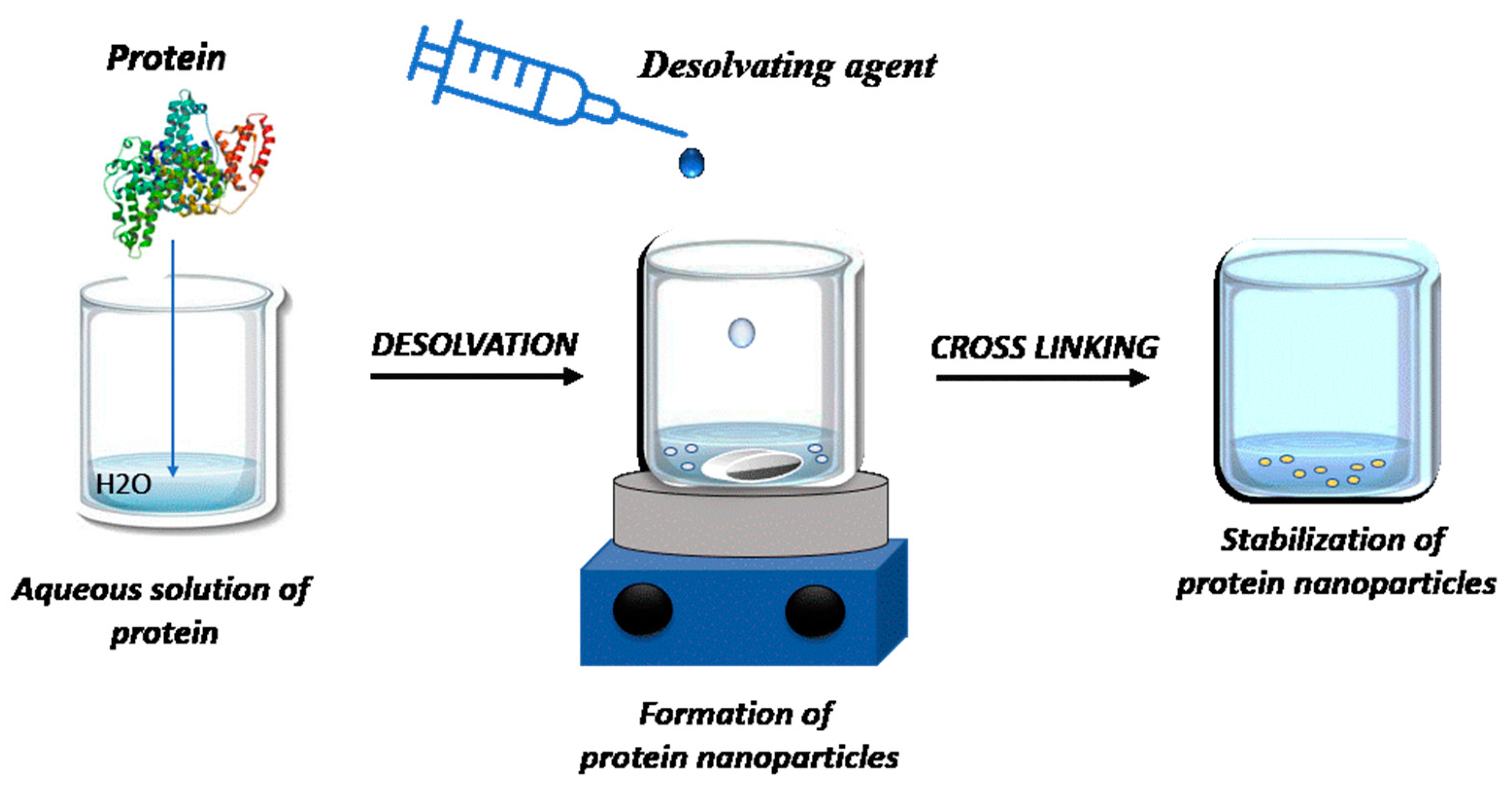
Figure 1. Schematic diagram of the desolvation method for preparing protein nanoparticles.
Coatings 13 00671 g001
Figure 2. Schematic representation of the preparation of albumin particles at room temperature (RT), using acetone (BSA NPs(1)) or EtOH (BSA NPs(2)) as desolvating agents [31]. Two types of eco-friendly crosslinkers were used: glucose (BSA NPs(3)) and ascorbic acid (vitamin C) (BSA NPs(4)). After preparation, the stability evaluation and spectral and morphological characterization were performed.
Coatings 13 00671 g002
Figure 3. Visualization of bovine serum albumin nanoparticles obtained by the desolvation method, using ethanol as a desolvating agent (BSA NPS(2)): (a) the first centrifugation, (b) the second centrifugation and (c) the third centrifugation.
Coatings 13 00671 g003
Figure 4. (A) UV–Vis spectra of glucose, vitamin C, BSA and all BSA NPs (1)–(4) obtained by the acetone and ethanol methods. (B) Time monitoring of the stability of BSA NPs obtained by the desolvating method.
Coatings 13 00671 g004
Figure 5. SEM micrographs of the BSA NPs(1) (A), BSA NPs(2) (B), BSA NPs(3) (C) and BSA NPs(4) (D); size distribution for BSA NPs(3) (E); and size distribution for BSA NPs(2) (F).
Coatings 13 00671 g005
Figure 6. The UV–Vis absorption (A) and fluorescence emission spectra (B) of Chla–liposomes (green line, ) and of biohybrids BSA NPs: BSA NPs(3)-1 (red line, ), BSA NPs(3)-2 (blue line, ) and BSA NPs(3)-3 (magenta line, ) (v/v).
Coatings 13 00671 g006
Figure 7. Three-dimensional AFM images (left) and the profiles (right) of BSA NPs(3) (A), liposomes (B) and BSA NPs(3)-3 (C). Samples were deposited on the mica substrate.
Coatings 13 00671 g007
Figure 8. The SEM micrographs of liposomes (left) and biohybrids BSA NPs—liposomes (right) at the magnification of 20,000×. The samples were deposited on Si substrates.
Coatings 13 00671 g008
Disclaimer/Publisher’s Note: The statements, opinions and data contained in all publications are solely those of the individual author(s) and contributor(s) and not of MDPI and/or the editor(s). MDPI and/or the editor(s) disclaim responsibility for any injury to people or property resulting from any ideas, methods, instructions or products referred to in the content.