
Biosensors in Food and Healthcare Industries: Bio-Coatings Based on Biogenic Nanoparticles and Biopolymers


Abstract
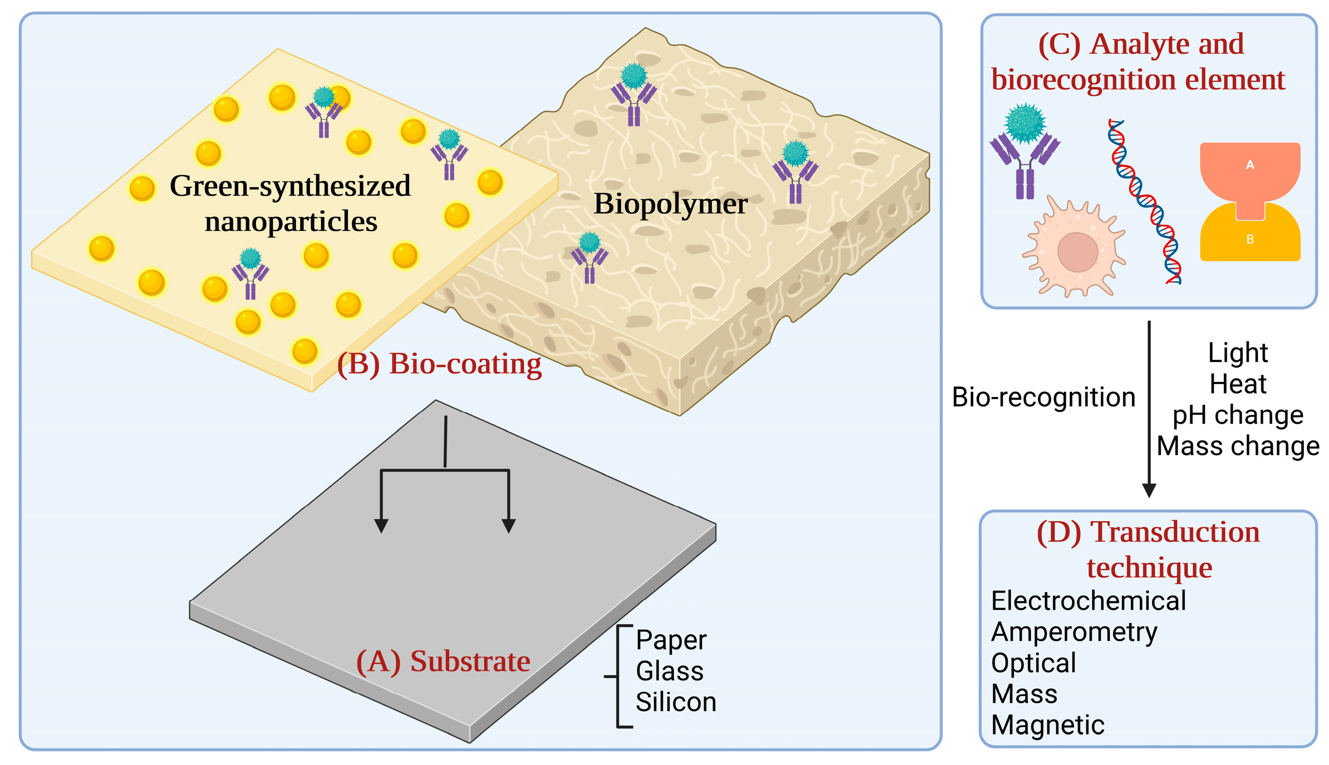
1. Introduction
2. Biosensors with Bio-Coatings and Applications in the Food Sector
3. Biosensors with Bio-Coatings and Applications in the Health Sector
4. Conclusions and Future Perspectives
Author Contributions
Funding
Institutional Review Board Statement
Informed Consent Statement
Data Availability Statement
Conflicts of Interest
Abbreviations
| Au | Gold |
| Ag | Silver |
| WHO | World Health Organization |
| QD | quantum dots |
| ASTM | American Society for Testing and Materials |
| NP | nanoparticle |
| mm | millimetre |
| nm | nanometre |
| NM | nanomaterial |
| HPLC-MS | High-performance liquid chromatography-mass spectrometry |
| PCR | Polymerase chain reaction |
| ELISA | Enzyme-linked immunosorbent assay |
| Pt | Platinum |
| Pd | Palladium |
| Zn | Zinc |
| Cd | Cadmium |
| Cu | Copper |
| Fe | Iron |
| Ni | Nickel |
| Co | Cobalt |
| HAuCl4 | Tetrachloroauric Acid |
| H2PtCl6 | Hexachloroplatinic acid |
| RhCl3 | Rhodium (III) chloride |
| PdCl2 | Palladium (II) chloride |
| cm | centimetre |
| TiO2 | Titanium dioxide |
| RF | radio frequency |
| K | Kelvin |
| kHz | kilohertz |
| MHz | megahertz |
| kW | kilowatt |
| MW | megawatt |
| atm | atmosphere |
| sec | seconds |
| N | Nitrogen |
| DMF | dimethylformamide |
| PEG | polyethylene glycol |
| UV | ultraviolet |
| AuNPs | gold nanoparticles |
| °C | degrees Celsius |
| min | minutes |
| ZnO | Zinc oxide |
| SnO2 | Tin oxide |
| PbO | Lead (II) oxide |
| EC-SPR | Electrochemical—surface plasmon resonance sensor |
| DNA | Deoxyribonucleic Acid. |
| LSPR | Localised surface plasmon resonance |
| SERS | Surface-enhanced Raman scattering |
| E. coli | Escherichia coli |
| PMNCs | polymeric nanocomposites |
| antibodies | ABs |
| GOX | glucose oxidase |
| PDA | polydopamine |
| DA | dopamine |
| CFU | colony-forming unit |
| mL | millilitre |
| PtNPs | platinum nanoparticles |
| PBNCs | polymeric bionanocomposites |
| L. monocytogenes | Listeria monocytogenes |
| μm | micrometre |
| LOD | Limit of detection |
| g | gram |
| β-Gal | β-galactosidase |
| S. typhimurium | Salmonella typhimurium |
| h | hours |
| PBS | phosphate buffered saline |
| EC | Commission Regulation |
| No | Number |
| S. boydii | Shigella boydii |
| ICS | immunochromatographic strip |
| S. aureus | Staphylococcus aureus |
| ATCC | American Type Culture Collection |
| MNPs | metal nanoparticles |
| MOs | metal oxides |
| CuO | copper oxide |
| Ag2O | silver oxide |
| CuNPs | Copper nanoparticles |
| pg | picograms |
| Fe3O4 | Iron oxide |
| SeNP | Selenium nanoparticle |
| FeNP | Iron nanoparticle |
| kg | kilogram |
| K | Potassium |
| Mg | Magnesium |
| Ca | Calcium |
| Hg | Mercury |
| IC | inhibition concentration |
| LC | lethal concentration |
| CMT | maximum permissible concentration |
| FDA | Food and Drug Administration |
| LOx | lactate oxidase |
| BC | Bio-cellulose |
| Co | Collagen |
| CuONPs | Copper oxide nanoparticles |
| fmol | femtomole |
| COVID-19 | Coronavirus Disease 2019 |
| SARS-CoV-2 | Severe acute respiratory syndrome coronavirus 2 |
| USD | The United States dollar |
| LDPE | Low-density polyethylene |
| RFID | Frequencies radio |
| EFSA | The European Food Safety Authority |
| MNTS | Micro- and Nanotechnologies |
| LDPE | Low-density polyethylene |
| OR | oil of oregano |
| RO | rosemary oil |
| SWNT | single walled carbon nanotube based |
| PLL | Poly-L-lysine |
| ESI | electrospray ionisation |
| GCE | glassy carbon electrode |
| PCL | polycaprolactone |
| PHB | polyhydroxy butyrate |
| PHV | polyhydroxy valerate |
| PE | polymers polyethylene |
| PVC | polyvinyl chloride |
| EVOH | ethylene vinyl alcohol |
| IgG | Immunoglobulin G |
| IgM | Immunoglobulin M |
| PBAT | poly (butylene adipate-co-terephthalate |
| TPS | cellulose-based thermoplastic starch |
| PLA | poly lactide |
| PHA | poly-hydroxyalkanoate |
| PHB | poly-hydroxybutyrate |
| PGA | poly-glutamic acid |
| MCF-7 | Michigan Cancer Foundation-7 |
References
- Chu, E.W.; Karr, J.R. Environmental Impact: Concept, Consequences, Measurement. In Reference Module in Life Sciences; Elsevier: Amsterdam, The Netherlands, 2017. [Google Scholar]
- McNeely, J.A. Nature and COVID-19: The Pandemic, the Environment, and the Way ahead. Ambio 2021, 50, 767–781. [Google Scholar] [CrossRef]
- Noah, N.M.; Ndangili, P.M. Green Synthesis of Nanomaterials from Sustainable Materials for Biosensors and Drug Delivery. Sens. Int. 2022, 3, 100166. [Google Scholar] [CrossRef]
- Moulahoum, H.; Ghorbanizamani, F.; Celik, E.G.; Timur, S. Nano-Scaled Materials and Polymer Integration in Biosensing Tools. Biosensors 2022, 12, 301. [Google Scholar] [CrossRef]
- Yasri, S.; Wiwanitkit, V. Sustainable Materials and COVID-19 Detection Biosensor: A Brief Review. Sens. Int. 2022, 3, 100171. [Google Scholar] [CrossRef]
- Ungureanu, C. Coatings with Natural Products—One Perspective on the Challenges Related to New Coatings’ Development. Coatings 2022, 12, 941. [Google Scholar] [CrossRef]
- Hosseini, E.S.; Dervin, S.; Ganguly, P.; Dahiya, R. Biodegradable Materials for Sustainable Health Monitoring Devices. ACS Appl. Bio. Mater. 2021, 4, 163–194. [Google Scholar] [CrossRef]
- Valencia, G.A.; Luciano, C.G.; Monteiro Fritz, A.R. Smart and Active Edible Coatings Based on Biopolymers. In Polymers for Agri-Food Applications; Springer International Publishing: Berlin/Heidelberg, Germany, 2019; pp. 391–416. [Google Scholar]
- Andriukov, B.G.; Lyapun, I.N.; Matosova, E.V.; Somova, L.M. Biosensor Technologies in Medicine: From Detection of Biochemical Markers to Research into Molecular Targets (Review). Sovrem. Tehnol. V Med. 2020, 12, 70–83. [Google Scholar] [CrossRef]
- Bhalla, N.; Jolly, P.; Formisano, N.; Estrela, P. Introduction to Biosensors. Essays Biochem. 2016, 60–61, 1–8. [Google Scholar] [CrossRef]
- Naresh, V.; Lee, N. A Review on Biosensors and Recent Development of Nanostructured Materials-Enabled Biosensors. Sensors 2021, 21, 1109. [Google Scholar] [CrossRef]
- Sharma, B.; Jain, P. Graphene Based Biopolymer Nanocomposites; Springer: Singapore, 2021; pp. 273–286. ISBN 978-981-15-9180-8. [Google Scholar]
- Cader Mhd Haniffa, M.A.; Ching, Y.C.; Abdullah, L.C.; Poh, S.C.; Chuah, C.H. Review of Bionanocomposite Coating Films and Their Applications. Polymers 2016, 8, 246. [Google Scholar] [CrossRef]
- Pradhan, S.; Brooks, A.K.; Yadavalli, V.K. Nature-Derived Materials for the Fabrication of Functional Biodevices. Mater. Today Bio. 2020, 7, 100065. [Google Scholar] [CrossRef]
- Barbinta-Patrascu, M.E.; Badea, N.; Bacalum, M.; Ungureanu, C.; Suica-Bunghez, I.R.; Iordache, S.M.; Pirvu, C.; Zgura, I.; Maraloiu, V.A. 3D Hybrid Structures Based on Biomimetic Membranes and Caryophyllus Aromaticus-“Green” Synthesized Nano-Silver with Improved Bioperformances. Mater. Sci. Eng. C 2019, 101, 120–137. [Google Scholar] [CrossRef]
- Barbinta-Patrascu, M.E.; Ungureanu, C.; Badea, N.; Bacalum, M.; Lazea-Stoyanova, A.; Zgura, I.; Negrila, C.; Enculescu, M.; Burnei, C. Novel Ecogenic Plasmonic Biohybrids as Multifunctional Bioactive Coatings. Coatings 2020, 10, 659. [Google Scholar] [CrossRef]
- Barbinta-Patrascu, M.E.; Ungureanu, C.; Iordache, S.M.; Iordache, A.M.; Bunghez, I.R.; Ghiurea, M.; Badea, N.; Fierascu, R.C.; Stamatin, I. Eco-Designed Biohybrids Based on Liposomes, Mint-Nanosilver and Carbon Nanotubes for Antioxidant and Antimicrobial Coating. Mater. Sci. Eng. C 2014, 39, 177–185. [Google Scholar] [CrossRef]
- Ungureanu, C.; Dumitriu, C.; Popescu, S.; Enculescu, M.; Tofan, V.; Popescu, M.; Pirvu, C. Enhancing Antimicrobial Activity of TiO2/Ti by Torularhodin Bioinspired Surface Modification. Bioelectrochemistry 2016, 107, 14–24. [Google Scholar] [CrossRef]
- Ungureanu, C.; Fierascu, I.; Fierascu, R.C.; Costea, T.; Avramescu, S.M.; Călinescu, M.F.; Somoghi, R.; Pirvu, C. In Vitro and in Vivo Evaluation of Silver Nanoparticles Phytosynthesized Using Raphanus sativus L. Waste Extracts. Materials 2021, 14, 1845. [Google Scholar] [CrossRef]
- Ungureanu, C.; Popescu, S.; Purcel, G.; Tofan, V.; Popescu, M.; Sǎlǎgeanu, A.; Pîrvu, C. Improved Antibacterial Behavior of Titanium Surface with Torularhodin-Polypyrrole Film. Mater. Sci. Eng. C 2014, 42, 726–733. [Google Scholar] [CrossRef]
- Ungureanu, C.; Tihan, G.T.; Zgârian, R.G.; Fierascu, I.; Baroi, A.M.; Răileanu, S.; Fierăscu, R.C. Metallic and Metal Oxides Nanoparticles for Sensing Food Pathogens—An Overview of Recent Findings and Future Prospects. Materials 2022, 15, 5374. [Google Scholar] [CrossRef]
- Boldeiu, A.; Simion, M.; Mihalache, I.; Radoi, A.; Banu, M.; Varasteanu, P.; Nadejde, P.; Vasile, E.; Acasandrei, A.; Popescu, R.C.; et al. Comparative Analysis of Honey and Citrate Stabilized Gold Nanoparticles: In Vitro Interaction with Proteins and Toxicity Studies. J. Photochem. Photobiol. B 2019, 197, 111519. [Google Scholar] [CrossRef]
- Ruta, L.L.; Banu, M.A.; Neagoe, A.D.; Kissen, R.; Bones, A.M.; Farcasanu, I.C. Accumulation of AG(I) by Saccharomyces Cerevisiae Cells Expressing Plant Metallothioneins. Cells 2018, 7, 266. [Google Scholar] [CrossRef]
- Onuki, Y.; Bhardwaj, U.; Papadimitrakopoulos, F.; Burgess, D.J. Biocompatibility of Implanted Diabetes Devices: Part 2: A Review of the Biocompatibility of Implantable Devices: Current Challenges to Overcome Foreign Body Response. J. Diabetes Sci. Technol. 2008, 2, 1003. [Google Scholar] [CrossRef]
- Neethirajan, S.; Ragavan, V.; Weng, X.; Chand, R. Biosensors for Sustainable Food Engineering: Challenges and Perspectives. Biosensors 2018, 8, 23. [Google Scholar] [CrossRef]
- Singh, J.; Dutta, T.; Kim, K.H.; Rawat, M.; Samddar, P.; Kumar, P. “Green” Synthesis of Metals and Their Oxide Nanoparticles: Applications for Environmental Remediation. J. Nanobiotechnol. 2018, 16, 84. [Google Scholar] [CrossRef]
- Jeevanandam, J.; Kiew, S.F.; Boakye-Ansah, S.; Lau, S.Y.; Barhoum, A.; Danquah, M.K.; Rodrigues, J. Green Approaches for the Synthesis of Metal and Metal Oxide Nanoparticles Using Microbial and Plant Extracts. Nanoscale 2022, 14, 2534–2571. [Google Scholar] [CrossRef]
- Beveridge, T.J.; Murray, R.G.E. Sites of Metal Deposition in the Cell Wall of Bacillus Subtilis. J. Bacteriol. 1980, 141, 876–887. [Google Scholar] [CrossRef]
- Patil, S.; Chandrasekaran, R. Biogenic Nanoparticles: A Comprehensive Perspective in Synthesis, Characterization, Application and Its Challenges. J. Genet. Eng. Biotechnol. 2020, 18, 67. [Google Scholar] [CrossRef]
- Koul, B.; Poonia, A.K.; Yadav, D.; Jin, J.O. Microbe-Mediated Biosynthesis of Nanoparticles: Applications and Future Prospects. Biomolecules 2021, 11, 886. [Google Scholar] [CrossRef]
- Johnston, C.W.; Wyatt, M.A.; Li, X.; Ibrahim, A.; Shuster, J.; Southam, G.; Magarvey, N.A. Gold Biomineralization by a Metallophore from a Gold-Associated Microbe. Nat. Chem. Biol. 2013, 9, 241–243. [Google Scholar] [CrossRef]
- Lloyd, J.R.; Yong, P.; Macaskie, L.E. Enzymatic Recovery of Elemental Palladium by Using Sulfate-Reducing Bacteria. Appl. Env. Microbiol. 1998, 64, 4607–4609. [Google Scholar] [CrossRef]
- Kalimuthu, K.; Suresh Babu, R.; Venkataraman, D.; Bilal, M.; Gurunathan, S. Biosynthesis of Silver Nanocrystals by Bacillus Licheniformis. Colloids Surf. B Biointerfaces 2008, 65, 150–153. [Google Scholar] [CrossRef]
- Baco-Carles, V.; Datas, L.; Tailhades, P. Copper Nanoparticles Prepared from Oxalic Precursors. ISRN Nanotechnol. 2011, 2011, 729594. [Google Scholar] [CrossRef]
- Gericke, M.; Pinches, A. Microbial Production of Gold Nanoparticles. Gold Bull. 2006, 39, 22–28. [Google Scholar] [CrossRef]
- Kowshik, M.; Ashtaputre, S.; Kharrazi, S.; Vogel, W.; Urban, J.; Kulkarni, S.K.; Paknikar, K.M. Extracellular Synthesis of Silver Nanoparticles by a Silver-Tolerant Yeast Strain. Nanotechnology 2002, 14, 95. [Google Scholar] [CrossRef]
- Pimprikar, P.S.; Joshi, S.S.; Kumar, A.R.; Zinjarde, S.S.; Kulkarni, S.K. Influence of Biomass and Gold Salt Concentration on Nanoparticle Synthesis by the Tropical Marine Yeast Yarrowia Lipolytica NCIM 3589. Colloids Surf. B Biointerfaces 2009, 74, 309–316. [Google Scholar] [CrossRef]
- Govender, Y.; Riddin, T.; Gericke, M.; Whiteley, C.G. Bioreduction of Platinum Salts into Nanoparticles: A Mechanistic Perspective. Biotechnol. Lett. 2009, 31, 95–100. [Google Scholar] [CrossRef]
- Velmurugan, P.; Shim, J.; You, Y.; Choi, S.; Kamala-Kannan, S.; Lee, K.J.; Kim, H.J.; Oh, B.T. Removal of Zinc by Live, Dead, and Dried Biomass of Fusarium Spp. Isolated from the Abandoned-Metal Mine in South Korea and Its Perspective of Producing Nanocrystals. J. Hazard. Mater. 2010, 182, 317–324. [Google Scholar] [CrossRef]
- Das, S.K.; Das, A.R.; Guha, A.K. Adsorption Behavior of Mercury on Functionalized Aspergillus Versicolor Mycelia: Atomic Force Microscopic Study. Langmuir 2009, 25, 360–366. [Google Scholar] [CrossRef]
- Das, S.K.; Das, A.R.; Guha, A.K. Gold Nanoparticles: Microbial Synthesis and Application in Water Hygiene Management. Langmuir 2009, 25, 8192–8199. [Google Scholar] [CrossRef]
- Mukherjee, P.; Ahmad, A.; Mandal, D.; Senapati, S.; Sainkar, S.R.; Khan, M.I.; Parishcha, R.; Ajaykumar, P.V.; Alam, M.; Kumar, R.; et al. Fungus-Mediated Synthesis of Silver Nanoparticles and Their Immobilization in the Mycelial Matrix: A Novel Biological Approach to Nanoparticle Synthesis. Nano Lett. 2001, 1, 515–519. [Google Scholar] [CrossRef]
- Cepoi, L.; Rudi, L.; Chiriac, T.; Valuta, A.; Zinicovscaia, I.; Duca, G.H.; Kirkesali, E.; Frontasyeva, M.; Culicov, O.; Pavlov, S.; et al. Biochemical Changes in Cyanobacteria during the Synthesis of Silver Nanoparticles. Can. J. Microbiol. 2014, 61, 13–21. [Google Scholar] [CrossRef]
- Parial, D.; Pal, R. Green Synthesis of Gold Nanoparticles Using Cyanobacteria and Their Characterization. Indian J. Appl. Res. 2011, 4, 69–72. [Google Scholar] [CrossRef]
- Govindaraju, K.; Kiruthiga, V.; Kumar, V.G.; Singaravelu, G. Extracellular Synthesis of Silver Nanoparticles by a Marine Alga, Sargassum Wightii Grevilli and Their Antibacterial Effects. J. Nanosci. Nanotechnol. 2009, 9, 5497–5501. [Google Scholar] [CrossRef]
- Dahoumane, S.A.; Yéprémian, C.; Djédiat, C.; Couté, A.; Fiévet, F.; Coradin, T.; Brayner, R. Improvement of Kinetics, Yield, and Colloidal Stability of Biogenic Gold Nanoparticles Using Living Cells of Euglena Gracilis Microalga. J. Nanoparticle Res. 2016, 18, 79. [Google Scholar] [CrossRef]
- Shankar, S.S.; Rai, A.; Ahmad, A.; Sastry, M. Rapid Synthesis of Au, Ag, and Bimetallic Au Core–Ag Shell Nanoparticles Using Neem (Azadirachta Indica) Leaf Broth. J. Colloid Interface Sci. 2004, 275, 496–502. [Google Scholar] [CrossRef]
- Lee, H.J.; Song, J.Y.; Kim, B.S. Biological Synthesis of Copper Nanoparticles Using Magnolia Kobus Leaf Extract and Their Antibacterial Activity. J. Chem. Technol. Biotechnol. 2013, 88, 1971–1977. [Google Scholar] [CrossRef]
- Song, J.Y.; Kwon, E.Y.; Kim, B.S. Biological Synthesis of Platinum Nanoparticles Using Diopyros Kaki Leaf Extract. Bioprocess Biosyst. Eng. 2010, 33, 159–164. [Google Scholar] [CrossRef]
- Thangavelu, R.M.; Ganapathy, R.; Ramasamy, P.; Krishnan, K. Fabrication of Virus Metal Hybrid Nanomaterials: An Ideal Reference for Bio Semiconductor. Arab. J. Chem. 2020, 13, 2750–2765. [Google Scholar] [CrossRef]
- Kobayashi, M.; Tomita, S.; Sawada, K.; Shiba, K.; Yanagi, H.; Yamashita, I.; Uraoka, Y.; Smith, D.R.; Padilla, W.J.; Vier, D.C.; et al. Chiral Meta-Molecules Consisting of Gold Nanoparticles and Genetically Engineered Tobacco Mosaic Virus. Opt. Express 2012, 20, 24856–24863. [Google Scholar] [CrossRef]
- Chaabouni, E.; Gassara, F.; Brar, S.K. Biopolymers Synthesis and Application. In Biotransformation of Waste Biomass into High Value Biochemicals; Springer: New York, NY, USA, 2014; pp. 415–443. ISBN 9781461480051. [Google Scholar]
- Sawant, S.N. Development of Biosensors from Biopolymer Composites. In Biopolymer Composites in Electronics; Elsevier Inc.: Amsterdam, The Netherlands, 2017; pp. 353–383. ISBN 9780081009741. [Google Scholar]
- Baranwal, J.; Barse, B.; Fais, A.; Delogu, G.L.; Kumar, A. Biopolymer: A Sustainable Material for Food and Medical Applications. Polymers 2022, 14, 983. [Google Scholar] [CrossRef]
- Song, J.; Winkeljann, B.; Lieleg, O. Biopolymer-Based Coatings: Promising Strategies to Improve the Biocompatibility and Functionality of Materials Used in Biomedical Engineering. Adv. Mater. Interfaces 2020, 7, 2000850. [Google Scholar] [CrossRef]
- Pagliano, G.; Ventorino, V.; Panico, A.; Pepe, O. Integrated Systems for Biopolymers and Bioenergy Production from Organic Waste and By-Products: A Review of Microbial Processes. Biotechnol. Biofuels 2017, 10, 1–24. [Google Scholar] [CrossRef]
- Vroman, I.; Tighzert, L. Biodegradable Polymers. Materials 2009, 2, 307. [Google Scholar] [CrossRef]
- Kim, D.S.; Yang, X.; Lee, J.H.; Yoo, H.Y.; Park, C.; Kim, S.W.; Lee, J. Development of GO/Co/Chitosan-Based Nano-Biosensor for Real-Time Detection of D-Glucose. Biosensors 2022, 12, 464. [Google Scholar] [CrossRef]
- Solanki, P.R.; Kaushik, A.; Ansari, A.A.; Tiwari, A.; Malhotra, B.D. Multi-Walled Carbon Nanotubes/Sol–Gel-Derived Silica/Chitosan Nanobiocomposite for Total Cholesterol Sensor. Sens. Actuators B Chem. 2009, 137, 727–735. [Google Scholar] [CrossRef]
- Leva-Bueno, J.; Meuskens, I.; Linke, D.; Millner, P.A.; Peyman, S.A. A Novel, Proof-of-Concept Electrochemical Impedimetric Biosensor Based on Extracellular Matrix Protein–Adhesin Interaction. Sens. Diagn. 2022, 1, 1003–1013. [Google Scholar] [CrossRef]
- Gomes, N.O.; Carrilho, E.; Machado, S.A.S.; Sgobbi, L.F. Bacterial Cellulose-Based Electrochemical Sensing Platform: A Smart Material for Miniaturized Biosensors. Electrochim. Acta 2020, 349, 136341. [Google Scholar] [CrossRef]
- Bougadi, E.T.; Kalogianni, D.P. Paper-Based DNA Biosensor for Food Authenticity Testing. Food Chem. 2020, 322, 126758. [Google Scholar] [CrossRef]
- Kaushik, A.; Solanki, P.R.; Ansari, A.A.; Sumana, G.; Ahmad, S.; Malhotra, B.D. Iron Oxide-Chitosan Nanobiocomposite for Urea Sensor. Sens. Actuators B Chem. 2009, 138, 572–580. [Google Scholar] [CrossRef]
- Zhao, L.; Wen, Z.; Jiang, F.; Zheng, Z.; Lu, S. Silk/Polyols/GOD Microneedle Based Electrochemical Biosensor for Continuous Glucose Monitoring. RSC Adv. 2020, 10, 6163–6171. [Google Scholar] [CrossRef]
- Kumar-Krishnan, S.; Chakaravarthy, S.; Hernandez-Rangel, A.; Prokhorov, E.; Luna-Bárcenas, G.; Esparza, R.; Meyyappan, M. Chitosan Supported Silver Nanowires as a Platform for Direct Electrochemistry and Highly Sensitive Electrochemical Glucose Biosensing. RSC Adv. 2016, 6, 20102–20108. [Google Scholar] [CrossRef]
- Silva, V.A.O.P.; Fernandes-Junior, W.S.; Rocha, D.P.; Stefano, J.S.; Munoz, R.A.A.; Bonacin, J.A.; Janegitz, B.C. 3D-Printed Reduced Graphene Oxide/Polylactic Acid Electrodes: A New Prototyped Platform for Sensing and Biosensing Applications. Biosens. Bioelectron. 2020, 170, 112684. [Google Scholar] [CrossRef]
- Cardoso, R.M.; Silva, P.R.L.; Lima, A.P.; Rocha, D.P.; Oliveira, T.C.; do Prado, T.M.; Fava, E.L.; Fatibello-Filho, O.; Richter, E.M.; Muñoz, R.A.A. 3D-Printed Graphene/Polylactic Acid Electrode for Bioanalysis: Biosensing of Glucose and Simultaneous Determination of Uric Acid and Nitrite in Biological Fluids. Sens. Actuators B Chem. 2020, 307, 127621. [Google Scholar] [CrossRef]
- Yang, B.; Kong, J.; Fang, X. Bandage-like Wearable Flexible Microfluidic Recombinase Polymerase Amplification Sensor for the Rapid Visual Detection of Nucleic Acids. Talanta 2019, 204, 685–692. [Google Scholar] [CrossRef]
- Park, T.J.; Yoo, S.M.; Keum, K.C.; Lee, S.Y. Microarray of DNA-Protein Complexes on Poly-3-Hydroxybutyrate Surface for Pathogen Detection. Anal. Bioanal. Chem. 2009, 393, 1639–1647. [Google Scholar] [CrossRef]
- Yazdanparast, S.; Benvidi, A.; Banaei, M.; Nikukar, H.; Tezerjani, M.D.; Azimzadeh, M. Dual-Aptamer Based Electrochemical Sandwich Biosensor for MCF-7 Human Breast Cancer Cells Using Silver Nanoparticle Labels and a Poly(Glutamic Acid)/MWNT Nanocomposite. Microchim. Acta 2018, 185, 405. [Google Scholar] [CrossRef]
- Perianes-Rodriguez, A.; Waltman, L.; van Eck, N.J. Constructing Bibliometric Networks: A Comparison between Full and Fractional Counting. J. Inf. 2016, 10, 1178–1195. [Google Scholar] [CrossRef]
- Deshpande, K.; Kanungo, L. Chemiluminescence and Fluorescence Biosensors for Food Application: A Review. Sens. Actuators Rep. 2023, 5, 100137. [Google Scholar] [CrossRef]
- Abdulhussein, S.K.; Fadhel, F.; Al-Kazazz, M.; Rheima, A.M. The Role of Nanomaterials in the Recent Development of Electrochemical Biosensors. Port. Electrochim. Acta 2023, 41, 211–221. [Google Scholar] [CrossRef]
- Khan, A.; Haque, M.N.; Kabiraz, D.C.; Yeasin, A.; al Rashid, H.; Sarker, A.C.; Hossain, G. A Review on Advanced Nanocomposites Materials Based Smart Textile Biosensor for Healthcare Monitoring from Human Sweat. Sens. Actuators A Phys. 2023, 350, 114093. [Google Scholar] [CrossRef]
- Deyasi, A.; Basak, A.; Sarkar, A. Application of Nanomaterial-Based Biosensors for Healthcare Diagnostics. Smart Innov. Syst. Technol. 2023, 322, 103–122. [Google Scholar] [CrossRef]
- Naghdi, T.; Ardalan, S.; Asghari Adib, Z.; Sharifi, A.R.; Golmohammadi, H. Moving toward Smart Biomedical Sensing. Biosens. Bioelectron. 2023, 223, 115009. [Google Scholar] [CrossRef]
- Yau, A.; Wang, Z.; Ponthempilly, N.; Zhang, Y.; Wang, X.; Chen, Y. Biosensor Integrated Tissue Chips and Their Applications on Earth and in Space. Biosens. Bioelectron. 2023, 222, 114820. [Google Scholar] [CrossRef]
- Soozanipour, A.; Ejeian, F.; Boroumand, Y.; Rezayat, A.; Moradi, S. Biotechnological Advancements towards Water, Food and Medical Healthcare: A Review. Chemosphere 2023, 312, 137185. [Google Scholar] [CrossRef]
- Herrmann, A.; Haag, R.; Schedler, U. Hydrogels and Their Role in Biosensing Applications. Adv. Healthc Mater. 2021, 10, 2100062. [Google Scholar] [CrossRef]
- Khalid, M.Y.; Arif, Z.U. Novel Biopolymer-Based Sustainable Composites for Food Packaging Applications: A Narrative Review. Food Packag. Shelf Life 2022, 33, 100892. [Google Scholar] [CrossRef]
- Food Packaging Is (Naturally) Getting Smarter-ACS Axial|ACS Publications. Available online: https://axial.acs.org/2022/11/18/food-packaging-is-naturally-getting-smarter/ (accessed on 6 January 2023).
- Purohit, J.; Chattopadhyay, A.; Singh, N.K. Green Synthesis of Microbial Nanoparticle: Approaches to Application. In Nanotechnology in the Life Sciences; Springer Science and Business Media B.V.: Berlin/Heidelberg, Germany, 2019; pp. 35–60. [Google Scholar]
- Nemiwal, M.; Zhang, T.C.; Kumar, D. Pectin Modified Metal Nanoparticles and Their Application in Property Modification of Biosensors. Carbohydr. Polym. Technol. Appl. 2021, 2, 100164. [Google Scholar] [CrossRef]
- Ashfaq, A.; Khursheed, N.; Fatima, S.; Anjum, Z.; Younis, K. Application of Nanotechnology in Food Packaging: Pros and Cons. J. Agric. Food Res. 2022, 7, 100270. [Google Scholar] [CrossRef]
- Biswas, R.; Alam, M.; Sarkar, A.; Haque, M.I.; Hasan, M.M.; Hoque, M. Application of Nanotechnology in Food: Processing, Preservation, Packaging and Safety Assessment. Heliyon 2022, 8, e11795. [Google Scholar] [CrossRef]
- Stewart, C.M.; Busta, F.F. Global Harmonization of the Control of Microbiological Risks. Ensuring Glob. Food Saf. 2010, 177, 461–474. [Google Scholar] [CrossRef]
- Sharma, P.; Pandey, V.; Sharma, M.M.M.; Patra, A.; Singh, B.; Mehta, S.; Husen, A. A Review on Biosensors and Nanosensors Application in Agroecosystems. Nanoscale Res. Lett. 2021, 16, 136. [Google Scholar] [CrossRef]
- Kaur, H.; Siwal, S.S.; Saini, R.V.; Singh, N.; Thakur, V.K. Significance of an Electrochemical Sensor and Nanocomposites: Toward the Electrocatalytic Detection of Neurotransmitters and Their Importance within the Physiological System. ACS Nanosci. Au 2022, 3, 1–27. [Google Scholar] [CrossRef]
- Ameta, S.K.; Rai, A.K.; Hiran, D.; Ameta, R.; Ameta, S.C. Use of Nanomaterials in Food Science. In Biogenic Nano-Particles and Their Use in Agro-Ecosystems; Springer: Singapore, 2020; pp. 457–488. [Google Scholar] [CrossRef]
- Toydemir, G.; Cekic, S.D.; Ozkan, G.; Uzunboy, S.; Avan, A.N.; Capanoglu, E.; Apak, R. Nanosensors for Foods. Food Eng. Ser. 2020, 327–375. [Google Scholar] [CrossRef]
- Tan, J.; Xu, J. Applications of Electronic Nose (e-Nose) and Electronic Tongue (e-Tongue) in Food Quality-Related Properties Determination: A Review. Artif. Intell. Agric. 2020, 4, 104–115. [Google Scholar] [CrossRef]
- Mairhofer, J.; Roppert, K.; Ertl, P. Microfluidic Systems for Pathogen Sensing: A Review. Sensors 2009, 9, 4804. [Google Scholar] [CrossRef]
- Chelliah, R.; Wei, S.; Daliri, E.B.M.; Rubab, M.; Elahi, F.; Yeon, S.J.; Jo, K.H.; Yan, P.; Liu, S.; Oh, D.H. Development of Nanosensors Based Intelligent Packaging Systems: Food Quality and Medicine. Nanomaterials 2021, 11, 1515. [Google Scholar] [CrossRef]
- Rhouati, A.; Bulbul, G.; Latif, U.; Hayat, A.; Li, Z.H.; Marty, J.L. Nano-Aptasensing in Mycotoxin Analysis: Recent Updates and Progress. Toxins 2017, 9, 349. [Google Scholar] [CrossRef]
- Dikshit, P.K.; Kumar, J.; Das, A.K.; Sadhu, S.; Sharma, S.; Singh, S.; Gupta, P.K.; Kim, B.S. Green Synthesis of Metallic Nanoparticles: Applications and Limitations. Catalysts 2021, 11, 902. [Google Scholar] [CrossRef]
- Singh, S.; Melnik, R. Coupled Multiphysics Modelling of Sensors for Chemical, Biomedical, and Environmental Applications with Focus on Smart Materials and Low-Dimensional Nanostructures. Chemosensors 2022, 10, 157. [Google Scholar] [CrossRef]
- Staples, M.; Daniel, K.; Cima, M.J.; Langer, R. Application of Micro- and Nano-Electromechanical Devices to Drug Delivery. Pharm. Res. 2006, 23, 847–863. [Google Scholar] [CrossRef]
- Kivirand, K.; Kagan, M.; Rinken, T.; Kivirand, K.; Kagan, M.; Rinken, T. Biosensors for the Detection of Antibiotic Residues in Milk. In Biosensors-Micro and Nanoscale Applications; BoD—Books on Demand: Paris, France, 2015. [Google Scholar] [CrossRef]
- Adrian, J.; Pasche, S.; Diserens, J.M.; Sánchez-Baeza, F.; Gao, H.; Marco, M.P.; Voirin, G. Waveguide Interrogated Optical Immunosensor (WIOS) for Detection of Sulfonamide Antibiotics in Milk. Biosens. Bioelectron. 2009, 24, 3340–3346. [Google Scholar] [CrossRef]
- Rodrigues, C.; Souza, V.G.L.; Coelhoso, I.; Fernando, A.L. Bio-Based Sensors for Smart Food Packaging—Current Applications and Future Trends. Sensors 2021, 21, 2148. [Google Scholar] [CrossRef]
- Taj, A.; Zia, R.; Iftikhar, M.; Younis, S.; Bajwa, S.Z. Nanosensors for Food Inspection. Nanosens. Smart Agric. 2022, 685–703. [Google Scholar] [CrossRef]
- The Regulation of Active and Intelligent Food Packaging in the U.S. and the EU*|PackagingLaw.com. Available online: https://www.packaginglaw.com/special-focus/regulation-active-and-intelligent-food-packaging-us-and-eu (accessed on 28 November 2022).
- Sustainable Management of Food Basics|US EPA. Available online: https://www.epa.gov/sustainable-management-food/sustainable-management-food-basics (accessed on 28 November 2022).
- EUR-Lex-32009R0450-EN-EUR-Lex. Available online: https://eur-lex.europa.eu/legal-content/EN/TXT/?uri=CELEX%3A32009R0450&qid=1669628352163 (accessed on 28 November 2022).
- Guidelines on Submission of a Dossier for Safety Evaluation by the EFSA of Active or Intelligent Substances Present in Active and Intelligent Materials and Articles Intended to Come into Contact with Food. EFSA J. 2009, 7, 1208.
- The Potential Risks Arising from Nanoscience and Nanotechnologies on Food and Feed Safety. EFSA J. 2009, 7, 958. [CrossRef]
- Guidance on the Risk Assessment of the Application of Nanoscience and Nanotechnologies in the Food and Feed Chain. EFSA J. 2011, 9, 2140. [CrossRef]
- Astuto, M.C. Overview of the EFSA Guidance on Particle-Technical Requirements and Proposed Appraisal Routes to Establish the Presence of Small Particles in Food and Feed Applications; European Food Safety Authority (EFSA): Parma, Italy, 2022.
- Ahmed, I.; Lin, H.; Zou, L.; Li, Z.; Brody, A.L.; Qazi, I.M.; Lv, L.; Pavase, T.R.; Khan, M.U.; Khan, S.; et al. An Overview of Smart Packaging Technologies for Monitoring Safety and Quality of Meat and Meat Products. Packag. Technol. Sci. 2018, 31, 449–471. [Google Scholar] [CrossRef]
- Nicoletti, M.; del Serrone, P. Intelligent and Smart Packaging. In Future Foods; InTech: London, UK, 2017. [Google Scholar]
- Onyeaka, H.; Passaretti, P.; Miri, T.; Al-Sharify, Z.T. The Safety of Nanomaterials in Food Production and Packaging. Curr. Res. Food Sci. 2022, 5, 763–774. [Google Scholar] [CrossRef]
- Bruna, T.; Maldonado-Bravo, F.; Jara, P.; Caro, N. Silver Nanoparticles and Their Antibacterial Applications. Int. J. Mol. Sci. 2021, 22, 7202. [Google Scholar] [CrossRef]
- Valipoor Motlagh, N.; Hamed Mosavian, M.T.; Mortazavi, S.A. Effect of Polyethylene Packaging Modified with Silver Particles on the Microbial, Sensory and Appearance of Dried Barberry. Packag. Technol. Sci. 2013, 26, 39–49. [Google Scholar] [CrossRef]
- Soltani, S.; Nourdahr, R. Study on the Antimicrobial Effect of Nanosilver Tray Packaging of Minced Beef at Refrigerator Temperature. Glob. Vet. 2012, 9, 284–289. [Google Scholar] [CrossRef]
- Metak, A.M. Effects of Nanocomposite Based Nano-Silver and Nano-Titanium Dioxideon Food Packaging Materials. Int. J. Appl. Sci. Technol. 2015, 5, 26–40. [Google Scholar]
- Durak, J.; Rokoszak, T.; Skiba, A.; Furman, P.; Styszko, K. Environmental Risk Assessment of Priority Biocidal Substances on Polish Surface Water Sample. Env. Sci. Pollut. Res. Int. 2021, 28, 1254. [Google Scholar] [CrossRef]
- Borkow, G.; Gabbay, J. Copper as a Biocidal Tool. Curr. Med. Chem. 2005, 12, 2163–2175. [Google Scholar] [CrossRef]
- Chen, Z.; Meng, H.; Xing, G.; Chen, C.; Zhao, Y.; Jia, G.; Wang, T.; Yuan, H.; Ye, C.; Zhao, F.; et al. Acute Toxicological Effects of Copper Nanoparticles in Vivo. Toxicol. Lett. 2006, 163, 109–120. [Google Scholar] [CrossRef]
- Espitia, P.J.P.; Soares, N.d.F.F.; Coimbra, J.S.d.R.; de Andrade, N.J.; Cruz, R.S.; Medeiros, E.A.A. Zinc Oxide Nanoparticles: Synthesis, Antimicrobial Activity and Food Packaging Applications. Food Bioproc. Tech. 2012, 5, 1447–1464. [Google Scholar] [CrossRef]
- Anvar, A.A.; Ahari, H.; Ataee, M. Antimicrobial Properties of Food Nanopackaging: A New Focus on Foodborne Pathogens. Front. Microbiol. 2021, 12, 690706. [Google Scholar] [CrossRef]
- Muthulakshmi, L.; Rajini, N.; Nellaiah, H.; Kathiresan, T.; Jawaid, M.; Varada Rajulu, A. Experimental Investigation of Cellulose/Silver Nanocomposites Using In Situ Generation Method. J. Polym. Environ. 2017, 25, 1021–1032. [Google Scholar] [CrossRef]
- Rai, M.; Wypij, M.; Ingle, A.P.; Trzcińska-Wencel, J.; Golińska, P. Emerging Trends in Pullulan-Based Antimicrobial Systems for Various Applications. Int. J. Mol. Sci. 2021, 22, 13596. [Google Scholar] [CrossRef]
- Khalaf, H.H.; Sharoba, A.M.; El-Tanahi, H.H.; Morsy, M.K.; Morsy, M.K. Stability of antimicrobial activity of pullulan edible films incorporated with nanoparticles and essential oils and their impact on turkey deli meat quality. J. Food Dairy Sci. 2013, 4, 557–573. [Google Scholar] [CrossRef]
- Incoronato, A.L.; Conte, A.; Buonocore, G.G.; del Nobile, M.A. Agar Hydrogel with Silver Nanoparticles to Prolong the Shelf Life of Fior Di Latte Cheese. J. Dairy Sci. 2011, 94, 1697–1704. [Google Scholar] [CrossRef]
- Longano, D.; Ditaranto, N.; Cioffi, N.; di Niso, F.; Sibillano, T.; Ancona, A.; Conte, A.; del Nobile, M.A.; Sabbatini, L.; Torsi, L. Analytical Characterization of Laser-Generated Copper Nanoparticles for Antibacterial Composite Food Packaging. Anal. Bioanal. Chem. 2012, 403, 1179–1186. [Google Scholar] [CrossRef]
- Bollella, P.; Schulz, C.; Favero, G.; Mazzei, F.; Ludwig, R.; Gorton, L.; Antiochia, R. Green Synthesis and Characterization of Gold and Silver Nanoparticles and Their Application for Development of a Third Generation Lactose Biosensor. Electroanalysis 2017, 29, 77–86. [Google Scholar] [CrossRef]
- Santhosh, A.; Theertha, V.; Prakash, P.; Smitha Chandran, S. From Waste to a Value Added Product: Green Synthesis of Silver Nanoparticles from Onion Peels Together with Its Diverse Applications. Mater. Today Proc. 2021, 46, 4460–4463. [Google Scholar] [CrossRef]
- Zamarchi, F.; Vieira, I.C. Determination of Paracetamol Using a Sensor Based on Green Synthesis of Silver Nanoparticles in Plant Extract. J. Pharm. Biomed. Anal. 2021, 196, 113912. [Google Scholar] [CrossRef]
- Majdi, H.; Salehi, R.; Pourhassan-Moghaddam, M.; Mahmoodi, S.; Poursalehi, Z.; Vasilescu, S. Antibody Conjugated Green Synthesized Chitosan-Gold Nanoparticles for Optical Biosensing. Colloid Interface Sci. Commun. 2019, 33, 100207. [Google Scholar] [CrossRef]
- Yu, T.; Xu, C.; Qiao, J.; Zhang, R.; Qi, L. Green Synthesis of Gold Nanoclusters Using Papaya Juice for Detection of L-Lysine. Chin. Chem. Lett. 2019, 30, 660–663. [Google Scholar] [CrossRef]
- Cheng, Y.; Zhang, Y.; Pei, R.; Xie, Y.; Yao, W.; Guo, Y.; Qian, H. Fast Detection of Bismerthiazol in Cabbage Based on Fluorescence Quenching of Protein-Capping Gold Nanoclusters. Anal. Sci. 2018, 34, 415–419. [Google Scholar] [CrossRef]
- Cheng, Y.; Kang, W.; Guo, Y.; Du, C.; Xie, Y.; Chen, Y.; Yao, W.; Qian, H. Visual Detection of Cu2+ Based on Fluorescence Quenching of Green-Synthesized Gold Nanoclusters Using Soy Protein as Template. Food Agric. Immunol. 2017, 28, 848–858. [Google Scholar] [CrossRef]
- Gayda, G.Z.; Demkiv, O.M.; Stasyuk, N.Y.; Serkiz, R.Y.; Lootsik, M.D.; Errachid, A.; Gonchar, M.V.; Nisnevitch, M. Metallic Nanoparticles Obtained via “Green” Synthesis as a Platform for Biosensor Construction. Appl. Sci. 2019, 9, 720. [Google Scholar] [CrossRef]
- Bagal-Kestwal, D.; Kestwal, R.M.; Chiang, B.H. Invertase-Nanogold Clusters Decorated Plant Membranes for Fluorescence-Based Sucrose Sensor. J. Nanobiotechnol. 2015, 13, 30. [Google Scholar] [CrossRef]
- Amouzadeh Tabrizi, M.; Varkani, J.N. Green Synthesis of Reduced Graphene Oxide Decorated with Gold Nanoparticles and Its Glucose Sensing Application. Sens. Actuators B Chem. 2014, 202, 475–482. [Google Scholar] [CrossRef]
- Dayakar, T.; Rao, K.V.; Bikshalu, K.; Rajendar, V.; Park, S.H. Novel Synthesis and Characterization of Pristine Cu Nanoparticles for the Non-Enzymatic Glucose Biosensor. J. Mater. Sci. Mater. Med. 2017, 28, 109. [Google Scholar] [CrossRef]
- Liu, Y.; Zhao, Y.; Zhang, Y. One-Step Green Synthesized Fluorescent Carbon Nanodots from Bamboo Leaves for Copper(II) Ion Detection. Sens. Actuators B Chem. 2014, 196, 647–652. [Google Scholar] [CrossRef]
- Sukumar, S.; Rudrasenan, A.; Padmanabhan Nambiar, D. Green-Synthesized Rice-Shaped Copper Oxide Nanoparticles Using Caesalpinia Bonducella Seed Extract and Their Applications. ACS Omega 2020, 5, 1040–1051. [Google Scholar] [CrossRef]
- Singh, P.; Singh, K.R.; Singh, J.; Singh, R.P. Biogenic Synthesis of Copper Oxide Nanoparticles: Characterization and Biosensing Application. ECS Trans. 2022, 107, 20127–20133. [Google Scholar] [CrossRef]
- Karthik, R.; Govindasamy, M.; Chen, S.M.; Mani, V.; Lou, B.S.; Devasenathipathy, R.; Hou, Y.S.; Elangovan, A. Green Synthesized Gold Nanoparticles Decorated Graphene Oxide for Sensitive Determination of Chloramphenicol in Milk, Powdered Milk, Honey and Eye Drops. J. Colloid Interface Sci. 2016, 475, 46–56. [Google Scholar] [CrossRef]
- Kaur, B.; Markan, M.; Singh, M. Green Synthesis of Gold Nanoparticles from Syzygium Aromaticum Extract and Its Use in Enhancing the Response of a Colorimetric Urea Biosensor. Bionanoscience 2012, 2, 251–258. [Google Scholar] [CrossRef]
- Masibi, K.K.; Fayemi, O.E.; Adekunle, A.S.; Sherif, E.S.M.; Ebenso, E.E. Electrochemical Determination of Caffeine Using Bimetallic Au−Ag Nanoparticles Obtained from Low-Cost Green Synthesis. Electroanalysis 2020, 32, 2745–2755. [Google Scholar] [CrossRef]
- Han, B.; Guan, H.; Song, Y.; Liu, Y. Radix Pueraria Flavonoids Assisted Green Synthesis of Reduced Gold Nanoparticles: Application for Electrochemical Nonenzymatic Detection of Cholesterol in Food Samples. ACS Omega 2022, 7, 43045–43054. [Google Scholar] [CrossRef]
- Manne, R.; Kumaradoss, M.M.R.M.; Iska, R.S.R.; Devarajan, A.; Mekala, N. Water Quality and Risk Assessment of Copper Content in Drinking Water Stored in Copper Container. Appl. Water Sci. 2022, 12, 127. [Google Scholar] [CrossRef]
- Pinheiro, T.; Cardoso, A.R.; Sousa, C.E.A.; Marques, A.C.; Tavares, A.P.M.; Matos, A.M.; Cruz, M.T.; Moreira, F.T.C.; Martins, R.; Fortunato, E.; et al. Paper-Based Biosensors for COVID-19: A Review of Innovative Tools for Controlling the Pandemic. ACS Omega 2021, 6, 29268–29290. [Google Scholar] [CrossRef]
- Choi, J.R. Development of Point-of-Care Biosensors for COVID-19. Front Chem. 2020, 8, 517. [Google Scholar] [CrossRef]
- Wang, J.; Drelich, A.J.; Hopkins, C.M.; Mecozzi, S.; Li, L.; Kwon, G.; Hong, S. Gold Nanoparticles in Virus Detection: Recent Advances and Potential Considerations for SARS-CoV-2 Testing Development. Wiley Interdiscip. Rev. Nanomed. Nanobiotechnol. 2022, 14, e1754. [Google Scholar] [CrossRef]
- Ardekani, L.S.; Thulstrup, P.W. Gold Nanoparticle-Mediated Lateral Flow Assays for Detection of Host Antibodies and COVID-19 Proteins. Nanomaterials 2022, 12, 1456. [Google Scholar] [CrossRef]
- WANTAI SARS-CoV-2 Ab Rapid Test-Letter of Authorization|Enhanced Reader. Available online: https://www.fda.gov/media/140035/download (accessed on 16 January 2023).
- Xu, L.; Li, D.; Ramadan, S.; Li, Y.; Klein, N. Facile Biosensors for Rapid Detection of COVID-19. Biosens. Bioelectron. 2020, 170, 112673. [Google Scholar] [CrossRef]
- Boutal, H.; Moguet, C.; Pommiès, L.; Simon, S.; Naas, T.; Volland, H. The Revolution of Lateral Flow Assay in the Field of AMR Detection. Diagnostics 2022, 12, 1744. [Google Scholar] [CrossRef]
- Coelho, J.H.; Eisele, A.P.P.; Valezi, C.F.; Mattos, G.J.; Schirmann, J.G.; Dekker, R.F.H.; Barbosa-Dekker, A.M.; Sartori, E.R. Exploring the Exocellular Fungal Biopolymer Botryosphaeran for Laccase-Biosensor Architecture and Application to Determine Dopamine and Spironolactone. Talanta 2019, 204, 475–483. [Google Scholar] [CrossRef]
- Ambrosetti, E.; Conti, M.; Teixeira, A.I.; Zilio, S.D. Patterned Carboxymethyl-Dextran Functionalized Surfaces Using Organic Mixed Monolayers for Biosensing Applications. ACS Appl. Bio. Mater. 2022, 5, 3310–3319. [Google Scholar] [CrossRef]
- Kownacka, A.E.; Vegelyte, D.; Joosse, M.; Anton, N.; Toebes, B.J.; Lauko, J.; Buzzacchera, I.; Lipinska, K.; Wilson, D.A.; Geelhoed-Duijvestijn, N.; et al. Clinical Evidence for Use of a Noninvasive Biosensor for Tear Glucose as an Alternative to Painful Finger-Prick for Diabetes Management Utilizing a Biopolymer Coating. Biomacromolecules 2018, 19, 4504–4511. [Google Scholar] [CrossRef]
- Wasik, D.; Mulchandani, A.; Yates, M.V. A Heparin-Functionalized Carbon Nanotube-Based Affinity Biosensor for Dengue Virus. Biosens. Bioelectron. 2017, 91, 811–816. [Google Scholar] [CrossRef]
- Hasanah, U.; Md Sani, N.D.; Heng, L.Y.; Idroes, R.; Safitri, E. Construction of a Hydrogel Pectin-Based Triglyceride Optical Biosensor with Immobilized Lipase Enzymes. Biosensors 2019, 9, 135. [Google Scholar] [CrossRef]
- Gao, J.; Wang, C.; Wang, C.; Chu, Y.; Wang, S.; Sun, M.Y.; Ji, H.; Gao, Y.; Wang, Y.; Han, Y.; et al. Poly-l-Lysine-Modified Graphene Field-Effect Transistor Biosensors for Ultrasensitive Breast Cancer MiRNAs and SARS-CoV-2 RNA Detection. Anal. Chem. 2022, 94, 1626–1636. [Google Scholar] [CrossRef]
- Soysa, H.S.M.; Rattanopas, S.; Teanphonkrang, S.; Quek, T.; Phomphrai, K.; Schulte, A. Biopolymer Cooperation for Sustainable High-Performance Oxidase-Based Biosensing with the Simplest Possible Readout of Substrate Conversion. Adv. Mater. Technol. 2021, 6, 2100096. [Google Scholar] [CrossRef]
- Castrovilli, M.C.; Tempesta, E.; Cartoni, A.; Plescia, P.; Bolognesi, P.; Chiarinelli, J.; Calandra, P.; Cicco, N.; Verrastro, M.F.; Centonze, D.; et al. Fabrication of a New, Low-Cost, and Environment-Friendly Laccase-Based Biosensor by Electrospray Immobilization with Unprecedented Reuse and Storage Performances. ACS Sustain. Chem. Eng. 2022, 10, 1888–1898. [Google Scholar] [CrossRef]
- Gigli, V.; Tortolini, C.; Capecchi, E.; Angeloni, A.; Lenzi, A.; Antiochia, R. Novel Amperometric Biosensor Based on Tyrosinase/Chitosan Nanoparticles for Sensitive and Interference-Free Detection of Total Catecholamine. Biosensors 2022, 12, 519. [Google Scholar] [CrossRef]
- Zhao, M.L.; Zeng, W.J.; Chai, Y.Q.; Yuan, R.; Zhuo, Y.; Zhuo, Y. An Affinity-Enhanced DNA Intercalator with Intense ECL Embedded in DNA Hydrogel for Biosensing Applications. Anal. Chem. 2020, 92, 11044–11052. [Google Scholar] [CrossRef]
- Li, F.; Lyu, D.; Liu, S.; Guo, W. DNA Hydrogels and Microgels for Biosensing and Biomedical Applications. Adv. Mater. 2020, 32, e1806538. [Google Scholar] [CrossRef]
- Mao, X.; Mao, D.; Chen, T.; Jalalah, M.; Al-Assiri, M.S.; Harraz, F.A.; Zhu, X.; Li, G. DNA Hydrogel-Based Three-Dimensional Electron Transporter and Its Application in Electrochemical Biosensing. ACS Appl. Mater. Interfaces 2020, 12, 36851–36859. [Google Scholar] [CrossRef]
- López Marzo, A.M.; Mayorga-Martinez, C.C.; Pumera, M. 3D-Printed Graphene Direct Electron Transfer Enzyme Biosensors. Biosens. Bioelectron. 2020, 151, 111980. [Google Scholar] [CrossRef]
- Muthuchamy, N.; Atchudan, R.; Edison, T.N.J.I.; Perumal, S.; Lee, Y.R. High-Performance Glucose Biosensor Based on Green Synthesized Zinc Oxide Nanoparticle Embedded Nitrogen-Doped Carbon Sheet. J. Electroanal. Chem. 2018, 816, 195–204. [Google Scholar] [CrossRef]
- Hojjati-Najafabadi, A.; Salmanpour, S.; Sen, F.; Asrami, P.N.; Mahdavian, M.; Khalilzadeh, M.A. A Tramadol Drug Electrochemical Sensor Amplified by Biosynthesized Au Nanoparticle Using Mentha Aquatic Extract and Ionic Liquid. Top. Catal. 2022, 65, 587–594. [Google Scholar] [CrossRef]
- Singh, S.; Sharma, M.; Singh, G. Recent Advancements in Urea Biosensors for Biomedical Applications. IET Nanobiotechnol. 2021, 15, 358. [Google Scholar] [CrossRef]
- Anbuvannan, M.; Ramesh, M.; Viruthagiri, G.; Shanmugam, N.; Kannadasan, N. Synthesis, Characterization and Photocatalytic Activity of ZnO Nanoparticles Prepared by Biological Method. Spectrochim. Acta A Mol. Biomol. Spectrosc. 2015, 143, 304–308. [Google Scholar] [CrossRef]
- Rajiv, P.; Rajeshwari, S.; Venckatesh, R. Bio-Fabrication of Zinc Oxide Nanoparticles Using Leaf Extract of Parthenium Hysterophorus L. and Its Size-Dependent Antifungal Activity against Plant Fungal Pathogens. Spectrochim. Acta A Mol. Biomol. Spectrosc. 2013, 112, 384–387. [Google Scholar] [CrossRef]
- Kalpana, V.N.; Rajeswari, V.D.; Fanizzi, F.P. A Review on Green Synthesis, Biomedical Applications, and Toxicity Studies of ZnO NPs. Bioinorg. Chem. Appl. 2018, 2018, 3569758. [Google Scholar] [CrossRef]
- Zheng, B.; Qian, L.; Yuan, H.; Xiao, D.; Yang, X.; Paau, M.C.; Choi, M.M.F. Preparation of Gold Nanoparticles on Eggshell Membrane and Their Biosensing Application. Talanta 2010, 82, 177–183. [Google Scholar] [CrossRef]


| Green Synthetic Method | Type of Nanoparticles/Source | Ref. |
|---|---|---|
| Bacteria-Mediated Synthesis of Nanoparticles | AuNPs/Delftia acidovorans | [31] |
| PdNPs/Escherichia coli | [32] | |
| AgNPs/Bacillus licheniform | [33] | |
| CuNPs/Morganella morganii | [34] | |
| Nanoparticle Synthesis Using Yeast | AuNPs/Pichia jadinii | [35] |
| AgNPs/Yeast strain MKY3 | [36] | |
| AuNPs/Yarrowia lipolytica NCIM3589 | [37] | |
| Nanoparticle Synthesis Using Fungi | PtNPs/Fusarium oxyporum | [38] |
| ZnNPs/Fusarium spp. | [39] | |
| HgNPs/Aspergillus versicolor mycelia | [40] | |
| AuNPs/Rhizopus oryzae | [41] | |
| AgNPs/Verticillum sp. | [42] | |
| Nanoparticles Synthesis Using Cyanobacteria | AgNPs/Spirulina platensis and Nostoclinckia | [43] |
| AuNPs/Lyngbya majuscula and Spirulina subsalsa | [44] | |
| Nanoparticle Synthesis Using Algae | AgNPs/Au-AgNPs/AuNPs/Sargassum wightii | [45] |
| AuNPs/Euglena gracilis | [46] | |
| Nanoparticle Synthesis Using Plants | AgNPs/Raphanus sativus L. | [19] |
| AgNPs/AuNPs/Azadirachta indica | [47] | |
| CuNPs/Magnolia kobus | [48] | |
| PtNPs/Diospyros kaki | [49] | |
| Nanoparticle Synthesis Using Viruses | AgNPs/AuNPs/China virus | [50] |
| AuNPs/Tobacco mosaic viruses | [51] |
| Biopolymer | Electrode Materials | Analyte | Transduction Method | Limit of Detection (LOD) | Ref. |
|---|---|---|---|---|---|
Natural polymers
| GOX/Co/chitosan | D-Glucose | Electrochemical | 2.7 nM | [58] |
| Carbon nanotubes/sol–gel-derived silica/chitosan | Cholesterol | Electrochemical | 12 mg/dL | [59] | |
| Collagen–Poc matrix | Escherichia coli | Electrochemical | 8 × 10⁴ CFU to 8 × 10⁷ CFU in 10 μL sample of YadA expressing E. coli | [60] | |
| Lox—BC | Lactate | Electrochemical | 1.31 mmol L−¹ | [61] | |
| Paper-based DNA biosensor | Cow, sheep, and goat yoghurt samples and adulterated food products (legumes, olive oil, meat) | Electrochemical | 1.6 fmol (cow and goat) 3.1 fmol (sheep) | [62] | |
| Iron oxide/chitosan | Urea | Electrochemical | 0.5 mg dL−1 | [63] | |
| Silk/polyols/glucose oxidase | D-Glucose | Electrochemical | 1.7 mM L−1 | [64] | |
| Chitosan/Silver Nanowires | D-Glucose | Electrochemical | 2.1 μM | [65] | |
Biosynthetic polymers
| Graphene Oxide/Polylactic Acid | Serotonin | Electrochemical | 0.032 μmol L−1 | [66] |
| Graphene/Polylactic Acid | Uric acid Nitrite | Electrochemical | 0.02 μmol L−1 0.03 μmol L−1 | [67] | |
| PBAT | Viral nucleic acid fragments (zika virus, Japanese encephalitis virus, West Nile virus, Dengue virus) | Optical | 10 copies/μL | [68] | |
| PHB | Acinetobacter baumannii, Escherichia coli, Klebsiella pneumoniae, Pseudomonas aeruginosa | Optical | 5 pM | [69] | |
| PGA | Michigan Cancer Foundation-7 (MCF-7) | Electrochemical | 25 cells | [70] |
| Nanomaterial | Source of Green Synthetic Method | Analyte | Transduction Method | The Limit of Detection (LOD) | Ref. |
|---|---|---|---|---|---|
| AgNPs | Quercetin | Lactose | Electrochemical | 3.5 μM | [126] |
| AgNPs | Onion peel | Mercury | Colorimetric | - | [127] |
| AgNPs | Pine-nut extract (Araucaria angustifolia) | Drugs | Electrochemical | 8.50 × 10⁻⁸ mol L−1 | [128] |
| AuNPs | Chitosan | the target antigen (Ag) | Optical | 1 μg/mL | [129] |
| AuNPs | Papaya juice | L-lysine | Fluorescence | 6.0 μmol/L | [130] |
| AuNPs | The protein from soybeans | Bismerthiazol | Fluorescence | 5 μg/mL | [131] |
| AuNPs | Soybean extract | Copper ions | Optical | 10 μM | [132] |
| PdNPs | Ogataea polymorpha | Bisphenol A | Amperometry | 0.145 mM | [133] |
| gold nanoclusters | Onion membranes | Sucrose | - | - | [134] |
| Graphene oxide decorated with AuNPs | Rose water | Glucose | Electrochemical | 10 μM | [135] |
| CuNPs | Ocimum tenuiflorum leaf extract | Glucose | Electrochemical | 0.038 μM | [136] |
| Carbon Quantum dots | Bamboo leaves | Copper ions | Optical | 115 nM | [137] |
| Copper oxide nanoparticles (CuONPs) | Caesalpinia bonducella seed extract | Riboflavin | Electrochemical | 1.04 nm | [138] |
| CuONPs | Stem latex of peepal (Ficus Religiosa) | Pesticides | Electrochemical | - | [139] |
| AuNPs | Bischofia javanica Blume leaves | Chloramphenicol determination in milk, powdered milk, honey, and eye drops | Amperometry | 0.25 µM | [140] |
| Au NPs | Syzygium aromaticum extract | Urease from milk | Electrochemical | - | [141] |
| Ag-AuNPs | Citrus × sinensis (L.) Osbeck peels | Caffeine | Electrochemical | 2.02 µM | [142] |
| AgNPS, AuNPS, PdNPS | O. polymorpha NCYC495-pGAP1-HsARG1 (leu2car1 Sc: LEU2) | Phenolic compounds and alcohols | Amperometry | - | [133] |
| AuNPs | Radix pueraria flavonoids | Cholesterol | Electrochemical | 0.259 μmol/L | [143] |
| Nanomaterial | Green Synthetic Method | Analyte | Transduction Method | The Limit of Detection (LOD) | Ref. |
|---|---|---|---|---|---|
| AgNPs | Allium cepa peels | toxic mercury | Optical | - | [127] |
| AgNPs | Araucaria angustifolia | paracetamol | Electrochemical | 8.50 × 10⁻⁸ M | [128] |
| AuNPs | chitosan | nitrocellulose | Optical | 1 μg/mL | [129] |
| CuNPs | Ocimum tenuiflorum leaf extract | glucose | Nonenzymatic electrochemical | 0.038 μM | [136] |
| CuONPs | Caesalpinia bonducella seed extract | riboflavin | Electrochemical | 1.04 nm | [138] |
| ZnO NPs | Peach extract | glucose | Amperometry | 4 μM | [165] |
| Graphene oxide decorated with AuNPs | Rose water | glucose | Electrochemical | 10 μM | [135] |
| Au NPs | Mentha aquatic extract | tramadol | Electrochemical | 6.0 nM | [166] |
| SeNPs | Bacillus subtilis | H2O2 | Electrochemical | 8 × 10⁻⁸ M | [134] |
| Au NPs | Syzygium aromaticum extract | urea | Optical | - | [167] |
| Chitosan nanoparticles | Chitosan | Catecholamine | Amperometry | 0.17 μM | [160] |
Disclaimer/Publisher’s Note: The statements, opinions and data contained in all publications are solely those of the individual author(s) and contributor(s) and not of MDPI and/or the editor(s). MDPI and/or the editor(s) disclaim responsibility for any injury to people or property resulting from any ideas, methods, instructions or products referred to in the content. |
© 2023 by the authors. Licensee MDPI, Basel, Switzerland. This article is an open access article distributed under the terms and conditions of the Creative Commons Attribution (CC BY) license (https://creativecommons.org/licenses/by/4.0/).
Share and Cite
Popescu, M.; Ungureanu, C. Biosensors in Food and Healthcare Industries: Bio-Coatings Based on Biogenic Nanoparticles and Biopolymers. Coatings 2023, 13, 486. https://doi.org/10.3390/coatings13030486
Popescu M, Ungureanu C. Biosensors in Food and Healthcare Industries: Bio-Coatings Based on Biogenic Nanoparticles and Biopolymers. Coatings. 2023; 13(3):486. https://doi.org/10.3390/coatings13030486
Chicago/Turabian StylePopescu, Melania, and Camelia Ungureanu. 2023. "Biosensors in Food and Healthcare Industries: Bio-Coatings Based on Biogenic Nanoparticles and Biopolymers" Coatings 13, no. 3: 486. https://doi.org/10.3390/coatings13030486
APA StylePopescu, M., & Ungureanu, C. (2023). Biosensors in Food and Healthcare Industries: Bio-Coatings Based on Biogenic Nanoparticles and Biopolymers. Coatings, 13(3), 486. https://doi.org/10.3390/coatings13030486