Article

Silane-Containing Universal Adhesives Influence Resin-Ceramic Microtensile Bond Strength

1
Department of Conservative Dental Sciences, College of Dentistry, Prince Sattam Bin Abdulaziz University, Al-Kharj 11942, Saudi Arabia
2
College of Dentistry, Prince Sattam Bin Abdulaziz University, Al-Kharj 11942, Saudi Arabia
3
Department of Restorative Dentistry, King Saud University College of Dentistry, Riyadh 11545, Saudi Arabia
4
Engr Abdullah Bugshan Research Chair for Dental and Oral Rehabilitation, King Saud University, Riyadh 11545, Saudi Arabia
*
Author to whom correspondence should be addressed.
Coatings 2023, 13(2), 477; https://doi.org/10.3390/coatings13020477
Submission received: 26 January 2023 / Revised: 13 February 2023 / Accepted: 14 February 2023 / Published: 20 February 2023

Abstract

:
Background: Silane-containing universal adhesives (UAs) are marketed as adhesion promotors for glass-ceramics. Objectives: This study aimed to evaluate the priming capacity of γ-methacryloxypropyltrimethoxysilane (γMPTS)-containing and γ-methacryloxypropyltriethoxysilane (γMPTES)/3-(aminopropyl)triethoxysilane (APTES)-containing universal adhesives (UAs) for lithium disilicate ceramic (LDC). Materials and Methods: Etched LDC discs were distributed into four groups according to the priming material used: (control), no priming; (MBN), LDC was primed with a universal primer (Monobond N); (SBU), γMPTS-containing UA (Single Bond Universal Adhesive) was used as a primer; and (SBP), γMPTES/APTES-containing UA (Scotchbond Universal Plus Adhesive) was used as a primer. LDC discs were cemented using a dual-cure resin cement, then sectioned into microbeams for microtensile bond strength (μTBS) evaluation. Failure modes were assessed. Results: MBN application showed the highest μTBS among all groups. γMPTES/APTES-containing UA (SBP) resulted in considerably higher μTBS compared with γMPTS-containing UA (SBU) or the control group. The mixed failures were the most predominant among all groups. Conclusions: The effect of silane-containing UAs on resin-ceramic μTBS is material dependent. Although γMPTES/APTES-containing UA improved bonding to LDC, the priming of LDC with either of the UAs tested cannot be considered as an alternative to a separate silanization (priming) step using a universal primer.

1. Introduction

Ceramic restorations are usually cemented using adhesive or self-adhesive resin cements which can effectively bond to both tooth structure and ceramic restoration while having enhanced esthetic and mechanical properties compared with conventional cements [1]. Robust resin-ceramic bond strength (adhesion) is crucial for the clinical outcome of indirect restorations made of lithium disilicate ceramic (LDC) [2,3]. Multiple steps are required to promote adequate bonding between LDC and methacrylate-based materials, such as adhesives or resin-based cements [4,5]. Ceramic surface treatments, such as priming in addition to using a type of luting cement, can influence resin-ceramic bond strength (adhesion) [6,7]. LDC is one of the most used glass-ceramic materials, which can be supplied in a fully or partly crystallized form, which requires crystallization (firing) or heat-pressing. Despite the wide range of chemical and microstructural differences among LDC materials, such materials are generally composed of a glass matrix supported by crystals [8]; hence, etching with hydrofluoric acid (HF) is required to dissolve the glass matrix [9] and increase the surface energy and wettability [10]. This step is usually followed by a priming procedure using a silane-containing or universal primer to promote chemical bonding (adhesion) between silane-treated LDC and methacrylate-based materials, such as adhesives or resin-based cements [9,11]. Dental silane primers usually contain organofunctional trialkoxysilanes, such as γ-methacryloxypropyltrimethoxysilane (γMPTS) and γ-methacryloxypropyltriethoxysilane (γMPTES), which are diluted and dissolved in ethanol and water at a specific pH [12]. Silane molecules chemically bond to methacrylate-based materials via the organofunctional group (methacrylate) and to HF-etched glass-ceramic via the silanol group resulting from silane activation (hydrolysis) [9,11,12]. Clinically, upon application of silane primer to a glass-ceramic restoration for a specific time (usually 60 s), an effective air-drying step is required to disperse any remaining excess of the primer solution and to evaporate the ethanol (solvent) and water content to form a homogenous silane layer onto the ceramic surface; otherwise, the resin-ceramic bond strength would be impaired [13], which would negatively affect the clinical performance of glass-ceramic restorations.
To simplify dental adhesive procedures, some universal adhesives (UAs) have been marketed as effective adhesion promotors (primers) to indirect materials owing to their silane and 10-methacryloyloxydecyl dihydrogen phosphate (10-MDP) content [14]. Such adhesives are claimed to be alternatives to 10-MDP-containing primers used for polycrystalline ceramics or silane-containing primers used for glass-ceramics. Recent studies indicated that although UAs can effectively promote bonding to zirconia ceramic or indirect composite [15], their ability to promote adequate bond strength (adhesion) to glass-ceramics is limited [15,16,17,18,19]. Similarly, the application of such silane-containing UAs as an adhesion promoter to hybrid ceramic failed to replace the use of commercially available silane-based primers [20]. Experimentally, the fresh mixing of silane compounds such as γMPTS with dental adhesives is more effective than adding silane compounds to an adhesive composition followed by long-term storage due to the acidity (pH) of the adhesive solution [21] and complexity of the contemporary UAs’ composition [20,22]. Such adhesives contained only organofunctional trialkoxysilane compounds. Recently, Scotchbond Universal Plus Adhesive (SBP; 3M Deutschland GmbH, Neuss, Germany) was introduced to the dental market as the first UA containing both organofunctional trialkoxysilane and amino–silane compounds, such as 3-(aminopropyl)triethoxysilane (APTES). Such optimized silane content is expected to enhance the priming efficacy of SBP adhesive to glass-ceramics due to its optimized silane content (γMPTES and APTES) [23]. Nevertheless, it is essential to compare the priming capacity of the new γMPTES/APTES-containing UA with commercially available silane-based or universal primers used clinically. The use of SBP as an alternative to silane-based or universal primer is assumed to promote adequate resin-ceramic bond strength (adhesion), which can save more clinical time and the additional financial cost of silane-containing or universal primers. In addition, γMPTES/APTES-containing UA can be applied in more versatile clinical situations, such as in the intra-oral repair of LDC restorations. This study aimed to assess whether silane-containing UAs can influence the resin-ceramic microtensile bond strength (µTBS). Therefore, the effect of two silane-containing UAs on the resin-ceramic µTBS was evaluated in comparison with silane-containing universal primer. The null hypotheses were that: (1) Ceramic surface treatments (primer and UAs) would have no significant effect on the resin-ceramic µTBS; (2) There would be no significant difference between the resin-ceramic µTBS obtained after the use of either of the two silane-containing UAs tested.

2. Materials and Methods

The materials used in the study and their compositions are described in Table 1.

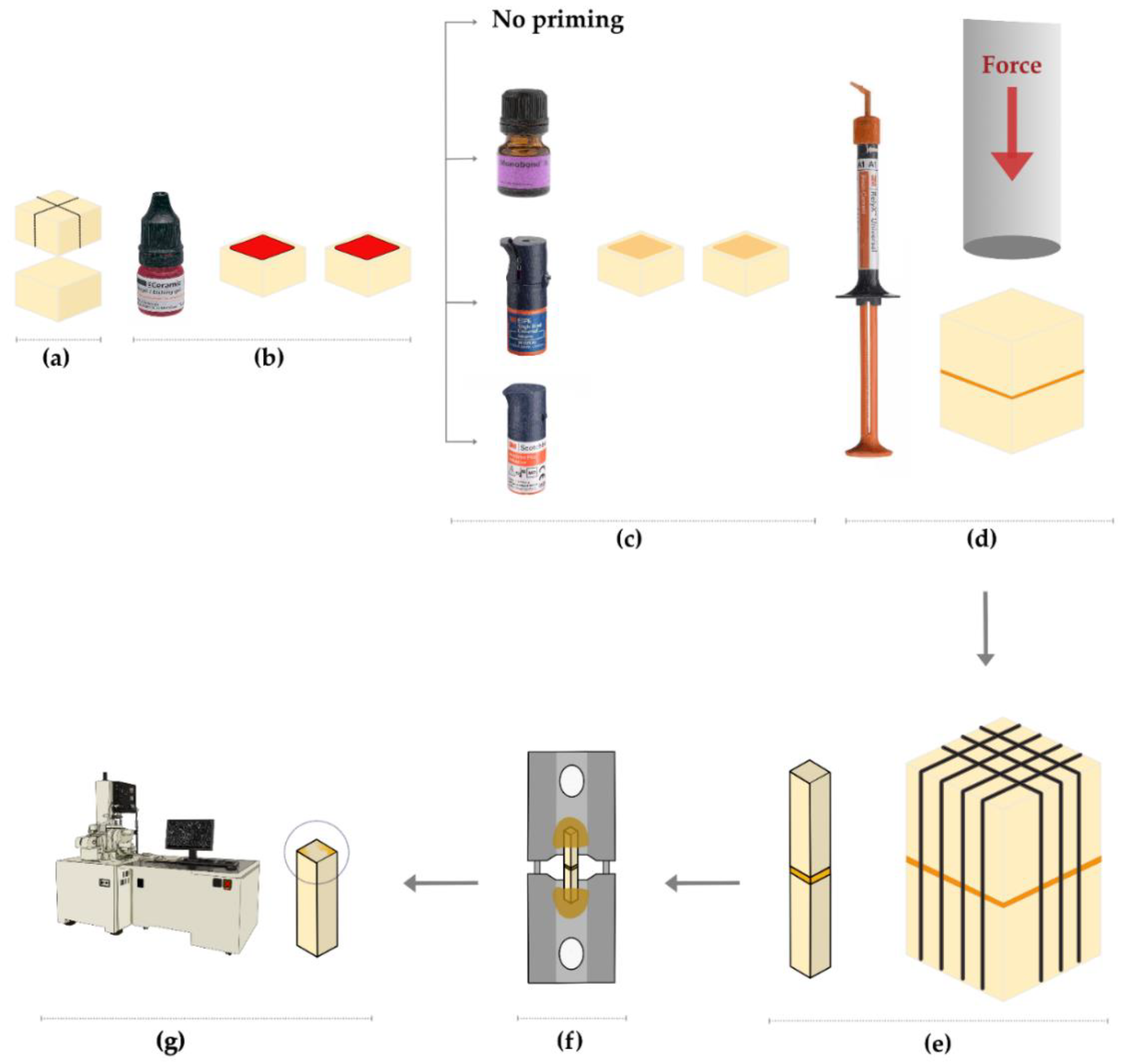
2.1. Specimens’ Preparation

LDC (GC InitialLiSi Block, GC Corporation, Tokyo, Japan) blocks were cut into a total of 40 (approximately 5.5 mm × 6.5 mm) 5 mm thick discs using a water-cooled diamond cutting disc mounted on a computer-controlled precision cutter (IsoMet 5000 Linear Precision Saw, Buehler, Lake Bluff, IL, USA). The top surface of each ceramic disc was wet-polished for 1 min using 600-grit silicon carbide papers mounted on a grinding machine (Automata, Jean Wirtz, Germany) performing 200 revolutions per minute (rpm), and then the discs were subjected to ultrasonic cleaning in distilled water for 5 min using an ultrasonicator (Sonicer, Yoshida Dental Manufacturing. Co., Ltd., Tokyo, Japan). The top surface of each ceramic disc was acid-etched with 5% HF (IPS Ceramic Etching Gel, Ivoclar Vivadent, Schaan, Liechtenstein) for 20 s, then thoroughly washed with water, before being air-dried and subjected to additional ultrasonic cleaning in distilled water for 5 min, and then air-dried and randomly distributed into four groups according to the priming material utilized. In Group 1 (control), the LDC discs were not primed. In Group 2, a universal primer (MBN) was applied to the top surface using a microbrush according to the manufacturer’s instructions, where it was left for 60 s; subsequently, any remaining excess was dispersed with a strong stream of air for approximately 5 s. In Group 3, γMPTS-containing UA (SBU) was mixed with one drop (1-to-1 ratio) of Scotchbond Universal DCA (3M Deutschland GmbH, Neuss, Germany) and immediately applied onto the ceramic surface with a microbrush for 20 s, before being air-dried for approximately 5 s without being light-cured. In Group 4, γMPTES/APTES-containing UA (SBP) was applied onto the ceramic surface with a microbrush for 20 s, before being air-dried for approximately 5 s without being light-cured. For each group, the two discs that received the same surface treatment were cemented using a dual-cure resin cement (RelyX Universal Resin Cement, 3M Oral Care, St. Paul, MN, USA). Equal amounts of resin cement were applied onto the HF-etched or surface-treated (primed) sides before both discs were vertically aligned using a custom-made metal cementation device; then, a cementation force of 1 kg was applied to the discs for 1 min (Figure 1) [24]. The excess cement was immediately and carefully removed with a small-size cotton pellet. The cemented blocks (n = 5/group) were light-cured from each side for 40 s. Then, additional light curing was performed from the top and bottom sides for an additional 40 s each using EliparTM S10 (3M ESPE, St. Paul, MN, USA) operated at 1000 mW/cm2, as verified by a hand-held radiometer (Bluephase Meter, Ivoclar Vivadent, Austria). The light-curing tip was kept at approximately 1 mm and at 0 angle to the cemented block. Then, the cemented blocks were kept in distilled water for 24 h at 37 °C before sectioning.

2.2. μTBS

The cemented ceramic blocks were sectioned into ceramic–resin-ceramic microbeams with a cross-sectional area of approximately 1 mm2 (Figure 1e) using a water-cooled diamond cutting disc mounted on a low-speed precision cutter (IsoMet 1000 Precision Cutter, Lake Bluff, IL, USA) for μTBS evaluation. Thirty microbeams were generated within each group. The generated microbeams were examined with a light microscope (Hirox Co., Ltd., Tokyo, Japan) at 15× magnification before μTBS testing. Defective microbeams were excluded. A digital micrometer (Mitutoyo, Kanagawa, Japan) was used to measure the microbeam cross-sectional surface area at the interface. Then, the microbeams were fixed (glued) to a custom-made metal jig using a cyanoacrylate glue (Loctite 416, Henkel, Düsseldorf, Germany) and attached to a universal testing machine (Instron 5965, Instron Corporation, Norwood, MN, USA) supplied with a 30 kN load cell. Before testing, it was ensured that the alignment of each microbeam was parallel to the direction of the tensile force. The microbeams were stressed under tension at a crosshead speed of 0.5 mm/min until failure or debonding. The μTBS (in megaPascal (MPa)) was calculated for each microbeam by dividing the maximum force (in Newton (N)) at fracture or debonding by the cross-sectional surface area (in mm2). The μTBS test was performed in accordance with the protocol described by the Academy of Dental Materials [25].

2.3. Failure/Fracture Mode Assessment

Failure/fracture modes were assessed at 15× magnification using a light microscope (Hirox Co., Ltd., Tokyo, Japan). Failed/fractured microbeams with unidentifiable failure-mode patterns were ultrasonically cleaned in distilled water for 5 min and dehydrated in ascending concentrations of ethanol, before being gold sputtered for 180 s at 40 mA using a JFC-1100 Fine Coat Ion Sputter (JEOL Ltd., Tokyo, Japan). Gold-sputtered microbeams were then assessed for the failure/fracture mode at low (75×, 80×) and high magnifications (500×) using a scanning electron microscope (JSM-6610LV; JEOL Ltd., Tokyo, Japan) operated at 20 kV. Failure/fracture mode patterns were classified as cohesive failure/fracture in the ceramic; adhesive failure/fracture at the resin-ceramic interface; cohesive failure/fracture the resin cement; or mixed failure, including failure/fracture at the ceramic and the resin cement, cohesive failure in the resin cement, and adhesive failure/fracture at the resin-ceramic interface. Defective microbeams or those failed/fractured away (>2 mm) from the resin-ceramic interface were excluded.

2.4. Statistical Analysis

The sample size was calculated using G*Power software, version 3.1.9.7 (Heinrich-Heine-Universität Düsseldorf, Düsseldorf, Germany). A total of 20 cemented blocks are required to provide 5 blocks per group. This design achieves 98% power and a 5% significance level. The µTBS values of microbeams generated from the same cemented ceramic block were averaged, thus the cemented ceramic block was considered as the statistical (experimental) unit [26]. Upon the evaluation of the normality of µTBS data using the Kolmogorov–Smirnov test, the one-way analysis of variance (ANOVA) and Tukey’s multiple comparison tests were applied in the statistical analysis considering the effect of the priming material on the resin-ceramic µTBS, with p < 0.05 considered as being statistically significant. The statistical analysis was performed using R software version 4.1.2 (R Foundation for Statistical Computing, Vienna, Austria).

3. Results

3.1. μTBS

The means ± standard deviations (SDs) μTBS for the tested groups are presented graphically and numerically in Figure 2. Universal primer (MBN) application showed the highest μTBS (32.21 ± 4.49 MPa) among all groups. γMPTES/APTES-containing UA (SBP) resulted in significantly higher μTBS (20.41 ± 6.26 MPa) compared with γMPTS-containing UA (SBU) (10.65 ± 3.5 MPa) or the control group (8.47 ± 3.11 MPa). There was no statistically significant difference between γMPTS-containing UA (SBU) and the control group. The results of Tukey’s multiple comparisons statistical analysis are illustrated in Table 2.

3.2. Failure Mode Evaluation

The frequencies of failure modes (expressed as percentages) recorded for the tested μTBS microbeams are presented in Figure 3. The mixed failures were the most predominant among all groups at 60% (15 microbeams) for the control group, 80% (20 microbeams) for MBN, 68% (17 microbeams) for SBU, and 72% (18 microbeams) for SBP. Adhesive failures occurred next to mixed failures, while cohesive failures were the least among all groups. No cohesive failures in ceramic, or pretest failures, were recorded for any group. The representative SEM photomicrographs of the recorded failure modes are presented in Figure 4. Most of the mixed failure-mode patterns involved an adhesive interfacial failure at the ceramic side and a cohesive failure within the resin cement (Figure 4c,d).

4. Discussion

Bond strength evaluations can be utilized to assess the effectiveness of adhesive procedures and materials [27] before clinical studies can be conducted. This study was designed to evaluate the effect of priming LDC with silane-containing UAs on resin-ceramic bond strength. In contrast to a previous study [23] in which a shear (macro) bond strength test was used to evaluate the bonding of light-cured (polymerized) silane-containing UAs, the current study utilized the μTBS test due to its better discriminative ability, more even stress distribution during testing, and less cohesive substrate failures compared with macro bond strength tests [28,29]. In addition, the ceramic discs were primed with UAs before they were cemented (under force) using a dual-cure resin-based cement to simulate the intra-oral cementation procedure.
The first null hypothesis was that ceramic surface treatments (universal primer or UAs) would have no significant effect on the resin-ceramic µTBS. The results of this study indicated that despite γMPTS-containing UA (SBU) having no effect on the resin-ceramic µTBS, both the universal primer (MBN) and γMPTES/APTES-containing UA (SBP) considerably improved the resin-ceramic µTBS. Thus, the first hypothesis was rejected.
Silane-containing universal primers, such as MBN, would not promote adequate adhesion to glass-ceramics without a prior HF etching step [30]. That is why the effect of MBN or silane-containing UAs should not be evaluated without prior HF etching. The application of HF on glass-ceramics results in the formation of tetrafluorosilane, which reacts with HF to form a soluble hydrofluorosilicic acid [12]. Then, a considerable amount of the glass matrix at the etched ceramic surface is dissolved and can be easily rinsed away with water. This not only results in the creation of surface microirregularities but also results in the formation of hydroxyl groups on the etched ceramic surface [9]. Upon the surface treatment of HF-etched glass-ceramic with silane-containing primers, a condensation reaction between such hydroxyl groups and the silanol groups of activated silane occurs, representing one aspect of ceramic bonding [9,31]. The other aspect is the reaction between organofunctional groups in the silane molecule and functional (methacrylate) groups of the methacrylate-based materials, such as resin-based cements or adhesives [9,31]. The universal primers utilized in this study, such as MBN, contain large percentages of organic solvents, such as ethanol or acetone [11,32]. Upon air-drying the primer layer applied on the ceramic surface, much of the solvent will be evaporated, resulting in the formation of a silane layer on the ceramic surface, which is a crucial factor affecting the resin-ceramic bond strength [31]. In contrast, compared with MBN, the composition of silane-containing UAs is much more complex because they are primarily manufactured to bond resin-based composite materials to the tooth structure [33]. To specify, other components within the UA composition, such as 10-MDP, Bis-GMA, HEMA, dimethacrylate resin, crosslinker monomers, and other components, cannot be eliminated after the application of UA. Thus, it is assumed that despite the ability of UAs to flow effectively into the microirregularities created by HF etching [21], the formation of a silane layer deposited onto the ceramic surface seems less likely, even with the effective air-drying of such UAs [20]. This can partly explain the inferior priming capacity of UAs compared with MBN. Meanwhile, UAs are acidic in nature [14,34], which might have affected the chemical stability of silane molecules within the UAs’ formulations, impairing their priming potential [21] and resulting in a low resin-ceramic bond strength. γMPTS-containing UA (SBU) did not have any impact on resin-ceramic bonding. This is in accordance with a recent meta-analysis that indicated that γMPTS-containing UAs do not have a priming capacity similar to silane-containing or universal primers [35]. In contrast, γMPTES/APTES-containing UA (SBP) not only significantly improved the resin-ceramic µTBS compared with the control group but also resulted in significantly higher μTBS than the γMPTS-containing UA (SBU). Therefore, the second null hypothesis, that there would be no significant difference between the resin-ceramic µTBS obtained after the use of either of the two silane-containing UAs tested, was also rejected. The different bond strengths obtained can be explained—in part—by the better priming capacity of SBP due to its optimized (γMPTES/APTES) silane technology. Combining the effects of the silanol-groups of both γMPTES and APTES molecules, as well as the amino groups of the APTES molecule, means they can simultaneously interact with the hydroxyl groups on the glass-ceramic surface [36], increasing UA’s priming capacity; thus, the resin-ceramic μTBS is enhanced. The APTES molecule within SBP forms hydrogen bonds at the HF-etched glass-ceramic surface and reacts with silanol groups, resulting in the formation of amino-silanol groups [37]. In addition, the APTES molecule can act as a siloxane intermolecular crosslinker [36], which can increase the density of the silane interaction layer and enhance the bonding to the glass-ceramic material [23]. Moreover, the Si–O–C2H5 group within the γMPTES molecule in SBP hydrolyzes slower than the Si–O–CH3 group within the γMPTS molecule in SBU [38], which may have decreased the dehydration condensation of silanol groups [23]. Another plausible explanation for the superior resin-ceramic µTBS obtained when SBP is used as a primer may be its improved dual-cure compatibility with the luting resin cement used (RelyX Universal Resin Cement). However, further in-depth investigations are required to prove this speculation.
Despite the precision of the μTBS test employed in this study, it is a technique-sensitive method associated with the generation of interfacial stress during specimens’ sectioning. Such stress can result in failure of low-performance adhesive interfaces or brittle substrates [29]. Multiple factors, such as specimens’ alignment in relation to the direction of tensile force, shape and dimensions, gripping jig, and testing machine, can also influence the outcome of the μTBS evaluation [25,39]. The outcome (μTBS data) should be interpreted in combination with the corresponding failure modes because a statistical evaluation of μTBS data can be affected by the inclusion or censoring microbeams with specific failure modes [40,41]. Both a light microscope and SEM were utilized to assess the failure mode patterns of the tested microbeams. The fractographic evaluation using SEM is more illustrative and can be performed in higher magnifications compared with using a light microscope [25]. We found that mixed failure patterns (Figure 4c,d) were the most predominant in all the groups. The control (no priming) group presented a higher incidence of adhesive failures compared with the other groups, which may be due to the relatively low μTBS. The failure mode patterns of the tested groups were affected by the setup of the μTBS test. Although sectioning the cemented ceramic blocks into microbeams is laboratory-intensive and technique-sensitive work, testing the ceramic-resin-ceramic microbeams employed in this study not only had the advantage of solely evaluating the resin-ceramic interface [24] but also eliminated the incidence of cohesive failures within the ceramic material, which can be explained by the high mechanical properties of LDC [42] and the homogenous stress distribution at the interface. Overall, the γMPTES/APTES-containing UA (SBP) showed promising performance as an LDC primer before cementation with a dual cure resin cement. However, this study has some limitations, including the lack of long-term artificial aging (water storage and thermocycling) of the microbeams and the use of one ceramic material and one resin cement. Moreover, mechanical loading before bond strength evaluation cannot be performed because of the geometry of the cemented blocks. Thus, the results of this study should be considered with much caution. In fact, in-depth chemical analyses of γMPTES/APTES ratio applied in SBP in addition to the reaction between γMPTES/APTES-containing UA (SBP) and HF-etched glass-ceramic are required to confirm its positive impact on resin-ceramic bond strength.

5. Conclusions

The effect of silane-containing UAs on resin-ceramic μTBS is material-dependent. γMPTS-containing UA (SBU) did not affect the resin-ceramic μTBS. Although γMPTES/APTES-containing UA (SBP) improved bonding to LDC compared with γMPTS-containing UA (SBU) or the control group (no priming), it resulted in considerably lower μTBS than a universal silane-containing primer (MBN). The priming of LDC with either of the UAs tested cannot be considered as an alternative to a separate silanization (priming) step using a universal silane-containing primer.

Author Contributions

Conceptualization, M.M.A.; methodology, M.M.A.; software, A.A. (Ali Alrahlah); validation, A.S.A.; formal analysis, M.M.A.; investigation, B.A. and S.A.; resources, A.A. (Abdullah Alshehri); data curation, M.M.A.; writing—original draft preparation, M.M.A. and F.A.; writing—review and editing, M.A.S.; visualization, M.M.A. and A.R.; supervision, A.A. (Ali Alrahlah); project administration, F.A.; funding acquisition, A.A. (Abdullah Alshehri). All authors have read and agreed to the published version of the manuscript.

Funding

The authors are grateful to the Deanship of Scientific Research, King Saud University, for funding this study through the Vice Deanship of Scientific Research Chairs and the Research Chair for Dental and Oral Rehabilitation, Engineer Abdullah Bugshan.

Institutional Review Board Statement

Not applicable.

Informed Consent Statement

Not applicable.

Data Availability Statement

The data presented in this study are available upon request from the corresponding author.

Conflicts of Interest

The authors declare no conflict of interest.

References

  1. van den Breemer, C.R.; Gresnigt, M.M.; Cune, M.S. Cementation of Glass-Ceramic Posterior Restorations: A Systematic Review. BioMed Res. Int. 2015, 2015, 148954. [Google Scholar] [CrossRef] [PubMed] [Green Version]
  2. Politano, G.; Van Meerbeek, B.; Peumans, M. Nonretentive Bonded Ceramic Partial Crowns: Concept and Simplified Protocol for Long-lasting Dental Restorations. J. Adhes. Dent. 2018, 20, 495–510. [Google Scholar] [CrossRef] [PubMed]
  3. Phark, J.-H.; Duarte, S., Jr. Microstructural considerations for novel lithium disilicate glass ceramics: A review. J. Esthet. Restor. Dent. 2022, 34, 92–103. [Google Scholar] [CrossRef] [PubMed]
  4. Awad, M.M.; Alqahtani, H.; Al-Mudahi, A.; Murayshed, M.S.; Alrahlah, A.; Bhandi, S.H. Adhesive Bonding to Computer-aided Design/ Computer-aided Manufacturing Esthetic Dental Materials: An Overview. J. Contemp. Dent. Pract. 2017, 18, 622–626. [Google Scholar] [CrossRef] [PubMed]
  5. Malysa, A.; Wezgowiec, J.; Grzebieluch, W.; Danel, D.P.; Wieckiewicz, M. Effect of Thermocycling on the Bond Strength of Self-Adhesive Resin Cements Used for Luting CAD/CAM Ceramics to Human Dentin. Int. J. Mol. Sci. 2022, 23, 745. [Google Scholar] [CrossRef]
  6. Malysa, A.; Wezgowiec, J.; Orzeszek, S.; Florjanski, W.; Zietek, M.; Wieckiewicz, M. Effect of Different Surface Treatment Methods on Bond Strength of Dental Ceramics to Dental Hard Tissues: A Systematic Review. Molecules 2021, 26, 1223. [Google Scholar] [CrossRef]
  7. Akar, T.; Dündar, A.; Kırmalı, Ö.; Üstün, Ö.; Kapdan, A.; Er, H.; Kuştarcı, A.; Er, K.; Yılmaz, B. Evaluation of the shear bond strength of zirconia to a self-adhesive resin cement after different surface treatment. Dent. Med. Probl. 2021, 58, 463–472. [Google Scholar] [CrossRef]
  8. Lubauer, J.; Belli, R.; Peterlik, H.; Hurle, K.; Lohbauer, U. Grasping the Lithium hype: Insights into modern dental Lithium Silicate glass-ceramics. Dent. Mater. 2022, 38, 318–332. [Google Scholar] [CrossRef]
  9. Tian, T.; Tsoi, J.K.-H.; Matinlinna, J.P.; Burrow, M.F. Aspects of bonding between resin luting cements and glass ceramic materials. Dent. Mater. 2014, 30, e147–e162. [Google Scholar] [CrossRef]
  10. Della Bona, A.; Anusavice, K.J.; Mecholsky, J.J., Jr. Failure analysis of resin composite bonded to ceramic. Dent. Mater. 2003, 19, 693–699. [Google Scholar] [CrossRef]
  11. Matinlinna, J.P.; Lassila, L.V.; Özcan, M.; Yli-Urpo, A.; Vallittu, P.K. An introduction to silanes and their clinical applications in dentistry. Int. J. Prosthodont. 2004, 17, 155–164. [Google Scholar] [PubMed]
  12. Lung, C.Y.; Matinlinna, J.P. Aspects of silane coupling agents and surface conditioning in dentistry: An overview. Dent. Mater. 2012, 28, 467–477. [Google Scholar] [CrossRef] [PubMed]
  13. Shen, C.; Oh, W.S.; Williams, J.R. Effect of post-silanization drying on the bond strength of composite to ceramic. J. Prosthet. Dent. 2004, 91, 453–458. [Google Scholar] [CrossRef] [PubMed]
  14. Nagarkar, S.; Theis-Mahon, N.; Perdigão, J. Universal dental adhesives: Current status, laboratory testing, and clinical performance. J. Biomed. Mater. Res. B Appl. Biomater. 2019, 107, 2121–2131. [Google Scholar] [CrossRef] [PubMed]
  15. Cuevas-Suárez, C.E.; de Oliveira da Rosa, W.L.; Vitti, R.P.; da Silva, A.F.; Piva, E. Bonding Strength of Universal Adhesives to Indirect Substrates: A Meta-Analysis of in Vitro Studies. J. Prosthodont. 2020, 29, 298–308. [Google Scholar] [CrossRef]
  16. Kalavacharla, V.K.; Lawson, N.C.; Ramp, L.C.; Burgess, J.O. Influence of Etching Protocol and Silane Treatment with a Universal Adhesive on Lithium Disilicate Bond Strength. Oper. Dent. 2015, 40, 372–378. [Google Scholar] [CrossRef] [Green Version]
  17. Makishi, P.; Andre, C.B.; Silva, J.L.; Bacelar-Sa, R.; Correr-Sobrinho, L.; Giannini, M. Effect of Storage Time on Bond Strength Performance of Multimode Adhesives to Indirect Resin Composite and Lithium Disilicate Glass Ceramic. Oper. Dent. 2016, 41, 541–551. [Google Scholar] [CrossRef] [Green Version]
  18. Yao, C.; Yang, H.; Yu, J.; Zhang, L.; Zhu, Y.; Huang, C. High Bond Durability of Universal Adhesives on Glass Ceramics Facilitated by Silane Pretreatment. Oper. Dent. 2018, 43, 602–612. [Google Scholar] [CrossRef]
  19. Yao, C.; Zhou, L.; Yang, H.; Wang, Y.; Sun, H.; Guo, J.; Huang, C. Effect of silane pretreatment on the immediate bonding of universal adhesives to computer-aided design/computer-aided manufacturing lithium disilicate glass ceramics. Eur. J. Oral Sci. 2017, 125, 173–180. [Google Scholar] [CrossRef]
  20. Awad, M.M.; Albedaiwi, L.; Almahdy, A.; Khan, R.; Silikas, N.; Hatamleh, M.M.; Alkhtani, F.M.; Alrahlah, A. Effect of universal adhesives on microtensile bond strength to hybrid ceramic. BMC Oral Health 2019, 19, 178. [Google Scholar] [CrossRef] [Green Version]
  21. Yoshihara, K.; Nagaoka, N.; Sonoda, A.; Maruo, Y.; Makita, Y.; Okihara, T.; Irie, M.; Yoshida, Y.; Van Meerbeek, B. Effectiveness and stability of silane coupling agent incorporated in ‘universal’ adhesives. Dent. Mater. 2016, 32, 1218–1225. [Google Scholar] [CrossRef] [PubMed] [Green Version]
  22. Alrahlah, A.; Awad, M.M.; Vohra, F.; Al-Mudahi, A.; Al Jeaidi, Z.A.; Elsharawy, M. Effect of self etching ceramic primer and universal adhesive on bond strength of lithium disilicate ceramic. J. Adhes. Sci. Technol. 2017, 31, 2611–2619. [Google Scholar] [CrossRef]
  23. Yao, C.; Ahmed, M.H.; De Grave, L.; Yoshihara, K.; Mercelis, B.; Okazaki, Y.; Van Landuyt, K.L.; Huang, C.; Van Meerbeek, B. Optimizing glass-ceramic bonding incorporating new silane technology in an experimental universal adhesive formulation. Dent. Mater. 2021, 37, 894–904. [Google Scholar] [CrossRef] [PubMed]
  24. Südbeck, J.M.; Jacker-Guhr, S.; Lührs, A.K. Do Different Pretreatment Methods Influence the Microtensile Bond Strength of Composite Cements to Silicate Ceramic? J. Adhes. Dent. 2021, 23, 335–345. [Google Scholar] [CrossRef] [PubMed]
  25. Armstrong, S.; Breschi, L.; Özcan, M.; Pfefferkorn, F.; Ferrari, M.; Van Meerbeek, B. Academy of Dental Materials guidance on in vitro testing of dental composite bonding effectiveness to dentin/enamel using micro-tensile bond strength (μTBS) approach. Dent. Mater. 2017, 33, 133–143. [Google Scholar] [CrossRef] [Green Version]
  26. Eckert, G.J.; Platt, J.A. A statistical evaluation of microtensile bond strength methodology for dental adhesives. Dent. Mater. 2007, 23, 385–391. [Google Scholar] [CrossRef]
  27. El Zohairy, A.A.; De Gee, A.J.; Mohsen, M.M.; Feilzer, A.J. Microtensile bond strength testing of luting cements to prefabricated CAD/CAM ceramic and composite blocks. Dent. Mater. 2003, 19, 575–583. [Google Scholar] [CrossRef]
  28. De Munck, J.; Mine, A.; Poitevin, A.; Van Ende, A.; Cardoso, M.V.; Van Landuyt, K.L.; Peumans, M.; Van Meerbeek, B. Meta-analytical review of parameters involved in dentin bonding. J. Dent. Res. 2012, 91, 351–357. [Google Scholar] [CrossRef]
  29. Salz, U.; Bock, T. Testing adhesion of direct restoratives to dental hard tissue—A review. J. Adhes. Dent. 2010, 12, 343–371. [Google Scholar] [CrossRef]
  30. Huang, T.H.; Kao, C.T. The shear bond strength of composite brackets on porcelain teeth. Eur. J. Orthod. 2001, 23, 433–439. [Google Scholar] [CrossRef] [Green Version]
  31. Matinlinna, J.P.; Lung, C.Y.K.; Tsoi, J.K.H. Silane adhesion mechanism in dental applications and surface treatments: A review. Dent. Mater. 2018, 34, 13–28. [Google Scholar] [CrossRef] [PubMed]
  32. Zakir, M.; Ashraf, U.; Tian, T.; Han, A.; Qiao, W.; Jin, X.; Zhang, M.; Tsoi, J.K.-H.; Matinlinna, J.P. The Role of Silane Coupling Agents and Universal Primers in Durable Adhesion to Dental Restorative Materials—A Review. Curr. Oral Health Rep. 2016, 3, 244–253. [Google Scholar] [CrossRef]
  33. Chen, C.; Niu, L.N.; Xie, H.; Zhang, Z.Y.; Zhou, L.Q.; Jiao, K.; Chen, J.H.; Pashley, D.H.; Tay, F.R. Bonding of universal adhesives to dentine--Old wine in new bottles? J. Dent. 2015, 43, 525–536. [Google Scholar] [CrossRef] [PubMed] [Green Version]
  34. Papadogiannis, D.; Dimitriadi, M.; Zafiropoulou, M.; Gaintantzopoulou, M.D.; Eliades, G. Universal Adhesives: Setting Characteristics and Reactivity with Dentin. Materials 2019, 12, 1720. [Google Scholar] [CrossRef] [Green Version]
  35. Lima, R.B.W.; Muniz, I.A.F.; Campos, D.E.S.; Murillo-Gómez, F.; Andrade, A.K.M.; Duarte, R.M.; de Souza, G.M. Effect of universal adhesives and self-etch ceramic primers on bond strength to glass ceramics: A systematic review and meta-analysis of in vitro studies. J. Prosthet. Dent. 2022. [Google Scholar] [CrossRef]
  36. Acres, R.G.; Ellis, A.V.; Alvino, J.; Lenahan, C.E.; Khodakov, D.A.; Metha, G.F.; Andersson, G.G. Molecular Structure of 3-Aminopropyltriethoxysilane Layers Formed on Silanol-Terminated Silicon Surfaces. J. Phys. Chem. C 2012, 116, 6289–6297. [Google Scholar] [CrossRef]
  37. Park, S.-J.; Jin, J.-S.; Lee, J.-R. Influence of silane coupling agents on the surface energetics of glass fibers and mechanical interfacial properties of glass fiber-reinforced composites. J. Adhes. Sci. Technol. 2000, 14, 1677–1689. [Google Scholar] [CrossRef]
  38. Xiong, M.; Zhang, K.; Chen, Y. ATRP of 3-(triethoxysilyl)propyl methacrylate and preparation of “stable” gelable block copolymers. Eur. Polym. J. 2008, 44, 3835–3841. [Google Scholar] [CrossRef]
  39. Roeder, L.; Pereira, P.N.; Yamamoto, T.; Ilie, N.; Armstrong, S.; Ferracane, J. Spotlight on bond strength testing--unraveling the complexities. Dent. Mater. 2011, 27, 1197–1203. [Google Scholar] [CrossRef]
  40. Comba, L.; Tichy, A.; Bradna, P. The Effect of Censoring on the Statistical Evaluation of Composite-to-Composite Bond Strength. J. Adhes. Dent. 2020, 22, 183–193. [Google Scholar] [CrossRef]
  41. Franz, A.; Lettner, S.; Watts, D.C.; Schedle, A. Should statistical analysis of bond-strength data include or exclude cohesive failures? Dent. Mater. 2022, 38, 1942–1946. [Google Scholar] [CrossRef] [PubMed]
  42. da Silva, S.E.G.; de Araújo, G.M.; Souza, K.B.; Moura, D.M.D.; Aurélio, I.L.; May, L.G.; Vila-Nova, T.E.L.; Zhang, Y.; de Assunção, E.S.R.O. Biaxial flexure strength and physicochemical characterization of a CAD/CAM lithium disilicate ceramic: Effect of etching time, silane, and adhesive applications. Clin. Oral Investig. 2022, 26, 6753–6763. [Google Scholar] [CrossRef] [PubMed]
Figure 1. Schematic illustration of specimens’ preparation and μTBS test. (a) Sectioning of ceramic blocks into discs; (b) acid etching with 5% HF; (c) priming of ceramic discs, control (no priming), MBN, SBU, SBP; (d) cementation of ceramic discs using RelyX Universal Resin Cement under 1 kg force; (e) sectioning of cemented discs into microbeams; (f) fixation of microbeams onto μTBS metal jig; (g) using a scanning electron microscope (SEM) to evaluate the failure/fracture mode of microbeams with failure-mode patterns unidentifiable with a light microscope.
Coatings 13 00477 g001
Figure 2. The means ± standard deviations (SDs) of resin-ceramic microtensile bond strength (μTBS) (expressed in MPa) of Group 1 (control), no priming; Group 2, primed with MBN; Group 3, primed with SBU; and Group 4, primed with SBP. Different superscript letters indicate statistically significant difference (p < 0.05) between groups.
Coatings 13 00477 g002
Figure 3. Frequencies of failure modes (expressed as percentages) recorded for tested μTBS microbeams of following groups: control (no priming), MBN, SBU, and SBP.
Coatings 13 00477 g003
Figure 4. Representative SEM photomicrographs at low and high magnifications of tested microbeams. (a), at 75× and (b), at 500×: adhesive failure; (c), at 75× and (d), at 500×: mixed failure; (e), at 80× and (f), at 500×: cohesive failure in resin cement. Cr: ceramic surface; gray arrow: ceramic surface covered by adhesive layer; white arrow: adhesive layer, and black arrow: resin cement.
Coatings 13 00477 g004
Table 1. Materials used in study.
Material (LOT Number)CodeComposition
Initial LiSi Block, GC, GC corporation, Tokyo, Japan.
(LOT: 2201201C)
LDCSilicon dioxide: 81%; phosphorus oxide 8.1%;
potassium oxide 5.9%; aluminum oxide 3.8%;
titanium oxide 0.5%; and cerium oxide 0.6%.
Single Bond Universal Adhesive, 3M Deutschland GmbH, Neuss, Germany.
(LOT: 10608B)
SBU10-MDP phosphate monomer, Vitrebond,
copolymer, HEMA, Bis-GMA, dimethacrylate resin, camphorquinone, silane (γMPTS),
ethanol, and water.
Scotchbond Universal Plus Adhesive, 3M Deutschland GmbH, Neuss, Germany.
(LOT: 7676509)
SBP10-MDP phosphate monomer, Vitrebond,
copolymer, HEMA, dimethacrylate resin,
crosslinking radiopaque monomer, dual-cure
accelerator, camphorquinone, optimized silane (γMPTES/APTES), ethanol, and water.
Monobond N, Ivoclar Vivadent, Schaan, Liechtenstein.
(LOT: Z02S7Z)
MBNAlcohol, silane methacrylate, phosphoric acid methacrylate, disulfide methacrylate.
RelyX Universal Resin Cement, 3M Oral Care, St. Paul, USA.
(LOT: 7756479)
-Dimethacrylate monomers, phosphorylated
dimethacrylate adhesion monomers,
photoinitiator system, novel amphiphilic redox initiator system, radiopaque fillers, and
rheological additives and pigments.
Bis-GMA, bisphenol A glycidyl methacrylate; 10-MDP, 10-methacryloyloxydecyl dihydrogen thiophosphate; HEMA, 2-hydroxyethyl methacrylate; γMPTS, γ methacryloxypropyltrimethoxysilane; γMPTES, γ-methacryloxypropyltriethoxysilane; APTES, 3-(aminopropyl)triethoxysilane.
Table 2. Results of Tukey’s multiple comparisons statistical analysis of μTBS data.
Pair-Wise Groups ComparisonsMean DifferenceLower BoundUpper Boundp-ValueSignificance
Control-MBN23.73915.58131.897<0.001*
Control-SBU2.181−5.97710.3390.869
Control-SBP11.9423.78420.1000.003*
MBN-SBU21.55713.39929.715<0.001*
MBN-SBP11.7973.63919.9550.004*
SBU-SBP−9.761−17.919−1.6030.017*
*: indicates statistically significant difference.
Disclaimer/Publisher’s Note: The statements, opinions and data contained in all publications are solely those of the individual author(s) and contributor(s) and not of MDPI and/or the editor(s). MDPI and/or the editor(s) disclaim responsibility for any injury to people or property resulting from any ideas, methods, instructions or products referred to in the content.

