1. Introduction
Patients’ aesthetic desire has significantly increased, leading to a high demand for dental restorative materials that are indistinguishable from natural teeth [
1]. Ceramics are increasingly being promoted as high-strength, tooth-colored materials for dental prostheses. The goal is the development of ceramics that have superior aesthetics with long-term durability [
2,
3]. The improvement in physical properties and excellent biocompatibility, in addition to aesthetic characteristics, resulted in the use of zirconia (Zr) ceramic restorations in the anterior and posterior region [
4]. New translucent monolithic zirconia (TZr) has been developed to merge strength with improved tooth-color matching [
1,
5]. However, color matching between aesthetic restorative materials and tooth still remains a challenge as the optical properties of the teeth go far beyond color itself [
1].
Several optical properties influence the appearance of restorations to resemble natural teeth, including the translucency parameter (TP) and contrast ratio (CR). Translucency defines a status between full transparency and opacity and is the amount of light passing through a material [
5,
6,
7,
8]. Zr ceramics are optically considered to be a semi-translucent material [
5]. Glazing, which is the final step in ceramic construction, reduces the translucency of Zr-based restorations [
9]. On the other hand, the CR is the ratio between the intensity of the spectral reflectance from an object against a black background to the reflectance of the same material against a white background [
1,
10,
11]. The measurement of the TP and CR of a material requires the use of a digital color system such as the CIE L*a*b* “Commission Internationale de l’Eclairage” system, which utilizes three co-ordinates: L*, a*, and b*. Each of these co-ordinates represent an axis which intersects with the others in the center, forming the CIE L*a*b* color space. The L* co-ordinate measures the lightness of an object, whereas the a* and b* co-ordinates represent the red–green axis and yellow–blue axis, respectively. Color change (ΔE) can then be calculated using a special formula [
10,
11,
12,
13]. The color of the dental ceramic and its optical properties mainly depend on the nature of the crystalline phase present. Other factors may influence the final color including type, thickness, and veneering technique [
14,
15].
Optical properties are also dependent on the material’s surface characteristics, such as surface texture or roughness. The surface roughness (Ra) of the restorative material plays a significant role in its appearance, as it influences the amount of light reflected [
16]. A rough surface texture will reflect an irregular or diffuse pattern, which will modify the color of the restoration [
17]. A very common treatment that might alter surface texture is dental bleaching [
18]. Bleaching can be performed in-office with a higher concentration of the active ingredient, such as a 35% concentration of carbamide peroxide, or as home-bleaching kits with concentrations of 10%–15% of carbamide peroxide [
19]. The higher the concentration, the faster the diffusion, and the more detrimental it can be to pulpal tissues. Therefore, home bleaching with lower-concentrated agents became popular since its introduction in the 1990s, as it may be less irritating to the tooth pulp without impairing its efficacy [
20]. Although home-bleaching protocols have been widely accepted as a treatment modality for brightening the color of natural teeth, their effect on restorative materials adjacent to the teeth being bleached should not be overlooked. It has been found that the translucency of CAD/CAM lithium disilicate ceramics has been altered after a bleaching procedure with 16% carbamide peroxide [
10]. With the launch of monolithic zirconia materials, which solved the veneer chipping problems of the porcelain-layered zirconia copings, considerable research has been carried out [
21] to look into the influence of different treatments and conditions on the appearance of these aesthetic materials. Furthermore, even though recently developed innovative ultra-translucent zirconia ceramics have demonstrated attractive aesthetic characteristics and the ability to match the shade of natural teeth, their color stability in reaction to outside treatments such as at-home bleaching/whitening still raises questions. There is a lack of information in the literature and research on how home bleaching alters the color of cutting-edge monolithic Zr and how it contrasts with more aesthetically pleasing lithium disilicate.
The goals of this in vitro study were to determine the effect of home bleaching (15% carbamide peroxide) on the optical properties, i.e., the translucency parameter (TP), contrast ratio (CR) and total color difference (ΔE) of three different types of aesthetic dental ceramics [(IPS Empress II (IPS); layered zirconia (LZr); and monolithic novel ultra-translucent zirconia (TZr)], and to evaluate the effect of the same home bleaching on the surface roughness (Ra) of the tested ceramic materials.
2. Materials and Methods
2.1. Ethical Approval
Before commencement of the study, ethical approval was obtained from the Institutional Review Board of King Saud University Medical City (research project No. E-20-4882).
2.2. Sample Size Determination
The sample size was calculated using power calculation software (G*Power Version 3.1.9.6; program written by Franz Faul, Universitat Kiel, Germany). At an alpha of 0.05 with an effect size of 0.61 and power of 0.82, the total sample size was determined to be 21 samples, with seven samples in each of the three test groups.
2.3. Specimen Preparation
A total of 21 ceramic specimens were fabricated using CAD/CAM technology following the manufacturer’s instructions [
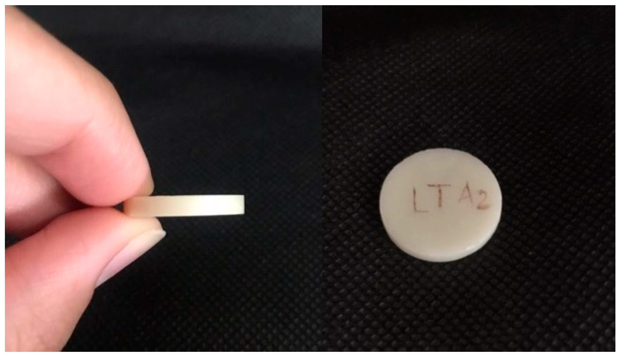
15]. The specimens were in the form of flat cylindrical disks measuring 20 mm in diameter and 3 mm thick (
Figure 1), and had a shade of A2. To standardize the dimensions of all the specimens, digitalized designing was carried out using 3D software 18.0.1931.0 (3D Builder, Microsoft Corporation, Washington, WA, USA) [
15]. The specimens were divided into three groups according to their material (n = 7): IPS, lithium disilicate; LZr, classic zirconia coping with feldspathic layering; and TZr, novel extra-translucent zirconia (
Table 1). All the specimens were glazed at temperatures recommended by the manufacturer. The details of the materials used in the study are listed in
Table 1.
2.4. Application of the Bleaching Material
Each specimen from the three groups was stored in a separate container filled with distilled water and was kept in an incubator at 37 °C for 24 h before starting the treatment. Before each bleaching procedure, samples were taken out from the distilled water and were dried with gauze. A common home-bleaching material (Opalescence PF 15%, Ultradent, South Jordan, UT) that contained 15% carbamide peroxide (CP) was used in the current study (
Table 1). The bleaching agent was applied with a brush applicator according to the manufacturer’s instructions at room temperature; then, samples were stored in the incubator at 37 °C for 6 h per day for 7 days. After each bleaching procedure, samples were rinsed for 1 min with distilled water to remove the bleaching agent. The specimens were stored in the incubator at 37 °C between each bleaching cycle.
2.5. Spectrophotometric Recordings
A spectrophotometer (LabScan XE
®, HunterLab, Sunset Hills Road, Reston, VA, USA) was used to measure the L*a*b* values at three different angles for each specimen against a black background and against a white background. The readings were recorded three times for each specimen. The L*a*b* values were used to calculate the ΔE, TP, and CR according to the following equations:
where the subscripts b and w indicate the black and white backgrounds, respectively, L* indicates lightness, a* indicates the green (−a) and red (+a) axes, and b* indicates the blue (−b) and yellow (+b) axes [
13].
where Y
n is equal to 100, and the subscripts B and W represent the black and white backgrounds, respectively [
11,
12].
CR values are ranged from 0 to 1, where 0 represents a transparent material and 1 indicates an opaque one [
1]. The ΔE, TP, and CR are measured for each sample before and after the application/exposure to the bleaching material.
2.6. Profilometry Analysis
Before and after the application of the bleaching material, each specimen disk was subjected to roughness measurements (Ra) using a 3D profilometer (ContourGT-X®, 3D Optical Microscope, Bruker Nano Surfaces Division, San Jose, CA, USA). The 3D surfaces before and after the bleaching were scanned using a 3D profiling system, and the Ra of the specimens was calculated using 3D software (Vision64®, Operation and Analysis Software, Bruker Nano Surfaces Division, San Jose, CA, USA).
2.7. Scanning Electron Microscope (SEM) Analysis
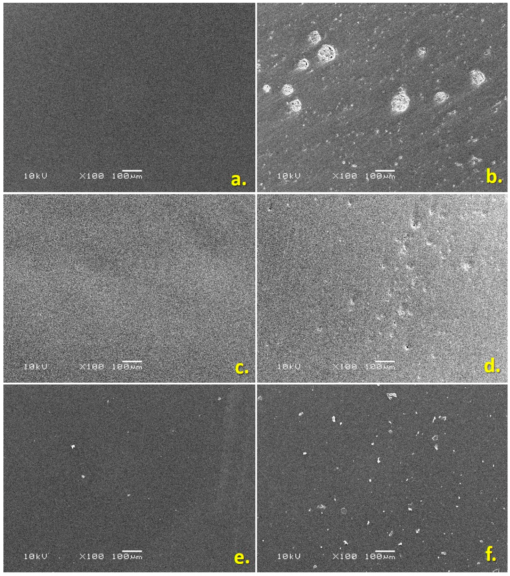
Samples of the specimens were analyzed with a scanning electron microscope (SEM) (JEOL, JSM-6360LV, Tokyo, Japan) to examine the surface characteristics of the three tested specimens (
Figure 2). For this purpose, the specimens were fixed on a stub, sputter coated, and analyzed with the SEM (10 kV, ×100, and 100 μm). The SEM analysis revealed an increase in the surface porosities after the bleaching as compared to the polished surfaces before the bleaching for the three tested groups of materials.
2.8. Data Analysis
Data were analyzed using the SPSS statistical software, version 26.0 for Windows. Due to the small size and skewed nature of the values of outcome variables, nonparametric statistical tests were used for analysis. The descriptive statistics (median and interquartile range) were used to describe the values of the outcome variables: TP, CR, ΔE, and Ra values in each of the three materials (IPS, LZr, and TZr). The Wilcoxon’s signed-rank test was used to compare the mean ranks of outcome variables between to the pre- and post-stages of bleaching in each of the three materials. The Kruskal–Wallis test, followed by the Dunn post hoc test, was used to compare the mean ranks of the TP, CR, ΔE, and Ra among the three test materials. Spearman’s rank correlation was used to quantify the relationship between the differences in the pre- and post-values of the TP, CR, ΔE, and Ra. A p-value of <0.05 was considered significant and used to report the statistical results.
3. Results
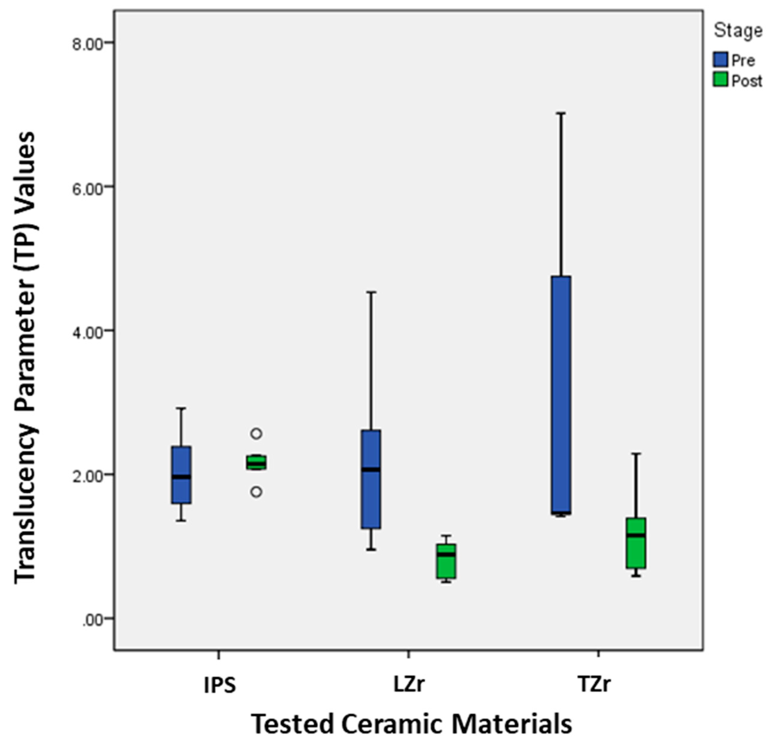
The comparison of the mean ranks of the TP before and after the bleaching for the IPS ceramic material showed no statistically significant difference (
p = 0.39). However, there was a statistically significant difference in the mean ranks of TP values before and after the bleaching for the LZr (
p = 0.01) and TZr (
p = 0.02) ceramic materials. For these two ceramic materials, the mean ranks of TP values after the bleaching were significantly lower than the mean ranks of TP values before the bleaching (
Table 2 and
Figure 3). The TP values of these two materials decreased significantly after the bleaching. This indicated that the bleaching had no effect on the translucency of the IPS ceramics, whereas the translucency of the LZr and TZr was affected by the bleaching.
According to the statistical analysis for the CR, no significant (
p = 0.39) changes were noted for the IPS ceramic material (
Table 2 and
Figure 4). On the other hand, the LZr and TZr ceramics showed significant changes in their CR. The CR values after exposure to the bleaching were significantly lower than the CR values before the bleaching. This indicated that the bleaching had no effect on the CR of the IPS ceramics, whereas the CR of the LZr and TZr was affected by the bleaching.
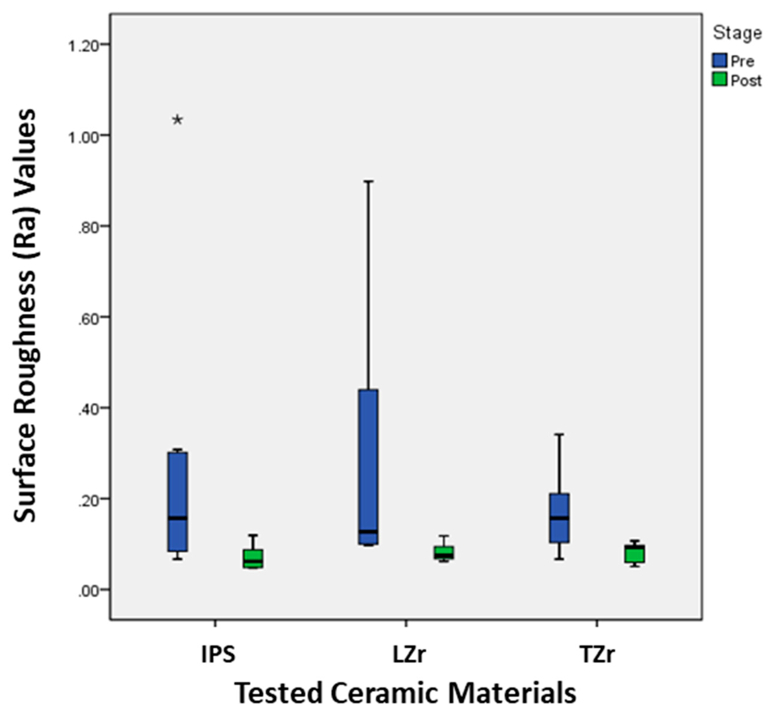
The bleaching of the three tested ceramics caused a significant increase in the Ra of these ceramics (
Table 2 and
Figure 5). The results revealed a significant increase in the Ra for all the tested ceramics after the bleaching process. Thus, it can be established that home bleaching will cause an increase in the Ra of the dental ceramics tested and make them more prone to plaque and bacterial adhesion.
The three tested ceramics’ surface porosities increased as a result of the bleaching process (
Figure 2). The SEM study of all three of the tested ceramics revealed changes in surface features with rising surface porosities, as shown in
Figure 2. It can be established that bleaching ceramic surfaces at home makes them more porous.
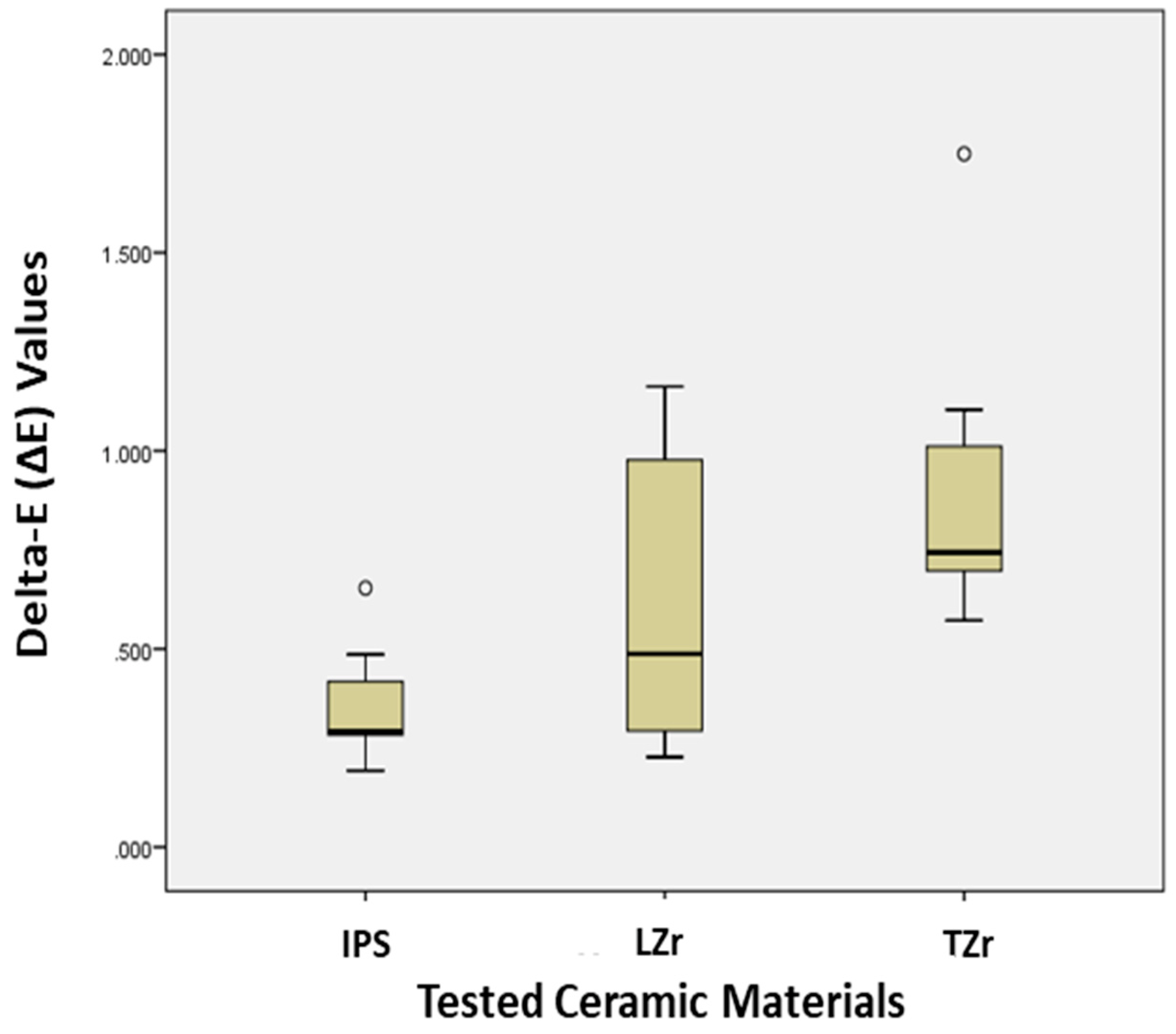
The results revealed statistically significant differences in the mean rank values (
p = 0.02) of the ΔE for the tested ceramics (
Figure 6). The post hoc test indicated that the mean rank values of the IPS material were significantly lower than the mean rank values of the TZr material (
p < 0.05), but not significantly different from the mean rank values of the classic LZr material (
p > 0.05). This indicated the presence of color differences between the different available ceramic materials.
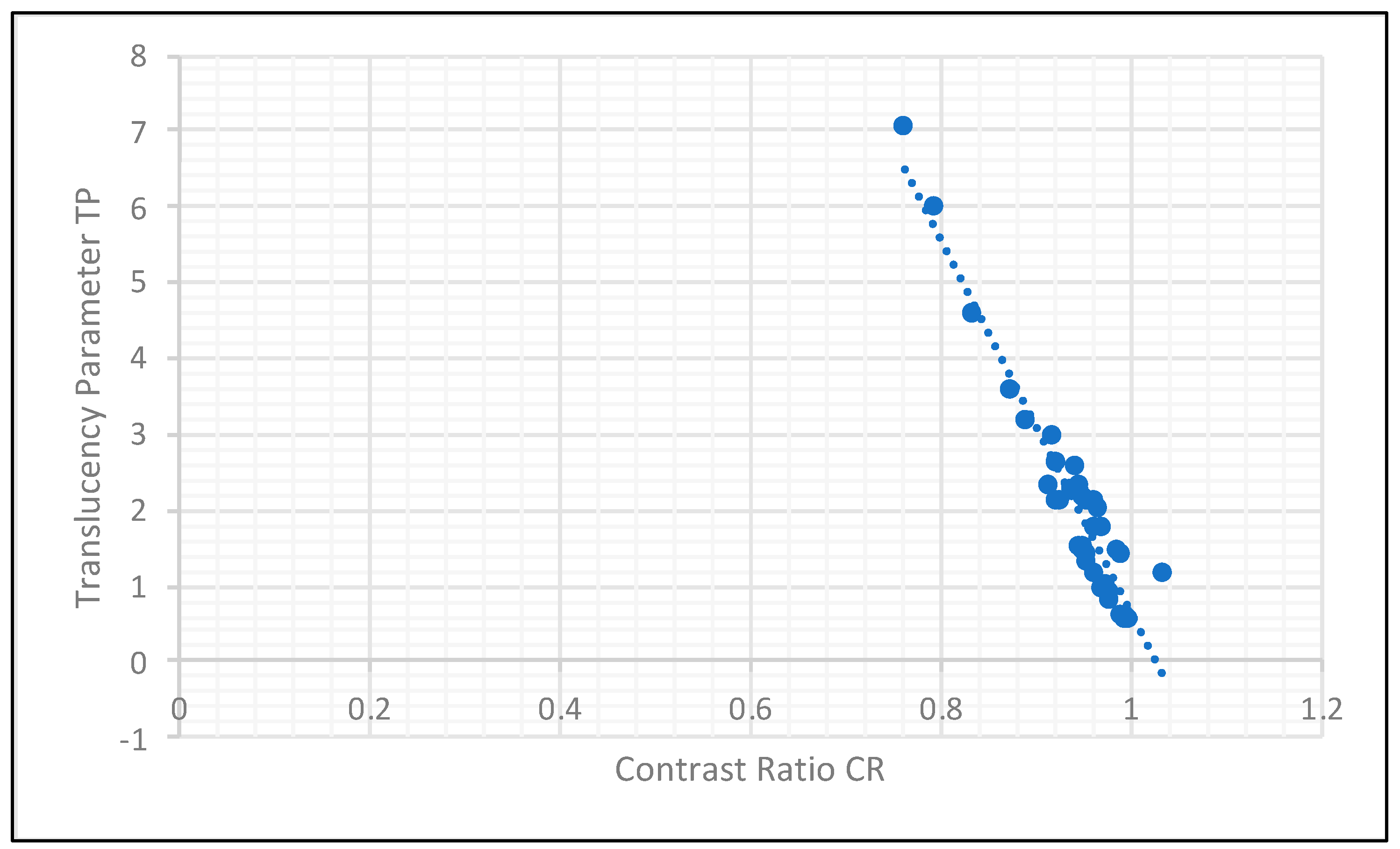
Spearman’s correlation between the differences in the pre- and post-values of the TP, CR, and Ra values in each of the three tested materials revealed a strong negative correlation between the translucency parameter and contrast ratio in all specimens tested (r = −0.92). That is, when the TP decreases, the CR increases (
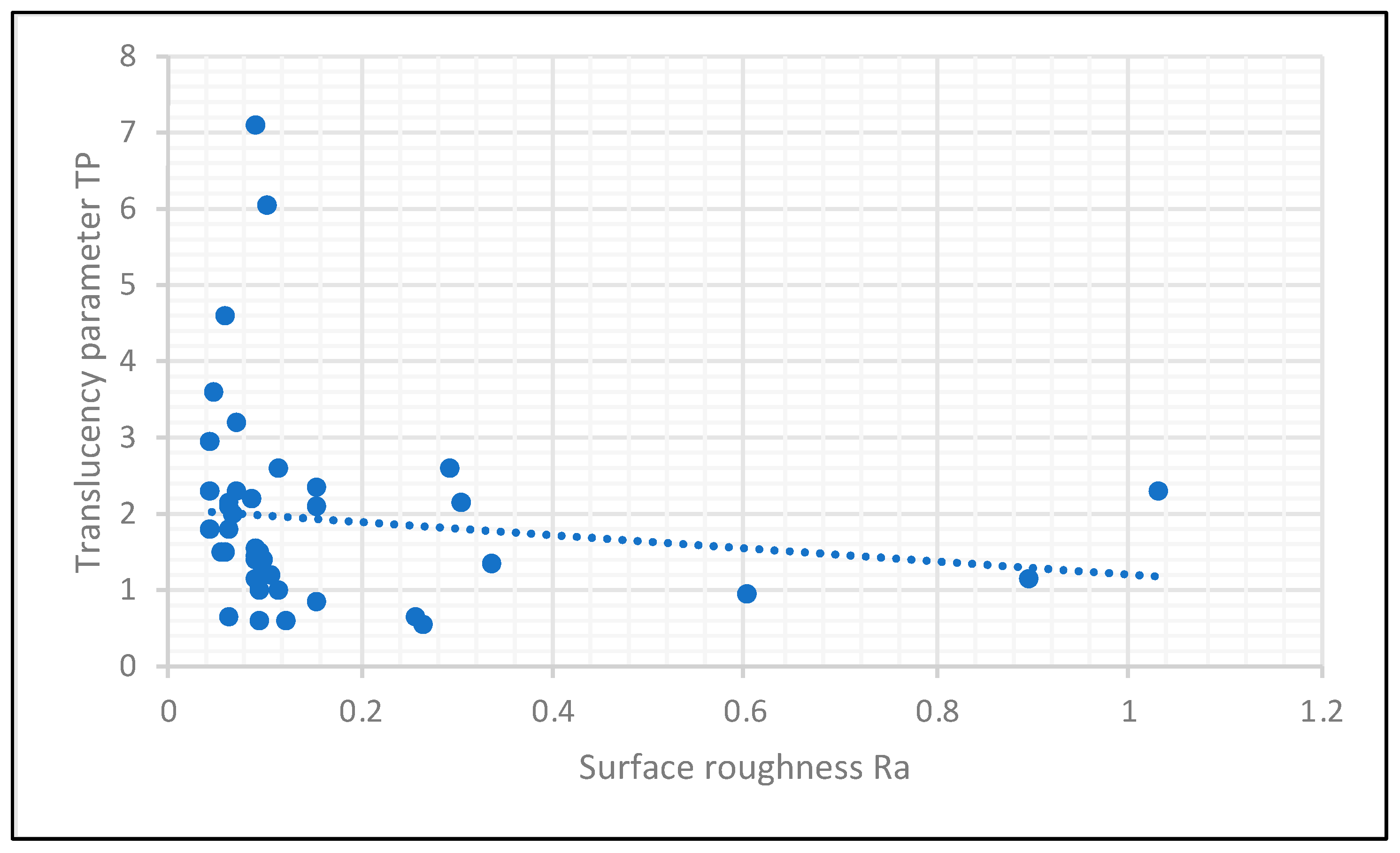
Figure 7). A similar pattern was observed between the surface roughness and the translucency parameter (
Figure 8). However, this negative correlation (r = −0.75) between Ra and TP values was mainly statistically significant in the LZr group.
4. Discussion
Home bleaching with 15% carbamide peroxide affected the optical properties (ΔE, TP, and CR) and increased the surface roughness (Ra) of the three tested aesthetic dental ceramic materials; therefore, the null hypotheses were rejected. The evaluation of the aforementioned optical properties was performed utilizing the CIE L*a*b* “Commission Internationale de l’Eclairage” numeric color system, in which the L*a*b* co-ordinates were obtained from a spectrophotometer. The use of a spectrophotometer for recording the CIE L*a*b* color co-ordinates has been widely accepted as a reliable tool in the field of color research of dental materials [
1,
10,
13,
15,
22,
23]. A recent study investigating the effect of home and office bleaching on the color of several CAD/CAM ceramic systems [
24] used the same color measurement tool as the current research and found a ΔE of 0.37 for the lithium disilicate samples (IPS e.max CAD) after bleaching with 20% carbamide peroxide, which is not too far from the observed color difference (ΔE = 0.29) in the current study for the lithium disilicate group (IPS e.max Press). The difference in the magnitude of ΔE between the two studies could be attributed to the difference in the concentration of carbamide peroxide used, as the current study used only 15% CP. The influence of home bleaching with carbamide peroxide concentrations close to the one used in the current study (15%) was investigated in another study by Karci and Demir, which focused on the translucency parameter and contrast ratio changes rather than the ΔE [
10]. The authors found significant changes in the TP and CR for the lithium disilicate material (IPS e.max CAD) after bleaching, which is different from what was seen in the present study [
10]. The lithium disilicate group (IPS e.max Press) in the present study did not demonstrate significant changes in the TP and CR. This disagreement between the two studies is not fully understood, as the crystals of both the IPS e.max Press and IPS e.max CAD are the same in composition, and both microstructures are 70% crystalline lithium disilicate; however, the size and length of these crystals are different, as stated by the manufacturer, which could partly explain the conflicting findings. On the other hand, the two zirconia materials (LZr and TZr) tested in the current study underwent significant changes in their optical properties after bleaching. The zirconia coping with feldspathic porcelain layering displayed the highest TP among the three materials before bleaching, which confirms the earlier findings of Heffernans et al. stating that conventional feldspathic porcelains are considered to exhibit higher TP values compared to other materials [
10,
25]. However, the TP values of the tested materials in the current study before bleaching were far less than some published values in previous studies [
10,
26,
27]. This could be attributed to the sample’s thickness of 3 mm in the present study as compared to a smaller thickness used in previous research (<2 mm), which is considered a limitation in the present study, as it is logical to notice color changes in materials if the thickness is decreased. In fact, Alkurt et al. found that the TP of the ultra-translucent multilayered zirconia with a 0.4 mm thickness was double that of the zirconia with a 1.5 mm thickness before bleaching (21.6 and 10.6, respectively) [
27]. Having said that, a significant decrease in the TP after bleaching was still noticeable in the present study for the LZr and the ultra-translucent monolithic zirconia (TZr) groups, which agrees with the earlier findings of Alkurt et al. who demonstrated a significant reduction in the TP for the monolithic zirconia materials tested after three days of bleaching. However, the authors demonstrated a later increase in the TP after extended periods of bleaching that lasted for seven and 14 days [
27].
Regarding the measurements and comparisons in relation to the opacity of the dental ceramics, recording of the CR is a viable option [
28]. According to Shono and Al-Nahedh, the CR and masking ability of a material are affected by the type of the dental ceramics used [
29]. This means there is variation in the CR between different dental ceramics. However, the results of our present study showed similar CR values between all the three tested ceramics before and after exposure to the home bleaching. The differences in the CR between the three tested materials was <0.06 and, according to Liu et al., only variations in CR values of >0.06 between ceramic restorations may be perceived by 50% of the population. Based on this information, although the difference in CR values observed between pre- and post-bleaching for the LZr and TZr was statistically significant, the resulting optical change might not be detected by the naked eye. This finding agrees with the earlier work of Karci et al., who found a CR difference <0.09 between pre- and post-bleaching readings for various dental ceramics [
10].
In the present study, the ceramic materials tested acted differently in response to the bleaching process, where the TP and CR values changed for the translucent zirconia and zirconia with feldspathic layering groups, whereas no significant change was found in the IPS e.max Press group. This finding suggests that the effect of bleaching is material-dependent. The modern-day glass ceramics are materials composed of a glass matrix and crystals. The dimension/size of these crystals, as well as their location and distribution, greatly affects the physical as well as optical properties of these ceramic materials [
10]. The LZr zirconia core contains a 3 mol% yttria partially stabilized tetragonal zirconia polycrystal with >15% cubic-phase zirconia, whereas TZr contains 5 mol% yttria partially stabilized zirconia with >50% cubic-phase zirconia [
2]. According to Zhang and Lawn, the incorporation of cubic-phase zirconia leads to an improvement in light transmittance. The IPS ceramic is composed of a homogeneous and dense distribution of lithium disilicate crystals that have a 70% crystal volume (Li
2SiO
3) embedded in a glassy matrix, and the diameter of the crystals is between 0.2 and 1 μm [
2,
10]. Generally, higher translucency is observed with a smaller crystalline ratio [
30]. The TP values observed in our study were higher for the IPS and LZr as compared to the TZr. It can be assumed that because of the smaller crystal size and ratio, these two materials performed better in terms of the TP [
10,
30].
In research related to dental color science, instruments that can calculate color co-ordinates and can help in finding the differences between them are commonly used [
15,
28]. Assessment of color is more than a numeric expression; usually, it is an assessment of the ΔE, which is used for the comparison between the colors of different objects. However, the perceptibility, acceptability, or rejection of the color differences between the dental materials may vary between different observers [
31]. Some researchers proposed a new color-difference formula that includes hue and chroma functions for accurate color discrimination [
32]. However, the use of the ΔE calculated from L*, a*, and b* values obtained from spectrophotometers is still considered as an accurate and viable method for quantifying and finding color differences between different dental materials [
15,
28]. The idea is that a ΔE of 1.0 is the smallest color difference the human eye can detect. Therefore, practically speaking, any ΔE less than 1.0 is not perceptible to the human eye [
32]. According to the results of this study, significant variations were found between the calculated ΔE values for the three tested ceramic materials. From a statistical point of view there is a significant difference, but from the clinical point of view, the color differences may not be significant as the differences in the ΔE for all the three tested dental ceramics are less than 1.0.
Additionally, the present study also evaluated the surface roughness and compared the Ra values of the tested dental ceramics before and after the bleaching, as the Ra is the most-used international parameter of roughness [
33]. The Ra parameters were recorded using a noncontact, 3D profilometer which offers an excellent resolution of the surface of the dental materials. Researchers have reported the use of the profilometer as the optimal technique, providing simple and practical methodology for the recording and evaluation of the Ra [
33,
34]. The surface roughness significantly increased in the present study after exposure of the tested ceramic specimens to a common home-bleaching material that contained 15% carbamide peroxide (CP), with Ra values in the range of 0.13–0.17 μm, which is close to the maximum value that is tolerable for a hard surface in the oral environment (0.18–0.2 μm) [
33]. The SEM analysis also confirmed the appearance of a rough porous surface after bleaching, indicating the penetration of the bleaching agent into deeper areas of the surface and resulting in an irregular surface with different levels of heights and depths. The results were in line with previous studies that confirmed an increase in the surface roughness of ceramics after bleaching [
35,
36,
37,
38]. The authors explained these higher roughness values to be due to the etching of the ceramic caused by the carbamide peroxide agent and have declared the influence of the bleaching agent to be linked to the depth of its penetration into the restorative materials [
35,
37,
38,
39]. Different penetration levels alter the surface topography of the dental ceramics, which may also affect the final color of the restorations [
35,
39]. With an increase in the surface roughness, the translucency of the material will be affected [
40] as the rough surface will cause variations in the light transmission and reflection [
32,
35,
37,
38,
39,
40]. A rough surface would also be more susceptible to staining as it has a higher potential for plaque accumulation, which could lead to discoloration [
16,
39]. Therefore, the surface roughness of the restorative materials plays a significant role in the appearance, and the changes in the optical properties observed in the present study (TP, CR, and ΔE) for the tested ceramic materials after bleaching may be the result of the increase in the surface roughness due to the changes in the surface topography caused by bleaching. Therefore, prior to the use of bleaching on patients with ceramic restorations in the aesthetic zone, clinicians should consider the effect of bleaching agents on the surfaces of these restorations. Ideally, the most up-to-date, novel aesthetic dental ceramic materials available on the market should possess similar optical and surface properties and should exhibit no changes in these properties when exposed to any external stimuli [
30].
The presence of saliva in the oral cavity plays a pivotal role in the appearance of restorative materials by adding glossiness and influencing the optical characteristics of the superficial surface of the materials [
10]. It also facilitates bleaching by increasing the wettability and dilution of the bleaching agent. Therefore, the lack of saliva in the current study may be considered an important limitation of the present investigation. Future studies with a larger sample size that simulate the oral environment are needed to evaluate the clinical implications of the current findings. Future work should also consider utilizing advanced surface analysis techniques, such as atomic force microscopy (AFM) and X-ray diffraction analysis (XRD), to provide detailed information about the crystallographic structure and chemical and physical changes that might appear on the surfaces of these ceramic materials after being treated with bleaching agents. Additionally, though, the literature has been enriched recently with related and comparable research studies that are similar to the present work. Another limitation of the current study, however, is that the available information is scarce when considering the vast number of new materials being launched into the market. Therefore, with the increase in the demand for highly aesthetic dental ceramics, new clinical studies and data are continuously required to monitor the consequences of external stimuli such as bleaching on the aesthetics of these new ceramic materials.
