Technical Note

Colorants Produced by Penicillium murcianum Are a Natural Moldicide against Trichoderma and Other Penicillium Species

by
Patricia Vega Gutierrez
1,*,
Vicente A. Hernández
2,3,
Nicole Sagredo
3 and
Seri C. Robinson
1,*
1
Wood Science and Engineering Department, College of Forestry, Oregon State University, Corvallis, OR 97331, USA
2
Facultad de Ciencias Forestales, University of Concepción, Victoria 631, Concepción 40730386, Chile
3
Centro de Biotecnología, University of Concepción, Barrio Universitario S/N, Concepción 40730386, Chile
*
Authors to whom correspondence should be addressed.
Coatings 2022, 12(6), 821; https://doi.org/10.3390/coatings12060821
Submission received: 16 May 2022 / Revised: 1 June 2022 / Accepted: 9 June 2022 / Published: 11 June 2022
(This article belongs to the Special Issue Science and Technology of Fabric Coatings)

Abstract

:
Mold growth is a continuing issue when it comes to human health, as well as a growing concern in localized wood decay, as numerous ‘traditional’ molds have been found to have soft rotting capabilities. Mold inhibitors on the market are often synthetic; however, the fungal kingdom has a wide range of more ‘natural’ options. Pigments produced by many fungi have been found to be toxic to other fungi, especially soft rotting fungi. This study looked at the pigments produced by Talaromyces australis (red) and Penicillium murcianum (yellow) and their effect upon the growth and pigment production of two species of Trichoderma and two species of Penicillium. Penicillium murcianum pigment inhibited growth and pigment production of all tested species at 3 mg/mL and higher. Results from this study indicate that P. murcianum colorants have the potential to inhibit growth and pigment production against other select ‘mold’ fungi. This holds potential not only for the wood preservation industry, but for the greater natural dye industry, especially in the area of antimicrobial textiles.

1. Introduction

Humans have a long history of using fungal pigments, both those derived from the fruiting form itself, and those produced by the hyphae directly. Some of the earliest uses include the red pigment from Monascus spp. for the red color in fermented rice products [1] and the blue-green spalted wood in German and Italian intarsia and marquetry, popular between the 1400s and 1600s [2,3,4].
While many fungi secrete extracellular pigments, the pigments vary in their structures and ‘abilities’. It is thought that many are anti-bacterial and anti-fungal, although some are known to play mechanical roles as well, such as protection from desiccation [5]. Many of the non-melanin based fungal pigments are quinones of some form, or very closely related. Some examples include yellow-brown fumigatin, a quinone from Aspergillus fumigatus Fresen [6,7,8,9], auroglaucin (orange-red), flavoglaucin (yellow), rubroglaucin (red) from Aspergillus glaucus (L.) [10], islandicin (green to red) from Penicillium islandicum Sopp. [11,12,13,14], xylindein (blue-green) from Chlorociboria species [15], and dramada (red-orange crystal) from Scytalidium cuboideum (Sacc. & Ellis) Sigler & Kang [16].
The antifungal properties of various Trichoderma species are so widely known that the pigments are utilized for applications such as textile dyeing [17,18], as well as Talaromyces australis C.M. Visagie, N. Yilmaz & J.C. Frisvad and Penicillium murcianum Ramírez & A.T. Martínez for wool textiles [19]. Antimicrobial pigments are of interest, especially to the textile industry, in terms of performance apparel that can prevent or minimize smells and potential fiber decay. Some fungal pigments, specifically the soft rotting ‘spalting’ pigments, have shown potential for colorfast textile dyes, such as the blue-green from Chlorociboria sp. and the red from Scytalidium cuboideum [20,21,22,23]. Some mold fungi have also been under investigation for textile dyes, among them Fusarium oxysporum Schlecht. emend. Snyder & Hansen) [24,25], Trichoderma virens [24], and Alternaria alternata (Fr.) Keissl. [17].
Mold fungi have traditionally been separated from decay fungi, although there is significant overlap between the two groups, particularly within the classification of soft rots (fungi that selectively decay the wood cell wall and cause localized damage) [26,27,28]. Specific pigments from soft rotting fungi are prized for their use in historic woodworks and are well known for their photostability. Unlike the stable blue-green of xylindein from the soft rotting fungi Chlorociboria species [15,29,30,31] or the melanin of soft rotting Xylaria polymorpha [32,33,34], the stability of these more traditional ‘mold’ pigments is understudied. Despite this, the benefit of using mold fungi pigments for dyeing instead of the more color-stable soft rotting pigments is in quantity generation. Mold fungi grow faster and produce more pigment in a shorter period than many soft rotting fungi [35]. There are also many more species to choose from. Hence, even though currently tested mold fungi may have less color stability, their growth rate and prevalence make them of continued interest. The purpose of this study was to look at the colorants produced by the common mold fungi Talaromyces australis (red) and Penicillium murcianum (yellow) for antifungal properties. If successful, the pigments, which have been tested for textile dying already (see previous citations), can move forward to testing for antimicrobial textiles. These colorants may also have use in wood preservation as a way to keep other mold fungi from potentially decaying wood in service.

2. Materials and Methods

2.1. Fungal Species

The fungal colorants used in this study were obtained from liquid cultures of Talaromyces australis C.M. Visagie, N. Yilmaz & J.C. Frisvad (strain TA2015) and Penicillium murcianum Ramírez & A.T. Martínez (strain PM2015). These microorganisms produced red and yellow extracellular colorants, respectively.
The cultures were obtained from Nothofagus sp. logs from central and southern Chile and isolated by the Hernandez lab in 2018. The extraction and quantification of the colorants followed protocols developed for textile dye production [19,28,36], i.e., cultivation in liquid media, followed by liquid/liquid extraction and drying to a powdered product. The colorants were re-solubilized to the five concentrations used in this study.
Four mold species were used: Penicillium verriculosum (strain M22), Penicillium magnanii (strain M38), Trichoderma sp. (strain M30), and Trichoderma sp. (strain M56). These species were selected based on their regular presence as a contaminating organism and their virulence. The species were sequenced, identified, and deposited in the publicly accessible Chilean Collection of Microbial Genetic Resources (CChRGM) of the Instituto de Investigaciones Agropecuarias (INIA Quilamapu, Chillán, Chile).

2.2. Incorporating Fungal Colorants to Culture Media

Colorants obtained from P. murcianum and T. australis were extracted with ethanol and then added to the media. A preliminary test identified that 1 mg/mL was the minimum concentration of colorants that exerted an inhibition effect on the test fungi. Consequently, five concentrations of colorant were used starting at 1 mg/mL with 1 mg/mL increments up to 5 mg/mL (Figure 1).
The dry colorants were diluted in 25 mL of ethanol and then added to 150 mL of 2% malt extract media. The media showed the typical color of each fungal colorant, red for T. australis and yellow for P. murcianum. The media was then autoclaved at 121 °C for 20 min, and 10 mL of media was distributed in Petri dishes 50 mm in diameter. Twelve 50 mm × 9 mm Petri dishes were prepared per concentration (Table 1).

2.3. Culturing and Monitoring

Three Petri dishes were cultured per concentration and three cultures without fungal pigment as the control. Each Petri dish was inoculated with a 3 mm diameter plug located in the center of the plate. The cultures were stored in an acclimatized chamber at 26 °C. The plates were incubated for 30 days. The growth area of the mycelium of each species tested was tested as the diameter of the area measured in millimeters in four stages: one day, three days, ten days, and thirty days after inoculation. This data was converted to an area in mm2. To compare the growth at the four stages of monitoring, the growth area was compared to the total area of the Petri dish, which was 1963.5 mm2. This value was analyzed in terms of the percentage of the total surface covered by the mycelium and based on the average of area per colorant concentration and colorant type.

2.4. Data Analysis

The dependent variable for this study was the growth of the mycelium of each of the four species tested, reported as an area in mm2. The independent variables were colorant type and colorant concentration. The colorant type was identified as PM for colorant obtained from P. murcianum and TA for colorant obtained from T. australis. Colorant concentration was expressed as 0 mg/mL for the control and from 1–5 mg/mL for the five concentrations used in this study. This data was analyzed using a two-way ANOVA, Tukey HSD, and Eta-Squared (η) for the effect size. The statistical analysis was conducted using R 4.2.0 [37] and RStudio 2022.02.0 [38].

3. Results and Discussion

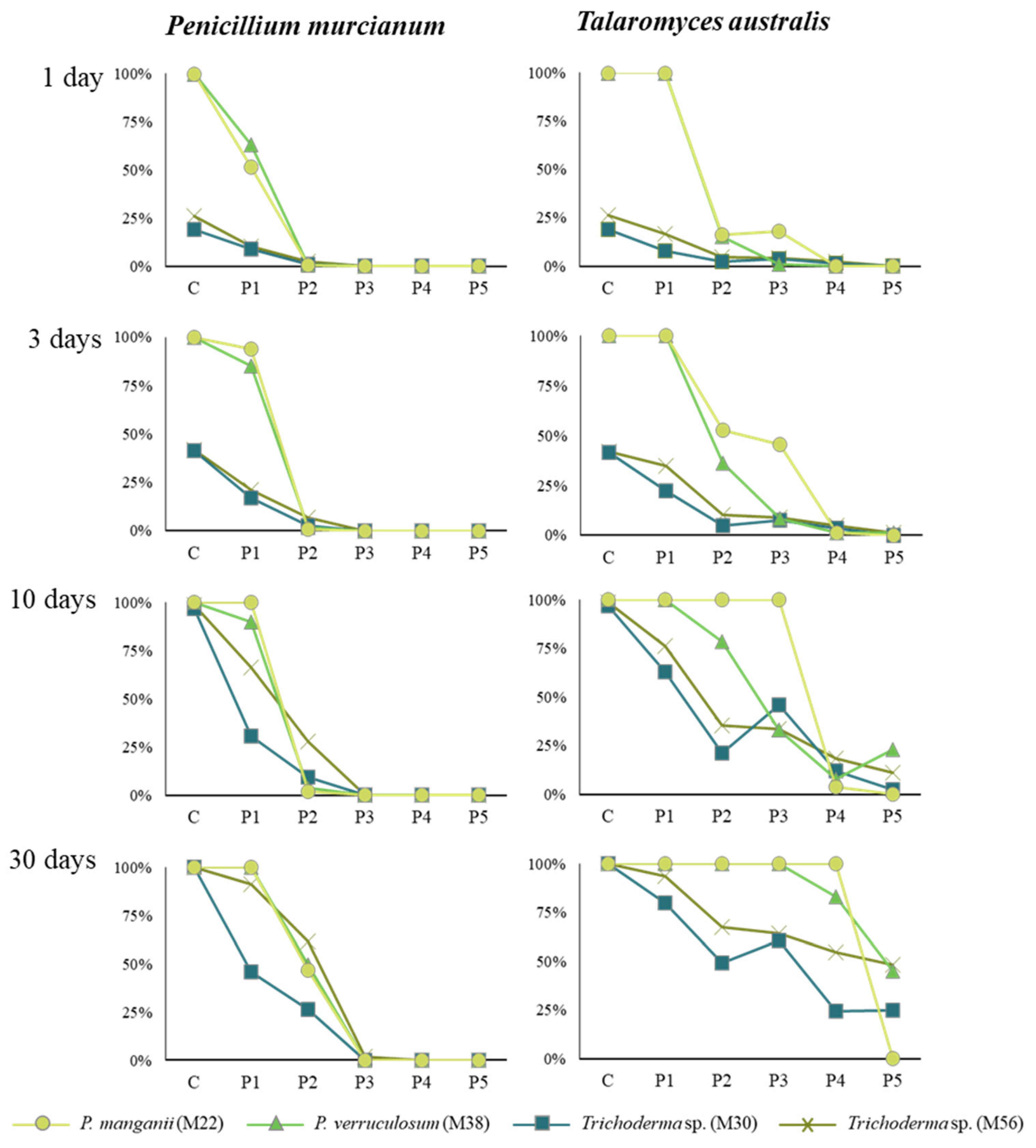
All controls reached 100% plate coverage, although at different rates: P. magninii at 10 days, P. verruculosum at 30 days, and both Trichoderma sp. at one day. This showcases the different growth rates of the fungi, as well as showing the health and pure monocultures of the samples. Statistical breakdowns of the data can be found in the Supplementary Materials, along with other relevant graphs and charts that detail the more minute details that would otherwise be redundant in the main text (Table S1, Figures S1 and S2).
The most significant inhibition came from treatments with P. murcianum (Figure 2). At concentrations of 3, 4, and 5 mg/mL this fungus completely inhibited the growth of all tested fungi. The pigment(s) produced by P. murcianum have not yet been elucidated, although they have been used in numerous textile dyeing tests [19,28]. The unpurified pigments, which likely contain other mycotoxins, were found to be suitable for contact with human skin but still biologically active [39]. Without further elucidation of the pigment(s) it is hard to draw conclusions as to the mechanism of inhibition. However, numerous other articles show that inhibition between Penicillium species can inhibit the growth of other fungal species ([39,40,41], etc.). The antifungal effects appear to be indicative of the genus, and as such, are not surprising.
Treatment with T. australis had no significant effect on P. magninii, P. verruculosum, and Trichoderma strain M56. It inhibited 55% of growth (allowing 45% of plate coverage) with the Trichoderma strain M30 (Figure 2). The results varied heavily according to the species and treatment. Unlike P. murcianum, which showed steady behavior across the four tested species, T. australis showed erratic inhibition. It did completely inhibit, after 30 days at the highest concentration, P. magninii. It did not have a statistically significant inhibitory effect on any other species. The uncharacterized pigment produced by this fungus appears to be less effective as a Penicillium inhibitor. This is likely due to Talaromyces being a teleomorph of many Penicillium species [42]. Some fungi, such as Xylaria polymorpha, are antagonistic towards their own species and even their own strains [32], while others seamlessly coexist. Being closely related may have affected the antifungal efficacy of T. australis.
In general, Penicillium magninii was highly resistant to T. australis. Only the highest concentration was successful in inhibiting the growth of this species. In contrast, the treatments with P. murcianum were highly effective at lower concentrations. P. verruculosum, showed a very similar behavior to P. magninii in terms of being inhibited by lower concentrations of P. murcianum. However, it was not inhibited by any treatment concentration of T. australis. It showed an inhibitory effect in that as the concentration increased the growth decreased, but not to the point of total inhibition. Even the highest concentration of T. australis could inhibit the growth of this species. These results show that P. murcianum has potential as an antimicrobial pretreatment, specifically against traditional ‘mold’ fungi.
It was noticeable that the properties of the fungal colorants used in this study remained vibrant after being exposed to high temperatures (121 °C) and pressure in the autoclave. This is not the case for the more commonly utilized soft rotting ‘spalting’ fungal pigments, which are known to decolor at high temperatures [43]. The water miscibility and the resistance to higher temperatures of these ‘mold’ pigments help counterbalance their poorer stability when compared to spalting pigments [44]. Similar to spalting pigments, the ‘mold’ pigments will need to eventually be formulated into inks if they are to be of use to the textile industry [45]. Unfortunately, many ‘mold’ fungi require mordants to permanently adhere to fabric [46,47,48], which is another hurdle that must be overcome.
Laboratory-level research has already shown P. murcianum to successfully dye wool and to be safe for contact with human skin [28]. At this stage, the efficacy of the pigment is less important of a question than production. As with the production of spalting fungal pigments, generating the volume of pigment needed for testing was difficult [49]. Although the tested fungi grow more quickly and produce more pigment than most spalting fungi, they still do not routinely produce ‘industrial’ amounts. Future work will need to develop liquid media batch reactors or other sources and continue to manipulate media to maximize pigment production so that the use of P. murcianum can move into industrial textile use. In the more immediate future, the effectiveness of P. murcianum to inhibit Trichoderma will need to be tested directly on fabric, to make sure the antifungal properties transfer across substrates. It is also possible than in vivo testing may require a lower concentration of pigment to enact antifungal properties, as many natural textiles, such as cotton and wool, have limited antimicrobial properties.

Supplementary Materials

The following supporting information can be downloaded at: https://www.mdpi.com/article/10.3390/coatings12060821/s1, Figure S1: The treatment with P. murcianum at concentrations of 3, 4, and 5 mg/mL were highly significant. The treatment of colorant from T. australis and a concentration of 5 mg/mL showed similar behavior. The concentration of the colorant shows a large effect (0.64), while the type of colorant and its interaction with the concentration had a medium effect (0.08 and 0.07 respectively). Below shows distribution of growth area at different concentration of colorants from T. australis (red) and P. murcinaum (yellow). The concentrations are Control = 0 mg/mL, P1 = 1 mg/mL, P2 = 2 mg/mL, P3 = 3 mg/mL, P4 = 4 mg/mL, P5 = 5 mg/mL. (a) Growth area distribution of P. magninii; (b) Growth area distribution of P. verriculosum, Figure S2: Growth Inhibition of Trichoderma sp. (M30). The colorant type, the colorant concentration, and their interaction were statistically significant strain M30 (S1). The treatment with P. murcianum and concentrations of 3, 4, and 5 mg/mL were strongly significant. The treatment with T. australis colorant at concentrations of 4 and 5 mg/mL where significant within the treatment, however not as significant as the treatment with P. murcianum colorants at similar concentration. The concentration of the colorant shows a large effect (0.68), while the type of colorant and its interaction with the concentration had a medium and small effect (0.06 and 0.02 respectively). Below shows distribution of growth area at different concentration of colorants from T. australis (red) and P. murcianum (yellow). The concentrations are Control = 0 mg/mL, P1 = 1 mg/mL, P2 = 2 mg/mL, P3 = 3 mg/mL, P4 = 4 mg/mL, P5 = 5 mg/mL. (a) Growth area distribution of Trichoderma sp. (M30); (b) Growth area distribution of Trichoderma sp. (M56); Table S1: Growth Inhibition of Penicillium magninii. The colorant type, the colorant concentration, and the interaction of both variables was statistically significant in inhibiting the growth of P. magninii. Two-way ANOVA results for the interaction of colorant and colorant concentration are shown below.

Author Contributions

Conceptualization, P.V.G.; methodology, P.V.G., N.S. and V.A.H.; formal analysis, P.V.G.; investigation, P.V.G. and N.S.; resources, V.A.H.; data curation, P.V.G.; writing—original draft preparation, P.V.G.; writing—review and editing, P.V.G. and S.C.R.; visualization, P.V.G.; supervision, V.A.H. and S.C.R.; project administration, V.A.H. and P.V.G.; funding acquisition, V.A.H. All authors have read and agreed to the published version of the manuscript.

Funding

This research was funded by the Project FONDEF—Chile, grant number ID15|10105 “Pigmentos Naturales de Alto Desempeño”.

Institutional Review Board Statement

Not applicable.

Informed Consent Statement

Not applicable.

Data Availability Statement

Not applicable.

Acknowledgments

The authors want to acknowledge the administration and research staff from the Center of Biotechnology of the University of Concepción—Chile (UDEC) for their support of this project at different stages. The authors also want to thank students José Muñoz and Isaac Saavedra from UDEC. V.H. and acknowledge the support from PAI Convocatoria Nacional Subvención a Instalación en la Academia 2018, 77180054 and Fodecyt 11180030.

Conflicts of Interest

The authors declare no conflict of interest.

References

  1. Erdoğrul, Ö.; Azirak, S. A review on the red yeast rice (Monascus purpureus). Turk. Electron. J. Biotechnol. 2004, 2, 37–49. [Google Scholar]
  2. Blanchette, R.A.; Wilmering, A.M.; Baumeister, M. The use of green-stained wood caused by the fungus Chlorociboria in intarsia masterpieces from the 15th century. Holzforsch.-Int. J. Biol. Chem. Phys. Technol. Wood 1992, 46, 225–232. [Google Scholar]
  3. Michaelsen, H.; Unger, A.; Fischer, C.-H. Blaugrüne Färbung an Intarsienhölzern des 16. bis 18. Jahrhunderts. 1992. Available online: http://www.openbibart.fr/item/display/10068/935304 (accessed on 9 April 2019).
  4. Robinson, S.C.; Michaelsen, H.; Robinson, J.C. Spalted Wood: The History, Science, and Art of a Unique Material; Schiffer Publishing: Atglen, PA, USA, 2016; 288p, Available online: http://www.schifferbooks.com/spalted-wood-the-history-science-and-art-of-a-unique-material-5920.html (accessed on 30 March 2017).
  5. Tudor, D.; Robinson, S.C.; Cooper, P. The influence of moisture content variation on fungal pigment formation in spalted wood. AMB Express 2012, 2, 1–10. [Google Scholar] [CrossRef] [PubMed] [Green Version]
  6. Waksman, S.A.; Horning, E.S.; Spencer, E.L. Two antagonistic fungi, Aspergillus fumigatus and Aspergillus clavatus, and their antibiotic substances. J. Bacteriol. 1943, 45, 233–248. [Google Scholar] [CrossRef] [Green Version]
  7. Waksman, S.A.; Geiger, W.B. The nature of the antibiotic substances produced by Aspergillus fumigatus. J. Bacteriol. 1944, 47, 391–397. [Google Scholar] [CrossRef] [Green Version]
  8. Packter, N.M.; Glover, J. Ubiquinone (50) and Ubichromenol in Aspergillus fumigatus, Fresenius. Nature 1960, 187, 413–414. [Google Scholar] [CrossRef]
  9. Packter, N.M. Studies on the biosynthesis of quinones in fungi. Incorporation of 6-methylsalicylic acid into fumigatin and related compounds in Aspergillus fumigatus IMI 89353. Biochem. J. 1965, 97, 321–332. [Google Scholar] [CrossRef] [Green Version]
  10. Gould, B.S.; Raistrick, H. Studies in the biochemistry of micro-organisms: The crystalline pigments of species in the Aspergillus glaucus series. Biochem. J. 1934, 28, 1640. [Google Scholar] [CrossRef] [Green Version]
  11. Howard, B.H.; Raistrick, H. Studies in the biochemistry of micro-organisms. 81. The colouring matters of Penicillium islandicum Sopp. Part 2. Chrysophanic acid, 4: 5-dihydroxy-2-methylanthraquinone. Biochem. J. 1950, 46, 49. [Google Scholar] [CrossRef] [Green Version]
  12. Howard, B.H.; Raistrick, H. Studies in the biochemistry of micro-organisms. 92. The colouring matters of Penicillium islandicum Sopp Part 4. Iridoskyrin, rubroskyrin and erythroskyrine. Biochem. J. 1954, 57, 212. [Google Scholar] [CrossRef] [Green Version]
  13. Takeda, N.; Seo, S.; Ogihara, Y.; Sankawa, U.; Iitaka, I.; Kitagawa, I.; Shibata, S. Studies on fungal metabolites—XXXI: Anthraquinonoid colouring matters of Penicillium islandicum sopp and some other fungi (–) luteoskyrin, (–) rubroskyrin, (+) rugulosin and their related compounds. Tetrahedron 1973, 29, 3703–3719. [Google Scholar] [CrossRef]
  14. Stark, A.A.; Townsend, J.M.; Wogan, G.N.; Demain, A.L.; Manmade, A.; Ghosh, A.C. Mutagenicity and antibacterial activity of mycotoxins produced by Penicillium islandicum Sopp and Penicillium rugulosum. J. Environ. Pathol. Toxicol. 1978, 2, 313–324. [Google Scholar] [PubMed]
  15. Saikawa, Y.; Watanabe, T.; Hashimoto, K.; Nakata, M. Absolute configuration and tautomeric structure of xylindein, a blue–green pigment of Chlorociboria species. Phytochemistry 2000, 55, 237–240. [Google Scholar] [CrossRef]
  16. Vega Gutierrez, S.M.; Van Court, R.C.; Stone, D.W.; Konkler, M.J.; Groth, E.N.; Robinson, S.C. Relationship between Molarity and Color in the Crystal (‘Dramada’) Produced by Scytalidium cuboideum, in Two Solvents. Molecules 2018, 23, 2581. [Google Scholar] [CrossRef] [PubMed] [Green Version]
  17. Sharma, D.; Gupta, C.; Aggarwal, S.; Nagpal, N. Pigment Extraction from Fungus for Textile Dyeing; NISCAIR-CSIR: New Delhi, India, 2012. [Google Scholar]
  18. Tuli, H.S.; Chaudhary, P.; Beniwal, V.; Sharma, A.K. Microbial pigments as natural color sources: Current trends and future perspectives. J. Food Sci. Technol. 2015, 52, 4669–4678. [Google Scholar] [CrossRef] [PubMed] [Green Version]
  19. Hernández, V.A.; Galleguillos, F.; Thibaut, R.; Müller, A. Fungal dyes for textile applications: Testing of industrial conditions for wool fabrics dyeing. J. Text. Inst. 2019, 110, 61–66. [Google Scholar] [CrossRef]
  20. Weber, G.; Chen, H.-L.; Hinsch, E.; Freitas, S.; Robinson, S.C. Pigments extracted from the wood-Staining fungi Chlorociboria aeruginosa, Scytalidium cuboideum, and S. ganodermophthorum show potential for use as textile dyes. Color Technol. 2014, 130, 445–452. [Google Scholar] [CrossRef]
  21. Hinsch, E.M. A Comparative Analysis of Extracted Fungal Pigments and Commercially Available Dyes for Colorizing Textiles. Ph.D. Thesis, Oregon State University, Corvallis, OR, USA, 2015. Available online: http://ir.library.oregonstate.edu/xmlui/handle/1957/55651 (accessed on 1 May 2017).
  22. Hinsch, E.M.; Weber, G.; Chen, H.-L.; Robinson, S.C. Colorfastness of Extracted Wood-staining Fungal Pigments on Fabrics: A new potential for textile dyes. J. Text. Appar. Technol. Manag. 2015, 9. Available online: https://ojs.cnr.ncsu.edu/index.php/JTATM/article/view/8018 (accessed on 25 September 2021).
  23. Palomino Agurto, M.E.; Vega Gutierrez, S.M.; Chen, H.L.; Robinson, S.C. Wood-Rotting Fungal Pigments as Colorant Coatings on Oil-Based Textile Dyes. Coatings 2017, 7, 152. [Google Scholar] [CrossRef] [Green Version]
  24. Nagia, F.A.; El-Mohamedy, R.S.R. Dyeing of wool with natural anthraquinone dyes from Fusarium oxysporum. Dye. Pigment. 2007, 75, 550–555. [Google Scholar] [CrossRef]
  25. El-Fakharany, E.M.; Hassan, M.A.; Taha, T.H. Production and Application of Extracellular Laccase Produced by Fusarium oxysporum EMT. Int. J. Agric. Biol. 2016, 18. [Google Scholar] [CrossRef]
  26. Galleguillos, F.; Hernández, V.; Figueroa, F.; Palfner, G.; Hernandez, V.A.; Robinson, S.C. Potential Use of Native Fungi for Value-Added Spalting in Chile. Prod. J. 2015. Available online: http://www.forestprodjournals.org/doi/abs/10.13073/FPJ-D-14-00093 (accessed on 12 April 2021). [CrossRef]
  27. Galleguillos, G.F.A. Hongos Asociados a Escarabajos Lignícolas en Bosque de Nothofagus su Aplicación en Control Biológico de Ophiostoma y en Spalting. Ph.D. Thesis, Universidad de Concepción, Concepción, Chile, 2016. [Google Scholar]
  28. Hernández, V.A.; Machuca, Á.; Saavedra, I.; Chavez, D.; Astuya, A.; Barriga, C. Talaromyces australis and Penicillium murcianum pigment production in optimized liquid cultures and evaluation of their cytotoxicity in textile applications. World J. Microbiol. Biotechnol. 2019, 35, 160. [Google Scholar] [CrossRef] [PubMed]
  29. Blackburn, G.M.; Todd, A.; Neilson, A. Structure of Xylindein. In Proceedings of the Chemical Society of London, Sheffield, UK, 4 April 1962. [Google Scholar]
  30. Edwards, R.L.; Kale, N. The structure of xylindein. Tetrahedron 1965, 21, 2095–2107. [Google Scholar] [CrossRef]
  31. Donner, C.D.; Cuzzupe, A.N.; Falzon, C.L.; Gill, M. Investigations towards the synthesis of xylindein, a blue-green pigment from the fungus Chlorociboria aeruginosa. Tetrahedron 2012, 68, 2799–2805. [Google Scholar] [CrossRef]
  32. Campbell, A.H. Zone lines in plant tissues I. The black lines formed by Xylaria polymorpha (Pers.) Grev. in hardwoods. Ann. Appl. Biol. 1933, 20, 123–145. [Google Scholar] [CrossRef]
  33. Bell, A.A.; Wheeler, M.H. Biosynthesis and functions of fungal melanins. Annu. Rev. Phytopathol. 1986, 24, 411–451. [Google Scholar] [CrossRef]
  34. Robinson, S.C.; Laks, P.E. Wood species and culture age affect zone line production of Xylaria polymorpha. Open Mycol. J. 2010, 4, 18–21. [Google Scholar] [CrossRef]
  35. ForIntek Canada Corporation. Discolorations on Wood Products. Forintek Canada Corp. 2002. Available online: https://cwc.ca/wp-content/uploads/assessingdecay-Discolorations_on_wood.pdf (accessed on 8 March 2022).
  36. Hernández, V.A.; Galleguillos, F.A.; Sagredo, N.; Machuca, Á. A note on the dyeing of wool fabrics using natural dyes extracted from rotten wood-inhabiting fungi. Coatings 2018, 8, 77. [Google Scholar] [CrossRef] [Green Version]
  37. R Core Team. R: A Language and Environment for Statistical Computing; R Foundation for Statistical Computing: Vienna, Austria, 2021; Available online: https://www.R-project.org/ (accessed on 1 May 2021).
  38. RStudio Team. In RStudio: Integrated Development Environment for R RStudio; PBC: Boston, MA, USA, 2022; Available online: http://www.rstudio.com/ (accessed on 1 May 2021).
  39. Marx, F.; Binder, U.; Leiter, É.; Pócsi, I. The Penicillium chrysogenum antifungal protein PAF, a promising tool for the development of new antifungal therapies and fungal cell biology studies. Cell. Mol. Life Sci. 2008, 65, 445–454. [Google Scholar] [CrossRef]
  40. Brown, A.G.; Smale, T.C.; King, T.J.; Hasenkamp, R.; Thompson, R.H. Crystal and molecular structure of compactin, a new antifungal metabolite from Penicillium brevicompactum. J. Chem. Soc. Perkin Trans. 1976, 1, 1165–1170. [Google Scholar] [CrossRef]
  41. Bladt, T.T.; Frisvad, J.C.; Knudensen, P.B.; Larsen, T.O. Anticancer and antifungal compounds from Aspergillus, Penicillium and other filamentous fungi. Molecules 2013, 18, 11338–11376. [Google Scholar] [CrossRef] [PubMed] [Green Version]
  42. Samson, R.A.; Yilmaz, N.; Houbracken, J.; Spierenburg, H.; Seifert, K.A.; Peterson, S.W.; Varga, J.; Frisvas, J.C. Phylogeny and nomenclature of the genus Talaromyces and taxa accommodated in Penicillium subgenus Biverticillium. Stud. Mycol. 2007, 70, 159–183. [Google Scholar] [CrossRef] [PubMed] [Green Version]
  43. Rodriguez de Oliveira, D.; Andersen, C.A.; Vega Gutierrez, S.M.; Kamke, F.A.; Robinson, S.C. Method of stabilizing heavily spalted big leaf maple as a decorative coating veneer layer for engineered wood flooring. Coatings 2019, 9, 132. [Google Scholar] [CrossRef] [Green Version]
  44. Hinsch, E.; Robinson, S.C. Comparing colorfastness of light of wood-staining fungal pigments and commercial dyes: An alternative method for colorfastness to light testing. Coatings 2018, 8, 189. [Google Scholar] [CrossRef] [Green Version]
  45. He, Y.; Cao, Y.; Hwang, H.-J.; Debarajb, H.; Vega Gutierrez, S.M.; Chen, H.-L.; Robinson, S.C.; Malhotra, R.; Chang, C.-H. Inkjet printing and in-situ crystallization of biopigments for eco-friendly and energy-efficient fabric coloration. Int. J. Precis. Eng. Manuf. Green Technol. 2021, 9, 941–953. [Google Scholar] [CrossRef]
  46. Räisänen, R.; Nousiainen, P.; Hynninen, P.H. Emodin and dermocybin natural anthraquinones as high-temperature disperse dyes for polyester and polyamide. Text. Res. J. 2001, 71, 922–927. [Google Scholar] [CrossRef]
  47. De Santis, D.; Moresi, M.; Gallo, A.M.; Petruccioli, M. Assessment of the dyeing properties of pigments from Monascus purpureus. J. Chem. Technol. Biotechnol. 2005, 80, 1072–1079. [Google Scholar] [CrossRef]
  48. Perumal, K.; Stalin, V.; Chandrasekarenthiran, S.; Sumathi, E.; Saravanakumar, A. Extraction and characterization of pigment from Sclerotinia sp. and its use in dyeing cotton. Text. Res. J. 2009, 79, 1178–1187. [Google Scholar] [CrossRef]
  49. Van Court, R.C.; Robinson, S.C. Stimulating production of pigment-type secondary metabolites from soft rotting wood decay fungi (“spalting” fungi). Adv. Biochem. Eng. Biotechnol. 2019, 169, 109–124. [Google Scholar]
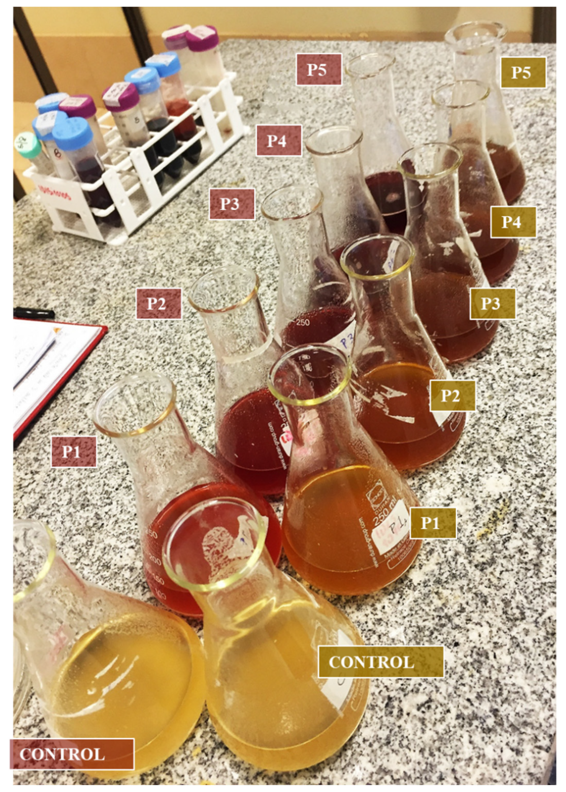
Figure 1. Dilutions of five different concentrations of T. australis (red labels) and P. murcianum (yellow labels). Control = 0 mg/mL, P1 = 1 mg/mL, P2 = 2 mg/mL, P3 = 3 mg/mL, P4 = 4 mg/mL, P5 = 5 mg/mL.
Coatings 12 00821 g001
Figure 2. Interaction of colorant concentration of P. murcianum and T. australis, and mean growth area percentage of four mold species after 1 day, 3 days, 10 days, and 30 days of inoculation. The concentrations of colorant were C = Control, P1 = 1 mg/mL, P2 = 2 mg/mL, P3 = 3 mg/mL, P4 = 4 mg/mL, and P5 = 5 mg/mL.
Coatings 12 00821 g002
Table 1. Treatments with five concentrations of colorants from Penicillium murcianum and Talaromyces australis incorporated in 2% MEA.
CodeConcentrationMold Species Tested at Each Concentration
Control2% MEAPenicillium magnanii (M22)
Penicillium verruculosum (M38)
Trichoderma sp. (M30)
Trichoderma sp. (M56)
P11 mg/mL colorant + 2% MEA
P22 mg/mL colorant + 2% MEA
P33 mg/mL colorant + 2% MEA
P44 mg/mL colorant + 2% MEA
P55 mg/mL colorant + 2% MEA
Publisher’s Note: MDPI stays neutral with regard to jurisdictional claims in published maps and institutional affiliations.