Article

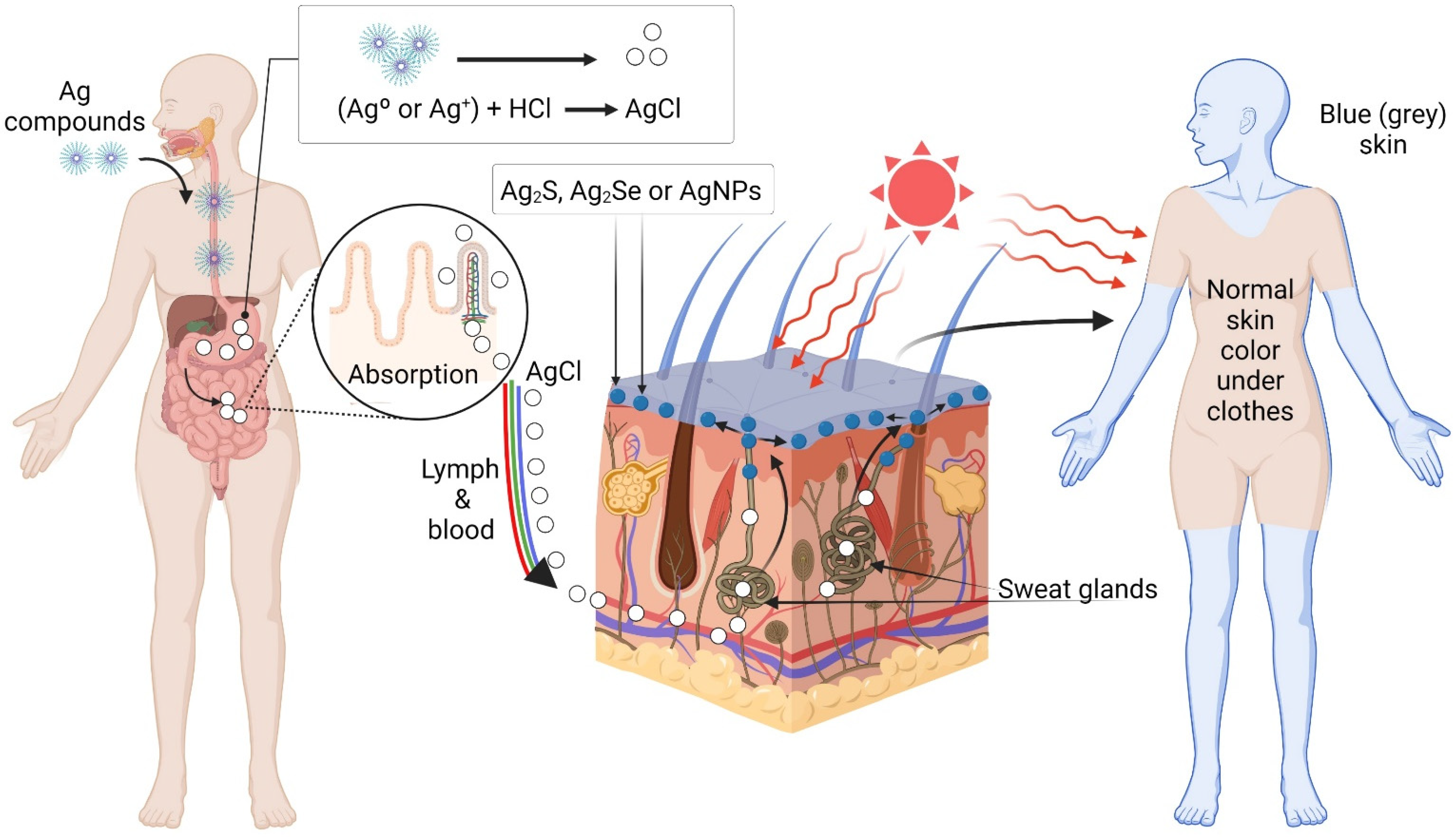
Hypothetical Mechanism of Skin Argyria

by
Vasily Burmistrov
1,
Alexander Burmistrov
2,
Galina Odegova
3,
Alexey Pestryakov
4,*,
Roberto Luna-Vázquez-Gómez
5,* and
Nina Bogdanchikova
6,*
1
Vector-Vita Ltd., 630098 Novosibirsk, Russia
2
Department of Mechanics and Mathematics, Computational Mathematics Section, Novosibirsk State University, 630090 Novosibirsk, Russia
3
Boreskov Institute of Catalysis, 630090 Novosibirsk, Russia
4
Research School of Chemistry and Applied Biomedical Sciences, Tomsk Polytechnic University, 634050 Tomsk, Russia
5
Escuela de Ciencias de la Salud, Universidad Autónoma de Baja California, Ensenada 22860, Mexico
6
Centro de Nanociencias y Nanotecnología, Universidad Nacional Autónoma de México, Ensenada 22860, Mexico
*
Authors to whom correspondence should be addressed.
Coatings 2022, 12(4), 532; https://doi.org/10.3390/coatings12040532
Submission received: 21 February 2022 / Revised: 3 April 2022 / Accepted: 8 April 2022 / Published: 15 April 2022

Abstract

:
Introduction. Argyria is an acquired skin condition that appears after the exposure or consumption of silver, leading to blue or grey coloration of the skin and mucosa. The aim of the present work was to draw researchers’ attention to two aspects of the argyria that until now have not received enough consideration. They are: (1) the process of delivering silver compound from the gastrointestinal tract to the skin and (2) the possibility for silver chloride to participate in this process along with the silver proteinates. Methodology. Illustrative experiments included the observation of color change (visual and using UV-Vis spectrometry) under different light exposure conditions of silver chloride sol in a sweat-simulating solution, in vials and under pig skin (in direct contact). Results and Discussion. A hypothetical mechanism based on a perspiration system for delivering the silver compounds from the gastrointestinal tract to the skin for argyria was proposed. It was also proposed not to completely exclude the partial participation of silver chloride along with the silver proteinates in this process.

1. Introduction

Currently, there is a notable increase in interest in preparations of silver nanoparticles. This is due to the extremely highly valued therapeutic properties inherent in modern nanosilver preparations [1,2,3,4,5,6,7,8,9,10,11,12,13]. For them, antiviral activity was revealed [6,14,15,16,17,18,19], which is especially important against the background of the emergence of new viral infections (atypical pneumonia, coronavirus infection). The anti-inflammatory and wound-healing effects of nanosilver have been shown [1,3,20]. The therapeutic efficacy of nanosilver in the treatment of drug-resistant forms of tuberculosis has been established [21,22]. The use of nanosilver preparations in the treatment of oncological diseases has been shown to be promising [7,11,23]. At the same time, many researchers and medical doctors note the insufficient knowledge about side effects and negative effects during the use of silver preparations.
One of the negative manifestations of silver preparation application is considered argyria or argyrosis [24,25,26,27]. The terms “argyria” or “argyrosis” (synonyms) were introduced into circulation at the beginning of the last century [1,28]. Patients with argyria usually does not have symptoms related to argyria. The main problem is the aesthetical nuisance, which can induce substantial embarrassment and social withdrawal [29]. When examining workers at silver mines and silver-processing enterprises, some workers who had worked at these enterprises for a long time (more than 15–20 years) experienced a change in skin color. Their skin acquired a bluish-grayish or bluish-brownish tone. Workers with skin argyria were thoroughly examined by doctors, but no significant pathologies or disruptions in the work of the internal systems of the body were identified. This color change was explained by the deposition of silver compounds (finely dispersed metallic silver, silver albuminates, or other silver compounds) in the skin. It was concluded that skin argyria is a consequence of the long-term intake of silver preparations into the body both from the inside (through the stomach) and from the outside (ingrained silver dust). Subsequently, cases of skin argyria were noted with the use of silver drugs (silver nitrate, collargol, and protargol) [30,31,32,33]. Thus, professional and medicinal skin argyria were distinguished [30]. The first is due to professional activity and is observed for persons who have been associated with the prolonged extraction and processing of silver ores and/or the production of different silver-containing compounds and products. It is important to mention that silver is used in the production of mirrors, silver-containing reagents, film and other chemicals for photographic processes, batteries, electrical equipment, coins, cutlery, jewelry, etc. Currently, due to the increased requirements for industrial safety and labor protection of workers, the risk of developing professional skin argyria was practically reduced to zero.
Medicinal skin argyria is associated with inadequate use of silver-containing drugs. Against the background of the growing interest in silver therapy and prevention, the likelihood of the emergence and development of drug-induced skin argyria increases. The mechanism of the appearance and development of drug-induced skin argyria is unknown. Many doctors treat skin argyria as an incurable disease with an unclear pathogenesis that is considered a serious risk factor for the manifestation and progression of various pathologies; skin argyria toxicity is also being actively discussed [34,35,36,37,38,39]. Nowadays, laser therapies are considered to be the most effective because they enable the almost complete removal of argyria pigment [40,41]. However, the main concern is severe pain accompanying the laser procedure and requiring anesthesia [40]. To prevent argyria, conservative options are recommended to avoid further exposure to Ag, as is sunlight protection [42].
The reason for the appearance and development of argyria is an excessive intake of silver to the body. Due to the increased interest in modern preparations of silver (in the form of silver nanoparticles), the processes of accumulation and distribution of silver in various internal organs have been studied in sufficient detail in vivo for certain silver- containing preparations [43,44,45]. For humans, studies dedicated to silver accumulation in internal organs are difficult for ethical reasons. In addition, there is evidence that the laboratory animal model does not fully reflect the complexity of the human skin argyria process. One such fact is skin pigmentation, which is observed in humans but not in laboratory animals. In other words, two aspects of argyria can be distinguished, conventionally designated as “internal argyria” (deposition of silver in internal tissues and organs) and “external argyria/skin argyria” (visually observed skin pigmentation). As already noted above, the deposition of silver in internal tissues and organs on laboratory animals was studied in many works [43,44,45] for different preparations of silver, and we will not discuss internal argyria in the present work. We only mention that in the case of internal argyria, silver can be deposited in the form of metallic silver nanoparticles, silver sulfide or selenide, silver complexes with protein thiol groups, etc. [46,47]. Unlike humans, skin pigmentation is not visually observed in common laboratory animals such as rabbits, guinea pigs, rats, mice, etc. (at least, we have not been able to find literature data on skin argyria pigmentation in common laboratory animals).
We found many publications in the literature dedicated to the study of the processes occurring with silver compounds in the human gastrointestinal system and in the human skin, i.e., in the initial stage of the appearance of silver-containing compounds in the body and in the last stage of their appearance on the skin, respectively. However, we could not find studies in the literature dedicated to the intermediate stage, which is delivering the silver compounds from the gastrointestinal tract to the skin. Neither could we find works exploring the possibility for silver chloride to participate in this process along with silver proteinates. So, the aim of the present work was to draw researchers’ attention to two aspects of the argyria that until now have not received enough consideration. They are: (1) the process of delivering silver compound from the gastrointestinal tract to the skin and (2) the possibility for silver chloride to participate in this process along with the silver proteinates.

2. Materials and Methods

As will be shown below, an essential place in our proposed mechanism is given to the process of perspiration. Since direct confirmation of the mechanism on a model of common laboratory animals (mice, rats, guinea pigs, etc.) is not possible, because these animals do not sweat, we used in vitro experiments simulating the stage of photoreduction. It is worth noting that some monkeys and apes, which have a developed sweating system, could be used as laboratory models for argyria studies. However, unfortunately they are much more expensive and require more maintenance than common laboratory animals such as rabbits, guinea pigs, rats, mice, etc.
As a solution simulating sweat, a mixture of physiological saline solution (0.8% NaCl, ReaChem, San Petersburg, Russia) and plasma-substituting solution Gemodez (JSC DalKhimPharm, Khabarovsk, Russia) was used in a ratio of 1:1. Gemodez is a drug prescribed as an intravenous infusion as a plasma-substituting and detoxifying agent. It is a water-salt solution containing 6% low molecular weight polivinilpirrolidone (PVP) (molecular weight of 12,600 ± 2700 Da) and sodium, potassium, calcium, magnesium, and chloride ions [48]. When AgNO3 (OlimpDM, Ekaterinburg, Russia) solution was added to this mixture (based on the final silver concentration of 1 mg/mL), a white silver chloride sol was formed. This sol was used as a solution simulating sweat with silver.
Experiment No. 1 “Effect of light exposure on silver chloride in solution”. The silver chloride sol was poured into 10 mL clear glass vials, which were hermetically sealed to prevent water evaporation. The concentration of silver in vials was 1 mg/mL. The vials were stored at room temperature under different light conditions: sample 1 was kept in the dark in a closed box; sample 2 was stored indoors in diffused sunlight away from direct sunlight; and sample 3 was exposed to direct sunlight. The solutions in vials were mixed (shaken) once a day to avoid the appearance of heterogeneity in color.
In order to reveal the effect of light irradiation on AgCl sol stabilized by PVP K-17 (Boai NKY Pharmaceuticals Ltd., Tianjin, China). The optical spectra were recorded in a UV-2501 PC spectrophotometer (Shimadzu, Kyoto, Japan) in the region of 300–900 nm. A 2 mm optical quartz cuvette was used. A silver chloride sol was prepared by mixing saline (0.85% NaCl, ReaChem, San Petersburg, Russia) and a solution of silver nitrate (OlimpDM, Ekaterinburg, Russia) with PVP (10 wt.%, Boai NKY Pharmaceuticals Ltd., Tianjin, China). A fairly stable suspension of milky-white silver chloride was formed. Then, this suspension was kept in sunlight.
Experiment No. 2 “Influence of light exposure on silver chloride under pig skin”. Pig skin was purchased from a butcher shop. The experiments were carried out at room temperature. During the experiments, signs of porcine skin decomposition were not observed. In this experiment, a 5 × 5 cm piece of raw pig skin was laid on top of cotton pad impregnated with silver chloride sol suspended in a sweat-simulating solution and remained for 5 days. Similar to the previous experiment, sample 1 was stored in the dark, sample 2 in diffused light, and sample 3 in direct sunlight.
Experiment No. 3. “Storage under different conditions of silver chloride sol in a solution simulating sweat”. Samples of silver chloride sol suspended in a solution simulating sweat were stored for three days under different conditions and with different concentrations of silver (Table 1).
Experiment No. 4. “Interaction of silver chloride sol with sodium thiosulfate solution”. A 30% solution of sodium thiosulfate was added to a solution of the “blue” sol of silver chloride; the solution became discolored and later transparent. Sodium thiosulfate is used as a complexing agent for silver compounds in photography.

3. Results

3.1. Experiment No. 1. Effect of Illumination on Silver Chloride in Solution

In Figure 1, photographs of samples of silver chloride sol after 7 days of storage under various conditions are shown. The initial colors of all three samples were the same as for sample 1 in Figure 1. After 7 days of storage in sample No. 1, which was kept in the dark, the colors of the solution had not changed. However, during storage, a change in the colors of the solutions in the samples stored in the light (samples 2 and 3) was noted: a slightly blueish-grey coloration with an increase in intensity during further storage for sample 2, stored in scattered light. Additionally, more intense brownish-grey coloration followed by further browning was observed for sample 3 stored in direct sunlight.
After sunlight irradiation of the solution prepared for the optical study, its color changed from white to purple. The optical spectra of AgCl sample before (dashed line) and after (solid line) sunlight irradiation are shown in Figure 2. Before irradiation, no peaks were observed, and only at lower than 400 nm the absorption increased. After irradiation, a peak with a maximum of 380 nm, and a broad peak with a maximum at 580 with a shoulder at 475 nm, appeared. The emergence of peaks with such characteristics indicates that under the influence of light, reduction of AgCl and the formation of AgNPs of various sizes are observed [49]. In this case, the presence of three absorption maxima indicates the formation of aggregates or large particles in the form of an ellipsoid [50,51,52].

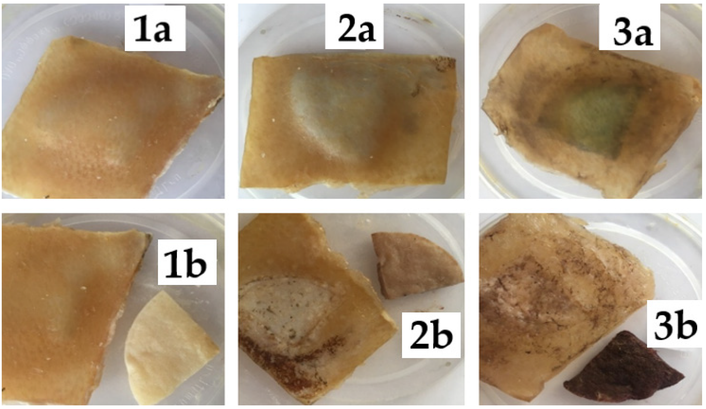
3.2. Experiment No. 2. Effect of Illumination on Silver Chloride under Pig Skin

In Figure 3, photographs of three samples after 5 days of storage are shown. Sample 1 (maintained in the dark) did not change its color, sample 2 (kept in diffused sunlight) turned slightly blue, and in sample 3 (kept in direct sunlight) the blue color change intensified. This experiment gives evidence that the pig skin does not protect the silver chloride sol from sunlight. The similarity of the color of the pig skin samples 2 and 3 with the color of the skin of patients with skin argyria is noted.

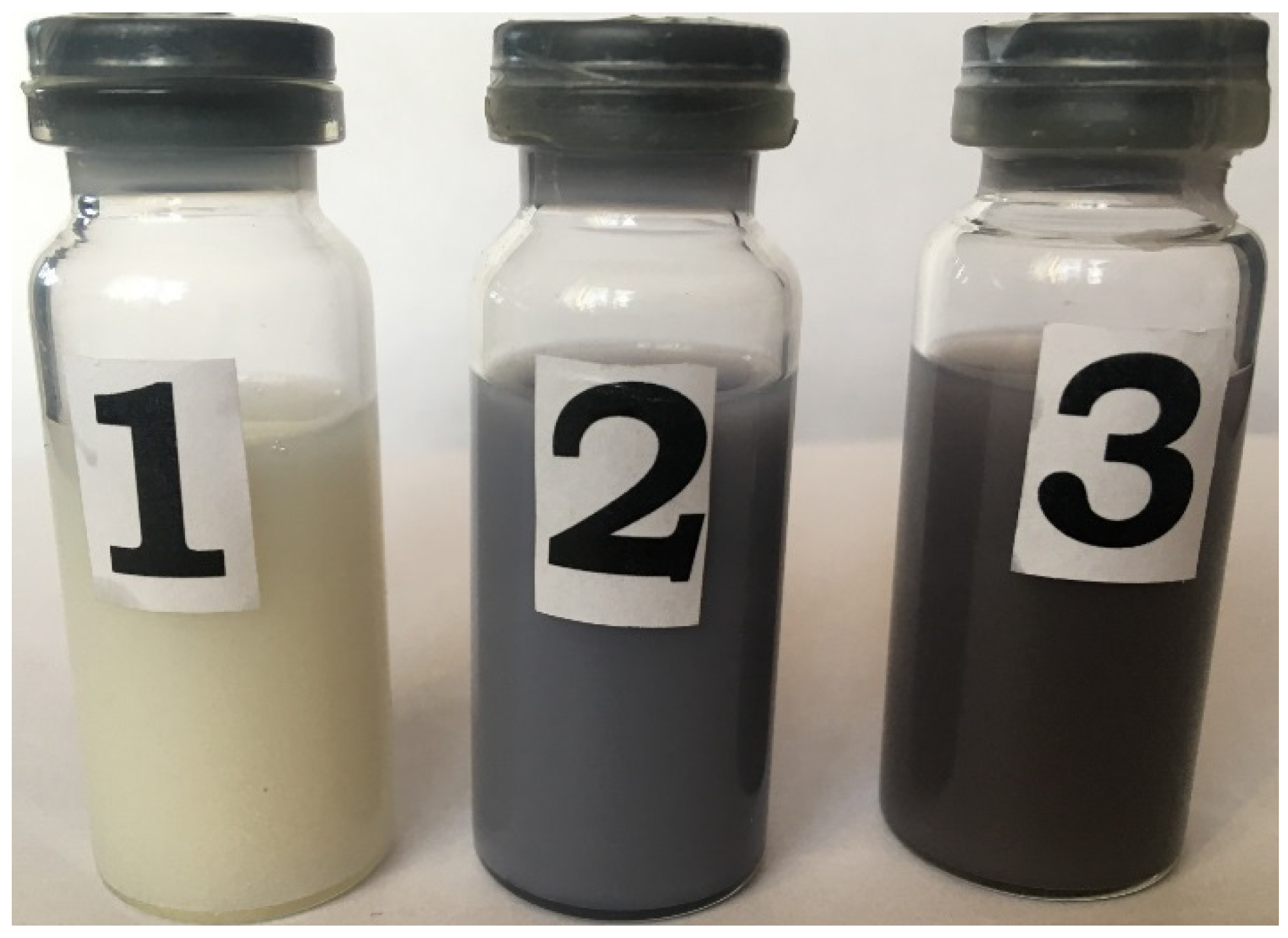
3.3. Experiment No. 3. Storage under Different Conditions of Silver Chloride Sol in a Solution Simulating Sweat

In Figure 4, photographs of samples of silver chloride sol in a solution simulating sweat, stored for three days under different conditions and with different concentrations of silver, are shown. The concentration and storage conditions are given in the Table 1. An increase in the color saturation of the solution is clearly seen, with an increase in the duration of exposure to sunlight (compare samples 0–3). The direct dependence of the color saturation of the solution on the concentration of silver is also noticeable (compare samples 3–4).

3.4. Experiment No. 4. “Interaction of Silver Chloride Sol with Sodium Thiosulfate Solution”

After adding 30% sodium thiosulfate solution to the blue sol of silver chloride solution, the solution became colorless and transparent, indicating that that sample color changes were due to the formation of reduced silver species. Sodium thiosulfate is used as a complexing agent for silver compounds in photography. It is also approved for use in medicine [48]. This experiment showed that 30% solution of sodium thiosulfate can be used for the elimination of undesirable skin coloration caused by silver particle formation.

4. Discussion

First, the cases of drug-induced skin argyria caused by long-term intake of silver preparations in the literature were analyzed. Due to this meta-analysis, the following key factors were identified:
  • Skin argyria is caused by the appearance in the surface layers of the skin, mainly in the dermis, of highly dispersed submicron particles of metallic silver [30,38,39,40,53,54].
  • Silver particles are predominantly located near the mouth and in the area of the sweat gland membranes [39]. This indicates that the delivery of silver compounds (from which metallic silver particles are formed) directly to the skin occurs along with sweat through the perspiration system.
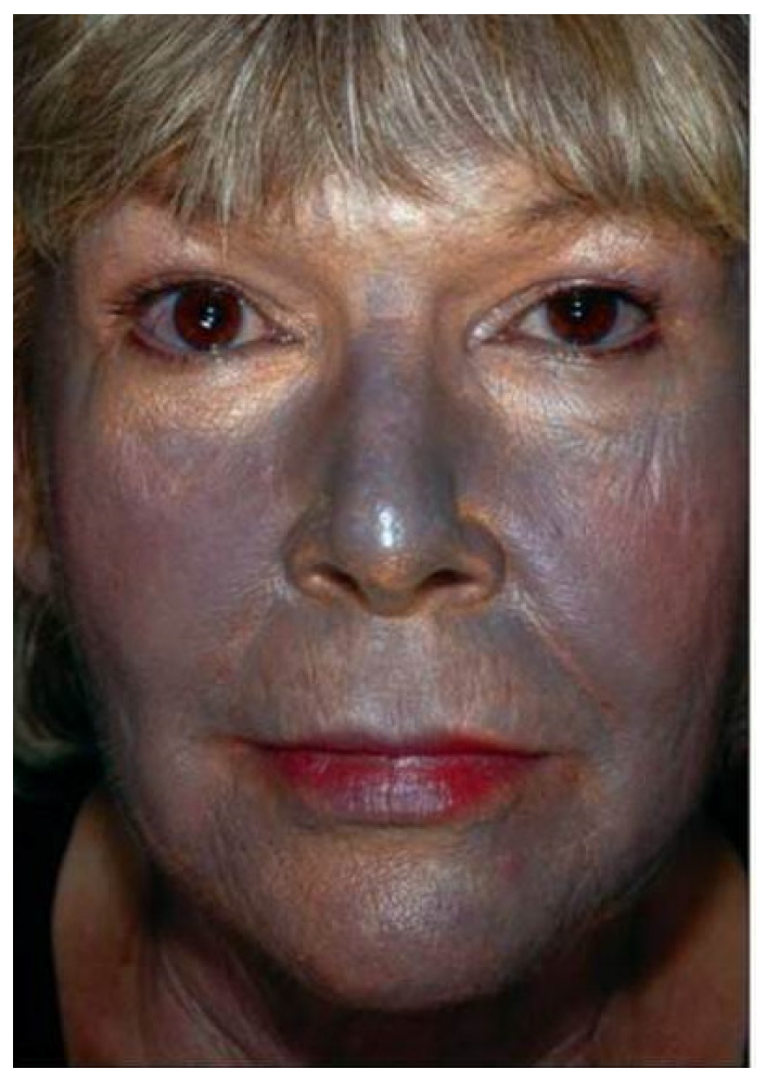
  • Skin discoloration was observed predominantly on the body areas exposed to direct sunlight. The skin on the parts of the body covered by clothing (buttocks, thighs, etc.) retained its natural flesh color [40]. This indicates that sunlight is involved in the emergence and development of skin argyria.
Additionally, the factor of high photosensitivity of ionic compounds of silver, namely, silver halides, was taken into account. Silver halides can be reduced by light to form clusters and particles of metallic silver. All classic silver photography is based on this [40,54,55,56,57,58].
Taking into account all these factors, the hypothetical mechanism of the appearance and development of skin argyria is suggested here. It involves the release of ionic silver in the form of a complex of silver chloride together with sweat into the near-surface skin layers and the subsequent reduction of ionic silver under the influence of sunlight, therefore forming metallic silver particles. The resulting silver particles cause skin coloration. We have chosen AgCl as an object of this study. All other silver compounds, which can be produced in the human body, such as complexes of AgNPs with protein thiol groups or nanoparticles of silver sulfides and selenide, etc., cannot be candidates because all these compounds are not decomposed under sunlight, which produces skin darkening, while AgCl is easily decomposed by light into metallic silver nanoparticles, which are dark colored. The reported experimental facts indicate that skin darkening in the case of argyria is observed predominantly on the body areas exposed to direct sunlight (hands, neck, and face). Among all silver containing compounds produced in the human body after silver consumption, silver chloride is the only light-sensitive compound and therefore is one of the most probable candidates for elucidating skin argyria mechanism.
The choice of a silver chloride sol as a model for silver-containing sweat is based on the following reasons. First, silver chloride is photosensitive. Secondly, chloride ions are present in all body fluids: blood, lymph, urine, gastric juice, sweat, tears, etc. For example, in human plasma the concentration of chloride ions is 95–110 mM [59], which is ~200 times higher than the concentration of total reduced thiols (~0.4–0.6 mM [60], which suggests that the interaction of silver compounds with chloride ions should not be excluded from the consideration of the process of delivering the silver compound from the gastrointestinal tract to the skin. AgNPs formed from AgCl under light should be considered, along with the accepted biochemical model of silver sulfide and silver selenide argyrial deposits [61].
When an ionic silver preparation, for example, silver nitrate, enters the stomach, silver chloride will be formed [62]. Note that skin argyria most often was observed when silver nitrate was used [1,30,32,63,64]. In the case of colloidal silver and silver nanoparticles, which slowly release silver ions into solution, these ions will also interact with chloride ions, forming silver chloride, that is, at least part of the silver will convert into silver chloride. Silver chloride is slightly soluble in water, but when obtained in situ, it easily forms highly dispersed stable sols, the stability of which increases in the presence of organic polymers (proteins, polypeptides). In addition, the solubility of silver chloride greatly increases in the presence of complexing agents in the solution, such as ammonia hydrate and excess chlorides [62].
We could not find any scientific publication describing the blue color of AgCl decomposed under light; instead, black and grey colors as in black and white photography are described. However, it is known that individuals with argyria have not only grey but also blue color skin (Figure 5).
Our experiments showed that after light exposure in sweat simulating solution medium and in cotton pad impregnated with this solution and placed under pig skin, blue color appeared (samples 2 and 3 of Figure 3 and samples 1 and 2 of Figure 4). So, it is interesting that AgCl under light radiation can obtain not only grey or black color but also blue color. How can this be explained? Ag/AgCl crystals stabilized by polivinilpirrolidone (PVP) as a capping agent and PVP and glycerol as reducing agents were formed as materials with distinct apparent colors (blue and fuchsia) [65]. It was revealed that the surface concentration of metallic silver measured by XPS was relatively low: 3.7 and 5.9% in AgNPs/AgCl-blue and AgNPs/AgCl-fuchsia, respectively [65]. Fuchsia color practically corresponds to body skin color, while blue color can be easily distinguished on the skin. So, in the presence of stabilizers such as PVP or proteins (in the case of skin), Ag produced from a reduction of AgCl can have blue and fuchsia colors. All abovementioned information suggests that AgCl participates in the development of the blue skin color characteristic in argyria.
Human sweat contains chlorides of sodium, potassium, magnesium, calcium, as well as urea, ammonia, lactic acid, uric acid, amino acids, fatty acids, proteins, polypeptides, pheromones. It also contains various toxic metabolites, as well as salts of trace metals such as iron, zinc, copper, nickel, cadmium, lead, and mercury [66,67]. For many of metals, sweating is an important mechanism for removing them from the body. When silver compounds enter the body, it is logical to assume that silver, like other metals, represents part of sweat, presumably in the form of a complex of silver chloride with sweat components and/or in the form of a sol. The uniqueness of silver in comparison with other metals lies in the high photosensitivity of its compounds.
According to literature data, the main part of silver is removed from the body by the liver and kidneys in urine; silver is also excreted in feces [1,3,30,36,68,69]. However, we note that this conclusion was made on the basis of the results obtained on laboratory animals that do not have a developed perspiration system, that is, on animals that do not sweat. The human body has a developed sweating system. The main function of sweating is body thermoregulation; an additional function is toxin elimination. Under normal conditions, at room temperature, a person secretes an average of 400–600 mL of sweat per day [66,67]. In hot weather, as well as during hard physical labor, the production of sweat increases to one or more liters per hour. This significantly exceeds the volume of urine. The amount of toxins released in sweat, including silver, can be significant. However, for a full study of the pharmacokinetic processes of silver release from the body, it is desirable in further experiments to use experimental animals with a developed sweating system, as in humans. It is also possible to involve volunteers (patients with skin argyria).
When a silver complex in sweat enters the near-surface layers of the skin, under the influence of sunlight, the ionic silver is reduced, forming the nuclei of metallic silver particles. With the further supply of ionic silver, these particles gradually grow, are visualized (appear, as in the development of a photograph), and the skin acquires the corresponding color. The color of the particles depends on the composition, size, shape, and ligand environment of the particles, and a number of other parameters [70]. The similarity of the skin coloration of patients with skin argyria and the coloration of silver chloride sol solutions after exposure to light indicates the identity of the formed silver particles in both systems. This indicates the reliability of the proposed mechanism for the emergence and development of skin argyria.
In Figure 6, a scheme of a hypothetical process of delivering silver compounds from the gastrointestinal tract to the skin for argyria is presented. First, silver compounds are orally consumed by an individual. In the stomach, these compounds (silver salts or nanoparticles) interact with HCl and are converted into AgCl and pass into the intestine, where they are absorbed into blood and lymphatic fluid as Ag-proteinates and AgCl. Blood and lymphatic fluid deliver these silver compounds into sweat glands. From sweat glands, they move through sweat pores with sweat fluid to the skin surface, converting into grey and blue compounds (Ag2S, Ag2Se, and AgNPs) under light irradiation.
The content of silver in the body depends on the ratio of the velocity of introduction of silver into the body and the velocity of its elimination from the body. With an increase in the velocity of introduction, the velocity of elimination of silver also increases. The body strives to maintain homeostasis. For maintaining homeostasis, apart from the main route of elimination of silver, which includes liver and feces, the route of elimination through the sweating system starts to work.
To prove the hypothetical mechanism of the process of delivering the silver compounds from the gastrointestinal tract to the skin for argyria, further studies of biopsy samples of the skin of volunteers with argyria should be done. On the one hand, biopsy microscopic analysis will allow for studding localization of argyria deposits close to the output or inside sweat glands. If argyria deposits are located predominantly there, this will confirm their main role in the process of delivering the silver compound from the gastrointestinal tract to the skin for argyria. On the other hand, analysis with an electron microscope with z-contrast will allow one to measure the chemical composition of these deposits (Ag2S, Ag2Se, AgNPs, etc.). It will allow one to estimate the relative contributions of Ag sulfides, Ag selenides, and Ag nanoparticles in argyria deposits.
Skin argyria implies the formation of submicron particles of metallic silver as a result of photoreduction. The formed metal particles, due to their size, can no longer diffuse through the epidermis and are localized at the place of their origin. If you protect your skin from sunlight (wearing a veil, hijab, gloves, dark glasses, etc.), then skin argyria will not develop. Ionic silver, along with sweat, will pass through the epidermis, and it will be safely washed off the skin during bath. Basically, skin argyria is just a kind of natural tattoo with submicron particles of metallic silver and can be perceived as a cosmetic defect. This silver, which has already been excreted from the body, does not practically participate in metabolism and can no longer harm the body. Thus, skin argyria is not a disease, it is an indicator of an overdose of ionic silver in the body that happened in the past. In other words, skin argyria itself does not pose any danger to the body, except for the appearance of psychological discomfort. However, an excessive intake of silver (overdose) can represent a health hazard. If ionic silver was not highly light sensitive, then no skin argyria problem would exist, but overdose problems would remain. For the diagnosis of silver overdose, there are objective indicators, in particular, a quantitative analysis for the silver content in blood.
In addition to danger of overdose, there is the danger of the inherent toxicity of the specific silver formulation. Silver formulations differ greatly in toxicity depending on the type, composition, production method, state of silver, and other parameters [1,2,3,35,69]. The most toxic formulations are those of ionic silver, and the least toxic formulations are those of silver nanoparticles containing highly dispersed particles of already reduced metallic silver [3]. At the present time, the design of safe, non-toxic, and effective silver nanoparticle formulations is conducted all over the world.
The high photosensitivity of silver compounds appearing in the process of metabolism and present in biological fluids and organs inside the body can produce artifacts in the results of laboratory studies, in particular, during biopsy and histological studies. The technology of working with light-sensitive materials has been known for a long time, and it has been well developed in classic silver photography. The work is carried out in a room protected from sunlight under artificial red light. The energy of red light is insufficient to reduce silver halides. It is possible that the same protective measure (red light application) will be required when studying the metabolic processes of silver formulations in the body.
The silver-removing ability of the sweat system can be used to detoxify the body by stimulating intense sweating (diaphoresis) such as those experienced in sauna, during intensive exercise, or during heat exposure. Naturally, this should be done under conditions of protection against photoreduction, that is, under red light.
The number of people with skin argyria is small, in fact, these are isolated cases, which are described in the literature in most publications [71,72,73]. That is why it is difficult to study skin argyria systematically, according to classical methods—with an experimental group, a comparison group, a placebo, etc. Nevertheless, a logical question arises: why did people taking silver formulations not stop taking them even after the first signs of skin argyria appeared? A critical analysis of cases with skin argyria showed that these people had health problems even before taking silver [38,39]. They tried to solve them, turning to conventional medicine, but it was without success. This prompted them to start taking silver preparations, which, according to their subjective feelings, helped them. In the hope of obtaining a greater positive effect, the periods of treatment were prolonged and the dosages were increased, but the situation was not controlled by medical personal.
As was shown in experiment No. 4, after adding sodium thiosulfate the blue color of the silver chloride sol disappeared. This discoloration indicates the possibility of using a sodium thiosulfate solution to remove skin argyria, for example, in the form of subcutaneous microinjections (mesotherapy). However, literature data show that the application of sodium thiosulfate in patients was not successful [74] or was partially successful [75]. So, this issue requires further experiments, taking into account safety and possible risks. Note that sodium thiosulfate solution is approved for use in medicine in the form of intravenous injections and oral administration [48].
Skin argyria is often perceived by many doctors as something terrible, incomprehensible, and impressive. This greatly prevents the introduction of modern silver preparations into medical practice. The results of the present work contribute to elucidate the mechanism of development of skin argyria, which can be considered as cosmetic defect. While internal argyria should draw the main attention for the toxicological studies, such as silver accumulation and removal from organs.

5. Conclusions

Earlier, the study of the mechanism and development of skin argyria, including the perspiration system, was impeded due to the following circumstances. On the one hand, the number of people with skin argyria is small, and therefore in the literature usually only isolated cases are described. On the other hand, the systematic study of a model of common laboratory animals (mice, rats, guinea pigs, etc.) is not possible because these animals do not sweat. The present work attracts researchers’ attention to two aspects of the argyria that until now have not received enough consideration: (1) the process of delivering silver compounds from the gastrointestinal tract to the skin, including the perspiration system; and (2) the possibility for silver chloride to participate in this process along with the silver proteinates. In this work, the hypothetical mechanism, based on the participation of the perspiration system for the elimination of silver from the human body and the ability of silver chloride to participate in this process, is suggested. This mechanism includes: (1) the inclusion of ionic silver in the composition of sweat and transportation to the near-surface layers of the skin through the perspiration system; (2) the reduction of ionic silver under sunlight in the surface layers of the skin with the formation of clusters and silver nanoparticles; (3) the growth and visualization (development) of silver particles, which causes a change in skin color. The proposed mechanism does not contradict but complements the accepted biochemical model of silver sulfide and silver selenide argyrial deposits produced from Ag-proteinates. The proposed hypothetical model should be experimentally verified in further experimental approaches.

Author Contributions

Conceptualization, V.B. and N.B.; methodology, V.B., A.B. and G.O.; validation, V.B., A.B. and G.O.; formal analysis, G.O.; investigation, V.B., A.B. and G.O.; resources, V.B. and A.P.; data curation, A.P. and V.B.; writing—original draft preparation, V.B., A.B. and A.P.; writing—review and editing, R.L.-V.-G. and N.B.; visualization, R.L.-V.-G.; supervision, V.B. and N.B.; project administration, V.B. and N.B.; and funding acquisition, V.B. and A.P. All authors have read and agreed to the published version of the manuscript.

Funding

This research received no external funding.

Institutional Review Board Statement

Not applicable.

Informed Consent Statement

Not applicable.

Data Availability Statement

Not applicable.

Acknowledgments

The authors acknowledge Tomsk Polytechnic University Development Program Priority 2030.

Conflicts of Interest

The authors declare no conflict of interest.

References

  1. Blagitko, E.M.; Burmistrov, V.A.; Kolesnikov, A.P.; Mikhailov, Y.u.I.; Rodionov, P.P. Silver in Medicine; Nauka-Centr: Novosibirsk, Russia, 2004. [Google Scholar]
  2. Ahamed, M.; AlSalhi, M.; Siddiqui, M. Silver nanoparticle applications and human health. Clin. Chim. Acta 2010, 411, 1841–1848. [Google Scholar] [CrossRef] [PubMed]
  3. Burmistrov, V.A.; Burmistrov, A.V. Biosilver Is Good to Health! 2014. Available online: http://vector-vita.narod.ru/Documents/papers/2014-biosilver.pdf (accessed on 18 December 2021).
  4. Zhang, C.; Hu, Z.; Deng, B. Silver nanoparticles in aquatic environments: Physiochemical behavior and antimicrobial mechanisms. Water Res. 2016, 88, 403–427. [Google Scholar] [CrossRef] [PubMed] [Green Version]
  5. Klippstein, R.; Fernandez-Montesinos, R.; Castillo, P.M.; Zaderenko Ana, P.; Pozo, D. Silver Nanoparticles Interactions with the Immune System: Implications for Health and Disease. In Silver Nanoparticles; Perez, I.D.P., Ed.; IntechOpen: Rijeka, Croatia, 2020; p. 309. [Google Scholar]
  6. Galdiero, S.; Falanga, A.; Vitiello, M.; Cantisani, M.; Marra, V.; Galdiero, M. Silver Nanoparticles as Potential Antiviral Agents. Molecules 2011, 16, 8894–8918. [Google Scholar] [CrossRef] [Green Version]
  7. Valenzuela-Salas, L.M.; Girón-Vázquez, N.G.; García-Ramos, J.C.; Torres-Bugarín, O.; Gómez, C.; Pestryakov, A.; Villarreal-Gómez, L.J.; Toledano-Magaña, Y.; Bogdanchikova, N. Antiproliferative and Antitumour Effect of Nongenotoxic Silver Nanoparticles on Melanoma Models. Oxidative Med. Cell. Longev. 2019, 2019, 4528241. [Google Scholar] [CrossRef]
  8. Rai, M.K.; Deshmukh, S.D.; Ingle, A.P.; Gade, A.K. Silver nanoparticles: The powerful nanoweapon against multidrug-resistant bacteria. J. Appl. Microbiol. 2012, 112, 841–852. [Google Scholar] [CrossRef]
  9. Vazquez-Munoz, R.; Avalos-Borja, M.; Castro-Longoria, E. Ultrastructural Analysis of Candida albicans When Exposed to Silver Nanoparticles. PLoS ONE 2014, 9, e108876. [Google Scholar] [CrossRef]
  10. Dos Santos, C.A.; Seckler, M.; Ingle, A.P.; Gupta, I.; Galdiero, S.; Galdiero, M.; Gade, A.; Rai, M. Silver Nanoparticles: Therapeutical Uses, Toxicity, and Safety Issues. J. Pharm. Sci. 2014, 103, 1931–1944. [Google Scholar] [CrossRef]
  11. Valenzuela-Salas, L.M.; Blanco-Salazar, A.; Perrusquía-Hernández, J.D.; Nequiz-Avendaño, M.; Mier-Maldonado, P.A.; Ruiz-Ruiz, B.; Campos-Gallegos, V.; Arellano-García, M.E.; García-Ramos, J.C.; Pestryakov, A.; et al. New Protein-Coated Silver Nanoparticles: Characterization, Antitumor and Amoebicidal Activity, Antiproliferative Selectivity, Genotoxicity, and Biocompatibility Evaluation. Pharmaceutics 2021, 13, 65. [Google Scholar] [CrossRef]
  12. Semenov, F.V.; Kachanova, O.A.; Fidarova, K.M. Experimental base for use of topical prescriptions containing silver nanoparticles in treatment of P. Aeruginosa and Acinetobacter Sp. infections in otorhinolaryngology. Russ. Otorhinolaryngol. 2013, 5, 88–90. [Google Scholar]
  13. Fidarova, K.M.; Babichev, S.A.; Kachanova, O.A. Perspectives of Ag-nanoparticles containing medications in the treatment of ENT infections caused by S. aureus. Russ. Otorhinolaryngol. 2015, 5, 127–129. [Google Scholar]
  14. Rai, M.; Deshmukh, S.D.; Ingle, A.P.; Gupta, I.R.; Galdiero, M.; Galdiero, S. Metal nanoparticles: The protective nanoshield against virus infection. Crit. Rev. Microbiol. 2016, 42, 46–56. [Google Scholar] [CrossRef] [PubMed]
  15. Lv, X.; Wang, P.; Bai, R.; Cong, Y.; Suo, S.; Ren, X.; Chen, C. Inhibitory effect of silver nanomaterials on transmissible virus-induced host cell infections. Biomaterials 2014, 35, 4195–4203. [Google Scholar] [CrossRef] [PubMed]
  16. Khandelwal, N.; Kaur, G.; Chaubey, K.K.; Singh, P.; Sharma, S.; Tiwari, A.; Singh, S.V.; Kumar, N. Silver nanoparticles impair Peste des petits ruminants virus replication. Virus Res. 2014, 190, 1–7. [Google Scholar] [CrossRef] [PubMed]
  17. Speshock, J.L.; Murdock, R.C.; Braydich-Stolle, L.K.; Schrand, A.M.; Hussain, S.M. Interaction of silver nanoparticles with Tacaribe virus. J. Nanobiotechnol. 2010, 8, 19. [Google Scholar] [CrossRef] [Green Version]
  18. Borrego, B.; Lorenzo, G.; Mota-Morales, J.D.; Almanza-Reyes, H.; Mateos, F.; López-Gil, E.; de la Losa, N.; Burmistrov, V.A.; Pestryakov, A.; Brun, A.; et al. Potential application of silver nanoparticles to control the infectivity of Rift Valley fever virus in vitro and in vivo. Nanomed. Nanotechnol. Biol. Med. 2016, 12, 1185–1192. [Google Scholar] [CrossRef]
  19. Almanza-Reyes, H. Evaluation of silver nanoparticles for the prevention of SARS-CoV-2 infection in health workers: In vitro and in vivo. PLoS ONE 2021, 16, e0256401. [Google Scholar] [CrossRef]
  20. Almonaci-Hernández, C.A.; Clinic, E.I.A.A.; Juarez-Moreno, K.O.; ECastañeda-Juarez, M.E.; Almanza-Reyes, H.; Pestryakov, A.; Bogdanchikova, N. Silver Nanoparticles for the Rapid Healing of Diabetic Foot Ulcers. Int. J. Med. Nano Res. 2017, 4, 19. [Google Scholar] [CrossRef] [Green Version]
  21. Gyusan, A.O.; Uraskulova, B.B.; Academy, F.S.B.E.I.O.H.V.E.N.C.S.H.T.; Hospital, C.R.S.B.P.A.T.F.R.C.; Dispensary, C.R.S.B.P.A.T.F.K.-C.R.A. The Problems of Tuberculosis in Otorhinolaryngology. Russ. Otorhinolaryngol. 2017, 89, 32–38. [Google Scholar] [CrossRef]
  22. Uraskulova, B.B.; Gyusan, A.O. The clinical and bacteriological study of the effectiveness of the application of silver nanoparticle for the treatment of tuberculosis. Vestnik Otorinolaringol. 2017, 82, 54–57. [Google Scholar] [CrossRef]
  23. Cruz-Ramirez, O.U. Antitumor Activity against Human Colorectal Adenocarcinoma of Silver Nanoparticles: Influence of [Ag]/[PVP] Ratio. Pharmaceutics 2021, 13, 1000. [Google Scholar] [CrossRef]
  24. Kwon, H.B.; Lee, J.H.; Lee, S.H.; Lee, A.Y.; Choi, J.S.; Ahn, Y.S. A Case of Argyria Following Colloidal Silver Ingestion. Ann. Dermatol. 2009, 21, 308–310. [Google Scholar] [CrossRef] [PubMed] [Green Version]
  25. Georgiadou, N.; Singh, S.; Wagner, B.; Goggin, P.; Katsarma, E.; Singh, M. Occupational Localized Cutaneous Argyria With Pseudo-Ochronosis in a Jeweler. Am. J. Dermatopathol. 2021, 43, 822–826. [Google Scholar] [CrossRef] [PubMed]
  26. Kim, Y.; Suh, H.S.; Cha, H.J.; Kim, S.H.; Jeong, K.S.; Kim, D.H. A case of generalized argyria after ingestion of colloidal silver solution. Am. J. Ind. Med. 2008, 52, 246–250. [Google Scholar] [CrossRef] [PubMed]
  27. Lee, J.; Korgavkar, K.; DiMarco, C.; Robinson-Bostom, L. Localized argyria with pseudo-ochronosis. J. Cutan. Pathol. 2020, 47, 671–674. [Google Scholar] [CrossRef] [PubMed]
  28. Kino, F.U. Argyria universalis. Frankf. Z. Path. 1909, 3, 398–412. [Google Scholar]
  29. Mota, L.; Dinis-Oliveira, R.J. Clinical and Forensic Aspects of the Different Subtypes of Argyria. J. Clin. Med. 2021, 10, 2086. [Google Scholar] [CrossRef] [PubMed]
  30. Lopatina, I.A.; Vinogradov, D.L. Argyros. Literature review and case description. Russ. Med. J. 2009. 17, 85–89.
  31. Kim, J.J.; Konkel, K.; McCulley, L.; Diak, I.-L. Cases of Argyria Associated With Colloidal Silver Use. Ann. Pharmacother. 2019, 53, 867–870. [Google Scholar] [CrossRef]
  32. Molina-Hernandez, A.I.; Diaz-Gonzalez, J.M.; Saeb-Lima, M.; Dominguez-Cherit, J. Argyria after silver nitrate intake: Case report and brief review of literature. Indian J. Dermatol. 2015, 60, 520. [Google Scholar] [CrossRef]
  33. Raimondo, L.; Garzaro, M.; Molinaro, L.; Bartoli, C.; Provenzano, E.; Pecorari, G. Iatrogenic Rhinopharyngeal Isolated Argyria Induced by Silver-Containing Nasal Drug. J. Craniofacial Surg. 2014, 25, e149–e151. [Google Scholar] [CrossRef]
  34. Seitz, I.P.; Kowarik, M.C.; Sartor-Pfeiffer, J.; Ziemann, U.; Wilhelm, H.; Bartz-Schmidt, K.U. Occurrence of primary progressive multiple sclerosis in a patient with argyria: Causality or coincidence? Mult. Scler. Relat. Disord. 2020, 46, 102465. [Google Scholar] [CrossRef]
  35. Drake, P.L.; Hazelwood, K.J. Exposure-Related Health Effects of Silver and Silver Compounds: A Review. Ann. Occup. Hyg. 2005, 49, 575–585. [Google Scholar] [CrossRef] [PubMed] [Green Version]
  36. Hadrup, N.; Sharma, A.K.; Loeschner, K. Toxicity of silver ions, metallic silver, and silver nanoparticle materials after in vivo dermal and mucosal surface exposure: A review. Regul. Toxicol. Pharmacol. 2018, 98, 257–267. [Google Scholar] [CrossRef] [PubMed] [Green Version]
  37. Zeinalov, O.; Kombarova, S.P.; Bagrov, D.V.; Petrosyan, A.M.; Tolibova, G.H.; Feofanov, A.V.; Shaitan, K.V. About the Influence of Silver Nanoparticles on Living Organisms Physiology. Rev. Clin. Pharmacol. Drug Ther. 2016, 14, 42–51. [Google Scholar] [CrossRef]
  38. Krivosheev, A.B.; Kondratova, M.A.; Khvan, L.A.; Kuzminykh, N.V. Liver cirrhosis and generalized argyria (review of literature and analysis of own observation). Med. Alph. 2017, 4, 28–32. [Google Scholar]
  39. Krivosheev, A.B.; Khwang, L.A.; Bobokhidze, D.N.; Krivosheeva, I.A.; Morozov, D.V.; Artyushin, V.A. Generalized argyria. Russ. J. Ski. Sex. Transm. Dis. 2018, 21, 101–105. Available online: https://rjsvd.com/1560-9588/article/view/37345#! (accessed on 7 April 2022). [CrossRef]
  40. Almurayshid, A.; Park, S.; Oh, S.H. Effective laser treatment options for argyria: Review of literatures. J. Cosmet. Dermatol. 2020, 19, 1877–1882. [Google Scholar] [CrossRef]
  41. Arunkajohnsak, S.; Thanomkitti, K.; Kasemsarn, P.; Pattanaprichakul, P.; Jiamton, S.; Eimpunth, S. Successful treatment of acupuncture-induced argyria using Q-switched 1064-nm Nd:YAG laser. JAAD Case Rep. 2020, 6, 984–987. [Google Scholar] [CrossRef]
  42. Gill, P.; Richards, K.; Cho, W.C.; Nagarajan, P.; Aung, P.P.; Ivan, D.; Curry, J.L.; Prieto, V.G.; Torres-Cabala, C.A. Localized cutaneous argyria: Review of a rare clinical mimicker of melanocytic lesions. Ann. Diagn. Pathol. 2021, 54, 151776. [Google Scholar] [CrossRef]
  43. Ferdous, Z.; Nemmar, A. Health Impact of Silver Nanoparticles: A Review of the Biodistribution and Toxicity Following Various Routes of Exposure. Int. J. Mol. Sci. 2020, 21, 2375. [Google Scholar] [CrossRef] [Green Version]
  44. Antsiferova, A.A.; Kopaeva, M.Y.; Kochkin, V.N.; Kashkarov, P.K. Kinetics of Silver Accumulation in Tissues of Laboratory Mice after Long-Term Oral Administration of Silver Nanoparticles. Nanomaterials 2021, 11, 3204. [Google Scholar] [CrossRef] [PubMed]
  45. Rudi, L.; Zinicovscaia, I.; Cepoi, L.; Chiriac, T.; Peshkova, A.; Cepoi, A.; Grozdov, D. Accumulation and Effect of Silver Nanoparticles Functionalized with Spirulina platensis on Rats. Nanomaterials 2021, 11, 2992. [Google Scholar] [CrossRef] [PubMed]
  46. Carnovale, C.; Guarnieri, D.; Di Cristo, L.; De Angelis, I.; Veronesi, G.; Scarpellini, A.; Malvindi, M.; Barone, F.; Pompa, P.; Sabella, S. Biotransformation of Silver Nanoparticles into Oro-Gastrointestinal Tract by Integrated In Vitro Testing Assay: Generation of Exposure-Dependent Physical Descriptors for Nanomaterial Grouping. Nanomaterials 2021, 11, 1587. [Google Scholar] [CrossRef] [PubMed]
  47. Cueva, C. Gastrointestinal digestion of food-use silver nanoparticles in the dynamic SIMulator of the GastroIntestinal tract (simgi®). Impact on human gut microbiota. Food Chem. Toxicol. 2019, 132, 110657. [Google Scholar] [CrossRef]
  48. Mashkovsky, M.D. Medicines; New Wave Publishing House LLC: Moscow, Russia, 2005; Volume 2, p. 1200. [Google Scholar]
  49. Daupor, H.; Chenea, A. Degradation of blue and red inks by Ag/AgCl photocatalyst under UV light irradiation. In AIP Conference Proceedings; AIP Publishing LLC: New York, NY, USA, 2017. [Google Scholar] [CrossRef] [Green Version]
  50. Karpov, S.V. Optical effects in metal nanocolloids. Photonics 2012, 32, 40–51. [Google Scholar]
  51. Vilamová, Z.; Konvičková, Z.; Mikeš, P.; Holišová, V.; Mančík, P.; Dobročka, E.; Kratošová, G.; Seidlerová, J. Ag-AgCl Nanoparticles Fixation on Electrospun PVA Fibres: Technological Concept and Progress. Sci. Rep. 2019, 9, 15520. [Google Scholar] [CrossRef] [Green Version]
  52. Petrov, Y.I. Small Particle Physics; Nauka: Moscow, Russia, 1982; p. 359. [Google Scholar]
  53. Cinotti, E.; Labeille, B.; Douchet, C.; Cambazard, F.; Perrot, J.-L. Dermoscopy, reflectance confocal microscopy, and high-definition optical coherence tomography in the diagnosis of generalized argyria. J. Am. Acad. Dermatol. 2017, 76, S66–S68. [Google Scholar] [CrossRef]
  54. Gülseren, D.; Arzberger, E.; Cerroni, L.; Richtig, E.; Hofmann-Wellenhof, R. Reflectance confocal microscopy and dermatopathologic findings of cutaneous argyria after colloidal silver ingestion. J. Eur. Acad. Dermatol. Venereol. 2017, 31, e178–e179. [Google Scholar] [CrossRef]
  55. Shelley, W.B.; Shelley, E.D.; Burmeister, V. Argyria: The intradermal “photograph,” a manifestation of passive photosensitivity. J. Am. Acad. Dermatol. 1987, 16, 211–217. [Google Scholar] [CrossRef]
  56. Photokinotechnics Encyclopedia. In Soviet Encyclopedia; Iophis, E.A. (Ed.) Macmillan: Moskow, Russia, 1981; p. 447. [Google Scholar]
  57. Hedgecoe, J. John Hedgecoe’s New Book of Photography; Dorling Kindersley: London, UK, 1994; 264p. [Google Scholar]
  58. Redko, A.V. Chemistry of photographic processes. In New Handbook of Chemist and Technologist; MMVI: St. Petersburg, Russia, 2007; pp. 836–918. [Google Scholar]
  59. Nagami, G.T. Hyperchloremia—Why and how. Nefrología 2016, 36, 347–353. [Google Scholar] [CrossRef] [Green Version]
  60. Turell, L.; Radi, R.; Alvarez, B. The thiol pool in human plasma: The central contribution of albumin to redox processes. Free Radic. Biol. Med. 2013, 65, 244–253. [Google Scholar] [CrossRef] [PubMed] [Green Version]
  61. Liu, J.; Wang, Z.; Liu, F.D.; Kane, A.B.; Hurt, R.H. Chemical Transformations of Nanosilver in Biological Environments. ACS Nano 2012, 6, 9887–9899. [Google Scholar] [CrossRef] [PubMed] [Green Version]
  62. Nikolsky, B.P. Handbook of Chemist; Khimiya: Leningrad, Russia, 1971; Volume 2, p. 1168. [Google Scholar]
  63. Simon, M.; Buchanan, J.A. Argyria, an Unexpected Case of Skin Discoloration From Colloidal Silver Salt Ingestion. J. Emerg. Med. 2020, 59, e39–e41. [Google Scholar] [CrossRef] [PubMed]
  64. Lee, S.M. Generalized Argyria after Habitual Use of AgNO3. J. Dermatol. 1994, 21, 50–53. [Google Scholar] [CrossRef]
  65. Fan, Y.; Bao, Y.; Song, Z.; Sun, Z.; Wang, D.; Han, D.; Niu, L. Controllable synthesis of coloured Ag0/AgCl with spectral analysis for photocatalysis. RSC Adv. 2018, 8, 24812–24818. [Google Scholar] [CrossRef] [Green Version]
  66. Encyclopedia, G.M. Sweat, Sweating, Sweat glands. In Great Medical Encyclopedia; Petrovsky, B.V., Ed.; Soviet Encyclopedia: Moscow, Russia, 1983; p. 560. [Google Scholar]
  67. Goldman, J.G. What Our Perspiration Reveals about Us. 2015. Available online: https://www.bbc.com/russian/science/2015/08/150821_vert_fut_what_our_perspiration_reveals (accessed on 1 December 2021).
  68. Buzulukov, Y.u.P.; Gmoshinskii, I.V.; Raspopov, R.V.; Dyomin, V.F.; Solov’yov, V.Y.; Kuzmin, P.G.; Shafeev, G.A.; Khotimchenko, S.A. Studies of Some Inorganic Nanoparticles after Intragastric Administration to Rats Using Radioactive Tracers. Med. Radiol. Radiat. Saf. 2012, 57, 5–12. [Google Scholar]
  69. Hadrup, N.; Sharma, A.K.; Loeschner, K.; Jacobsen, N.R. Pulmonary toxicity of silver vapours, nanoparticles and fine dusts: A review. Regul. Toxicol. Pharmacol. 2020, 115, 104690. [Google Scholar] [CrossRef]
  70. Krutyakov, Y.A.; Kudrinskiy, A.A.; Olenin, A.Y.; Lisichkin, G.V. Synthesis and properties of silver nanoparticles: Advances and prospects. Russ. Chem. Rev. 2008, 77, 233. [Google Scholar] [CrossRef]
  71. Boix-Vilanova, J.; Del Pozo, L.J.; Martinez, M.; Ramos, D.; Izquierdo, N. Dermoscopy of localised argyria: Apropos of five cases. Australas. J. Dermatol. 2019, 61, e122–e123. [Google Scholar] [CrossRef]
  72. Isak, V.; Beerli, T.; Cozzio, A.; Flatz, L. A Rare Case of Localized Argyria on the Face. Case Rep. Dermatol. 2019, 11, 23–27. [Google Scholar] [CrossRef]
  73. McCague, A.; Joe, V.C. A Case of Argyria and Acute Leukopenia Associated with the Use of an Antimicrobial Soft Silicone Foam Dressing. J. Burn Care Res. 2016, 37, e493–e496. [Google Scholar] [CrossRef] [PubMed]
  74. Rojanapanthu, P.; Sampattavanich, N.; Aunhachoke, K. Argyria from Overindulgence of a Breath Freshener. Thai J. Dermatol. 2018, 34, 217–224. [Google Scholar]
  75. Fung, M.C.; Bowen, D.L. Silver products for medical indications: Risk-benefit assessment. J. Toxicol. Clin. Toxicol. 1996, 34, 119–126. [Google Scholar] [CrossRef]
Figure 1. Colors of silver chloride sol in a solution simulating sweat after 7 days of storage: sample 1—in a dark place, sample 2—in diffused sunlight, and sample 3—in direct sunlight.
Coatings 12 00532 g001
Figure 2. The optical spectra of AgCl sample before (dashed line) and after (solid curve) sunlight irradiation.
Coatings 12 00532 g002
Figure 3. Cotton pad impregnated with silver chloride sol suspension in sweat-simulating solution after 5 days’ storage under pig skin. Sample 1: under the skin in a dark place (1a); the disc sector color has not changed (1b). Sample 2: under the skin in diffused sunlight (2a), the disc sector has darkened (2b). Sample 3: under the skin in direct sunlight (3a), the disc sector has significantly darkened (3b).
Coatings 12 00532 g003
Figure 4. Samples of silver chloride sol. The photo was taken after three days of storage under different conditions. The concentration and storage conditions are shown in the second column of the Table 1.
Coatings 12 00532 g004
Figure 5. Typical skin pigmentation due to argyria. Photograph obtained from [29].
Coatings 12 00532 g005
Figure 6. Scheme of a hypothetical process of delivering the silver compounds from the gastrointestinal tract to the skin for argyria. Created with BioRender.com (accessed on 7 April 2022).
Coatings 12 00532 g006
Table 1. Storage conditions for samples of silver chloride sol in experiment No. 3.
SampleSilver Concentration, mg/mLStorage, Days
In the DarkIn the Sunlight
0130
1121
2112
3103
40.203
Publisher’s Note: MDPI stays neutral with regard to jurisdictional claims in published maps and institutional affiliations.

