Article

Antifungal Properties of Zinc Oxide Nanoparticles on Candida albicans

by
Sinouvassane Djearamane
1,*,
Lin-Jia Xiu
1,
Ling-Shing Wong
2,*,
Ranjithkumar Rajamani
3,
Devaraj Bharathi
4,
Saminathan Kayarohanam
5,
Alice Escalante De Cruz
6,
Lai-Hock Tey
7,
Ashok Kumar Janakiraman
8,
Mohammod Aminuzzaman
7,9,* and
Siddharthan Selvaraj
10
1
Department of Biomedical Science, Universiti Tunku Abdul Rahman (UTAR), Kampar 31900, Malaysia
2
Life Science Division, Faculty of Health and Life Sciences, INTI International University, Nilai 71800, Malaysia
3
Viyen Biotech LLP, Coimbatore 641034, Tamil Nadu, India
4
Department of Biotechnology, Hindusthan College of Arts and Science, Coimbatore 641028, Tamil Nadu, India
5
Faculty of Bioeconomics and Health sciences, Geomatika University College, Kuala Lumpur 54200, Malaysia
6
School of Applied Sciences, Faculty of Engineering, Science & Technology, Nilai University, Nilai 71800, Malaysia
7
Department of Chemical Sciences, Universiti Tunku Abdul Rahman, Kampar 31900, Malaysia
8
Faculty of Pharmaceutical Sciences, UCSI University, Cheras 56000, Malaysia
9
Centre for Photonics and Advanced Materials Research (CPAMR), Universiti Tunku Abdul Rahman (UTAR), Sungai Long Campus, Jalan Sungai Long, Bandar Sungai Long, 43000 Kajang, Malaysia
10
Faculty of Dentistry, AIMST University, Bedong, Kedah 08100, Malaysia
*
Authors to whom correspondence should be addressed.
Coatings 2022, 12(12), 1864; https://doi.org/10.3390/coatings12121864
Submission received: 26 August 2022 / Revised: 2 October 2022 / Accepted: 17 October 2022 / Published: 30 November 2022

Abstract

:
This paper reports the antifungal properties of zinc oxide nanoparticles (ZnO NPs) on Candida albicans ATCC 1023 through the study of growth inhibitory effects of ZnO NPs on C. albicans and the effect of the nanoparticles on the surface of C. albicans. The growth inhibitory effects of ZnO NPs (5, 10, 20, 40, 80, and 160 µg/mL) on C. albicans at 24 h were determined through the reduction in suspension turbidity and colony count. Fourier transform infrared (FTIR) analysis was carried out to establish the functional groups associated with the interaction of ZnO NPs on the yeast cell wall, while scanning electron microscopy (SEM) with energy dispersive X-ray (SEM-EDX) analysis was utilised to determine the surface accumulation of ZnO NPs on the yeast cells and the consequential morphological alterations on C. albicans. The results exhibited a significant (p < 0.05) growth inhibition for all tested concentrations except for 5 µg/mL of ZnO NPs at 24 h as compared to negative control. FTIR analysis revealed the possible involvement of alcohol, amide A, methyl, alkynes, amide I and II, and phosphate groups from the cell wall of C. albicans in the surface interaction with ZnO NPs. Finally, SEM-EDX revealed a considerable accumulation of ZnO NPs on the yeast cells and consequential morphological alterations on C. albicans, including the damage of hyphae, pitting of the cell wall, invagination, and rupture of the cell membrane. The current study demonstrated that ZnO NPs possess antifungal properties against C. albicans in a dose-dependent manner, and the surface interaction of ZnO NPs on fungal cells caused alterations in cell membrane integrity that might have resulted in cell death.

1. Introduction

Fungal contaminations and infections cause serious public health problems. The increasing rate of fungal infections in recent times needs immediate intervention [1]. Candida albicans is one of the most important fungal pathogens that threaten human and animal health [2]. C. albicans is known as an “opportunistic” pathogen and the infection caused by C. albicans is known as candidiasis. Surprisingly, at least 75% of women have suffered from Candida infection in their lifetime [3]. The therapeutic options for treating fungal infections are quite limited considering the available antibacterial agents. Fungi are eukaryotic microorganisms with similar biological and biochemical processes as the host cells. Therefore, antifungal drugs need to be specially designed to target specific structures of fungal cells, otherwise, molecules that are toxic to fungal cells will also show toxicity toward host cells [4]. However, most current fungistatic drugs, while inhibiting the growth of fungi, at the same time raise the resistance of fungi towards the drugs [5,6]. Therefore, there is an urgent need to develop new and novel antifungals with superior properties [7].
Currently, nanomaterials have received increasing attention due to their unique physical, chemical, and biological properties, which differ significantly from their conventional counterparts [8]. Recent studies have demonstrated antifungal properties of various nanomaterials, including zinc oxide [9], silver [10], gold [11], copper oxide [12], and iron oxide [13]. Among the nanomaterials reported, zinc oxide nanoparticles (ZnO NPs) are one of the most promising agents and can be used for antifungal, antibacterial, and other biomedical applications due to their high physiochemical stability and novel surface attraction properties. ZnO NPs are the safest NP as recognized by the FDA-USA [14,15,16,17,18].
Several studies have been done to determine the antifungal properties of ZnO NPs on different fungi such as Candida albicans, Saccharomyces cerevisiae, Microsporum canis, and Aspergillus brasiliensis with low toxicity towards human cells [19,20,21,22,23]. The toxicity mechanisms of ZnO NPs towards bacteria or fungi mainly depended on the size, shape, and concentration of ZnO NPS, and the type of media used. Generally, the smaller the particle size, the larger the surface area to volume ratio, leading to higher antibacterial effects [24]. The direct contact of ZnO NPs on the bacterial cell wall destroys the integrity of the cell wall [25] and stimulates the overproduction of reactive oxygen species (ROS), such as the hydroxyl group, superoxide anions radicals, and hydrogen peroxide, in the cells, which can lead to cell death. The level of ROS production is directly proportional to the surface area of the organism that is exposed to the ZnO NPs. Moreover, several studies also indicated that ZnO NPs work under dose-dependent mechanisms, which means, the higher the concentration of ZnO NPs, the greater the antimicrobial activity [23,26,27].
Although many studies have been carried out to test the antibacterial activity of ZnO NPs, information on their antifungal properties is still lacking in the literature, especially for C. albicans, which normally causes mucosal infections. The aim of the present study was to determine the antifungal effect on the dose-dependent mechanism by exposing C. albicans to a wide range of concentrations of ZnO NPs from 5, to 10, 20, 40, 80, and 160 µg/mL. The antifungal effects were evaluated using the turbidity method, iodonitrotetrazolium chloride (INT) assay, and the colony count method. The study also investigated the involvement of functional groups from the cell wall of C. albicans in the interaction of ZnO NPs on the cell surface as well as the morphological changes caused by ZnO NPs on C. albicans.

2. Materials and Methods

2.1. ZnO NPs Suspension Preparation

The ZnO NPs (<100 nm) were acquired from Sigma-Aldrich, United States of America. Initially, ZnO NPs (320 μg/mL) suspension was prepared in Sabouraud dextrose broth (SDB) using ultra-sonication for 30 min at 37 kHz. Then, this initial dispersion was diluted with further amounts of SDB to prepare the different concentrations of ZnO NPs.

2.2. Characterization of ZnO NPs

The physico-chemical characterization of the ZnO NPs was confirmed by various analytical tools such as SEM-EDX, XRD, and FTIR, as reported in our previous study [28]. The results showed that ZnO NPs have a crystalline hexagonal wurtzite structure with an average particle size of 59.1 nm. The ZnO NPs were then tested for their antifungal properties against C. albicans.

2.3. Determination of Growth Pattern

A growth curve was used to determine the mid-log phase of Candida albicans ATCC 10231. The fungus was grown in SDB in 15 mL falcon tubes at 35 °C without shaking. Turbidity was measured with a spectrophotometer (Libra S4, Biochrom, United Kingdom) at a wavelength of 600 nm for every 1 h interval from 0 to 8 h; which was followed by further tests at 24, 48, and 72 h.

2.4. Exposure to ZnO NPs

A 12 mL sample of C. albicans was incubated overnight and then further diluted to an OD (optical density) of 0.100 at 600 nm as the initial OD for the mid-log phase. After 4 h of mid-log phase incubation, 5 mL of the C. albicans was exposed to 5 mL of six distinct concentrations of ZnO NPs. The exposed cells were then incubated for 24 h at 35 °C without shaking. C. albicans without treatment with ZnO NPs was used as a negative control, while the positive control was the cells treated with 60 μg/mL amphotericin B.

2.5. Growth Inhibition Test

2.5.1. Turbidity Determination

The turbidity of yeast suspensions treated with ZnO NPs, and the positive and negative controls, were analysed utilizing a spectrophotometer at 600 nm after 24 h of incubation. SDB was used as the blank for this method. The results were used to investigate the effect of different concentrations of ZnO NPs on the growth of the yeast.

2.5.2. Colony Count

The colony count method was also utilised to estimate the antifungal effect of ZnO NPs. The ZnO NPs-treated bacteria suspensions and controls were streaked on Sabouraud dextrose agar (SDA) using cotton swabs after 24 h incubation at 35 °C. The plates were observed for growth inhibition of bacterial cells through colony counting.

2.5.3. INT Assay

An INT (2-(p-iodophenyl)-3-(p-nitrophenyl)-5-phenyl tetrazolium chloride) assay was performed to estimate the growth inhibition effect of ZnO NPs. After 24 h incubation, 1 mL samples of the ZnO NPs-treated suspensions and controls were added to microcentrifuge tubes and washed twice with 1 mL of 1× phosphate buffer saline (PBS) at an RCF of 6000× g for 10 min and then resuspended with 1 mL of 1× PBS. A 100 μL volume of the washed yeast suspension was added with 20 μL of 0.4 mg/mL INT dye in the 96 wells plate and incubated for 20 min. The colour changes were then recorded.

2.6. Surface Interaction Analysis of ZnO NPs on the Yeast Cell Wall

FTIR analysis (Spectrum RX1, Perkin-Elmer Spectrum, Waltham, MA, USA) was conducted to identify the involvement of surface functional groups from the fungus cell wall in the binding of the yeast cells to the ZnO NPs. After 24 h incubation, 25 mL samples of the negative control and yeast suspension treated with 160 μg/mL of ZnO NPs were centrifuged for 10 min at an RCF of 6000× g. The pellet formed was then washed three times with 1× PBS and freeze-dried to remove moisture. The samples were then scanned in the range of 4000 to 400 cm−1.

2.7. Scanning Electron Microscope and Energy Dispersive X-ray Analysis

After 24 h incubation, the negative control and the yeast cells treated with 80 and 160 g/mL of ZnO NPs were fixed with 2.5% glutaraldehyde in PBS. The samples were washed three times with distilled water for ten minutes at an RCF of 6000 g in 0.1 M PBS. The samples were then treated using three different concentrations of ethanol (75%, 95%, and 100%). The final 100% ethanol dehydration procedure was repeated three times and followed by critical point drying and sputter coating. The materials were imaged and then analysed using SEM-EDX (JEOL JSM 6710F, 701F -JSM, JOEL, Tokyo, Japan).

2.8. Statistical Analysis

To analyse the variation that ZnO NPs caused in the yeast cells, a statistical analysis was done. The experiments were carried out in triplicates (n = 3), and the mean and standard deviation of the data are shown. One-way analysis of variance (ANOVA) was used to analyse all data, with a significance level of p < 0.05.

3. Results

3.1. The growth Curve of C. albicans

A graph was plotted to determine the growth pattern of C. albicans. The log phase of C. albicans started at 2 h and lasted until 6 h, and therefore the mid-log phase was identified at 4 h. The growth of C. albicans started to decline from 24 h to 72 h, indicating the decline phase of the growth curve (Figure 1).

3.2. Growth Inhibition Test

3.2.1. Turbidity Method

After 24 h of incubation with various additions of ZnO NPs, the optical density of the yeast suspension at OD600 was measured using the turbidity technique. The following formula was used to determine the percentage of C. albicans growth inhibition following treatment with various concentrations of ZnO NPs.
Growth   inhibition   percentage = OD   negative   control     OD   test OD   negative   control × 100 %
where OD is the optical density at 600 nm.
The results demonstrated a significant (p < 0.05) difference in C. albicans growth inhibition for all the tested concentrations of 10, 20, 40, 80, and 160 μg/mL ZnO NPs at 24 h as compared to the negative control, but not for the 5 μg/mL concentration. The values were 9.14 ± 1.37, 15.22 ± 1.04, 30.05 ± 1.70, 56.78 ± 0.91, 89.4 ± 2.29, and 95.86 ± 2.5% for 5, 10, 20, 40, 80, and 160 μg/mL ZnO NPs, respectively. For the positive control, the growth inhibition was 98.6 ± 3.89% (Figure 2). Based on the calculated growth inhibition on C. albicans by treatment with ZnO NPs, the EC50 value for ZnO NPs against C. albicans was estimated to be 35.59 μg/mL. As Figure 2 shows, the increasing concentrations of ZnO NPs caused increased growth inhibition in the yeast cells, indicating a dose-dependent inhibitory effect of ZnO NPs. This can also be visually observed in Figure 3, where the turbidity or flocculation of yeast cells decreased as the concentration of ZnO NPs increased.

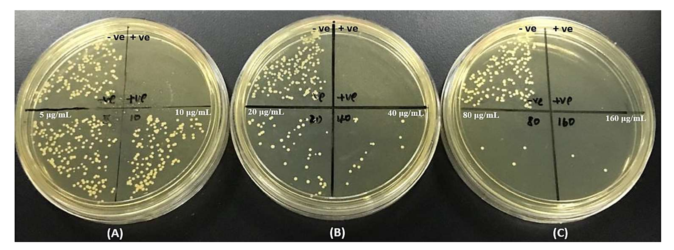
3.2.2. Colony Count Method
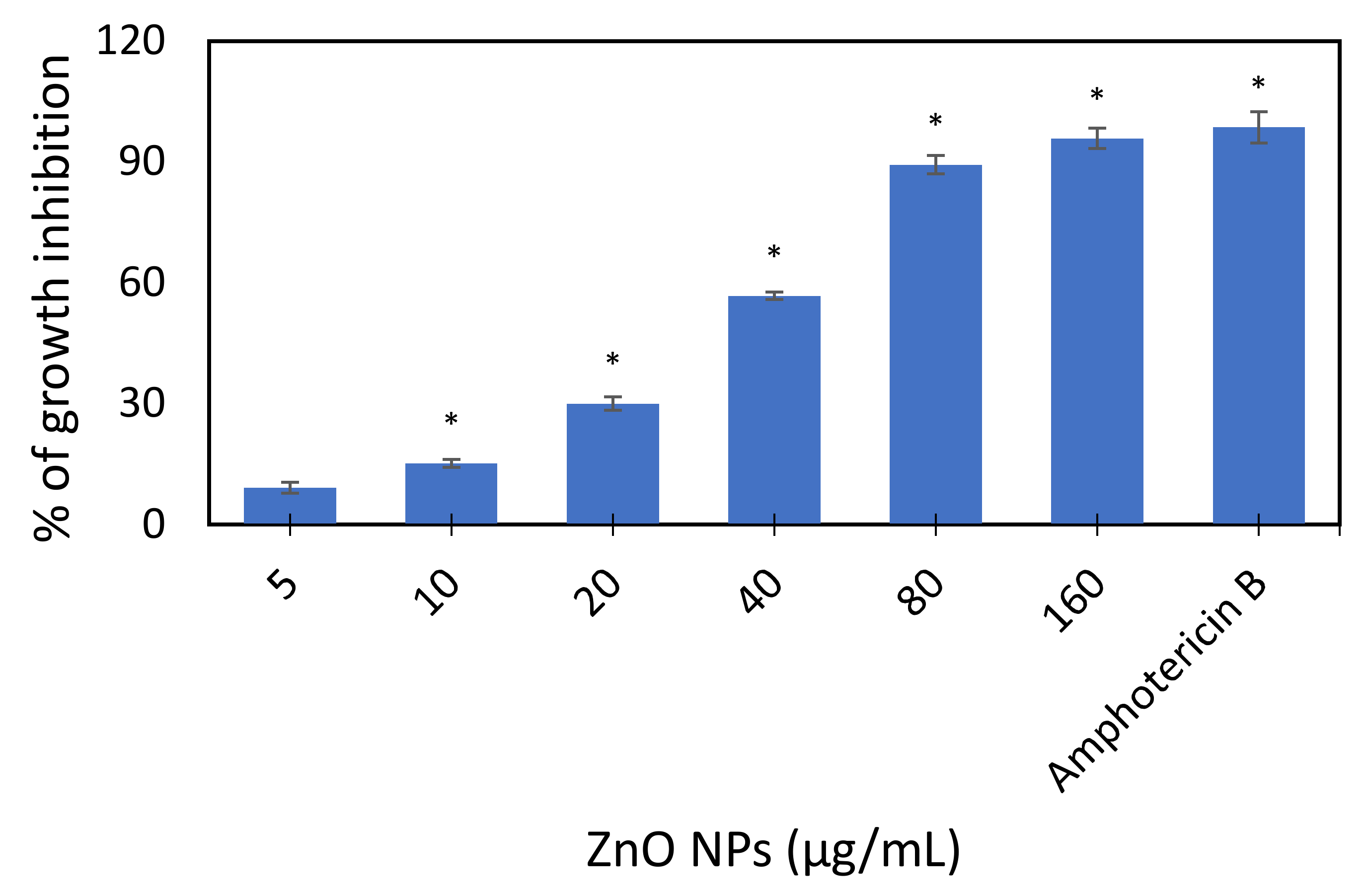
The colony count technique was utilised to investigate the number of viable cells formed on SDA. The colonies were counted for yeast suspensions treated with different concentrations of ZnO NPs (5, 10, 20, 40, 80, and 160 μg/mL) after 24 h incubation at 35 °C. Amphotericin B with a concentration of 60μg/mL was used as a positive control. The number of colonies that formed decreased as the concentration of ZnO NPs increased (Figure 4). In addition, the positive control showed no colony growth. The number of colonies formed on SDA is tabulated in Table 1.

3.2.3. INT Assay

An INT assay was used to determine the minimum inhibitory concentration (MIC) of ZnO NPs that can inhibit the growth of C. albicans by observing the appearance of colour formation for each washed yeast suspension treated with different concentration of ZnO NPs (5, 10, 20, 40, 80, and 160 µg/mL). As shown in Figure 5, no colour formation was observed for both columns E and F after being incubated with INT dye for 20 min, identifying 80 µg/mL as the MIC value that can inhibit the growth of C. albicans.

3.3. Surface Interaction and Cellular Accumulation of ZnO NPs on the Yeast Cell Wall

3.3.1. Fourier Transform Infrared (FTIR) Analysis

FTIR analysis was used to identify the functional groups that were responsible for the interaction between ZnO NPs and the surface of C. albicans.
The FTIR spectrum of ZnO nanopowder showed peaks at 3425, 1628, and 524 cm−1. Earlier studies have reported O–H stretching vibrations between 3400 and 3600 cm−1 [29], Zn–O stretching at 1634.00 cm−1 [30], and ZnO NPs stretching at 400–800 cm−1 [31]. The FTIR spectrum obtained from the negative control demonstrated O–H and N–H stretching at 3436 cm−1, C–H stretching at 2378 and 2345 cm−1, C=O stretching at 1641 cm−1, C–O stretching at 1405 cm−1, PO2 and C–O stretching at 1076 cm−1, and glycogen stretching at 570 cm−1. The peaks that shifted in the ZnO NPs-treated yeast cells after 24 h incubation were the O–H and N–H stretching (3436 to 3447 cm−1), indicating an alcohol or amide group, the CH2 stretching of the methyl group (2930 to 2923 cm−1), the C C stretching of the alkynes (2078 to 2067 cm−1), and the C–O vibration in polysaccharides and PO2-absorption of phosphate groups from phospholipids (1076 to 1048 cm−1) (Figure 6). Hence, our FTIR investigation indicates the possible involvement of alcohol and amide A (3447 cm−1), methyl (2923 cm−1), alkynes (2067 cm−1), amide I and II (1638 cm−1), and phosphate groups (1048 cm−1) in the surface interaction of NPs onto the yeast cell wall (Table 2).

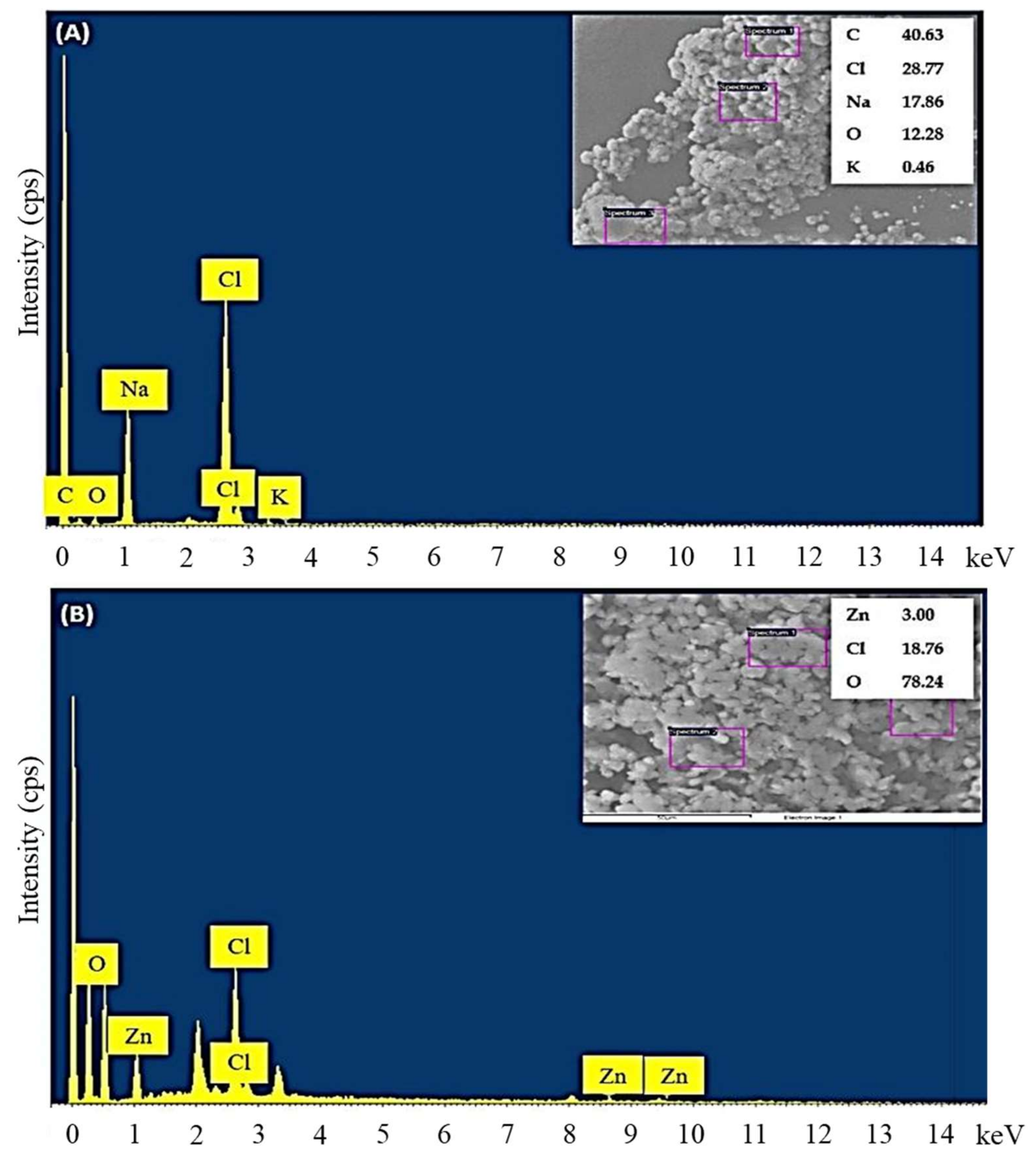
3.3.2. Energy Dispersive X-ray Analysis

Energy dispersive X-ray (EDX) analysis was used to identify the surface accumulation of ZnO NPs on the yeast cells treated with ZnO NPs. The EDX spectrum of the negative control without the treatment of ZnO NPs (Figure 7A) showed the presence of sodium, potassium, carbon, oxygen, and chlorine; while the yeast suspension treated with 160 µg/mL ZnO NPs showed the presence of zinc, oxygen, and chlorine (Figure 7B), demonstrating the surface accumulation of ZnO NPs on the yeast cells.

3.3.3. SEM Micrographs

The surface morphological changes in the C. albicans after treatment with 80 and 160 µg/mL ZnO NPs were examined by SEM. Figure 8A,B are the SEM images of the negative control at 2000× and 5000× magnification, respectively. The negative control images showed a smooth cell surface and uncompromised cell membrane in the yeast cells. Conversely, alterations in cell structure were observed after the exposure of C. albicans with 80 and 160 µg/mL of ZnO NPs at 24 h. Figure 8C–F show that the harvested yeast suspension showed breakage of hyphae, pitting and regional invagination of the cell surface, and rupture of cell membrane.

4. Discussion

4.1. Growth Inhibitory Effect of ZnO NPs

The results of turbidity measurements and colony counts in this current study showed a dose-dependent inhibitory effect of ZnO NPs on C. albicans with the increase in the concentration of ZnO NPs producing incremental antifungal effects. In addition, the INT assay reported 80 µg/mL of ZnO NPs as the MIC against C. albicans. Minimum inhibitory concentration (MIC) is the minimum concentration of an antimicrobial agent that can cause growth inhibition of a microorganism, while minimum fungicidal concentration (MFC) indicates the minimum concentration of an antifungal agent that can kill the fungi. Similar with our findings, earlier studies have reported the antifungal effects of ZnO NPs on C. albicans. The dose-dependent growth inhibitory effects of ZnO NPs were demonstrated against C. albicans using disc diffusion [32] and cell counting [33] methods, where an incremental increase in the diameter inhibition zone and the decrease in the number of viable cells were noticed as the concentration of ZnO NPs increased. Moreover, our results show a lower EC50 value for ZnO NPs (35.59 μg/mL) on C. albicans compared with the 131 mg/L identified as the EC50 of ZnO NPs against Saccharomyces cerevisiae after 24 h incubation, indicating a higher antifungal potential of ZnO NPs on C. albicans [34].
An investigation of the antifungal activity of different NPs, such as MgO, ZnO, SiO2, and CuO NPs, on C. albicans, showed the lowest MIC and MFC for ZnO NPs indicating the superior antifungal property of ZnO NPs against other NPs [23]. A study by [35] investigated Ag NPs for their antifungal activity on different Candida strains such as C. albicans, C. glabrata, and C. tropicalis, and reported a MIC of 60 µg/mL for C. albicans, and a MIC of 30 µg/mL for both C. glabrata and C. tropicalis.

4.2. Surface Interaction and Cellular Accumulation of ZnO NPs

The surface interaction of ZnO NPs with the yeast cell wall of C. albicans was evaluated using FTIR spectroscopy. Generally, two important elements can be obtained from the FTIR spectrum, the identification of functional groups and conformational freedom. The functional groups can be determined from the peak position, while the conformational freedom can be predicted from the bandwidth. In the present study, the treatment of C. albicans with ZnO NPs resulted in peak shifts from 3434 cm−1 to 3447 cm−1, dominated by the O–H and N–H group stretching vibration from the proteins and polysaccharides and intermolecular hydrogen bond [36,37,38], 2930 to 2923 cm−1, representing the stretching of the methyl group from lipids, 2078 to 2067 cm−1, corresponding to C≡C stretching of alkynes and from 1076 cm−1 to 1048 cm−1, indicating the involvement of sugars (mannans moieties, β-glucans, arabinose, mannose, etc.) and phosphate group from the phospholipids of the yeast cell wall. A minor peak shift from 1641 cm−1 to 1638 cm−1 was due to the amide I bands of β-pleated sheet structures, dominated by C=O stretching of the polypeptide and protein backbone. The disappearance of the peak at 524 cm−1 from the FTIR spectrum of ZnO NPs may arise from the masking of the ZnO group due to the adhesion of membrane biomolecules from the cell surface of yeast cells [37,38,39,40,41,42]. In common with the present results, our earlier studies have reported the possible involvement of hydroxyl and methylene groups from lipids, amide I from proteins, phosphate from phospholipids and phosphated proteins, and mannans in the interaction of ZnO NPs with bacterial cell membranes [9,14,43].
The surface accumulation of ZnO NPs on the yeast cell is demonstrated by the results of the EDX analysis (Figure 7). To explain the accumulation of ZnO NPs on the surface of the yeast cells, and subsequent cell damage or death, it is important to consider the composition of the fungal cell wall. The cell wall is composed of glycoproteins, polysaccharides (especially glucan and chitin), hydrophobins, and amphipathic proteins, which are the proteins in fungi involved in their interaction with the environment. Upon the exposure of fungal cells to the ZnO NPs, dynamic physicochemical interactions, kinetic and thermodynamic exchanges, and attractive van der Waals forces occur between the surface of the NPs and the surface of the biological component, such as the membrane and proteins. All these interactions are believed to cause an interaction between the ZnO NPs and the cell membrane of fungal cells, leading to the accumulation of NPs on the cell membrane and the subsequent membrane disruption [44,45,46,47]. Further, the process of adhesion of NPs on the surface of the microorganism is also believed to be an important reason for the observed cytotoxicity. The surface biomolecules of microorganisms can dominate in the cell adhesion process due to their functional groups, highly charged structure, and the bridging effect with the surface of the nanoparticles. Important biomolecules such as lipopolysaccharides and phospholipids from the cell surface were found to adsorb on ZnO NPs surfaces during NP exposure, leading to structural changes in the proteins and phospholipids, which are the most likely reason in addition to membrane damage for the observed cytotoxicity towards microorganisms [48,49,50,51].
Scanning electron microscopy (SEM) was used to observe the morphological changes of C. albicans by comparing a negative control and C. albicans after 24 h exposure to ZnO NPs. The C. albicans without ZnO NPs treatment showed smooth and healthy ovoid and spherical conidia, hyphae, and pseudohyphae (Figure 8A,B). In contrast, breakage of hyphae, rupture of the membrane, and pitting and invagination of the cell surface were observed on the C. albicans exposed to 80 and 160 µg/mL ZnO NPs (Figure 8C–F). Similar findings were reported in earlier studies on C. albicans, where the formation of cavities on the cell surface was observed when C. albicans exposed to 15 µg/mL EtZnO NPs (ZnO NPs synthesised using egg white) [52]; while destruction of the external cell wall and the formation of a wrinkled, bumpy and irregular external cell surface were demonstrated when treated with 40 µg/mL Ag NPs [53]. The formation of cavities could be due to the production of apoptosome in the yeast cell that could lead to cell death. In addition, the production of hydrogen peroxide (H2O2) due to the interaction of NPs on the cell surface can lead to rupture of yeast cell membrane [52]. Drastic damage on the hyphae surface was observed due to the condensation of the nucleus and hyphae injury when Aspergillus flavus and Aspergillus niger were treated with 200 mg/L molybdenum trioxide nanoparticles for 10 days [54]. In addition, the treatment of the plant pathogen Colletotrichum species that can cause pepper anthracnose with 100 ppm Ag NPs was found to have a detrimental effect on the growth of hyphae and germination of conidia [55].
The antimicrobial mechanism of metallic NPs is still being discussed in many aspects. However, based on the scientific reports, the principal mechanisms proposed are the formation of reactive oxidative species (ROS) and the release of metal ions from the NPs due to the interaction of NPs with the cell membrane causing inhibition of cell wall synthesis, enzyme activities, and cell signalling, DNA damage, ribosome disassembly, inactivation of protein synthesis, and structure modification of essential proteins (Figure 9). In addition to the membrane dysfunction caused by the accumulation of positively charged Zn2+ from the dissolution of ZnO NPs on the surface of the cell membrane, the internalisation of ZnO NPs disrupts microbial metabolic activity, eventually causing microbial cell death [56,57,58,59].

5. Conclusions

The current work demonstrated a significant dose-dependent growth inhibitory effect of ZnO NPs on C. albicans through turbidity measurements and colony counting, with MIC and EC50 values of 80 µg/mL and 39.59 µg/mL ZnO NPs, respectively. The FTIR spectrum of ZnO NPs-treated C. albicans indicated the possible involvement of alcohol, amide A, alkynes, amide 1 & II, and phosphate groups in the surface interaction of ZnO NPs with the fungal cells. Further, EDX analysis confirmed the surface accumulation of ZnO NPs on the yeast cells, and SEM micrographs displayed the breakage of hyphae, pitting of the cell wall, invagination, and cell membrane rupture in C. albicans upon treating with ZnO NPs. These changes to the cell membrane could lead to cell death. Based on the results obtained from the present study, it is possible to conclude that ZnO NPs can be utilised as an effective antifungal agent against C. albicans.

Author Contributions

Supervision, study design, review and editing, S.D., investigation L.-J.X. and S.D. study design, review and editing L.-S.W., M.A. and L.-H.T., writing, review, R.R., D.B. and S.S. review, editing, S.K., A.E.D.C. and A.K.J. All authors have read and agreed to the published version of the manuscript.

Funding

This research work was funded by the Universiti Tunku Abdul Rahman Research Fund (UTARRF/2020-C2/S06) and the Ministry of Higher Education (MOHE), Malaysia (Grant No. FRGS-1-2014-SG03-INTI-02-1).

Institutional Review Board Statement

Not applicable.

Informed Consent Statement

Not applicable.

Data Availability Statement

Not applicable.

Conflicts of Interest

Authors declare no conflict of interest.

References

  1. Reddy, G.K.K.; Padmavathi, A.R.; Nancharaiah, Y.V. Fungal infections: Pathogenesis, antifungals and alternate treatment approaches. Curr. Res. Microb. Sci. 2022, 3, 100137. [Google Scholar] [CrossRef] [PubMed]
  2. Iyer, K.R.; Robbins, N.; Cowen, L.E. The role of Candida albicans stress response pathways in antifungal tolerance and resistance. iScience 2022, 25, 103953. [Google Scholar] [CrossRef] [PubMed]
  3. Kabir, M.A.; Hussain, M.A.; Ahmad, Z. Candida albicans: A model organism for studying fungal pathogens. ISRN Microbiol. 2012, 2012, 538694. [Google Scholar] [CrossRef] [PubMed] [Green Version]
  4. Roemer, T.; Krysan, D.J. Antifungal drug development: Challenges, unmet clinical needs, and new approaches. Cold Spring Harb. Perspect. Med. 2014, 4, a019703. [Google Scholar] [CrossRef] [PubMed]
  5. Graybill, J.R.; Burgess, D.S.; Hardin, T.C. Key issues concerning fungistatic versus fungicidal drugs. Eur. J. Clin. Microbiol. Infect. Dis. 1997, 16, 42–50. [Google Scholar] [CrossRef] [PubMed]
  6. Fisher, M.C.; Alastruey-Izquierdo, A.; Berman, J.; Bicanic, T.; Bignell, E.M.; Bowyer, P.; Bromley, M.; Brüggemann, R.; Garber, G.; Cornely, O.A.; et al. Tackling the emerging threat of antifungal resistance to human health. Nat. Rev. Microbiol. 2022, 1–15. [Google Scholar] [CrossRef] [PubMed]
  7. Armengol, E.S.; Harmanci, M.; Laffleur, F. Current strategies to determine antifungal and antimicrobial activity of natural compounds. Microbiol. Res. 2021, 252, 126867. [Google Scholar] [CrossRef] [PubMed]
  8. Bhavyasree, P.G.; Xavier, T.S. Green synthesised copper and copper oxide-based nanomaterials using plant extracts and their application in antimicrobial activity. Curr. Res. Green Sustain. Chem. 2022, 5, 100249. [Google Scholar]
  9. Djearamane, S.; Ravintharan, T.; Liang, S.X.; Wong, L.S. Sensitivity of Proteus vulgaris to Zinc Oxide Nanoparticles. Sains Malays. 2022, 51, 1353–1362. [Google Scholar] [CrossRef]
  10. Owaid, M.N.; Rabeea, M.A.; Aziz, A.A.; Jameel, M.S.; Dheyab, M.A. Mycogenic fabrication of silver nanoparticles using Picoa, Pezizales, characterization and their antifungal activity. Environ. Nanotechnol. Monit. Manag. 2022, 17, 100612. [Google Scholar] [CrossRef]
  11. Nidhin, M.; Saneha, D.; Hans, S.; Varghese, A.; Fatima, Z.; Hameed, S. Studies on the antifungal activity of biotemplated gold nanoparticles over Candida albicans. Mater. Res. Bull. 2019, 119, 110563. [Google Scholar]
  12. Hermida-Montero, L.A.; Pariona, N.; Mtz-Enriquez, A.I.; Carrión, G.; Paraguay-Delgado, F.; Rosas-Saito, G. Aqueous-phase synthesis of nanoparticles of copper/copper oxides and their antifungal effect against Fusarium oxysporum. J. Hazard. Mater. 2019, 380, 120850. [Google Scholar] [CrossRef] [PubMed]
  13. Parveen, S.; Wani, A.H.; Shah, M.A.; Devi, H.S.; Bhat, M.Y.; Koka, J.A. Preparation, characterization and antifungal activity of iron oxide nanoparticles. Microb. Pathogen. 2018, 115, 2087–2292. [Google Scholar] [CrossRef]
  14. Dhanasegaran, K.; Djearamane, S.; Liang, S.X.; Wong, L.S.; Kasivelu, G.; Lee, P.F.; Lim, Y.M. Antibacterial properties of zinc oxide nanoparticles on Pseudomonas aeruginosa (ATCC 27853). Sci. Iran. 2021, 28, 3806–3815. [Google Scholar]
  15. Vijayakumar, S.; Vidhya, E.; Nilavukkarasi, M.; Punitha, V.N.; Praseetha, P.K. Potential eco-friendly Zinc Oxide nanomaterials through Phyco-nanotechnology—A review. Biocatal. Agric. Biotechnol. 2021, 35, 102050. [Google Scholar] [CrossRef]
  16. Abhishek, T.; Sharma, A.; Tejwan, N.; Ghosh, N.; Das, J. A state of the art review on the synthesis, antibacterial, antioxidant, antidiabetic and tissue regeneration activities of zinc oxide nanoparticles. Adv. Colloid Interface Sci. 2021, 295, 102495. [Google Scholar]
  17. Dutta, G.; Sugumaran, A. Bioengineered zinc oxide nanoparticles: Chemical, green, biological fabrication methods and its potential biomedical applications. J. Drug Deliv. Sci. Technol. 2021, 66, 102853. [Google Scholar]
  18. Karthikeyan, C.; Tharmalingam, N.; Varaprasad, K.; Mylonakis, E.; Yallapu, M.M. Biocidal and biocompatible hybrid nanomaterials from biomolecule chitosan, alginate and ZnO. Carbohydr. Polym. 2021, 274, 118646. [Google Scholar] [CrossRef] [PubMed]
  19. Kasemets, K.; Ivask, A.; Dubourguier, H.C.; Kahru, A. Toxicity of nanoparticles of ZnO, CuO and TiO2 to yeast Saccharomyces cerevisiae. Toxicol. Vitr. 2009, 23, 1116–1122. [Google Scholar] [CrossRef]
  20. El-Diasty, E.M.; Ahmed, M.A.; Okasha, N.; Mansour, S.F.; El-Dek, S.I.; El-Khalek, H.M.; Youssif, M.H. Antifungal activity of zinc oxide nanoparticles against dermatophytic lesions of cattle. Rom. J. Biophys. 2013, 23, 191–202. [Google Scholar]
  21. Singh, P.; Nanda, P.A. Antimicrobial and antifungal potential of zinc oxide nanoparticles in comparison to conventional zinc oxide particles. J. Chem. Pharm. Res. 2013, 5, 457–463. [Google Scholar]
  22. Pasquet, J.; Chevalier, Y.; Couval, E.; Bouvier, D.; Noizet, G.; Morliere, C.; Bolzinger, M.A. Antimicrobial activity of zinc oxide particles on five micro-organisms of the Challenge Tests related to their physicochemical properties. Int. J. Pharm. 2014, 460, 92–100. [Google Scholar] [CrossRef] [PubMed]
  23. Karimiyan, A.; Najafzadeh, H.; Ghorbanpour, M.; Hekmati-Moghaddam, S.H. Antifungal effect of magnesium oxide, zinc oxide, silicon oxide and copper oxide nanoparticles against Candida albicans. Zahedan J. Res. Med. Sci. 2015, 17, e2179. [Google Scholar] [CrossRef] [Green Version]
  24. Azam, A.; Ahmed, A.S.; Oves, M.; Khan, M.S.; Habib, S.S.; Memic, A. Antimicrobial activity of metal oxide nanoparticles against Gram-positive and Gram-negative bacteria: A comparative study. Int. J. Nanomed. 2012, 7, 6003–6009. [Google Scholar] [CrossRef] [Green Version]
  25. Reddy, K.M.; Feris, K.; Bell, J.; Wingett, D.G.; Hanely, C.; Punnoose, A. Selective toxicity of zinc oxide nanoparticles to prokaryotic and eukaryotic systems. Appl. Phys. Lett. 2007, 90, 213902. [Google Scholar] [CrossRef] [PubMed] [Green Version]
  26. Raghupathi, K.R.; Koodali, R.T.; Manna, A.C. Size-dependent bacterial growth inhibition and mechanism of antibacterial activity of zinc oxide nanoparticles. Langmuir 2011, 27, 4020–4028. [Google Scholar] [CrossRef]
  27. Djearamane, S.; Wong, L.S.; Lim, Y.M.; Lee, P.F. Oxidative stress effects of zinc oxide nanoparticles on freshwater microalga Haematococcus pluvialis. Ecol. Environ. Conserv. 2020, 26, 663–668. [Google Scholar]
  28. Djearamane, S.; Loh, Z.C.; Lee, J.J.; Wong, L.S.; Ranjithkumar, R.; Luque, P.A.; Gupta, P.K.; Liang, S.X.T. Remedial Aspect of Zinc Oxide Nanoparticles Against Serratia Marcescens and Enterococcus Faecalis. Front. Pharmacol. 2022, 13, 891304. [Google Scholar] [CrossRef] [PubMed]
  29. Sukri, S.N.A.M.; Shameli, K.; Wong, M.M.-T.; Teow, S.-Y.; Chew, J.; Ismail, N.A. Cytotoxicity and antibacterial activities of plant-mediated synthesized zinc oxide (ZnO) nanoparticles using Punica granatum (pomegranate) fruit peels extract. J. Mol. Struct. 2019, 1189, 57–65. [Google Scholar] [CrossRef]
  30. Kumar, H.; Rani, R. Structural and Optical Characterization of ZnO Nanoparticles Synthesized by Microemulsion Route. Int. Lett. Chem. Phys. Astron. 2013, 14, 26. [Google Scholar] [CrossRef] [Green Version]
  31. Ramesh, M.; Anbuvannan, M.; Viruthagiri, G.J.S.A.P.A.M. Green synthesis of ZnO nanoparticles using Solanum nigrum leaf extract and their antibacterial activity. Spectrochim. Acta Part A Mol. Biomol. Spectrosc. 2015, 136, 864–870. [Google Scholar] [CrossRef] [PubMed]
  32. Abomuti, M.A.; Danish, E.Y.; Firoz, A.; Hasan, N.; Malik, M.A. Green Synthesis of Zinc Oxide Nanoparticles Using Salvia officinalis Leaf Extract and Their Photocatalytic and Antifungal Activities. Biology 2021, 10, 1075. [Google Scholar] [CrossRef] [PubMed]
  33. Sharma, R.K.; Ghose, R. Synthesis of zinc oxide nanoparticles by homogeneous precipitation method and its application in antifungal activity against Candida albicans. Ceram. Int. 2015, 41, 967–975. [Google Scholar] [CrossRef]
  34. Lipovsky, A.; Nitzan, Y.; Gedanken, A.; Lubart, R. Antifungal activity of ZnO nanoparticles-the role of ROS mediated cell injury. Nanotechnology 2011, 22, 105101. [Google Scholar] [CrossRef]
  35. Monteiro, D.R.; Gorup, L.F.; Silva, S.; Negri, M.; de Camargo, E.R.; Oliveira, R.; Barbosa, D.B.; Henriques, M. Silver colloidal nanoparticles: Antifungal effect against adhered cells and biofilms of Candida albicans and Candida glabrata. Biofouling 2011, 27, 711–719. [Google Scholar] [CrossRef] [PubMed]
  36. Khatoon, N.; Mishra, A.; Alam, H.; Manzoor, N.; Sardar, M. Biosynthesis, characterization, and antifungal activity of the silver nanoparticles against pathogenic Candida species. BioNanoScience 2015, 5, 65–74. [Google Scholar] [CrossRef]
  37. Melin, A.M.; Perromat, A.; Déléris, G. Pharmacologic application of Fourier transform IR spectroscopy: In vivo toxicity of carbon tetrachloride on rat liver. Biopolym. Orig. Res. Biomol. 2000, 57, 160–168. [Google Scholar] [CrossRef]
  38. Naumann, D. FT-infrared and FT-Raman spectroscopy in biomedical research. Appl. Spectrosc. Rev. 2001, 36, 239–298. [Google Scholar] [CrossRef]
  39. Banyay, M.; Sarkar, M.M.; Gräslund, A. A library of IR bands of nucleic acids in solution. Biophys. Chem. 2003, 104, 477–488. [Google Scholar] [CrossRef]
  40. Kačuráková, M.; Mathlouthi, M. FTIR and laser-Raman spectra of oligosaccharides in water: Characterization of the glycosidic bond. Carbohydr. Res. 1996, 284, 145–157. [Google Scholar] [CrossRef] [PubMed]
  41. Galichet, A.; Sockalingum, G.D.; Belarbi, A.; Manfait, M. FTIR spectroscopic analysis of Saccharomyces cerevisiae cell walls: Study of an anomalous strain exhibiting a pink-colored cell phenotype. FEMS Microbiol. Lett. 2001, 197, 179–186. [Google Scholar] [CrossRef] [PubMed] [Green Version]
  42. Plata, M.R.; Koch, C.; Wechselberger, P.; Herwig, C.; Lendl, B.B. Determination of carbohydrates present in Saccharomyces cerevisiae using mid-infrared spectroscopy and partial least squares regression. Anal. Bioanal. Chem. 2013, 405, 8241–8250. [Google Scholar] [CrossRef] [PubMed] [Green Version]
  43. Lianga, S.X.; Wong, L.S.; Lim, Y.M.; Djearamane, S.; Lee, P.F. Effects of Zinc Oxide nanoparticles on Streptococcus pyogenes. S. Afr. J. Chem. Eng. 2020, 34, 63–71. [Google Scholar] [CrossRef]
  44. Gow, N.A.R.; Latge, J.-P.; Munro, C.A. The fungal cell wall: Structure, biosynthesis, and function. Microbiol. Spectr. 2017, 5, 3. [Google Scholar]
  45. Gow, N.A.R.; Latge, J.P.; Munro, C.A. The Fugal Kingdom; ASM Press: Washington, DC, USA, 2017. [Google Scholar]
  46. Feofilova, E.P. The fungal cell wall: Modern concepts of its composition and biological function. Microbiology 2010, 79, 711–720. [Google Scholar] [CrossRef]
  47. Nel, A.E.; Mädler, L.; Velegol, D.; Xia, T.; Hoek, E.M.V.; Somasundaran, P.; Klaessig, F.; Castranova, V.; Thompson, M. Understanding biophysicochemical interactions at the nano-bio interface. Nat. Mater. 2009, 8, 543–557. [Google Scholar] [CrossRef]
  48. Min, Y.; Akbulut, M.; Kristiansen, K.; Golan, Y.; Israelachvili, J. The role of interparticle and external forces in nanoparticle assembly. Nat. Mater. 2008, 7, 527–538. [Google Scholar] [CrossRef]
  49. Anselm, O.; Chorover, J. Spectroscopic study of extracellular polymeric substances from Bacillus subtilis: Aqueous chemistry and adsorption effects. Biomacromolecules 2004, 5, 1219–1230. [Google Scholar]
  50. Parikh Sanjai, J.; Chorover, J. ATR-FTIR spectroscopy reveals bond formation during bacterial adhesion to iron oxide. Langmuir 2006, 22, 8492–8500. [Google Scholar] [CrossRef]
  51. Wei, J.; Yang, K.; Vachet, R.W.; Xing, B. Interaction between oxide nanoparticles and biomolecules of the bacterial cell envelope as examined by infrared spectroscopy. Langmuir 2010, 26, 18071–18077. [Google Scholar]
  52. Shoeb, M.; Singh, B.R.; Khan, J.A.; Khan, W.; Singh, B.N.; Singh, H.B.; Naqvi, A.H. ROS-dependent anticandidal activity of zinc oxide nanoparticles synthesized by using egg albumen as a biotemplate. Adv. Nat. Sci. Nanosci. Nanotechnol. 2013, 4, 035015. [Google Scholar] [CrossRef]
  53. Radhakrishnan, V.S.; Mudiam, M.K.R.; Kumar, M.; Dwivedi, S.P.; Singh, S.P.; Prasad, T. Silver nanoparticles induced alterations in multiple cellular targets, which are critical for drug susceptibilities and pathogenicity in fungal pathogen (Candida albicans). Int. J. Nanomed. 2018, 13, 2647. [Google Scholar] [CrossRef] [PubMed] [Green Version]
  54. Nguyen, H.N.; Oliveira, R.C.; Nadres, E.T.; Paparella, A.; Rodrigues, D.F. A morphological, enzymatic and metabolic approach to elucidate apoptotic-like cell death in fungi exposed to h- and α-molybdenum trioxide nanoparticles. Nanoscale 2018, 10, 20702–20716. [Google Scholar]
  55. Lamsal, K.; Kim, S.W.; Jung, J.H.; Kim, Y.S.; Kim, K.S.; Lee, Y.S. Application of silver nanoparticles for the control of colletotrichum species in vitro and pepper anthracnose disease in field. Mycobiology 2011, 39, 194–199. [Google Scholar] [CrossRef] [PubMed] [Green Version]
  56. Sirelkhatim, A.; Mahmud, S.; Seeni, A.; Kaus, N.H.M.; Ann, L.C.; Bakhori, S.K.M.; Hasan, H.; Mohamad, D. Review on Zinc Oxide Nanoparticles: Antibacterial Activity and Toxicity Mechanism. Nano-Micro Lett. 2015, 7, 219–242. [Google Scholar] [CrossRef] [Green Version]
  57. Nisar, P.; Ali, N.; Rahman, L.; Ali, M.; Shinwari, Z.K. Antimicrobial activities of biologically synthesized metal nanoparticles: An insight into the mechanism of action. JBIC J. Biol. Inorg. Chem. 2019, 24, 929–941. [Google Scholar] [CrossRef]
  58. Ghorbani, F.; Gorji, P.; Mobarakeh, M.S.; Mozaffari, H.R.; Masaeli, R.; Safaei, M. Optimized Synthesis of Xanthan gum/ZnO/TiO2Nanocomposite with High Antifungal Activity against Pathogenic Candida albicans. J. Nanomater. 2022, 2022, 7255181. [Google Scholar] [CrossRef]
  59. Gharpure, S.; Yadwade, R.; Ankamwar, B. Non-antimicrobial and Non-anticancer Properties of ZnO Nanoparticles Biosynthesized Using Different Plant Parts of Bixa orellana. ACS Omega 2022, 7, 1914–1933. [Google Scholar] [CrossRef] [PubMed]
Figure 1. Growth curve of C. albicans in SDB at 35 °C.
Coatings 12 01864 g001
Figure 2. Growth inhibition determined by the turbidity method on C. albicans after treatment with various concentrations of ZnO NPs for 24 h at 35 °C in SDB. An asterisk (*) indicates the significance difference at p < 0.05 between the control and treatments.
Coatings 12 01864 g002
Figure 3. The turbidity of C. albicans in SDB at 35 °C after 24 h treatment with (A) 5 μg/mL, (B) 10 μg/mL, (C) 20 μg/mL, (D) 40 μg/mL, (E) 80 μg/mL, and (F) 160 μg/mL of ZnO NPs, and a negative control (G), and a positive control (H).
Coatings 12 01864 g003
Figure 4. Colonies of C. albicans formed on SDA at 35 °C after 24 h incubation in a negative control, a positive control, and yeast suspensions treated with 5 μg/mL and 10 μg/mL ZnO NPs (A), 20 μg/mL and 40 μg/mL ZnO NPs (B), and 80 μg/mL and 160 μg/mL ZnO NPs (C).
Coatings 12 01864 g004
Figure 5. The colour changes of C. albicans after treatment with (A) 5 µg/mL, (B) 10 µg/mL, (C) 20 µg/mL, (D) 40 µg/mL, (E) 80 µg/mL, and (F) 160 µg/mL of ZnO NPs, compared to a negative control (G), and a positive control (H), (ac) three sets of data).
Coatings 12 01864 g005
Figure 6. FTIR spectra of ZnO NPs, C. albicans negative control, and C. albicans treated with 160 μg/mL ZnO NPs (test).
Coatings 12 01864 g006
Figure 7. EDX spectra of the negative control after 24 h incubation (A), and a yeast suspension treated with 160 µg/mL ZnO NPs after 24 h of incubation (B).
Coatings 12 01864 g007
Figure 8. SEM images of C. albicans without ZnO NPs treatment (negative control) (A,B); treatment with 80 µg/mL of ZnO NPs showed breakage of hyphae (red arrow) and pitting of yeast surface (black arrow) (C,D); treatment with 160 μ g/mL of ZnO NPs showed invagination of yeast cell surface (blue arrow) and rupture of the cell membrane (yellow arrow) (E,F) after 24 h incubation.
Coatings 12 01864 g008
Figure 9. Possible antimicrobial mechanisms of nanoparticles.
Coatings 12 01864 g009
Table 1. Number of colonies formed on SDA after 24 h incubation.
ZnO NPs (μg/mL)Number of Colonies
Mean ± Standard Deviation
0197 ± 7
5200 ± 6
10120 ± 29
2052 ± 9
4010 ± 6
802 ± 2
1602 ± 1
Amphotericin B0 ± 0
Table 2. Possible involvement of functional groups based on the FTIR analysis.
Absorption (cm−1)Molecular MotionFunctional GroupBiomolecules
3436 3447
2930 2923
O–H and N–H stretching
CH2 stretching
Alcohol, amide A
Methyl group
Proteins, polysaccharides, chitin
Lipids
2078 2067C C stretchingAlkynes groupHydrocarbon
1076 1048C–O mainly by vibrations and absorptions of polysaccharides and phosphate groupsPhosphate groupPolysaccharides, mainly glucans and mannans, phospholipids
1641 1638C=O stretching, Zn–O stretchingAmide I and amide II bands, respectively due to the C=O stretching and the NH bending of the peptide bondPolypeptide, protein backbone
Publisher’s Note: MDPI stays neutral with regard to jurisdictional claims in published maps and institutional affiliations.

