
Universal Adhesives: Evaluation of the Relationship between Bond Strength and Application Strategies—A Systematic Review and Meta-Analyses

 ,
,  , , and
Abstract
1. Introduction
2. Materials and Methods
2.1. Inclusion and Exclusion Criteria
2.2. Search Strategy
2.2.1. Sources of Information
2.2.2. Search Terms
2.2.3. Study Selection
2.2.4. Study Data
2.3. Quality Assessment
3. Results
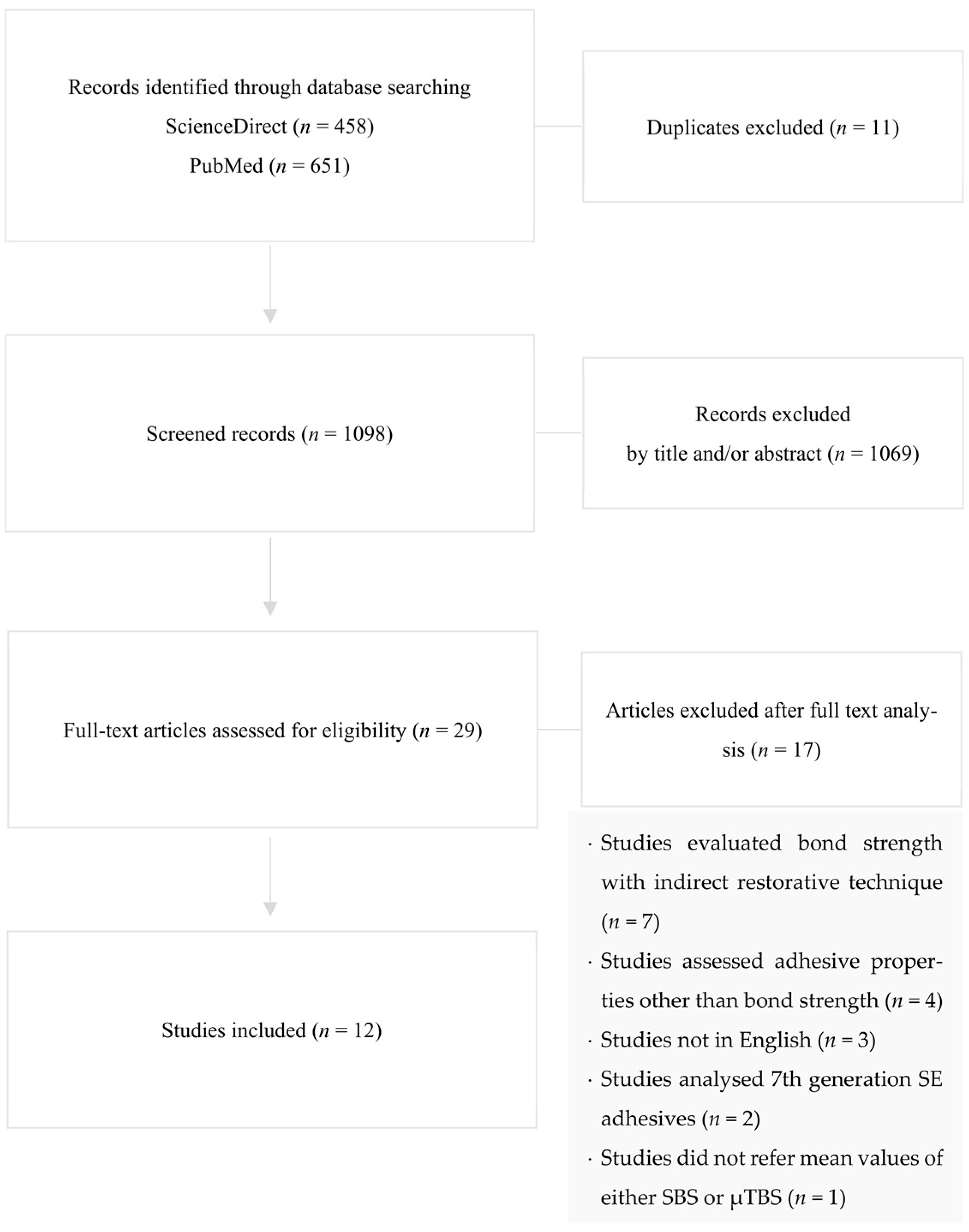
3.1. Study Selection and Flow Diagram
3.2. Study Characteristics
Study Type
3.3. Quality Assessment
3.4. Study Results
3.4.1. Mean Microtensile Bond Strength (µTBS) Analysis
3.4.2. Mean Shear Bond Strength (SBS) Analysis
4. Discussion
5. Conclusions
Author Contributions
Funding
Institutional Review Board Statement
Informed Consent Statement
Conflicts of Interest
References
- Nagarkar, S.; Theis-Mahon, N.; Perdigão, J. Universal dental adhesives: Current status, laboratory testing, and clinical performance. J. Biomed. Mater. Res. Part B Appl. Biomater. 2018, 107, 2121–2131. [Google Scholar] [CrossRef] [PubMed]
- Perdigão, J.; Araujo, E.; Ramos, R.Q.; Gomes, G.; Pizzolotto, L. Adhesive dentistry: Current concepts and clinical considerations. J. Esthet. Restor. Dent. 2020, 33, 51–68. [Google Scholar] [CrossRef] [PubMed]
- Van Meerbeek, B.; Yoshihara, K.; Yoshida, Y.; Mine, A.; De Munck, J.; Van Landuyt, K.L. State of the art of self-etch adhesives. Dent. Mater. 2011, 27, 17–28. [Google Scholar] [CrossRef]
- Perdigão, J.; Swift, J.E. Contemporary Issues-Universal Adhesives. J. Esthet. Restor. Dent. 2015, 27, 331–334. [Google Scholar] [CrossRef] [PubMed]
- Carrilho, E.; Cardoso, M.; Ferreira, M.M.; Marto, C.M.; Paula, A.; Coelho, A.S. 10-MDP Based Dental Adhesives: Adhesive Interface Characterization and Adhesive Stability—A Systematic Review. Materials 2019, 12, 790. [Google Scholar] [CrossRef] [PubMed]
- Yoshihara, K.; Nagaoka, N.; Okihara, T.; Kuroboshi, M.; Hayakawa, S.; Maruo, Y.; Nishigawa, G.; De Munck, J.; Yoshida, Y.; Van Meerbeek, B. Functional monomer impurity affects adhesive performance. Dent. Mater. 2015, 31, 1493–1501. [Google Scholar] [CrossRef]
- Yoshihara, K.; Yoshida, Y.; Hayakawa, S.; Nagaoka, N.; Irie, M.; Ogawa, T.; Van Landuyt, K.L.; Osaka, A.; Suzuki, K.; Minagi, S.; et al. Nanolayering of phosphoric acid ester monomer on enamel and dentin. Acta Biomater. 2011, 7, 3187–3195. [Google Scholar] [CrossRef] [PubMed]
- Pashley, D.H.; Carvalho, R.M.; Sano, H.; Nakajima, M.; Yoshiyama, M.; Shono, Y.; A Fernandes, C.; Tay, F. The microtensile bond test: A review. J. Adhes. Dent. 1999, 1, 299–309. [Google Scholar]
- De Munck, J.; Van Landuyt, K.L.; Peumans, M.; Poitevin, A.; Lambrechts, P.; Braem, M.; Van Meerbeek, B. A critical review of the durability of adhesion to tooth tissue: Methods and results. J. Dent. Res. 2005, 84, 118–132. [Google Scholar] [CrossRef]
- Shamseer, L.; Moher, D.; Clarke, M.; Ghersi, D.; Liberati, A.; Petticrew, M.; Shekelle, P.; Stewart, L.A.; PRISMA-P Group. Preferred reporting items for systematic review and meta-analysis protocols (PRISMA-P) 2015: Elaboration and explanation. BMJ 2015, 350, g7647. [Google Scholar] [CrossRef] [PubMed]
- Faggion, C.M. Guidelines for Reporting Pre-clinical In Vitro Studies on Dental Materials. J. Évid. Based Dent. Pract. 2012, 12, 182–189. [Google Scholar] [CrossRef] [PubMed]
- De Goes, F.; Shinohara, M.S.; Freitas, M.S. Performance of a New One-Step Multi-mode Adhesive on Etched vs. Non-etched Enamel on Bond Strength and Interfacial Morphology. J. Adhes. Dent. 2014, 16, 243–250. [Google Scholar] [PubMed]
- Beltrami, R.; Chiesa, M.; Scribante, A.; Allegretti, J.; Poggio, C. Comparison of Shear Bond Strength of Universal Adhesives on Etched and Nonetched Enamel. J. Appl. Biomater. Funct. Mater. 2016, 14, 78–83. [Google Scholar] [CrossRef] [PubMed]
- Kharouf, N.; Mancino, D.; Haikel, Y.; Rapp, G.; Zghal, J.; Arntz, Y.; Reitzer, F. Does Etching of the Enamel with the Rubbing Technique Promote the Bond Strength of a Universal Adhesive System? J. Contemp. Dent. Pract. 2020, 21, 1117–1121. [Google Scholar] [CrossRef] [PubMed]
- Giacomini, M.C.; Scaffa, P.M.C.; Gonçalves, R.S.; Zabeu, G.S.; Vidal, C.D.M.P.; Carrilho, M.R.D.O.; Honório, H.M.; Wang, L. Profile of a 10-MDP-based universal adhesive system associated with chlorhexidine: Dentin bond strength and in situ zymography performance. J. Mech. Behav. Biomed. Mater. 2020, 110, 103925. [Google Scholar] [CrossRef] [PubMed]
- Ahmed, M.H.; Yoshihara, K.; Mercelis, B.; Van Landuyt, K.; Peumans, M.; Van Meerbeek, B. Quick bonding using a universal adhesive. Clin. Oral Investig. 2019, 24, 2837–2851. [Google Scholar] [CrossRef]
- Marchesi, G.; Frassetto, A.; Mazzoni, A.; Apolonio, F.; Diolosà, M.; Cadenaro, M.; Di Lenarda, R.; Pashley, D.H.; Tay, F.; Breschi, L. Adhesive performance of a multi-mode adhesive system: 1-Year in vitro study. J. Dent. 2013, 42, 603–612. [Google Scholar] [CrossRef]
- Cardoso, G.C.; Nakashini, L.; Isolan, C.P.; Jardim, P.S.; Moraes, R.R. Bond stability of universal adhesives applied to dentin using etch-and-rinse or self-etch strategies. Braz. Dent. J. 2019, 30, 467–475. [Google Scholar] [CrossRef] [PubMed]
- Song, L.; Sarikaya, R.; Ye, Q.; Misra, A.; Tamerler, C.; Spencer, P. Multifunctional monomer acts as co-initiator and crosslinker to provide autonomous strengthening with enhanced hydrolytic stability in dental adhesives. Dent. Mater. 2020, 36, 284–295. [Google Scholar] [CrossRef] [PubMed]
- Scotti, N.; Giovanni, C.; Massimo, G.; Breschi, L. New adhesives and bonding techniques. Why and when? Int. J. Esthet. Dent. 2017, 12, 524–535. [Google Scholar] [PubMed]
- Da Rosa, W.L.; Piva, E.; da Silva, A.F. Bond strength of Universal Adhesives: A systematic review and meta-analysis. J. Dent. 2015, 43, 765–776. [Google Scholar] [CrossRef]
- Jacker-Guhr, S.; Sander, J.; Luehrs, A.-K. How “Universal” is Adhesion? Shear Bond Strength of Multi-mode Adhesives to Enamel and Dentin. J. Adhes. Dent. 2019, 21, 87–95. [Google Scholar]
- Jang, J.-H.; Lee, M.G.; Woo, S.U.; Lee, C.O.; Yi, J.-K.; Kim, D.-S. Comparative study of the dentin bond strength of a new universal adhesive. Dent. Mater. J. 2016, 35, 606–612. [Google Scholar] [CrossRef] [PubMed]
- Frattes, F.C.; Augusto, M.G.; Torres, C.R.G.; Pucci, C.R.; Borges, A. Bond Strength to Eroded Enamel and Dentin Using a Universal Adhesive System. J. Adhes. Dent. 2017, 19, 121–127. [Google Scholar] [PubMed]
- Cruz, J.; Sousa, B.; Coito, C.; Lopes, M.; Vargas, M.; Cavalheiro, A. Microtensile bond strength to dentin and enamel of self-etch vs. etch-and-rinse modes of universal adhesives. Am. J. Dent. 2019, 32, 174–182. [Google Scholar] [PubMed]
- Burrer, P.; Dang, H.; Par, M.; Attin, T.; Tauböck, T.T. Effect of Over-Etching and Prolonged Application Time of a Universal Adhesive on Dentin Bond Strength. Polymers 2020, 12, 2902. [Google Scholar] [CrossRef] [PubMed]
- Sano, H.; Chowdhury, A.F.M.A.; Saikaew, P.; Matsumoto, M.; Hoshika, S.; Yamauti, M. The microtensile bond strength test: Its historical background and application to bond testing. Jpn. Dent. Sci. Rev. 2019, 56, 24–31. [Google Scholar] [CrossRef]
- Goracci, C.; Sadek, F.T.; Monticelli, F.; Cardoso, P.E.; Ferrari, M. Influence of substrate, shape, and thickness on microtensile specimens’ structural integrity and their measured bond strengths. Dent. Mater. 2004, 20, 643–654. [Google Scholar] [CrossRef]
- Sirisha, K.; Ravishankar, Y.; Ravikumar, P.; Rambabu, T. Validity of bond strength tests: A critical review-Part II. J. Conserv. Dent. 2014, 17, 420–426. [Google Scholar] [CrossRef] [PubMed]
- Abu-Hann, A.; Gordan, V.V.; Mjor, I. The effect of variation in etching times in dentin bonding. Gen. Dent. 2004, 52, 28–33. [Google Scholar]
- Wang, Y.; Spencer, P. Effect of acid etching time and technique on interfacial characteristics of the adhesive-dentin bond using differenctial staining. Eur. J. Oral Sci. 2004, 112, 293–299. [Google Scholar] [CrossRef] [PubMed]
- Zafar, M.S.; Ahmed, N. The effects of acid etching time on surface mechanical properties of dental hard tisses. Dent. Mater. J. 2015, 34, 315–320. [Google Scholar] [CrossRef] [PubMed]
- Zhao, S.-J.; Zhang, L.; Tang, L.-H.; Chen, J.-H. Nanoleakage and microtensile bond strength at the adhesive-dentin interface after different etching times. Am. J. Dent. 2010, 23, 335–340. [Google Scholar] [PubMed]
- Oldak-Moradian, J.; George, A. Biomineralization of enamel and dentin mediated by matrix proteins. J. Dent. Res. 2021, 100, 1020–1029. [Google Scholar] [CrossRef] [PubMed]
- Cadenaro, M.; Maravic, T.; Comba, A.; Mazzoni, A.; Fanfoni, L.; Hilton, T.; Ferracane, J.; Breschi, L. The role of polymerization in adhesive dentistry. Dent. Mater. 2019, 35, e1–e22. [Google Scholar] [CrossRef] [PubMed]
- Jin, X.; Han, F.; Wang, Q.; Yuan, X.; Zhou, Q.; Xie, H.; Niu, L.; Chen, C. The roles of 10-methacryloyloxydecyl dihydrogen phosphate and its calcium salt in preserving the adhesive–dentin hybrid layer. Dent. Mater. 2022, 38, 1194–1205. [Google Scholar] [CrossRef]
- Cuevas-Suárez, C.; Rosa, W.L.D.O.D.; Lund, R.G.; da Silva, A.F.; Piva, E. Bonding Performance of Universal Adhesives: An Updated Systematic Review and Meta-Analysis. J. Adhes. Dent. 2019, 21, 7–26. [Google Scholar] [PubMed]
- Sezinando, A. Looking for the ideal Adhesive–A Review. Rev. Port. De Estomatol. Med. Dentária E Cir. Maxilofac. 2014, 55, 194–206. [Google Scholar] [CrossRef]
- Sofan, E.; Sofan, A.; Palaia, G.; Tenore, G.; Romeo, U.; Migliau, G. Classification review of dental adhesive systems: From the IV generation to the universal type. Ann. Di Stomatol. 2017, VIII, 1–17. [Google Scholar]
- Perdigão, J.; Sezinando, A.; Monteiro, P.C. Laboratory bonding ability of a multi-purpose dentin adhesive. Am. J. Dent. 2012, 25, 153–158. [Google Scholar] [PubMed]
- Hashimoto, M. A review–Micromorphological evidence of degradation in resin-dentin bonds and potential preventional solutions. J. Biomed. Mater. Res. 2010, 92, 268–280. [Google Scholar] [CrossRef] [PubMed]
- Breschi, L.; Maravic, T.; Cunha, S.R.; Comba, A.; Milena, C.; Tjaderhane, L.; Pashley, D.H.; Tay, F.R.; Mazzoni, A. Dentin bonding systems: From dentin collagen stracture to bond preservation and clinical applications. Dent. Mater. 2018, 34, 78–96. [Google Scholar] [CrossRef] [PubMed]
- Manarte-Monteiro, P.; Domingues, J.; Teixeira, L.; Gavinha, S.; Manso, M.C. Universal Adhesives and Adhesion Modes in Non-Carious Cervical Restorations: 2-Year Randomised Clinical Trial. Polymers 2021, 14, 33. [Google Scholar] [CrossRef] [PubMed]
- Cruz, J.; Silva, A.; Eira, R.; Coito, C.; Sousa, B.R.; Lopes, M.; Cavalheiro, A. 24-month clinical performance of a universal adhesive on non-carious cervical lesions: Self-etch and etch-and-rinse techniques. J. Adhes. Dent. 2020, 23, 379–387. [Google Scholar] [CrossRef]


| SEARCH FIELD | MESH TERMS OR KEYWORDS |
|---|---|
| Search field 1 | (“Dental Bonding” OR “Dental Adhesives“ OR “Universal Adhesives” OR “Multi-mode Adhesives”) |
| AND | |
| Search field 2 | (“Composite Resins” OR “Resin-based Composite” OR “Dental Resins” OR “Dental Materials”) |
| AND | |
| Search field 3 | (“Bond Strength” OR “Shear Bond Strength” OR “Microtensile Bond Strength” OR “Bonding Performance”) |
| Author, Year | Sample Size (N) Total (Per Group) | Intervention Group | Control Group | Bond Strength Evaluation | |||||
|---|---|---|---|---|---|---|---|---|---|
| UA Applied Name (Brand) | Application Strategy | Adhesive Applied Name (Brand) | Application Strategy | Bond Strength Test Applied (Tissue) | Testing Machine Name (Brand) | Mean SBS or µTBS Mean (SD) (MPa) | Main Outcomes | ||
| Marchesi et al., 2014 | 60 (15) (Human molars) | Scotchbond™ Universal (3M ESPE) | ER SE | Prime&Bond® NT (Dentsply) | ER | µTBS (Dentin) | NR | Intervention Groups 24 h—SE: 35.5 (9.7); ER dry bonding: 41.6 (10.3); ER wet bonding: 34.8 (9.4) 6 months—SE: 27.6 (8.8); ER dry bonding: 24.7 (7.7); ER wet bonding: 24.3 (7.1) 1 year—SE: 26.8 (9.5); ER dry bonding: 21.8 (9.4); ER wet bonding: 21.9 (9.5) ControlGroups 24 h: 34.8 (11.4); 6 months: 32.6 (10.7); 1 year: 32.4 (11.7) | SE group showed higher µTBS values compared to other groups |
| De Goes, Shinohara and Freitas, 2014 | 30 (8) (Human molars) | Scotchbond™ Universal (3M ESPE) | ER SE | Clearfil™ SE Bond (Kurakay); Scotchbond™ Multi-Purpose (3M ESPE); Excite® F (Ivoclar) | ER SE | µTBS (Enamel) | EZ-Test™ (Shimadzu®) | Intervention Groups ER: 33.6 (9.3); SE: 27.4 (8.5) ControlGroups Clearfil™ SE Bond: 28.5 (8.3) Etched Clearfil™ SE Bond: 34.2 (9.0) Scotchbond™ Multi-Purpose: 30.4 (11.0) Excite® F: 23.3 (8.2) | Enamel pre-etching increases bond strength values of UAs |
| Beltrami et al., 2016 | 160 (80) (Bovine incisors) | Scotchbond™ Universal (3M ESPE); Futurabond® M+ (VOCO); Adhese® Universal (Ivoclar); Clearfil™ Universal Bond (Kurakay); GBU-500 (GC Corporation); Peak™ Universal Bond (Ultradent) | ER SE | Clearfil™ SE Bond 2 (Kurakay); Optibond™ XTR (Kerr) | ER SE | SBS (Enamel) | Universal Testing Machine Model 3343 (Instron®) | Intervention Groups Scotchbond™ Universal—ER: 11.68 (2.41); SE: 2.90 (0.86) Futurabond® M+—ER: 11.42 (3.66); SE: 2.83 (1.22) Adhese® Universal—ER: 9.28 (2.97); SE: 3.44 (1.55) Clearfil™ Universal Bond—ER: 7.13 (4.64); SE: 3.23 (1.64) GBU-500 (GC Corporation)—ER: 10.61 (1.86); SE: 4.30 (3.39) Peak™ Universal Bond—ER: 11.74 (2.26); SE: 2.75 (0.55) Control Groups Clearfil™ SE Bond 2—ER: 12.45 (2.94); SE: 4.35 (2.12); Optibond™ XTR—ER: 9.51 (5.87); SE: 3.90 (1.32) | All adhesives showed similar bond strength values when enamel pre-etching was performed |
| Jang et al., 2016 | 24 (4) (Human molars) | All-Bond Universal® (Bisco) | ER SE | Clearfil™ SE Bond (Kurakay); One-Step Plus® (Bisco); Adper™ Single Bond Plus (3M ESPE); Optibond FL™ (Kerr) | ER SE | µTBS (Dentin) | EZ-Test™ (Shimadzu®) | Intervention Groups ER: 38.81 (7.75); SE: 39.02 (10.81) Control Groups Clearfil™ SE Bond: 39.33 (6.95) One-Step Plus®: 26.05 (4.42) Adper™ Single Bond Plus: 27.06 (9.05) Optibond FL™: 38.44 (7.57) | UAs create reliable bonds to dentin, regardless of application strategy |
| Frattes et al., 2017 | 44 (11) (Bovine incisors) | Single Bond™ Universal (3M ESPE) | ER SE | NA | NA | µTBS (Enamel and dentin, both sound and eroded) | Universal Testing Machine DL-200MF (Instron®) | Intervention Groups Sound Enamel—ER: 28.00 (6.40); SE: 22.04 (3.27); Eroded Enamel—ER: 29.16 (6.32); SE: 27.75 (5.70) Sound Dentin—ER: 23.34 (4.06); SE: 25.85 (5.53) Eroded Dentin—ER: 29.29 (6.04); SE: 29.17 (5.22) | Erosion and surface pre-etching increased UAs bond strength to enamel but not to dentin |
| Cruz et al., 2019 | 208 (13) (Human molars) | Scotchbond™ Universal (3M ESPE); Adhese® Universal (Ivoclar); Clearfil™ Universal Bond (Kurakay); Optibond™ XTR (Kerr) | SE | Scotchbond™ Universal (3M ESPE); Adhese® Universal (Ivoclar); Clearfil™ Universal Bond Quick (Kurakay); Optibond™ XTR (Kerr) | ER | µTBS (Enamel and dentin) | Universal Testing Machine Model 4502 (Instron®) | Intervention Groups vs. Control Groups -Enamel Scotchbond™ Universal—ER: 14.62 (6.29); SE: 14.62 (6.29) Optibond™ XTR—ER: 11.99 (3.87); SE: 11.99 (3.87) Adhese® Universal—ER: 10.69 (5.21); SE: 10.69 (5.21) Clearfil™ Universal Bond—ER: 16.09 (7.09); SE: 16.09 (7.09) -Dentin Scotchbond™ Universal—ER: 23.44 (5.45); SE: 23.11 (11.12) Optibond™ XTR—ER: 21.23 (5.97); SE: 18.76 (4.78) Adhese® Universal—ER: 22.23 (6.91); SE: 22.27 (7.36) Clearfil™ Universal Bond—ER: 17.82 (8.08); SE: 17.30 (8.00) | Dentin: adhesives resulted in statistically different µTBS means; no difference was found between SE and ER strategies Enamel: both the mean µTBS and the application strategy were statistically different |
| Jacker-Guhr, Sander and Luehrs, 2019 | 180 (10) (Bovine molars) | Scotchbond™ Universal (3M ESPE); Prime&Bond Elect® (Dentsply); All-Bond Universal® (Bisco); iBond® Universal (Kulzer) | ER SE | Optibond FL™ (Kerr) | ER | SBS (Enamel and dentin) | Type 20 K (UTSTESTER®) | Intervention Groups Scotchbond™ Universal—Enamel ER: 41.2 (2.5); Enamel ER + TC: 42.9 (6.9); Enamel SE: 21.9 (7.5); Enamel SE + TC: 25.7 (7.3); Dentin ER: 34.9 (10.4); Dentin ER + TC: 35.5 (8.5); Dentin SE: 22.5 (6.3); Dentin SE + TC: 20.5 (10.6); Prime&Bond Elect®—Enamel ER: 39.1 (7.0); Enamel ER + TC: 41.6 (6.2); Enamel SE: 16.1 (7.2); Enamel SE + TC: 18.6 (9.8); Dentin ER: 32.3 (10.4); Dentin ER + TC: 37.0 (11.6); Dentin SE: 22. 4 (6.9); Dentin SE + TC: 23.4 (8.9); All-Bond Universal®—Enamel ER: 41.6 (4.2); Enamel ER + TC: 43.1 (5.0); Enamel SE: 19.2 (3.1); Enamel SE + TC: 23.9 (7.9); Dentin ER: 35.1 (10.1); Dentin ER + TC: 37.3 (8.8); Dentin SE: 19.6 (6.1); Dentin SE + TC: 21.3 (6.4); iBond® Universal—Enamel ER: 33.8 (4.8); Enamel ER + TC: 32.1 (7.4); Enamel SE: 13.4 (3.7); Enamel SE + TC: 14.0 (7.5); Dentin ER: 30.6 (4.9); Dentin ER + TC: 21.5 (6.1); Dentin SE: 17.1 (3.5); Dentin SE + TC: 17.5 (5.2) Control Groups Optibond FL™—Enamel ER: 36.9 (2.5); Enamel ER + TC: 38.8 (8.29; Dentin ER: 32.0 (6.0); Dentin ER + TC: 36.9 (6.7) | UAs benefit from pre-etching as bond strength values increase, namely in enamel |
| Cardoso et al., 2019 | 120 (5) (Bovine incisors) | Ambar Universal™ (FGM); G-Bond™ (GC Corporation); Single Bond™ Universal (3M ESPE); Tetric® N-Bond Universal (Ivoclar); Ybond Universal™ (Yller) | ER SE | Clearfil™ SE Bond (Kurakay); Scotchbond™ Multi-Purpose (3M ESPE) | ER SE | µTBS (Dentin) | Universal Testing Machine DL500 (Instron®) | Intervention Groups Ambar Universal™ ER—24 h: 30.0 (12.1); 6 months: 28.1 (8.0) SE—24 h: 40.2 (16.5); 6 months: 25.4 (7.7) G-Bond™ ER—24 h: 25.8 (5.2); 6 months: 11.8 (6.2) SE—24 h: 21.8 (3.0); 6 months: 21.0 (9.8) Single Bond™ Universal ER—24 h: 34.8 (8.7); 6 months: 28.9 (9.7) SE—24 h: 31.9 (14.5); 6 months: 27.5 (6.2) Tetric® N-Bond Universal ER—24 h: 36.0 (9.3); 6 months: 21.4 (3.1) SE—24 h: 34.4 (13.0); 6 months: 32.3 (5.4) Ybond Universal™ ER—24 h: 27.9 (7.4); 6 months: 31.6 (9.3) SE—24 h: 23.8 (8.1); 6 months: 23.1 (3.7) Control Groups Clearfill™ SE Bond: SE—24 h: 34.4 (13.0); 6 months: 28.1 (10.6) Scotchbond™ Multi-Purpose: ER—24 h: 34.0 (6.9); 6 months: 27.5 (9.4) | ER strategy: all adhesives showed similar results SE strategy: the use of UAs, in dentin, should not be preceded by acid etching |
| Ahmed et al., 2020 | 40 (5) (Human molars) | Clearfil™ Universal Bond (Kurakay) | SE | Scotchbond™ Universal (3M ESPE); Clearfil™ SE Bond (Kurakay) | ER SE | µTBS (Dentin) | Universal Testing Machine LRX (Deguma-Schutz® GmbH) | Intervention Groups Clearfil™ Universal Bond SE: 0 s wait—1 week: 37.48 (12.65); 6 months: 39.62 (14.42); 20 s wait—1 week: 53.37 (18.94); 6 months: 38.83 (14.20); ER: 0 s wait—1 week: 44.01 (17.46); 6 months: 38.93 (18.10); 20 s wait—1 week: 62.88 (28.83); 6 months: 44.50 (14.34) ControlGroups Scotchbond™ Universal SE: 1 week: 34.81 (22.88); 6 months: 36.32 (23.00); ER: 1 week: 46.14 (24.82); 6 months: 33.07 (18.31) Clearfil™ SE Bond SE: 1 week: 56.29 (17.76); 6 months: 57.62 (22.71); ER: 1 week: 64.50 (21.89); 6 months: 57.49 (21.19) | Clearfil™ Universal Bond Quick applied in the quick strategy did not underperform. Superior bonding effectiveness was attained by Clearfil™ SE Bond in both SE and ER application strategies |
| Burrer et al., 2020 | 90 (10) (Human molars) | Scotchbond™ Universal (3M ESPE) | ER | Scotchbond™ Universal (3M ESPE) | ER | µTBS (Dentin) | Universal Testing Machine Z010 (ZwickRoell®) | Intervention Groups 10 s acid/7.5 s adhesive: 1.51 (1.27); 20 s acid/7.5 s adhesive: 14.16 (5.01); 20 s acid/30 s adhesive: 12.68 (4.84); 20 s acid/60 s adhesive: 15.82 (4.03); 20 s acid/120 s adhesive: 13.84 (3.23); 40 s acid/30 s adhesive: 17.40 (2.33); 80 s acid/60 s adhesive: 18.84 (5.50); 160 s acid/120 s adhesive: 15.88 (4.58) ControlGroups 20 s acid/15 s adhesive: 15.14 (5.46) | Recommended application time of an UA of at least 20 s when bonding to over-etched dentin |
| Giacomini et al., 2020 | 102 (12) (Human molars) | Adper™ Single Bond Universal (3M ESPE) | ER SE | Adper™ Single Bond 2 (3M ESPE) | SE | µTBS (Dentin) | Universal Testing Machine Model 3342 (Instron®) | Water Intervention Groups—ER—Initial: 31.62 (8.20); 6 months: 32.05 (7.04); SE—Initial: 45.62 (12.39); 6 months: 40.15 (14.77) Control Groups—Initial: 33.35 (9.01); 6 months: 32.59 (9.44) Chlorhexidine Intervention Groups—ER—Initial: 33.66 (7.79); 6 months: 33.79 (6.24); SE- Initial: 37.47 (10.68); 6 months: 34.25 (11.21) Control Groups—Initial: 28.41 (7.64); 6 months: 31.55 (6.15) | Highest bond strength values resulted of the use of the universal adhesive in the self-etching mode |
| Kharouf et al., 2020 | 30 (20) (Human molars) | Ybond Universal™ (Yller) | ER | Ybond Universal™ (Yller) | ER SE | SBS (Enamel) | Universal Testing Machine Model 3345 (Instron®) | Intervention Groups: 25.98 (5.70) Control Groups: SE: 9.96 (2.98); ER: 22.07 (5.27) | ER strategy with rubbing technique resulted in statistically significant greater SBS values |
| Modified CONSORT Checklist of Items for Reporting In Vitro Studies of Dental Materials | |||||||||||||||
|---|---|---|---|---|---|---|---|---|---|---|---|---|---|---|---|
| 1 | 2a | 2b | 3 | 4 | 5 | 6 | 7 | 8 | 9 | 10 | 11 | 12 | 13 | 14 | |
| Marchesi et al., 2014 | * | * | * | * | * | * | * | * | |||||||
| De Goes, Shinohara and Freitas, 2014 | * | * | * | * | * | * | * | ||||||||
| Beltrami et al., 2016 | * | * | * | * | * | * | * | * | |||||||
| Jang et al., 2016 | * | * | * | * | * | * | * | * | |||||||
| Frattes et al., 2017 | * | * | * | * | * | * | * | ||||||||
| Jacker-Guhr, Sander and Luehrs, 2019 | * | * | * | * | * | * | * | * | |||||||
| Cruz et al., 2019 | * | * | * | * | * | * | * | ||||||||
| Cardoso et al., 2019 | * | * | * | * | * | * | * | * | |||||||
| Ahmed et al., 2020 | * | * | * | * | * | * | * | * | * | ||||||
| Burrer et al., 2020 | * | * | * | * | * | * | * | * | |||||||
| Giacomini et al., 2020 | * | * | * | * | * | * | * | * | |||||||
| Kharouf et al., 2020 | * | * | * | * | * | * | * | * | |||||||
Publisher’s Note: MDPI stays neutral with regard to jurisdictional claims in published maps and institutional affiliations. |
© 2022 by the authors. Licensee MDPI, Basel, Switzerland. This article is an open access article distributed under the terms and conditions of the Creative Commons Attribution (CC BY) license (https://creativecommons.org/licenses/by/4.0/).
Share and Cite
Triani, F.; Pereira da Silva, L.; Ferreira Lemos, B.; Domingues, J.; Teixeira, L.; Manarte-Monteiro, P. Universal Adhesives: Evaluation of the Relationship between Bond Strength and Application Strategies—A Systematic Review and Meta-Analyses. Coatings 2022, 12, 1501. https://doi.org/10.3390/coatings12101501
Triani F, Pereira da Silva L, Ferreira Lemos B, Domingues J, Teixeira L, Manarte-Monteiro P. Universal Adhesives: Evaluation of the Relationship between Bond Strength and Application Strategies—A Systematic Review and Meta-Analyses. Coatings. 2022; 12(10):1501. https://doi.org/10.3390/coatings12101501
Chicago/Turabian StyleTriani, Federico, Lígia Pereira da Silva, Bernardo Ferreira Lemos, Joana Domingues, Liliana Teixeira, and Patrícia Manarte-Monteiro. 2022. "Universal Adhesives: Evaluation of the Relationship between Bond Strength and Application Strategies—A Systematic Review and Meta-Analyses" Coatings 12, no. 10: 1501. https://doi.org/10.3390/coatings12101501
APA StyleTriani, F., Pereira da Silva, L., Ferreira Lemos, B., Domingues, J., Teixeira, L., & Manarte-Monteiro, P. (2022). Universal Adhesives: Evaluation of the Relationship between Bond Strength and Application Strategies—A Systematic Review and Meta-Analyses. Coatings, 12(10), 1501. https://doi.org/10.3390/coatings12101501