Article

Fabrication of Fe‒Al Coatings with Micro/Nanostructures for Antifouling Applications

1
School of Mechanical and Electrical Engineering, Guangdong University of Petrochemical Technology, Maoming 525000, China
2
Guangdong Petrochemical Equipment Engineering and Technology Research Centre, Guangdong University of Petrochemical Technology, Maoming 525000, China
*
Author to whom correspondence should be addressed.
Coatings 2020, 10(9), 902; https://doi.org/10.3390/coatings10090902
Submission received: 7 August 2020 / Revised: 16 September 2020 / Accepted: 17 September 2020 / Published: 20 September 2020

Abstract

:
Fouling is one of the common problems in heat-transfer applications, resulting in higher fouling resistance, and lower heat-transfer coefficient. This paper introduces the design and fabrication of an Fe–Al coating with micro/nanostructures on low-carbon steel by electrical discharge coating (EDC) technology to improve the antifouling property. The Fe–Al coating with micro/nanostructures is characterized by a large number of micro/nanostructures and superior anti-fouling property, which is attributed to its hydrophobic surface. The antifouling property, fouling induction period and contact angle of the Fe–Al coating with micro/nanostructures increase with the increasing gap voltage. Compared with the polished surface of low-carbon steel, the Fe–Al coating with micro/nanostructures extends the induction period from 214 to 1350 min, with a heat flux of 98 kW·m−2. After 50 adhesion tests, the contact angle of the Fe–Al coating with micro/nanostructures decreases from 6.81% to 27.52%, which indicates that the Fe–Al coating with micro/nanostructures is durable and suitable for industrial applications.

1. Introduction

In industrial applications, the heat-transfer surface of a heat exchanger is in direct contact with a heat-transfer medium, which could lead to fouling deposited on the heat-transfer surface, such as inorganic crystallization fouling (ICF) [1,2], organic fouling [3], biofouling [4,5], etc. Because of the inhibition effect from deposited fouling on the heat-transfer surface, higher fouling resistance (Rf) and lower heat-transfer coefficient appeared on the heat exchanger during the operation. In past decades, researchers investigated antifouling coating, geometric topography surface, and micro/nanostructure surfaces, which modified the surface property, to mitigate the adhesion of fouling [6,7,8].
Compared with a plain surface, a hydrophobic surface with micro/nanostructures shows a better antifouling property [9,10]. Bogacz et al. [11] found that the mass of crystalline deposits grew with the increased wettability of the surface. Thus, the adhesion behavior of fouling on the heat-transfer surface can be inhibited by increasing the hydrophobicity of the heat-transfer surface [12]. Moreover, the anti-corrosion property of the heat-transfer surface also affects the antifouling property [12,13]. The new area of the heat-transfer surface that appears after corrosion can be adhered easily by the free fouling crystalline in the heat-transfer medium, which results in the decreased heat-transfer coefficient of heat exchanger [14,15]. Therefore, the antifouling property of the heat-transfer surface can be enhanced by the improvement of hydrophobicity and anti-corrosion.
The Fe–Al intermetallic compound shows superior mechanical, anti-corrosion, and antioxidizing properties, as compared with the stainless-steel and high-temperature alloy. As a metal material, the Fe–Al intermetallic compound also shows good heat-transfer performance. Thus, the Fe–Al intermetallic compound can be used as a traditional engineering material in many applications [16]. In general, the Fe–Al coating can be fabricated by surface modification methods, such as the pack aluminizing process [16], self–propagation high–temperature synthesis (SHS) technology [17], the detonation gun spray method [18], and so on. Although an Fe–Al coating of a certain thickness can be fabricated by these methods, a long process and high temperature (above 650 °C) in fabrication process are required for a stable Fe–Al phase [16,19,20], which hinders the application of the Fe–Al coating. In addition, few studies about the hydrophobicity of the Fe–Al coating have been reported. Therefore, the fabrication and hydrophobic property of the Fe–Al coating require deeper investigation.
Electrical discharge coating (EDC) technology is one of the common surface modification methods. Because of the electrical discharge process between the electrode and workpiece, the materials of the electrode move to the surface of the workpiece as a coating [21]. Meanwhile, the high temperature during the electrical discharge process allows for simultaneous coating of multiple materials with different melting points [21,22], which addresses the feasibility of stable Fe–Al phase formation in the coating. Additionally, some reports pointed out that a defect-free coating surface can be obtained through EDC by proper selection of the process parameters [23,24]. The EDC process is convenient, and the dielectric fluid can be recycled without polluting the environment. Thus, it is worth investigating the fabrication of the Fe–Al coating by EDC technology. Furthermore, the topography of coating fabricated by EDC involves a large number of micro/nanostructures, which are attracted to the topography fabricated by electrical discharge machining (EDM) [24]. The micro/nanostructures contribute to the improved hydrophobicity of the Fe–Al coating.
The aim of this paper is to investigate the antifouling property of the Fe–Al coating in a fouling medium, which is fabricated on a low-carbon steel surface by EDC technology. The micro/nanostructure and hydrophobic property of the Fe–Al coating fabricated by EDC technology are also investigated. The antifouling mechanism of the Fe–Al coating is discussed. This study gives new ideas for the design of antifouling coatings or devices in heat-transfer surface regions.

2. Materials and Methods

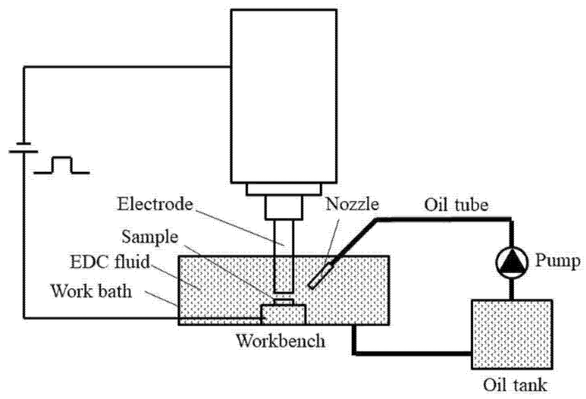
2.1. Preparation of Fe–Al Electrode

The Fe–Al electrode was sintered by pure powders of Fe (99.5 wt.%, 500 grids) and Al (99.5 wt.%, 500 grids). First, the Fe and Al powders were mixed thoroughly at a weight ratio of 10:1. Then, the mixed powders and grinding balls were put into a ball mill at a weight ratio of 1:2. The analytical nitrogen and ethanol were put into the mill together to prevent the mixed powders from oxidizing. The rolling speed of the ball mill was 300 r/min, and the roll time was 60 min. After ball milling, the mixed powders were shaped into an Fe–Al cylinder with a diameter of 25 mm under a pressure of 150 MPa. The Fe–Al cylinder was sintered at 650 °C and 400 MPa for 90 min. At the point, the Fe–Al electrode was finished.

2.2. Fe–Al Coating Fabrication Process

The selected substrate was low-carbon steel. The steel samples were cut to dimensions of 10 mm × 10 mm × 3 mm and sanded with grape 320 SiC paper to remove the oxidation layer from the steel surface. The structure of the EDC machine (D7145, Jiangsu Sanxing Machinery Manufacture Co., Ltd., Taizhou, China) is shown in Figure 1.
In this study, the Fe–Al coating fabrication parameters of EDC technology were as shown in Table 1. The as-coated substrates were ultrasonically cleaned by ethanol, acetone, and deionized water for degreasing and removing surface impurities, then dried in a vacuum oven at 35 °C for 2 h.

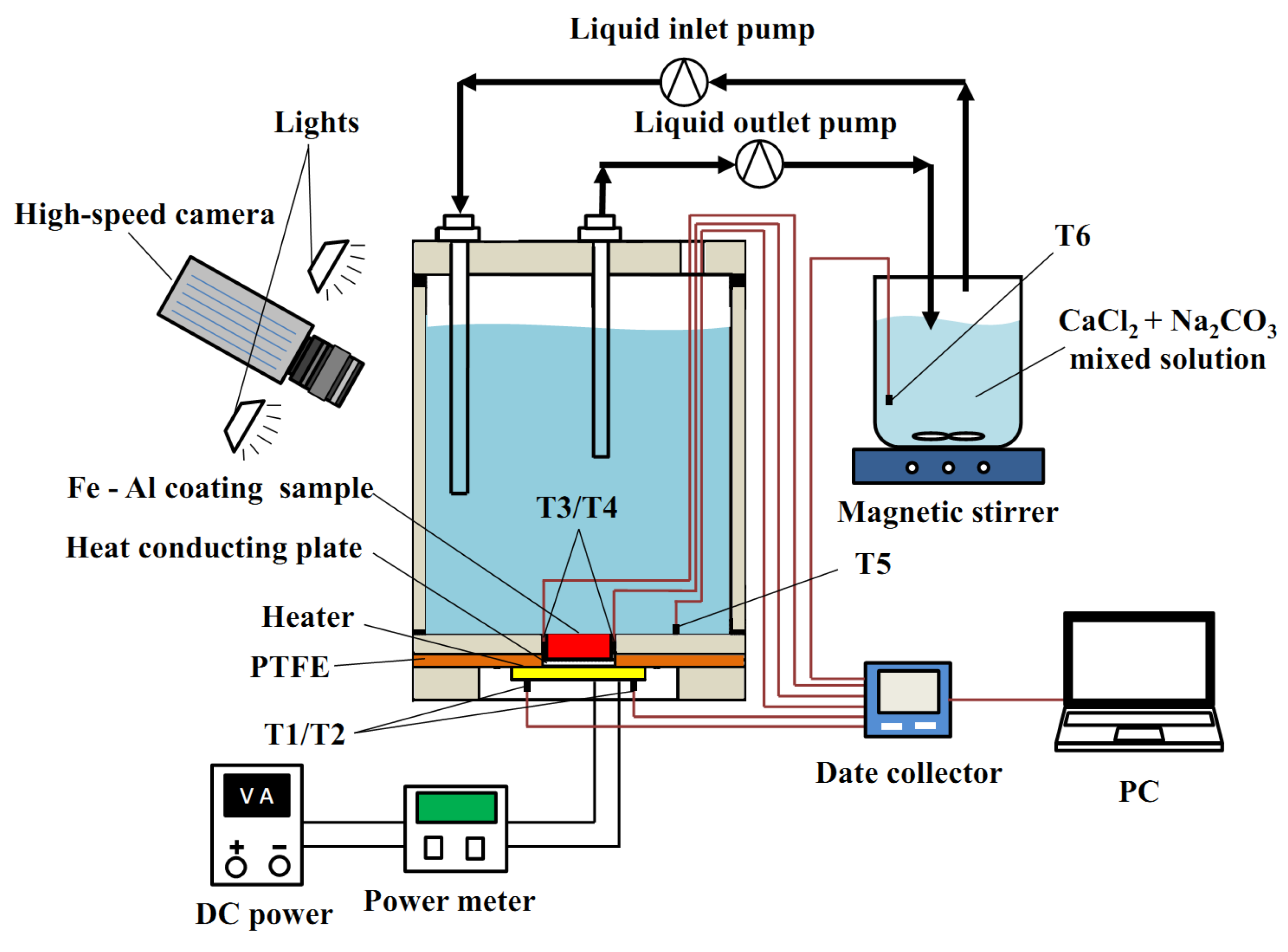
2.3. Antifouling Property Experiment

The CaCl2 + NaHCO3 mixed solution with a concentration of 0.1 mol·L−1 was used to characterize the antifouling performance of the polished and modified surfaces [1]. All chemical reagents were analytical grade, and the solvent was deionized water. The antifouling experiment was carried out on a self-made heat-transfer test device, as shown in Figure 2. All thermocouples are type K. The antifouling experiment samples, including polished surface and Fe–Al coating samples, were immersed in the fouling solution and kept in the self-made heat-transfer test device for 72 h. The input heat flux in the experiment was 98 kW·m−2 and the experiment lasted 72 h. The data collector recorded the values of all thermocouples every 5 min. During the antifouling experiment, a high-speed camera (5KF20C, FuHuang AgileDevice, Hefei, China) was employed to record the transformation procedure of the tested surface at 100 frames/s.

2.4. Characterization

The morphologies of the modified surface and the fouling were characterized by a scanning electron microscope (SEM, EVO-10, Carl Zeiss, Cambridge, UK) and the chemical compositions were analyzed via energy-dispersive spectroscopy (EDS, SmartEDX, Carl Zeiss, Cambridge, UK) equipped with SEM. The crystalline structure of the modified surface before the antifouling experiment was investigated by X-ray diffraction (XRD) using Cu Kα radiation with a wavelength of 0.15418 nm (Ultima IV, Rigaku, Japan). The surface roughness (Ra, arithmetic mean roughness) was measured via a surface roughness measuring instrument (JB-1C, Shanghai Cany Precision Instrument Co., Ltd., Shanghai, China) and the average Ra value from three random regions for each sample was reported with their error. A contact angle test was carried out by a contact angle measuring instrument (SD-CAZ2, Dongguan Sindin Precision Instrument, Incs., Dongguan, China) and the deionized water was used as the testing medium. Test drops (4 μL) were placed on the surface, and the average contact angle values measured at five different positions of the same sample, were reported.

3. Results

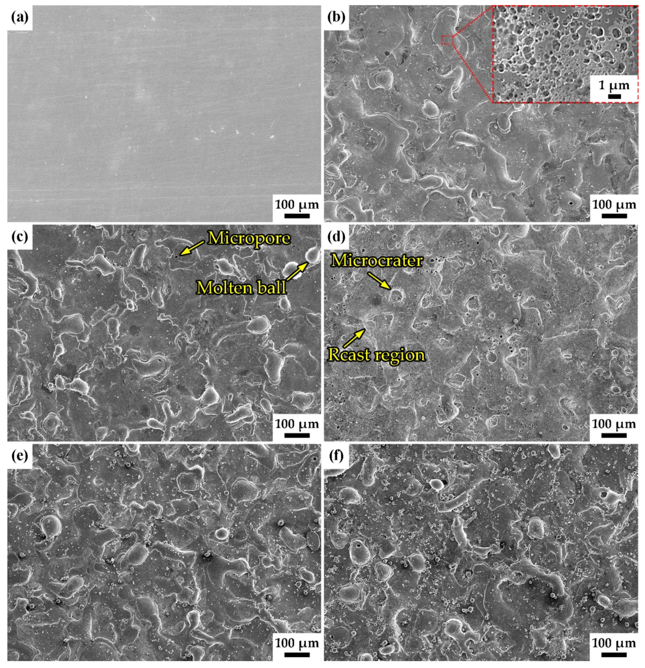
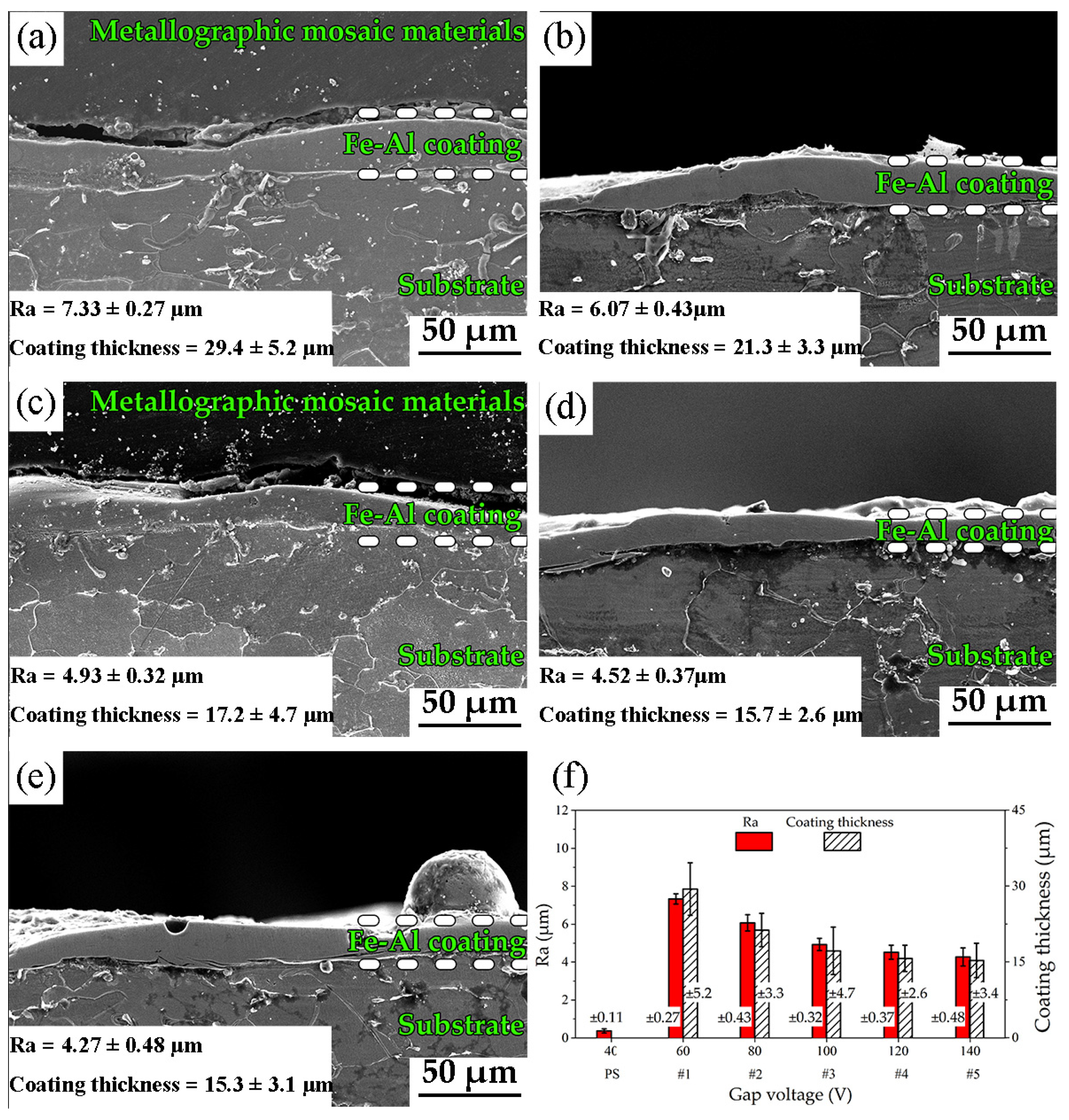
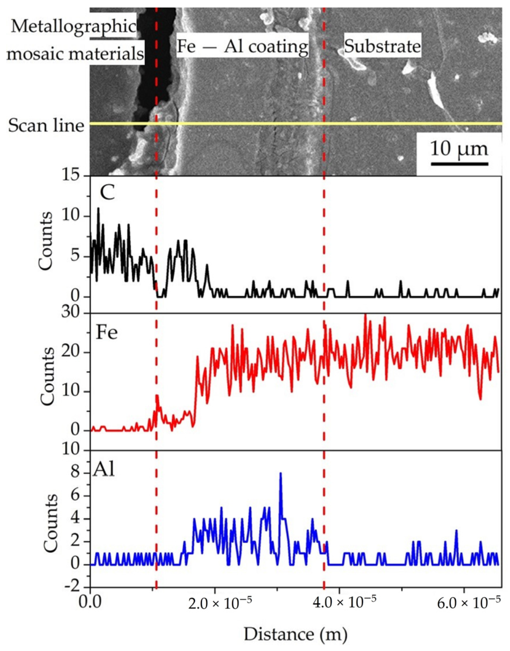
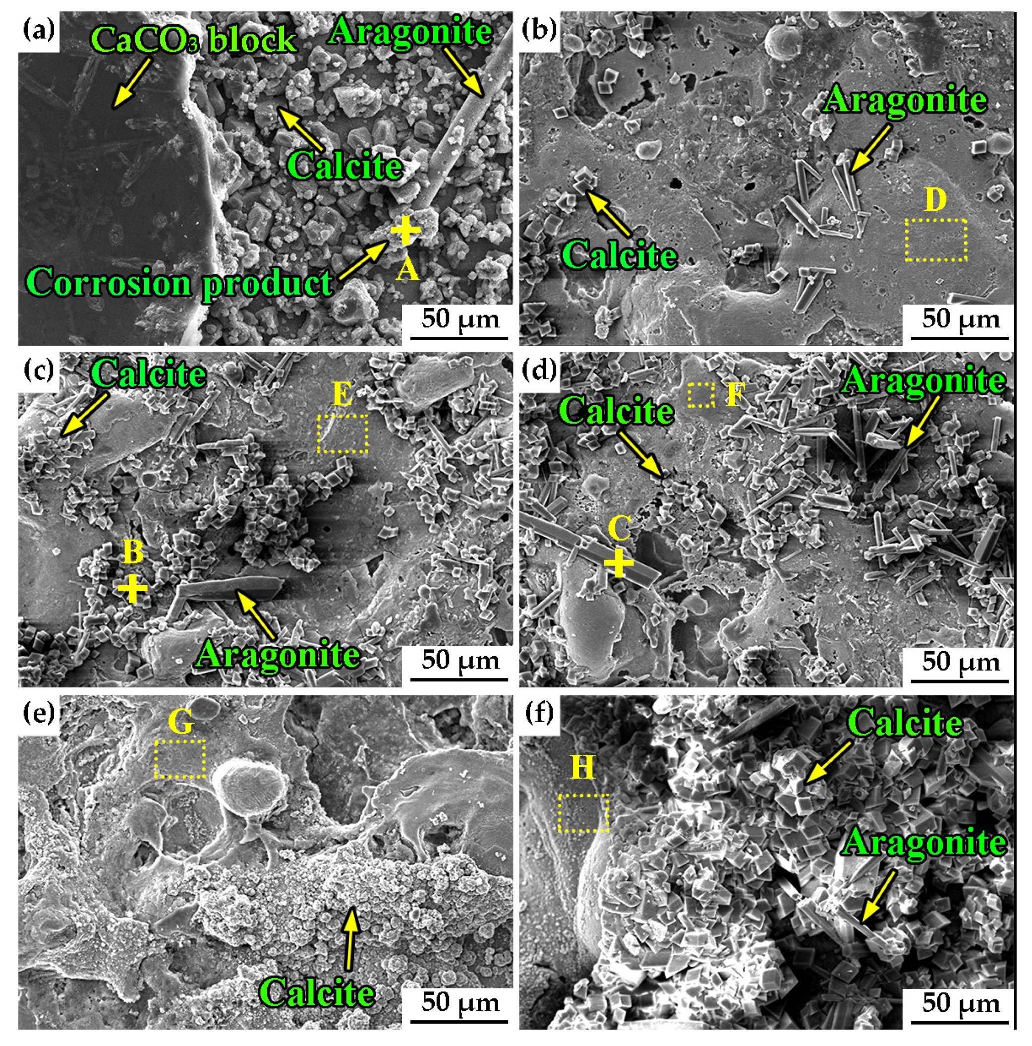
3.1. Surface Morphology and Chemical Composition

Figure 3 shows the morphology of low-carbon steel and Fe–Al coating before antifouling experiment. As shown in Figure 3a, the low-carbon steel surface is a flat surface with some scratches after polishing (PS). The Fe–Al coating samples shown in Figure 3b–f show that there are many micro–characteristics, such as micro–craters, micro–porous, molten balls, and recast regions, which are distributed homogeneously on the sample surface fabricated by EDC technology (EDCed coating). The micro-structures on the Fe–Al coating surface are typical EDM characteristics [24]. As the gap voltage increases, the number and dimension of the micro–EDM characteristics in the EDCed coatings increase, indicating that the morphology of the Fe–Al coating has become more complicated. Moreover, according to the high magnification SEM image of the Fe–Al coating sample shown as the inset in Figure 3b, there are many nanopores on the Fe–Al coating. The porosity of the Fe–Al coating was measured by Image J software (National Institutes of Health, Rockville, MD, USA), and the results are shown in Figure 4. As shown in Figure 4f, the porosity of Fe–Al coatings was reduced as the gap voltage increased from #1 to #5, and there was a sharp decrease from low gap voltage to high gap voltage.
The chemical compositions of all EDCed coatings detected by EDS are shown in Table 2. According to Table 2, the Fe, Al, and C elements, but not O, were present in the EDCed coating. The Fe and Al elements were from the electrode and the C element was from the electrical fluid, which was gasified due to the high temperature during the electrical discharge process. The results shown in Table 2 indicate that there is no oxidation or passive film formed on the EDCed coating. The reason could be the electrical discharge process of the Fe–Al electrode in the EDM fluid—there was no air in direct contact with the coating surface. The average Fe/Al ratio by weight is approximately 12.71:1, and the Al content in the Fe–Al coating decreased with the increased gap voltage. According to the results of Figure 3 and Table 2, the Fe–Al coating with micro/nanostructures, (Fe–Al MNS), is fabricated by EDC technology on the low-carbon steel substrate.
The coating thickness and roughness of Fe–Al MNS are shown in Figure 5. As shown in Figure 5a–e, the Fe–Al MNSs with different coating thicknesses are formed continuously on the substrates and no cracks appeared in the Fe–Al MNS, which indicates that Fe–Al MNS was a compact coating. According to Figure 5f, the average thickness of Fe–Al MNS decreased with the increasing gap voltage. Meanwhile, the roughness values of Fe–Al MNS samples were higher than those of PS with 0.37 ± 0.11 μm of Ra, as shown in Figure 5. The roughness values of Fe–Al MNS samples also decreased with the increase of gap voltage. The reason may be that the number of recast regions on Fe–Al MNS increased with the increasing gap voltage, which covered the coating surface to reduce the roughness.
According to the line scan result of EDS from the section of Fe–Al MNS sample shown in Figure 6, the Al element only exists in the Fe–Al MNS and the Fe element exists in both the Fe–Al MNS and the substrate. The results shown in Figure 6 indicate that there is a metallurgical layer between Fe–Al MNS and the substrate, which could enhance the bonding strength between them. Moreover, C mainly exists on the surface of Fe–Al MNS and the contents in both Fe–Al MNS and substrate are low. Because the high temperature during the electrical discharge process of the Fe–Al electrode resulted in the gasification of EDM fluid, the carbon element of EDM fluid combined with the Fe and Al elements to form carbides in the coating. Thus, some carbides were formed on the surface of Fe–Al MNS, which was consistent with the results of Table 2.

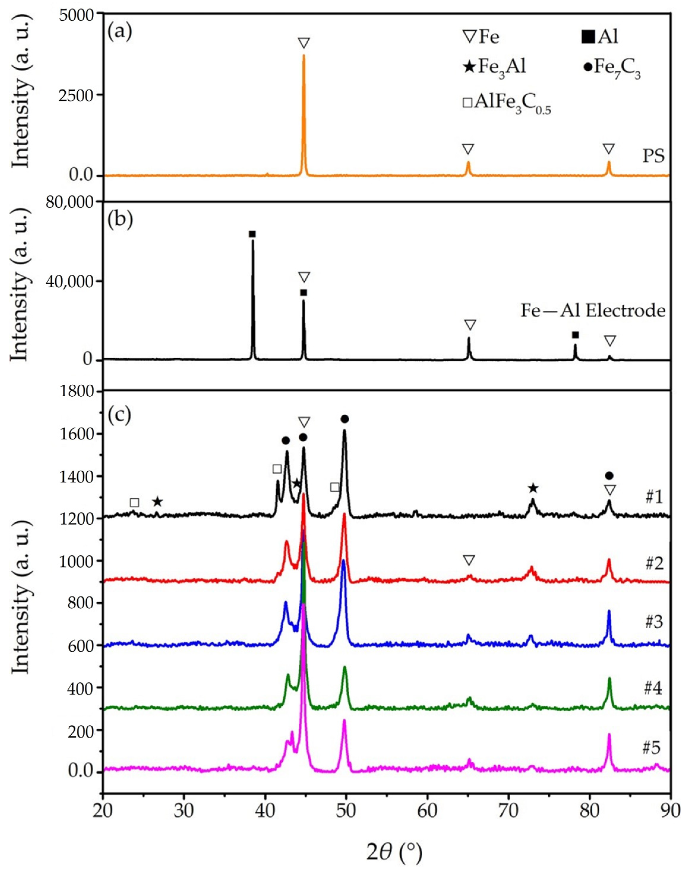
3.2. Phase Combination of Fe–Al Micro/Nanostructures (MNS)

Figure 7 shows the XRD results of PS, the Fe–Al electrode and Fe–Al MNS samples. As shown in Figure 7a, there is only Fe element characteristic peaks in the XRD results. According to the XRD results of the Fe–Al electrode shown in Figure 7b, only Fe and Al phases exist, without the Fe–Al phase. The reason could be that the sintering conditions of the Fe–Al electrode described in Section 2.1 cannot achieve the formation conditions of Fe–Al composition [25,26], and the powders of Fe and Al only adhered in the Fe–Al electrode. The XRD results of Fe–Al MNS samples #1, #2, and #3 are shown in Figure 7c. According to the XRD results of Figure 7c, the characteristic peaks of Fe and Al also appeared in all Fe–Al MNS samples, and there are some typical characteristic peaks of Fe3Al phase and carbides, such as Fe7C3 and AlFe3C0.5 phases. The XRD results shown in Figure 7c indicate that the Fe3Al phase was formed in the Fe–Al MNS. Because of the high temperature and pressure during the electrical discharge process of the Fe–Al electrode [27,28], the gap between the Fe–Al electrode and working piece acted like a sintering furnace, which promoted the formation of the Fe3Al phase in the coating.
In addition, the intensity of characteristic peaks for the AlFe3C0.5 phase, showing in Figure 7c, decrease with the increasing gap voltage, indicating that the content of AlFe3C0.5 phase in Fe–Al MNS decreases. This is because the impact energy of electrical discharge from the Fe–Al electrode in the high gap voltage condition was so high that the electrical discharge had blown up the coating, and the formed carbides in the coating were widespread [29]. Therefore, the content of carbides in the coating was reduced with the increasing gap voltage. A similar phenomenon for the Fe7C3 phase was also observed in all Fe–Al MNS samples.

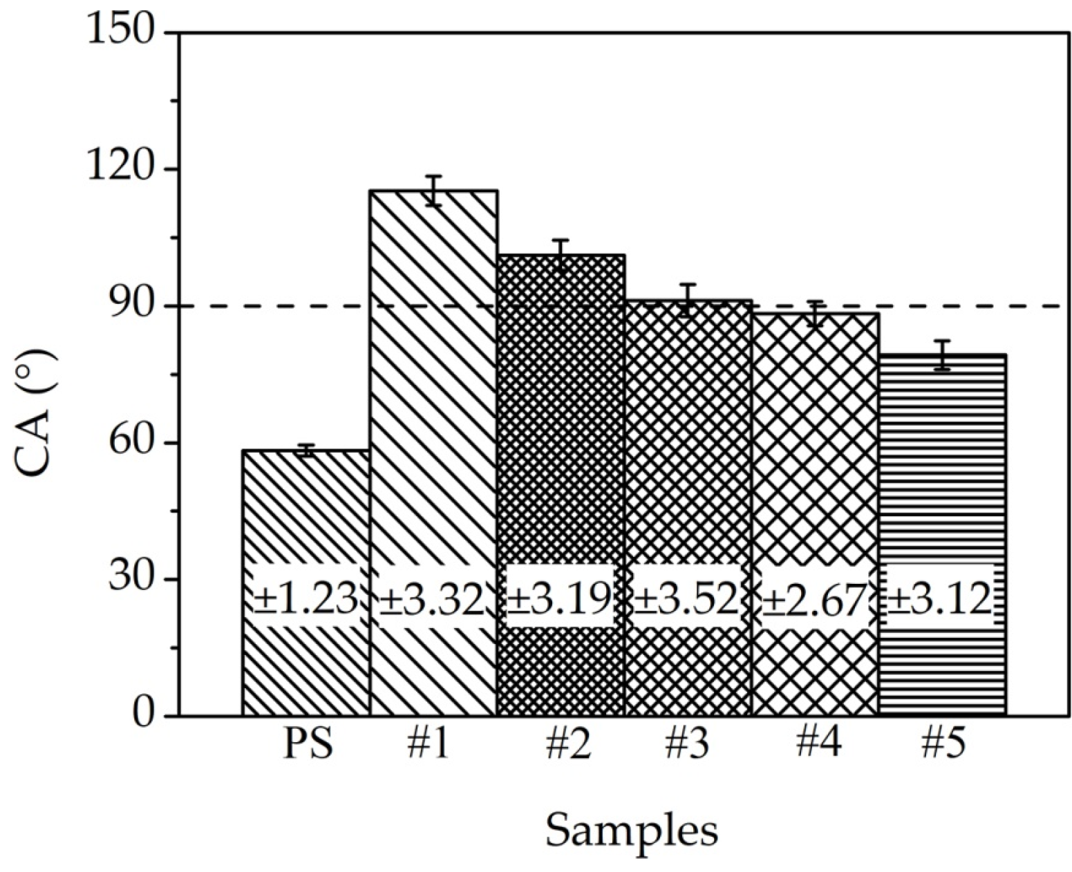
3.3. Surface Wettability

Figure 8 shows the contact angles of PS and Fe–Al MNS samples. As shown in Figure 8, the contact angle (CA) of PS is below 90°, indicating that PS is the hydrophilic surface. Although the CAs of Fe–Al MNS samples are all higher than those of PS, but CAs of #4 and #5 Fe–Al MNS are below 90°, which indicate that #4 and #5 Fe–Al MNS are also the hydrophilic surfaces. The CAs of Fe–Al MNS samples #1, #2, and #3 with low gap voltage (<100 V) are over 90°, indicating that the Fe–Al MNS samples with a low gap voltage are hydrophobic surfaces. Meanwhile, the CAs of Fe–Al MNS samples decreased with the increasing gap voltage. With the increasing gap voltage, the morphologies of Fe–Al MNS become increasingly complex, according to Figure 3b–f. A lot of micro/nanostructures on the surface were covered by the recast region, causing the surface roughness of Fe–Al MNS to decrease, as shown in Figure 5. According to the Wenzel [30] and Cassie-Baxter [31] wettability mechanism, the contact between the water droplet and the Fe–Al MNS is complex. The water droplet could infiltrate into the micro/nanostructures or be isolated by the trapped air in the micro/nanostructures. There are not enough micro-pores in the Fe–Al MNS with high gap voltage to trap the air for the isolation of water droplet, as shown in Figure 3f and Figure 4. Thus, the #5 Fe–Al MNS fabricated with a high gap voltage shows the lowest CA.

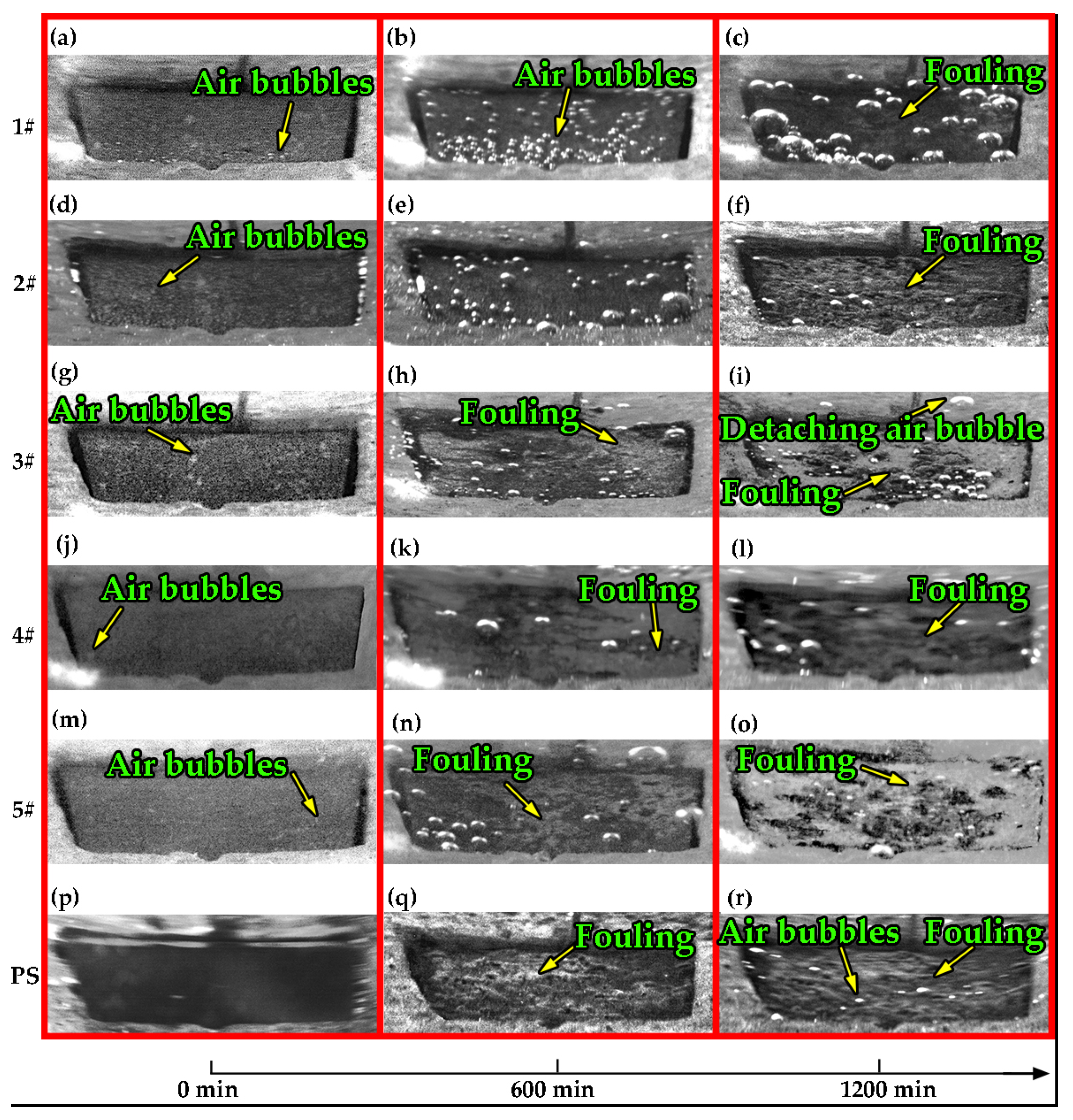
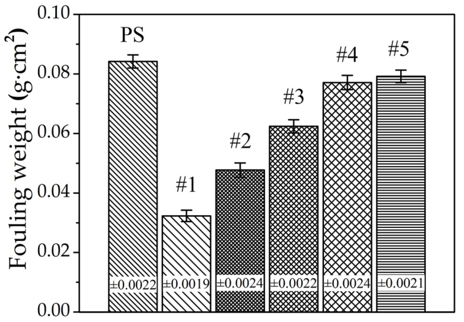
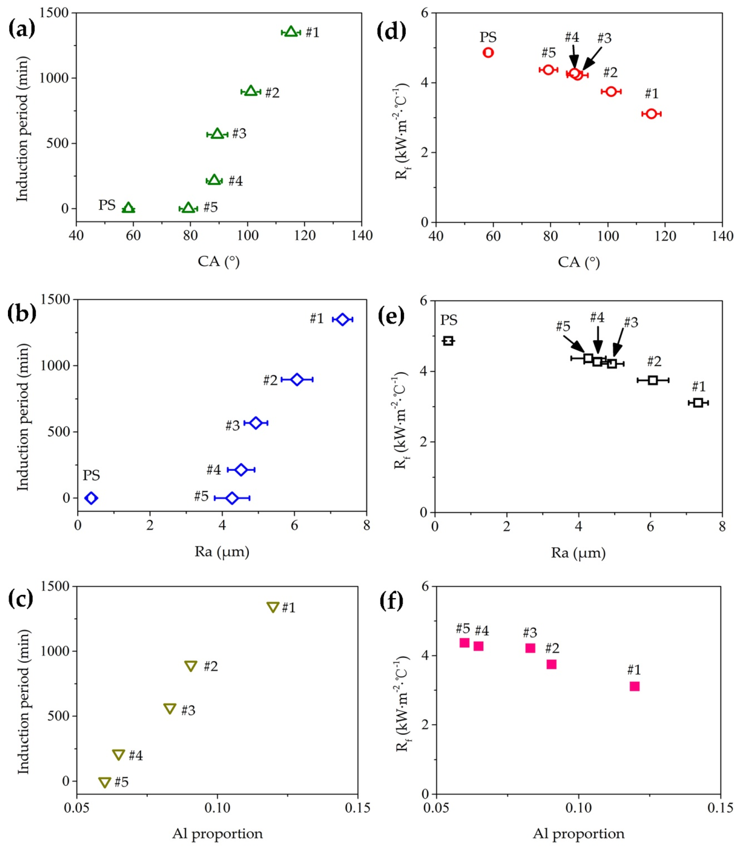
3.4. Antifouling Property of Fe–Al MNS Sample

The antifouling property can be investigated by the fouling resistance Rf, according to Equation (1) [29]:
R f = 1 U t 1 U 0
where Ut is the overall heat-transfer coefficient at generic time t, while U0 is the overall heat-transfer coefficient at the start of the operation, i.e., when the heat-transfer surfaces are supposed to be clean.
After the antifouling experiment, the Rf plots of PS and Fe–Al MNS samples were as shown in Figure 9. As shown in Figure 9, the Rf plots of the PS sample increased with time and reached a constant value (Rf max) rapidly, which indicates that the fouling deposited on the heat-transfer surface was stable. Compared with the Rf plot of PS, the Rf plots of the Fe–Al MNS samples shown in Figure 9 are different. There is an obvious period with a zero value of Rf, which is defined as an induction period of the heat-transfer surface, appearing in the Rf plots of #1, #2, #3, and #4. There is no induction period in the Rf plot of #5 Fe–Al MNS. This result indicates that there is no induction period for PS and there are different induction periods for each Fe–Al MNS sample, which is similar to previous research [32]. Bogacz [33] found that there was a significant influence between the wettability of the heat-transfer surface and the fouling adhesion, which was more significant than the influence of roughness. For the heat-transfer surface with the lower contact angle, the lower removal rate appeared for the deposited fouling on the surface [34,35]. Thus, it was difficult for the fouling crystals deposited on the PS to be removed, leading the Rf of PS to increase to the Rf maximum in a short period. Moreover, the hydrophobic property of #1, #2, and #3 Fe–Al MNS inhibits the adhesion of fouling crystals. The reason is that the air can be trapped in the micro/nanostructures of Fe–Al MNS, which inhibits contact with the fouling on the Fe–Al MNS. While #4 and #5 Fe–Al MNS are hydrophilic surfaces, the droplet can infiltrate the micro/nanostructures, resulting in an increased deposition area of fouling. But the porosity of #4 Fe–Al MNS is higher than that of #5 Fe–Al MNS shown in Figure 4, which can reduce the contact between the surface and the fouling solution by trapped air in the micro/nanostructures. As a result, there is a short induction period in the Rf plot of #4 Fe–Al MNS as shown in Figure 9, and without induction period for #5 Fe–Al MNS. In addition, the micro/nanostructures of Fe–Al MNS also increase the nuclear area of boiling bubbles [36,37], which affects the adhesion of fouling crystals near the heat-transfer surface. Thus, because of the combined action of surface wettability and boiling bubbles, there are different slope values of Rf plot for each Fe–Al MNS samples. Without the combined action, the slope value of PS Rf plot is highest.
A high-speed camera was employed to capture the surface morphologies of Fe–Al MNS during the antifouling experiment, which are shown in Figure 10. As shown in Figure 10a,d,g,j,m,p, there were some air bubbles trapped on the Fe–Al MNS, and no air bubbles were trapped on the PS at 0 min. When the Fe–Al MNS sample was immersed in the fouling solution, the trapped air appeared as bubbles on the Fe–Al MNS surface [38]. Compared with Fe–Al MNS, there were no micro/nanostructures on the surface of PS to trap air, so no air bubbles appeared on the surface. Additionally, the number and dimension of air bubbles that adhered on the surface of #4 and #5 Fe–Al MNS were less than on the other Fe–Al MNS. Over time, as shown in Figure 10b,e,h,k,n,q, a growing number of air bubbles appeared on the Fe–Al MNS due to the enhancement of boiling bubble activity [39,40]. The detachment of boiling bubbles from the Fe–Al MNS helped to dislodge the loose fouling from the surface with a wiping action [32]. Thus, there was no fouling deposited on the surface of #1 and #2 and some fouling deposited on the surface of #3, #4, and #5 where there were no boiling bubbles. Compared with Fe–Al MNS, the PS was totally different, with a lot of fouling deposited, which covered the whole heat-transfer surface with a fouling layer. Because of the hydrophilic property of PS, it was convenient that the fouling crystals in the fouling solution were deposited on the surface. Furthermore, there was no wiping action on the surface of PS due to there being no detachment of boiling bubbles, resulting in the deposited fouling layer being strongly adhered on the substrate. At 1200 min, as shown in Figure 10c,f,i,l,o,r, the fouling layer adhered on the PS was still stable and fouling also appeared on all Fe–Al MNS samples. The fouling deposited on the #5 Fe–Al MNS was the greatest of all the Fe–Al MNS samples, while on the other Fe–Al MNS it was reduced gradually from #4 to #1. As shown in Figure 10c, there were a lot of large air bubbles on the #1 Fe–Al MNS, which could isolate the Fe–Al MNS from the fouling solution. In addition, the wipe action from the detachment of bubbles continued to affect the deposition of fouling. These phenomena were also observed on the other Fe–Al MNS in Figure 10f,i,l,o. However, the average dimension of detaching air bubbles was gradually decreased from #2 to #5 Fe–Al MNS. According to Figure 8, the hydrophobic property of #2, #3, #4, and #5 Fe–Al MNS was weaker than that of #1 Fe–Al MNS, resulting in the fouling crystal nucleus being able to adhere in the micro/nano pores of #2, #3, #4, and #5 Fe–Al MNS to impede the formation of boiling bubbles. Thus, the wiping action from the detachment of boiling bubbles was weakened, so fouling could be easily deposited on the #2, #3, #4, and #5 Fe–Al MNS compared with #1 Fe–Al MNS.
The fouling weight per area of all samples after the antifouling experiment is shown in Figure 11, indicating that the fouling deposited on the PS was the highest. Although there was fouling on the Fe–Al MNS samples, the amount was less than that on the PS and gradually increased. As shown in Figure 9, the induction periods of #1, #2, #3, and #4 Fe–Al MNS were 1350, 896, 568, and 214 min, respectively. Moreover, the Rf max of all Fe–Al MNS samples was lower than that of the PS sample. Moreover, the Rf max of #1 Fe–Al MNS was lowest in all samples and the Rf max of the other Fe–Al MNS samples were close, a situation that also can be observed for the thickness of the deposited fouling layer in Figure 10c,f,i,l,o.
Figure 12 shows the fouling morphologies on the PS and Fe–Al MNS samples after 72-h antifouling experiments. As shown in Figure 12a, a large amount of fouling was deposited on the PS and some of the fouling crystals were large, which increased the Rf of PS to the highest value. The #1, #2, #3, #4, and #5 Fe–Al MNS samples after antifouling experiments are shown in Figure 12b–f, respectively. As shown in Figure 12b–f, there was also a lot of fouling deposited on the surface of Fe–Al MNS after the antifouling experiment. Compared with the fouling morphology on PS, the fouling crystals deposited on the Fe–Al MNS were smaller and distributed randomly. According to the EDS results of all samples after the antifouling experiment shown in Table 3 and Figure 12, the composition marked A is the corrosion product of the substrate. The compositions marked B with a cube-like shape and C with a needle-like shape show typical CaCO3 fouling [41], which also existed on the PS after the antifouling experiment showing in Figure 12a. There is no Ca element detected in the test regions without deposition of fouling crystals on the surface of Fe–Al MNS, which are marked D, E, F, G and H in Figure 12b–f. This indicates that there are no fouling crystals deposited on the whole surface of Fe–Al MNS, but there may be a passive film or oxidation film on the surface of Fe–Al MNS. In addition, the number of fouling crystals deposited on the Fe–Al MNS increased gradually from #1 to #5 shown in Figure 12b–f. The results indicate that Fe–Al MNS shows better antifouling property in this study and the antifouling property can be affected by the surface characteristics. Moreover, although there are a lot of Cl ions in the fouling solution, which can react with the Fe element in the Fe–Al MNS, the activity of Al is so strong that a passive film forms preferentially to protect the Fe–Al MNS [25]. Thus, there is no corrosion product, so the fouling crystals are easily absorbed, as observed obviously on the surface of Fe–Al MNS.

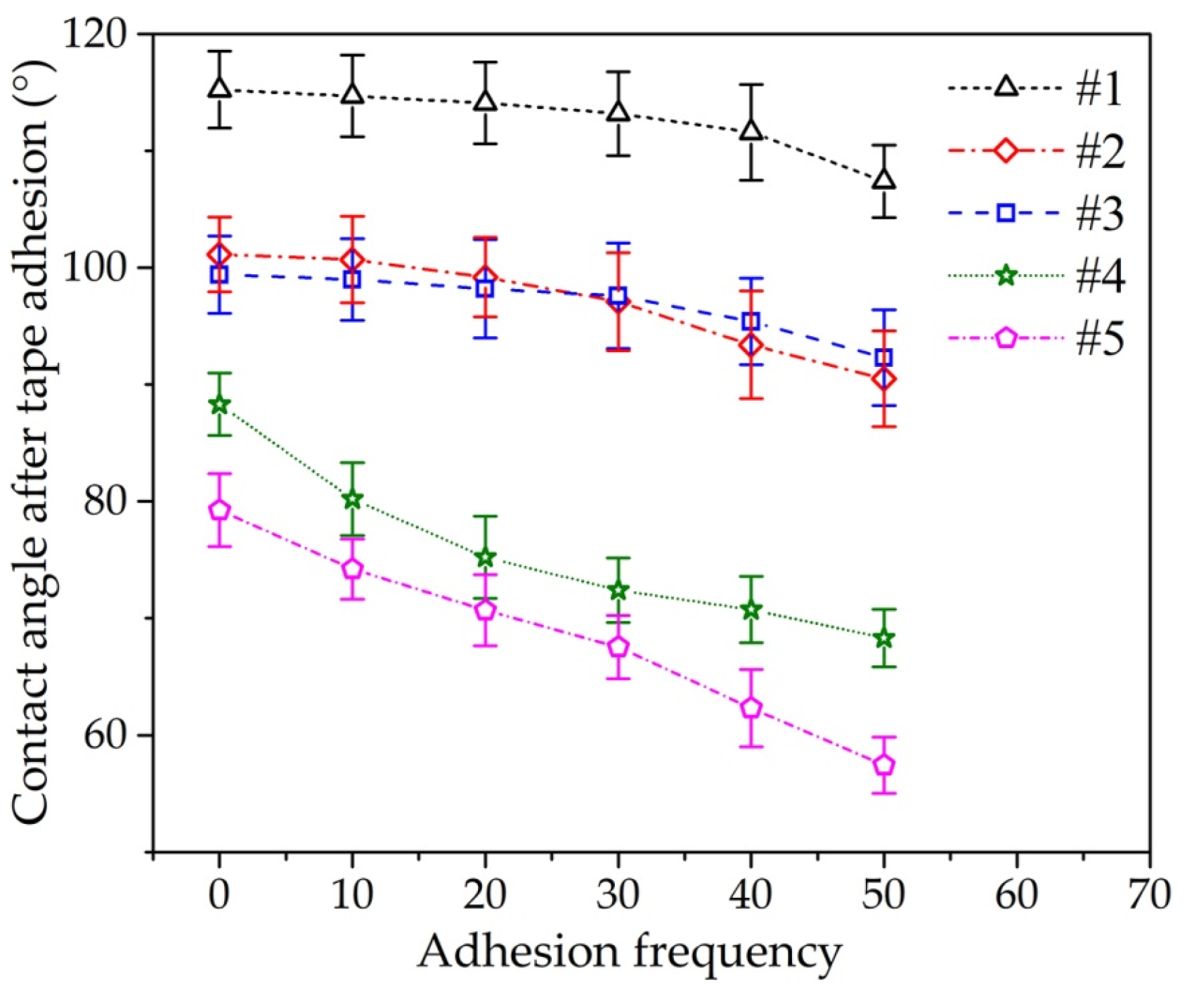
3.5. Durability Test of Fe–Al MNS

The durability is an important factor in the industrial application of Fe–Al MNS. According to the ASTM standard (ASTM D3359–17) [42], the hydrophobic property of an Fe–Al MNS sample after semi-transparent tape adhesions is measured by a contact angle measuring instrument. The tape test for the Fe–Al MNS was repeated three times per condition. The contact angle of Fe–Al MNS after the tape adhesion is shown in Figure 13. As shown in Figure 13, the contact angle values of Fe–Al MNS decreased with the increase in tape adhesion frequency, with the reduction in CA for #1, #2, #3, #4, and #5 being 6.81%, 10.51%, 7.15%, 22.64%, and 27.52%. This may be because the micro/nanostructures, such as molten balls and nanopores, are destroyed when the tape is torn off from the surface. In addition, after the durability test, Fe–Al MNS sample #1 remained in a hydrophobic surface state, which can improve the antifouling property of the surface. Fe–Al MNS samples #2 and #3 retained their hydrophobic property after 40 tests, but it was difficult for these two Fe–Al MNS samples to maintain the hydrophobic property after 50 tests. That is, the antifouling property of Fe–Al MNS is durable and there is a prospect for industrial application.

4. Discussion

According to the above results, Fe–Al MNS shows a superior antifouling property. The surface characteristics of Fe–Al MNS affected the antifouling property of Fe–Al MNS, and the relations between the surface characteristics and antifouling property are shown in Figure 14. As shown in Figure 14a–c, with the increase in CA, Ra, and Al proportion, the induction period increased. The trend of induction period versus CA, Ra, and Al proportion increased sharply at first, but the rate of increase decreased, as shown in Figure 14a–c, which indicates that there is a limit to the impact of induction period for the Al proportion in the coating. As shown in Figure 14d–f, the trend plots of Rf max versus CA, Ra, and Al proportion is different from the induction period. The Rf max decreased with the increase in CA, Ra, and Al proportion.
Ozden et al. [40] observed that the better the hydrophobicity of the heat-transfer surface, the lower the amount of fouling deposited on the surface. In this study, as the hydrophobicity of Fe–Al MNS reduces with the increasing gap voltage, the contact area between the fouling solution and Fe–Al MNS is increased according to the Cassie–Baxter model [31]. This means that the amount of fouling deposited on the Fe–Al MNS increased with the increasing gap voltage, and as shown in Figure 5f, the CA and Ra of Fe–Al MNS is reduced with increasing gap voltage. Therefore, the Rf max is increased with reduced CA and Ra shown in Figure 14d,e, which can also be demonstrated by the amount of fouling deposited on the Fe–Al MNS shown in Figure 10c,f,i,l,o. On the other hand, due to the slow flow rate of the test vessel, the fouling solution flowed like a laminar flow, and then the fouling crystal nucleus in the solution was deposited on the heat-transfer surface by the molecular transport mechanism, which limited the deposition rate of fouling on the Fe–Al MNS [11]. Hence, the induction period of Fe–Al MNS is extended, as shown in Figure 9. Moreover, the micro/nanostructures increased the boiling bubble activity, which creates a porous surface [41,42], and the detachment of the boiling bubble dislodged the loose fouling from the surface with wiping action [9,43]. As shown in Figure 14c,f, the Al proportion in the coating also influenced the antifouling property. A higher Al proportion can help with the formation of the passive film and effectively inhibit the diffusion of Fe2+ and Al2+ ions from the coating [44], which induce the formation and deposition of fouling in the fouling solution [45]. As described above, the co-effect among the hydrophobic property of Fe–Al MNS, the wiping action of the detachment of boiling bubbles, and the Al passive film affects the molecular transfer behavior of the fouling crystal nucleus to mitigate fouling deposition. As a result, compared with PS, the Fe–Al MNS shows superior antifouling property. Meanwhile, due to the above effects, the induction periods of different Fe–Al MNS samples are extended and different, as shown in Figure 14. The antifouling mechanism of Fe–Al MNS is shown in Figure 15.

5. Conclusions

The Fe–Al coating with micro/nanostructures (Fe–Al MNS) is fabricated by electrical discharge machining (EDC) technology. The micro/nanostructures, chemical composition, wettability, and antifouling behavior of the resulting surface are evaluated and compared with the polished surface of low-carbon steel. The conclusions are as follows:
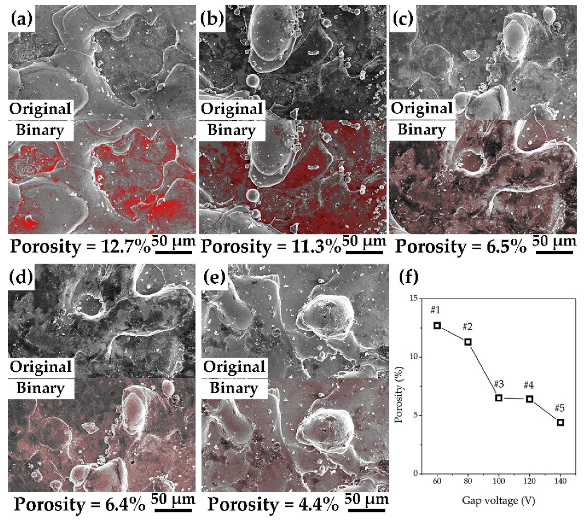
  • According to the EDC technology and Fe–Al electrode, the Fe–Al coating with micro/nanostructures is fabricated on a low-carbon steel substrate. The roughness and coating thickness of Fe–Al MNS decreased with the increasing gap voltage of EDC. The porosity of Fe–Al MNS decreased sharply from a low gap voltage to a high gap voltage. The main phases of Fe–Al MNS were combined by Fe3Al, Fe7C3, and AlFe3C0.5.
  • The Fe–Al MNS showed a hydrophobic property. The contact angle of Fe–Al MNS decreased with the increasing EDC gap voltage. Due to the effect of micro/nanostructures of Fe–Al MNS, water droplets could be isolated by tapped air or infiltration in the coating, resulting in the regulation of the hydrophobic property.
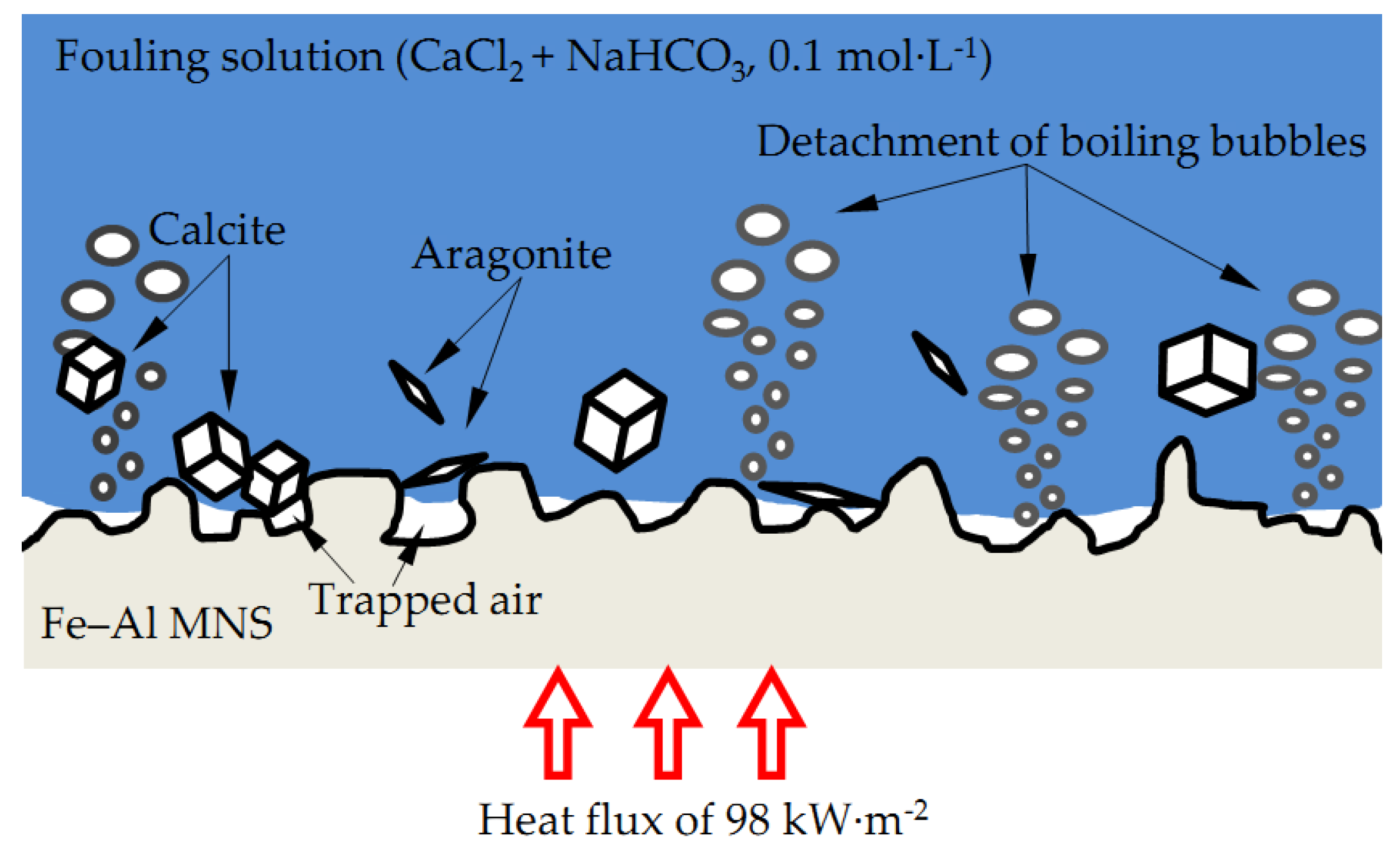
  • Compared with the polished surface of low-carbon steel, Fe–Al MNS shows better antifouling performance, with an extended induction period and lower Rf max value. The antifouling property of Fe–Al MNS samples decreased with the increasing EDC gap voltage. One potential reason is that the hydrophobic property of the Fe–Al MNS inhibited the adhesion of fouling crystals. Another reason could be that the micro/nanostructures of Fe–Al MNS also increased the nuclear area of boiling bubbles, which can dislodge the loose fouling crystals from the Fe–Al MNS surface by a wiping action when the boiling bubbles detach. Finally, due to the activity of the Al element in the Fe–Al MNS, a passive film was formed on the Fe–Al MNS surface to mitigate the corrosion products, which could easily absorb the fouling crystals. As a result, the adhesion behavior of the fouling crystals was inhibited. Thus, the co-effect of the above reasons supports the fact that Fe–Al MNS showed a superior antifouling property. Meanwhile, after the durability test, the hydrophobic property of Fe–Al MNS decreased slightly, indicating that there is an industrial application prospect for Fe–Al MNS.
  • According to the results of this study, micro/nanostructures enhance the antifouling property of Fe–Al MNS. This indicates that there is a relationship between the scale effect of micro/nanostructures and the antifouling property, which is worth investigating further.

Author Contributions

Conceptualization, Z.H.; Methodology, Z.H. and Z.F.; Software, Z.H., D.W., and Y.C.; Validation, Z.H., Z.F., D.W., and Y.C.; Formal Analysis, Z.F.; Investigation, Z.H. and D.W.; Resources, Z.H., S.L., and C.M.; data curation, Z.H. and S.L.; Writing—Original Draft Preparation, Z.H.; Writing—Review and Editing, Z.H. and Z.F.; Visualization, Z.H.; Supervision, D.W.; Project Administration, Z.H.; Funding Acquisition, Z.H. and Y.C. All authors have read and agreed to the published version of the manuscript.

Funding

This research was funded by the Featured Innovative Natural Science Foundation via the Innovative and Strong School Project for Ordinary University of Guangdong Province of China (Grant No. 2017KTSCX126), the Opening Foundation of Guangdong Petrochemical Equipment Engineering and Technology Research Center of China (Grant No. 2017JJ517010), the Science and Technology Planning Project of Maoming City (Grant No. 2020508), the Natural Science Foundation of Guangdong Province (Grant No. 2017A030307007), and the Science and Technology Planning Project of Guangdong Province (Grant No. 2015A030401103).

Conflicts of Interest

The authors declare no conflict of interest.

References

  1. Wei, L.; Kan, Z.; Manglik, R.M.; Li, G.Q.; Bergles, A.E. Investigation of CaCO3 fouling in plate heat exchangers. Heat Mass Transfer. 2016, 52, 2401–2414. [Google Scholar]
  2. Kazi, S.N. Fouling and fouling mitigation of calcium compounds on heat exchangers by novel colloids and surface modifications. Rev. Chem. Eng. 2019, 36, 653–685. [Google Scholar] [CrossRef]
  3. Hu, M.; Zheng, S.; Mi, B. Organic fouling of graphene oxide membranes and its implications for membrane fouling control in engineered osmosis. Environ. Sci. Technol. 2016, 50, 685–693. [Google Scholar] [CrossRef] [PubMed]
  4. Perreault, F.O.; Jaramillo, H.; Xie, M.; Ude, M.; Nghiem, L.D.; Elimelech, M. Biofouling mitigation in forward osmosis using graphene oxide functionalized thin-film composite membranes. Environ. Sci. Technol. 2016, 50, 5840–5848. [Google Scholar] [CrossRef] [PubMed] [Green Version]
  5. Wang, Z.; Fei, G.; Xia, H.; Zuilhof, H. Dual water-healable zwitterionic polymer coatings for anti-biofouling surfaces. J. Mater. Chem. B. 2018, 6, 6930–6935. [Google Scholar] [CrossRef] [PubMed] [Green Version]
  6. He, Z.R.; Liu, C.S.; Jie, X.H.; Lian, W.Q.; Luo, S.T. Preparation of anti-fouling heat transfer surface by magnetron sputtering a-C film on electrical discharge machining Cu surface. Surf. Coat. Tech. 2019, 369, 44–51. [Google Scholar] [CrossRef]
  7. Weeranoppanant, N.; Amar, L.; Tong, E.; Faria, M.; Hill, M.I.; Leonard, E.F. Modeling of fouling in cross-flow microfiltration of suspensions. AIChE J. 2019, 65, 207–213. [Google Scholar] [CrossRef] [Green Version]
  8. Malayeri, M.R.; Evangelidou, M. Enhanced heat transfer and fouling propensity of DLC coated smooth and finned tubes during external nucleate boiling. J. Heat Transfer. 2016, 138, 081502. [Google Scholar] [CrossRef]
  9. He, Z.R.; Liu, C.S.; Gao, H.Y.; Jie, X.H.; Lian, W.Q. Experimental study on the anti-fouling effects of EDM machined hierarchical micro/nano structure for heat transfer surface. Appl. Therm. Eng. 2019, 162, 114248. [Google Scholar] [CrossRef]
  10. He, Z.R.; Luo, S.T.; Liu, C.S.; Jie, X.H.; Lian, W.Q. Hierarchical micro/nano structure surface fabricated by electrical discharge machining for anti-fouling application. J. Mater. Res. Technol. 2019, 8, 3878–3890. [Google Scholar] [CrossRef]
  11. Bogacz, W.; Lemanowicz, M.; Rashed, M.H.A.; Nakonieczny, D.; Piotrowski, T.; Wójcik, J. Impact of roughness, wettability and hydrodynamic conditions on the incrustation on stainless steel surfaces. Appl. Therm. Eng. 2017, 112, 352–361. [Google Scholar] [CrossRef]
  12. Lv, Y.; Liu, M.; Xu, Y. Corrosion and Fouling Behaviors on modified stainless steel surfaces in simulated oilfield geothermal water. Prot. Met. Phys. Chem. Surf. 2018, 54, 526–535. [Google Scholar] [CrossRef]
  13. Lv, Y.; Liu, M. Corrosion and fouling behaviours of copper-based superhydrophobic coating. Surf. Eng. 2018, 35, 542–549. [Google Scholar] [CrossRef]
  14. Steinhagen, H.M.; Malayeri, M.R.; Watkinson, A.P. Fouling of heat exchangers-new approaches to solve an old problem. Heat Transfer Eng. 2005, 26, 1–4. [Google Scholar] [CrossRef]
  15. Steinhagen, H.M.; Malayeri, M.R.; Watkinson, A.P. Heat exchanger fouling: Environmental impacts. Heat Transfer Eng. 2009, 30, 773–776. [Google Scholar] [CrossRef]
  16. Li, N.N.; Wang, M.Z.; Li, Y.S.; Chen, G.; Li, P. Corrosion behavior of Fe–Al coatings fabricated by pack aluminizing method. Acta Metall. Sin. 2016, 29, 813–819. [Google Scholar] [CrossRef] [Green Version]
  17. Reichardt, A.; Shapiro, A.A.; Otis, R.; Dillon, R.P.; Borgonia, J.P.; McEnerney, B.W.; Hosemann, P.; Beese, A.M. Advances in additive manufacturing of metal-based functionally graded materials. Int. Mater. Rev. 2020, 1–29. [Google Scholar] [CrossRef]
  18. Senderowski, C.; Chodala, M.; Bojar, Z. Corrosion behavior of detonation gun sprayed Fe–Al type intermetallic coating. Materials 2015, 8, 1108–1123. [Google Scholar] [CrossRef] [Green Version]
  19. Vasylyev, M.A.; Mordyuk, B.N.; Sidorenko, S.I.; Voloshko, S.M.; Burmak, A.P. Corrosion of 2024 alloy after ultrasonic impact cladding with iron. Surf. Eng. 2017, 34, 324–329. [Google Scholar] [CrossRef]
  20. Gao, H.; Xie, W.; Zhang, H.; Shen, W.; He, Y. Modification of the reactive synthesis of porous FeAl with addition of Si. Mater. High Temp. 2019, 36, 1–8. [Google Scholar] [CrossRef]
  21. Tyagi, R.; Mahto, N.K.; Das, A.K.; Mandal, A. Preparation of MoS2 + Cu coating through the EDC process and its analysis. Surf. Eng. 2020, 36, 86–93. [Google Scholar] [CrossRef]
  22. Xie, Z.J.; Mai, Y.J.; Lian, W.Q.; He, S.L.; Jie, X.H. Titanium carbide coating with enhanced tribological properties obtained by EDC using partially sintered titanium electrodes and graphite powder mixed dielectric. Surf. Coat Tech. 2016, 300, 50–57. [Google Scholar] [CrossRef]
  23. So, J.Y.; Bae, W.G. Fabrication of superhydrophobic metallic surface by wire electrical discharge machining for seamless roll-to-roll printing. Metals 2018, 8, 228. [Google Scholar]
  24. Tyagi, R.; Das, A.K.; Mandal, A. Electrical discharge coating using WS2 and Cu powder mixture for solid lubrication and enhanced tribological performance. Tribol. Int. 2018, 120, 80–92. [Google Scholar] [CrossRef]
  25. Mandal, P.; Mondal, S.C. Surface characteristics of mild steel using EDM with Cu-MWCNT composite electrode. Mater. Manuf. Process. 2019, 34, 1326–1332. [Google Scholar] [CrossRef]
  26. Wismogroho, A.S.; Widayatno, W.B.; Suryadi; Zaini Thosin, K.A.; Rochman, N.T.; Sueyoshi, H. Iron aluminide coating on Al by mechanical alloying. Surf. Eng. 2011, 27, 126–133. [Google Scholar] [CrossRef]
  27. Muralidharan, B.; Chelladurai, H.; Singh, P.; Roy, M.K. Single-spark analysis of electro-discharge deposition process. Mater. Manuf. Process. 2015, 31, 1853–1864. [Google Scholar] [CrossRef]
  28. Peng, Z.; Feng, T.; Wei, Z.; Zhang, Y.; Li, Y. Directly writing patterning of conductive material by high voltage induced weak electric arc machining (HV-μEAM). Coatings 2019, 9, 538. [Google Scholar] [CrossRef] [Green Version]
  29. Yue, X.; Yang, X. Molecular dynamics simulation of single pulse discharge process: Clarifying the function of pressure generated inside the melting area in EDM. Mol. Simulat. 2017, 43, 935–944. [Google Scholar] [CrossRef]
  30. Robert, N.W. Resistance of solid surfaces to weting by water. Ind. Eng. Chem. 1936, 28, 988–994. [Google Scholar]
  31. Cassie, A.B.D.; Baxter, S. Wettability of porous surfaces. Trans. Faraday Soc. 1944, 40, 546–551. [Google Scholar] [CrossRef]
  32. Wang, L.L.; Liu, M.Y. Pool boiling fouling and corrosion properties on liquid-phase-deposition TiO2 coatings with copper substrate. AIChE J. 2011, 57, 1710–1718. [Google Scholar] [CrossRef]
  33. Oldani, V.; Sergi, G.; Pirola, C.; Bianchi, C.L. Use of a sol-gel hybrid coating composed by a fluoropolymer and silica for the mitigation of mineral fouling in heat exchangers. Appl. Therm. Eng. 2016, 106, 427–431. [Google Scholar] [CrossRef] [Green Version]
  34. Xu, Z.; Zhao, Y.; Han, Z.; Wang, J. Numerical simulation of calcium sulfate (CaSO4) fouling in the plate heat exchanger. Heat Mass Transfer. 2018, 54, 1867–1877. [Google Scholar] [CrossRef]
  35. Zou, A.; Maroo, S.C. Critical height of micro/nano structures for pool boiling heat transfer enhancement. Appl. Phys. Lett. 2013, 103, 221602. [Google Scholar] [CrossRef]
  36. Yao, Z.; Lu, Y.-W.; Kandlikar, S.G. Pool boiling heat transfer enhancement through nanostructures on silicon microchannels. J. Nanotechnol. Eng. Med. 2013, 3, 031002. [Google Scholar] [CrossRef]
  37. Kumar, S.; Chang, Y.W.; Chen, P.-H. Pool-boiling heat-transfer enhancement on cylindrical surfaces with hybrid wettable patterns. JOVE-J. Vis. Exp. 2017, 122, e55387. [Google Scholar] [CrossRef]
  38. Arya, M.; Khandekar, S.; Pratap, D.; Ramakrishna, S.A. Pool boiling of water on nano-structured micro wires at sub-atmospheric conditions. Heat Mass Transfer. 2016, 52, 1725–1737. [Google Scholar] [CrossRef]
  39. Roy, P.; Kisslinger, R.; Farsinezhad, S.; Mahdi, N.; Bhatnagar, A.; Hosseini, A.; Bu, L.; Hua, W.; Wiltshire, B.D.; Eisenhawer, A.; et al. All-solution processed, scalable superhydrophobic coatings on stainless steel surfaces based on functionalized discrete titania nanotubes. Chem. Eng. J. 2018, 351, 482–489. [Google Scholar] [CrossRef]
  40. Ozden, H.O.; Puri, V.M. Computational analysis of fouling by low energy surfaces. J. Food. Eng. 2010, 99, 250–256. [Google Scholar] [CrossRef]
  41. Xu, P.; Li, Q.; Xuan, Y. Enhanced boiling heat transfer on composite porous surface. Int. J. Heat Mass Tran. 2015, 80, 107–114. [Google Scholar] [CrossRef]
  42. Rishi, A.M.; Gupta, A.; Kandlikar, S.G. Improving aging performance of electrodeposited copper coatings during pool boiling. Appl. Therm. Eng. 2018, 140, 406–414. [Google Scholar] [CrossRef]
  43. Esawy, M.; Malayeri, M.R. Modeling of CaSO4 crystallization fouling of finned tubes during nucleate pool boiling. Chem. Eng. Res. Des. 2017, 118, 51–60. [Google Scholar] [CrossRef]
  44. Wang, G.G.; Zhu, L.Q.; Liu, H.C.; Li, W.P. Galvanic corrosion of Ni–Cu–Al composite coating and its anti-fouling property for metal pipeline in simulated geothermal water. Surf. Coat. Tech. 2012, 206, 3728–3732. [Google Scholar] [CrossRef]
  45. Kazi, S.N.; Duffy, G.G.; Chen, X.D. Fouling and fouling mitigation on heated metal surfaces. Desalination 2012, 288, 126–134. [Google Scholar] [CrossRef]
Figure 1. Structure of the electrical machine.
Coatings 10 00902 g001
Figure 2. Structure of self-made heat-transfer test device. T1/T2 are thermocouples for the heater, T3/T4 are thermocouples for the surface of the test sample, T5 is a thermocouple for fouling solution in the test vessel, and T6 is a thermocouple for the fouling solution in the beaker.
Coatings 10 00902 g002
Figure 3. Morphologies of (a) surface after polishing (PS), (b) #1, (c) #2, (d) #3, (e) #4, and (f) #5.
Coatings 10 00902 g003
Figure 4. Porosity values of (a) #1, (b) #2, (c) #3, (d) #4, and (e) #5 Fe–Al coatings, and (f) porosity of Fe–Al coatings versus gap voltage.
Coatings 10 00902 g004
Figure 5. Section morphologies of Fe–Al micro/nanostructures (MNS): (a) #1, (b) #2, and (c) #3. (d) The change trend of roughness and coating thickness for all samples with the increasing gap voltage.
Coatings 10 00902 g005
Figure 6. Distribution of chemical elements in the Fe–Al MNS.
Coatings 10 00902 g006
Figure 7. X-ray diffraction (XRD) results of (a) PS, (b) Fe–Al electrode, and (c) Fe–Al MNS samples.
Coatings 10 00902 g007
Figure 8. Contact angle (CA) value of PS and Fe–Al MNS samples.
Coatings 10 00902 g008
Figure 9. Fouling resistance Rf plots of PS and Fe–Al MNS samples with time in the heat flux of 98 kW·m−2.
Coatings 10 00902 g009
Figure 10. High-speed captures of PS and Fe–Al MNS samples during the antifouling experiment at 0 min, 600 min, and 1200 min. (a) #1, 0 min, (b) #1, 600 min, (c) #1, 1200 min, (d) #2, 0 min, (e) #2, 600 min, (f) #2, 1200 min, (g) #3, 0 min, (h) #3, 600 min, (i) #3, 1200 min, (j) #4, 0 min, (k) #4, 600 min, (l) #4, 1200 min, (m) #5, 0 min, (n) #5, 600 min, (o) #5, 1200 min, (p) PS, 0 min, (q) PS, 600 min, and (r) PS, 1200 min.
Coatings 10 00902 g010
Figure 11. Fouling weight per area of all samples after antifouling experiment.
Coatings 10 00902 g011
Figure 12. Fouling morphologies of (a) PS, (b) #1, (c) #2, (d) #3, (e) #4, and (f) #5 after antifouling experiments for 72 h. The crosses marked A, B, and C are the spot analyses of energy-dispersive spectroscopy (EDS), and the dashed rectangles marked D, E, F, G, and H are the surface analyses of EDS.
Coatings 10 00902 g012
Figure 13. Curves of contact angles of Fe–Al MNS samples after the tape adhesion experiment.
Coatings 10 00902 g013
Figure 14. Trend plots of induction period versus (a) CA, (b) surface roughness (Ra), and (c) Al proportion. Trend plots of Rf max versus (d) CA, (e) Ra and (f) Al proportion.
Coatings 10 00902 g014
Figure 15. Antifouling mechanism of Fe–Al MNS for CaCO3 fouling.
Coatings 10 00902 g015
Table 1. Fe–Al coating fabrication parameters of electrical discharge coating (EDC) technology.
SampleCurrent (A)Pulse Width (μs)Duty Ratio (%)Gap Voltage (V)
#112502060
#212502080
#3125020100
#4125020120
#5125020140
Table 2. Chemical composition of Fe–Al coating samples by weight (%).
SampleCFeAlOFe:Al
#189.3010.708.35:1
#28.1684.227.6211.05:1
#33.6588.967.3912.04:1
#410.6482.135.4415.43:1
#511.2183.775.0216.69:1
Average value of Fe/Al ratio12.71:1
“–” means this element was not detected in the sample.
Table 3. Energy-dispersive spectroscopy (EDS) results of PS and Fe–Al MNS samples after antifouling experiment.
MarksC (wt.%)Fe (wt.%)Al (wt.%)O (wt.%)Ca (wt.%)
A spot0.5072.590.4726.44
B spot4.004.589.8738.3243.23
C spot5.794.383.6643.6442.53
D region2.2871.826.4519.45
E region2.3167.356.5623.78
F region2.2380.308.488.99
G region2.9270.436.8919.76
H region1.5072.595.8720.04
“–” means this element was not detected in these samples.

