Article

Healing Performance of a Self-Healing Protective Coating According to Damage Width

Department of Chemistry, Yonsei University, Wonju, Gangwon-do 26493, Korea
*
Author to whom correspondence should be addressed.
Coatings 2020, 10(6), 543; https://doi.org/10.3390/coatings10060543
Submission received: 30 April 2020 / Revised: 29 May 2020 / Accepted: 29 May 2020 / Published: 3 June 2020
(This article belongs to the Special Issue Self-Healing Coatings and Films)

Abstract

:
Although self-healing protective coatings have been widely studied, systematic research on healing performance of the coating according to damage width has been rare. In addition, there has been rare reports of self-healing of the protective coating having damage width wider than 100 µm. In this study, self-healing performance of a microcapsule type self-healing protective coating on cement mortar was studied for the coating with damage width of 100–300 µm. The effect of capsule-loading (20 wt%, 30 wt% and 40 wt%), capsule size (65-, 102- and 135-µm-mean diameter) and coating thickness (50-, 80- and 100-µm-thick undercoating) on healing efficiency was investigated by water sorptivity test. Accelerated carbonation test, chloride ion penetration test and scanning electron microscope (SEM) study were conducted for the self-healing coating with a 300-µm-wide damage. Healing efficiency of the self-healing coating decreased with increasing damage width. As capsule-loading, capsule size or coating thickness increased, healing efficiency of the self-healing coating increased. Healing efficiency of 76% or higher was achieved using the self-healing coating with a 300-µm-wide scratch. The self-healing coating with a 200-µm-wide crack showed healing efficiency of 70% or higher. The self-healing coating having a 300-µm-wide scratch showed effective protection of the substrate mortar from carbonation and chloride ion penetration, which was supported by SEM study.

1. Introduction

A protective coating is used to protect a substrate material from various environmental deterioration substances such as water, carbon dioxide and chloride ions. If the coating is damaged by scratching or microcracking, however, those substances can penetrate into the substrate through the damaged region. This can cause deterioration of the substrate material, resulting in significant reduction of its serviceability. If the protective coating has the ability to self-heal the damage, it could effectively protect the substrate material from the deterioration. It is expected that the self-healing technology can extend lifetime of material, reduce maintenance cost and enhance public safety [1,2,3,4,5].
Microcapsule type self-healing protective coatings have been extensively studied because of their ease of preparation, healing of relatively large damage volume and applicability in a variety of coating matrix [5,6]. A microencapsulated liquid healing agent is embedded in matrix of the protective coating. When damage occurs in the coating, the healing agent is released from ruptured microcapsules and polymerizes to repair the damaged region. The self-healing of the coatings was reported to be triggered by UV light [7,8,9], atmospheric oxygen [10,11,12,13], catalyst [14,15], moisture [16,17], calcium ions [18] or crosslinking agent [19]. Most of the self-healing coatings have been developed for metal protection [7,9,11,12,13,14,15,16,17,18,19], and a few self-healing coatings for application to cementitious materials have been reported [8,10].
One of the most important performances of the microcapsule-type self-healing coating is healing efficiency, which means a quantitative measure of the restoration and recovery of a lost or degraded property [5]. Another important performance of the self-healing coating is healable width of damage. So far, most microcapsule-type self-healing coatings have been reported to repair damage having below 100-µm width [7,8,9,10,11,12,13,14,15,16,17,18,19]. In addition, the research on the healing performance of the coating according to damage width has been rare. Therefore, systematic research on the effect of damage width (>100 µm) on healing performance is needed to develop more efficient self-healing protective coatings.
In this study, self-healing performance of a microcapsule type self-healing protective coating for cementitious material was studied for the coating with damage width of 100–300 µm. The effect of capsule-loading, capsule size and coating thickness on healing efficiency was investigated by water sorptivity test. Accelerated carbonation test, chloride ion penetration test and scanning electron microscope (SEM) study were conducted for self-healing coating with a 300-µm-wide damage.

2. Materials and Methods

2.1. Materials

Urea, formaldehyde aqueous solution (37 wt%), poly(ethylene-alt-maleic anhydride) (EMA), resorcinol, 1-octanol and ammonium chloride were purchased from Sigma-Aldrich Korea (Seoul, Korea). Linseed oil was purchased from Shinhan Art Materials (Seoul, Republic of Korea). Acrylic undercoating and top-coating formulations (Wrapping Coat®) were kindly donated from Samjooncnc Co. (Pocheon, Korea). Mortar specimens were prepared with a cement:sand:water mass ratio of 2:6:1 according to the KS F 2476 and KS F 4936 standard methods. The resulting mixture was poured in a mold (40 mm × 40 mm × 120 mm for water sorptivity test, 100 mm × 100 mm × 100 mm for accelerated carbonation test or Φ100 mm × 50 mm for chloride ion penetration test). According to the KS F 4936, mortar paste was first cured in the mold for 48 h at room temperature. Each mortar was further cured for 5 days in water and then finally cured for 7 days under ambient conditions. Cellulose-fiber-reinforced cement (CRC) board for coating samples and carbon tape for microcapsules were used for the SEM study.

2.2. Instruments

A mechanical stirrer (NZ-1000, Eyela, Tokyo, Japan) equipped with a propeller-type impeller was used for microencapsulation. A microscope (BX-51, Olympus, Tokyo, Japan) was used to obtain images of microcapsules. Microcapsule size was analyzed using a charge coupled device (CCD) camera (HK6U3Cool, Koptic, Seoul, Korea) equipped in the microscope and image analysis software (HKBasic, Koptic). Mean diameter of the microcapsules was determined from data set of at least 500 measurements. A field emission scanning electron microscope (FE-SEM, SU-70, Hitachi, Tokyo, Japan) was used to examine the morphology of the microcapsules, and the damaged and healed area of the coatings. Scratch was generated in surface of coated mortars using scratchers with widths of 100, 150, 200, 250 and 300 µm. Microcrack was generated in coated mortars by three-point bending mode using a universal testing machine (UTM) (QC-505M1, Cometech Testing Machine, Taichung, Taiwan). The scratch and crack width was measured by a portable USB microscope.

2.3. Microencapsulation of Linseed Oil

Water (20 mL) and a 2.5 wt% aqueous solution of EMA (5 mL) were added to a 100 mL beaker. The beaker was placed in a water bath. Urea (0.503 g), ammonium chloride (0.050 g) and resorcinol (0.050 g) were added under agitation at 300 rpm. The pH of the resulting mixture was adjusted to 3.5 using a 10 wt% NaOH solution. The mixture was stirred at 1000 rpm with a mechanical stirrer and 2 drops of 1-octanol was added. Then, 8 mL of linseed oil was added and the mixture was stirred at 1000, 1500 or 2000 rpm for 20 min to form a stable emulsion. After adding a 37 wt% formaldehyde solution (1.456 g), the temperature of the mixture was increased to 60 °C, which was maintained for 4.5 h. The resulting suspension was cooled to room temperature, and microcapsules were filtered using vacuum filtration. The microcapsules were washed with water and ethyl alcohol.

2.4. Preparation of Coating Samples

The linseed oil-loaded microcapsules with mean diameter of 65, 102 or 135 µm were added into the commercial undercoating formulation with a capsule: formulation mass ratio of 20:80, 30:70 or 40:60 (Table 1). Three different mean diameter microcapsules were used for water sorptivity test and for accelerated carbonation and chloride ion penetration tests, 135 µm mean diameter microcapsules were used. The resulting formulation was applied to one side of mortar specimen. The coated surface was dried for 3 h at room temperature. The thickness of the undercoating was controlled to be 50, 80 or 100 µm. The undercoating thickness was carefully measured at capsule-free undercoating regions (about 10 points). Then the top-coating formulation was applied to the undercoating surface, followed by drying for 1 h at room temperature. Additional top-coating formulation was applied and dried for 2 days. The thickness of top-coating layer was measured to be about 200 µm. Control coating samples were prepared in a similar method without microcapsules. The coated mortar samples were used for water sorptivity test, accelerated carbonation test and chloride ion penetration test. The coated CRC board samples were prepared in a similar fashion and used for SEM study.

2.5. Water Sorptivity Test

The control or self-healing coating was applied to one rectangular side of the mortar. The surface of the control and self-healing coatings were scratched by scratchers with thickness of 100, 150, 200, 250 or 300 µm (Table 1). For the other specimens, the coated surface of the mortar specimens were cracked by pressing the center part of opposite side of the coated side at a rate of loading of 500 N/min using the UTM. Scratch or crack width was measured at 10 points along the length of each scratch or crack, and the values were averaged. After generating scratch or crack, four side surfaces adjacent to the coated side were covered with epoxy resin to prevent water permeation through the adjacent surfaces. After storing the specimens for 48 h at room temperature to induce self-healing, the damaged surface was immersed in water at room temperature for 48 h. After 48 h, the increase in mass of the mortar due to water uptake was determined. Three specimens were tested, and an average of the measured values was calculated.

2.6. Determination of Healing Efficiency

Healing efficiency was calculated using the following equation,
Healing efficiency (%) = (1−Uself-healing/Ucontrol) × 100
where Uself-healing is water uptake of self-healing coating sample and Ucontrol is water uptake of control coating sample [5].

2.7. Accelerated Carbonation Test

An undercoating formulation with 40-wt%-capsule-loading was prepared and applied to one side of the cubic mortars. The top-coating formulation was applied on the undercoating to prepare self-healing coating. Control coating specimens were prepared in a similar fashion without using microcapsules. Five side surfaces except the coated side were covered with epoxy resin. Scratches were applied to the control and self-healing coatings with a 300-µm-thick scratcher and the specimens were left for 48 h at room temperature to induce self-healing. The carbonation test was conducted according to KS F 4936 standard method using an acceleration tester at 20 ℃ and 65% relative humidity for 28 days. The concentration of carbon dioxide was 5%. The depth of carbonation was measured by use of 1% phenolphthalein solution. Three specimens were tested, and an average of the measured values was calculated.

2.8. Chloride Ion Penetration Test

An undercoating formulation with 40-wt%-capsule-loading was prepared and applied to one circular side of the cylindrical mortar. The side surface adjacent to coated side was covered with epoxy resin. Scratches were applied to the control and self-healing coatings with a 300-µm-thick scratcher, and the specimens were left for 48 h at room temperature to induce self-healing. The chloride ion penetration test was conducted using 3.0% NaCl aqueous solution and 0.3 N NaOH aqueous solution according to KS F 2711 standard method. The specimens were subjected to 60-V potential for 6 h and current values were recorded every 30 min. The total charge that passed through each coated mortar specimen was measured and used to evaluate the chloride ion penetrability of the coating. Three specimens were tested, and an average of the measured values was calculated.

3. Results and Discussion
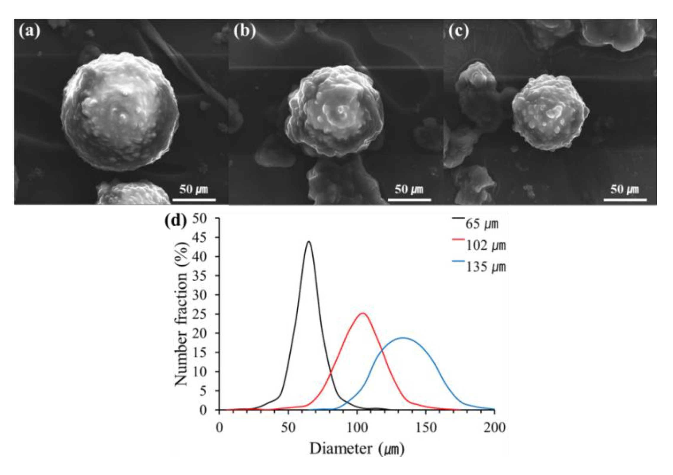
3.1. Microcapsules

Linseed oil is widely used as a healing agent for microcapsule-type self-healing coatings due to its environment-friendly nature, good film-forming property and low cost [8,16,18,19]. Linseed oil was microencapsulated using urea-formaldehyde polymer (UF) by in situ polymerization. The morphology of the microcapsules was observed by SEM (Figure 1). Spherical microcapsules were formed at agitation rates of 1000, 1500 and 2000 rpm (Figure 1a,b,c, respectively) and the surface of the microcapsules was relatively rough. The size distributions of the microcapsules were investigated by optical microscopy (Figure 1d). The mean diameter of the microcapsules prepared at 1000, 1500 or 2000 rpm was 135, 102 or 65 µm, respectively. The microcapsules were not sieved by size and used as-prepared.

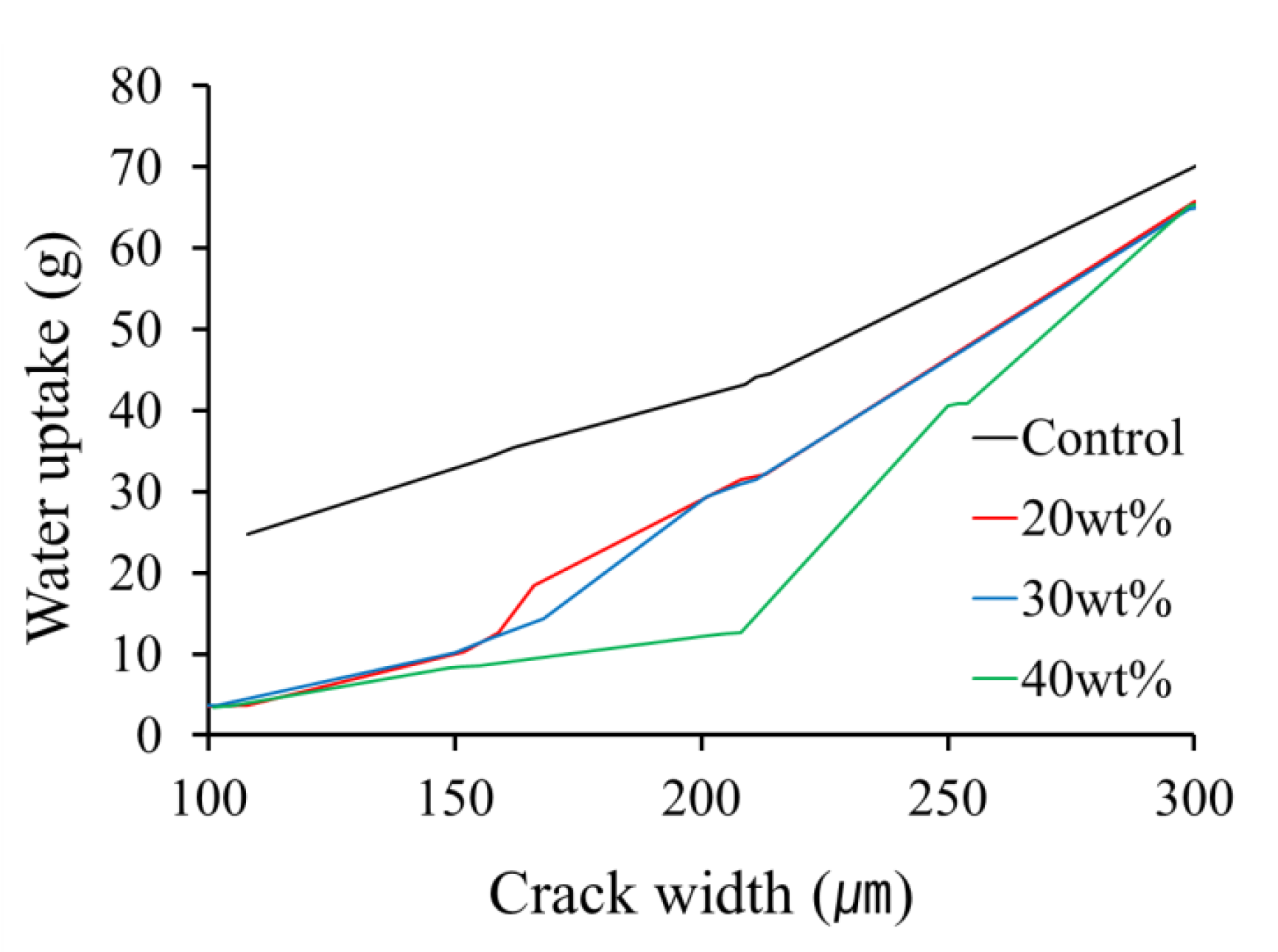
3.2. Water Sorptivity Test

To evaluate healing efficiency of the linseed oil microcapsule-based self-healing coating coated on cementitious material, a water sorptivity test was conducted using coated mortar specimens. Each specimen was prepared by applying the control or self-healing coating formulation to one rectangular side of a mortar and damage was generated in the coated surface of the mortar. Scratch was generated with scratchers having 100, 150, 200, 250 or 300-µm thickness. It was confirmed that each cut was deep enough to reach the mortar surface. On the other hand, protective coatings can also be damaged by microcracking. Microcracks can occur in the coating itself and microcracks occurring in the cementitious materials may cause microcracking of the protective coating. In this study, microcracks were generated in both the mortar and the protective coating by pressing the center part of opposite side of the coated side using a UTM. Although the crack width could be controlled by varying the pressing force, it was not easy to reach the desired exact crack width (Table 1). The damaged self-healing coatings were allowed to heal for 48 h at room temperature and then subjected to the water sorptivity test. Healing efficiency of the damaged self-healing coatings was calculated using the Equation (1). In the cases of cracked coatings, healing efficiency was only roughly estimated because of the difficulty in exact control of crack width.

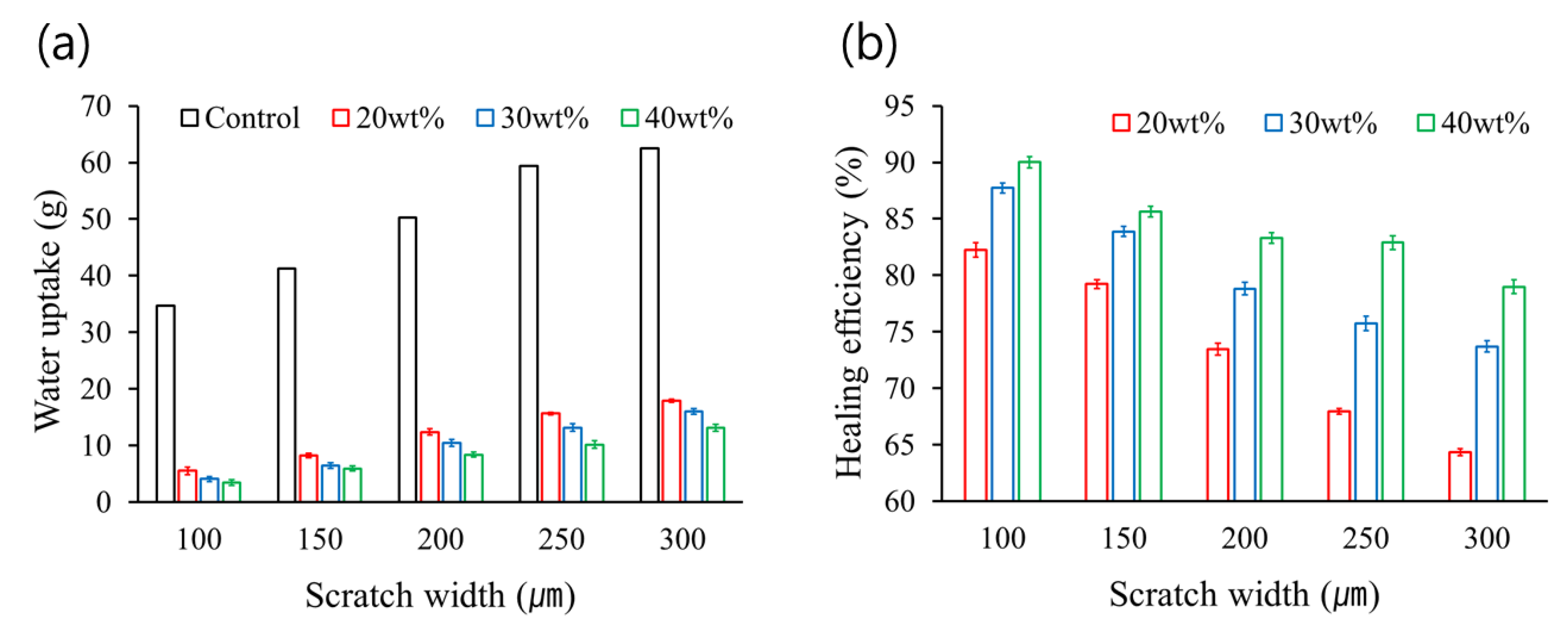
3.2.1. Water Sorptivity According to Damage Width and Capsule-loading

The results of the water sorptivity test according to damage width and capsule-loading were shown in Figure 2 and Figure 3. The capsule-loading was 20 wt%, 30 wt% or 40 wt% and mean microcapsule diameter was 65 µm. The undercoating thickness was controlled to be 50µm. The water uptake amount increased with increasing scratch width for each capsule-loading (Figure 2a). Because the amount of linseed oil that flows out of broken capsules upon being damaged is limited, the sealing of wider damage region would be less efficient. The water uptake amount decreased with increasing capsule-loading (Figure 2a). This is due to that the amount of healing agent released from microcapsules would increase with increasing capsule-loading. Figure 2b shows the healing efficiency of the self-healing coatings that was estimated based on the data of Figure 2a. When capsule-loading was 20 wt%, healing efficiency drastically decreased with increasing scratch width. However, in the case of 40-wt%-capsule-loading, healing efficiency slowly decreased according to increasing scratch width. It should be noted that a 300-µm-wide scratch self-healed in 79% healing efficiency when capsule-loading was 40 wt%.
Figure 3 shows the water uptake of the self-healing coating specimens according to crack width and capsule-loading. As expected, water uptake increased with increasing crack width. The coating samples with 40-wt%-capsule-loading showed lower water uptake values than those with 20-wt%- or 30-wt%- capsule-loading. Figure 3 also shows that the increasing rate of water uptake according to increasing crack width is much higher than that according to increasing scratch width. It is considered that crack is generated in both the mortar and the coating, leading to the formation of larger damage volume than scratch. In addition, water probably can penetrate more easily through crack of the mortar. For a 200-µm-wide crack, the self-healing coating with 40-wt%-capsule-loading roughly showed 70% healing efficiency, but the other coatings with 20-wt%-ror 30-wt%-capsule-loading showed much lower healing efficiency of around 30%. For a 150-µm-wide crack, all the self-healing coatings showed healing efficiency of about 75% or higher. Based on the results in Figure 2 and Figure 3, under the conditions of mean capsule diameter of 65 µm and undercoating thickness of 50µm, the best self-healing performance was obtained at 40-wt%-capsule-loading.

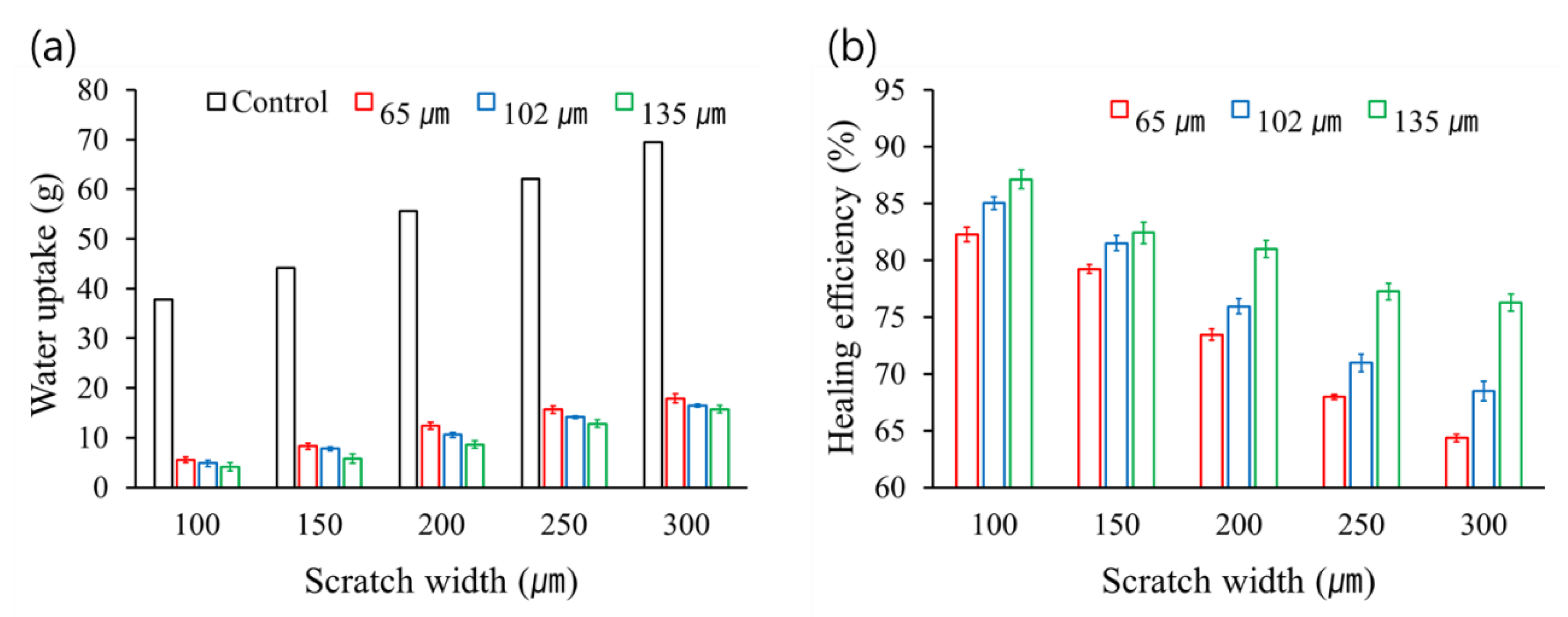
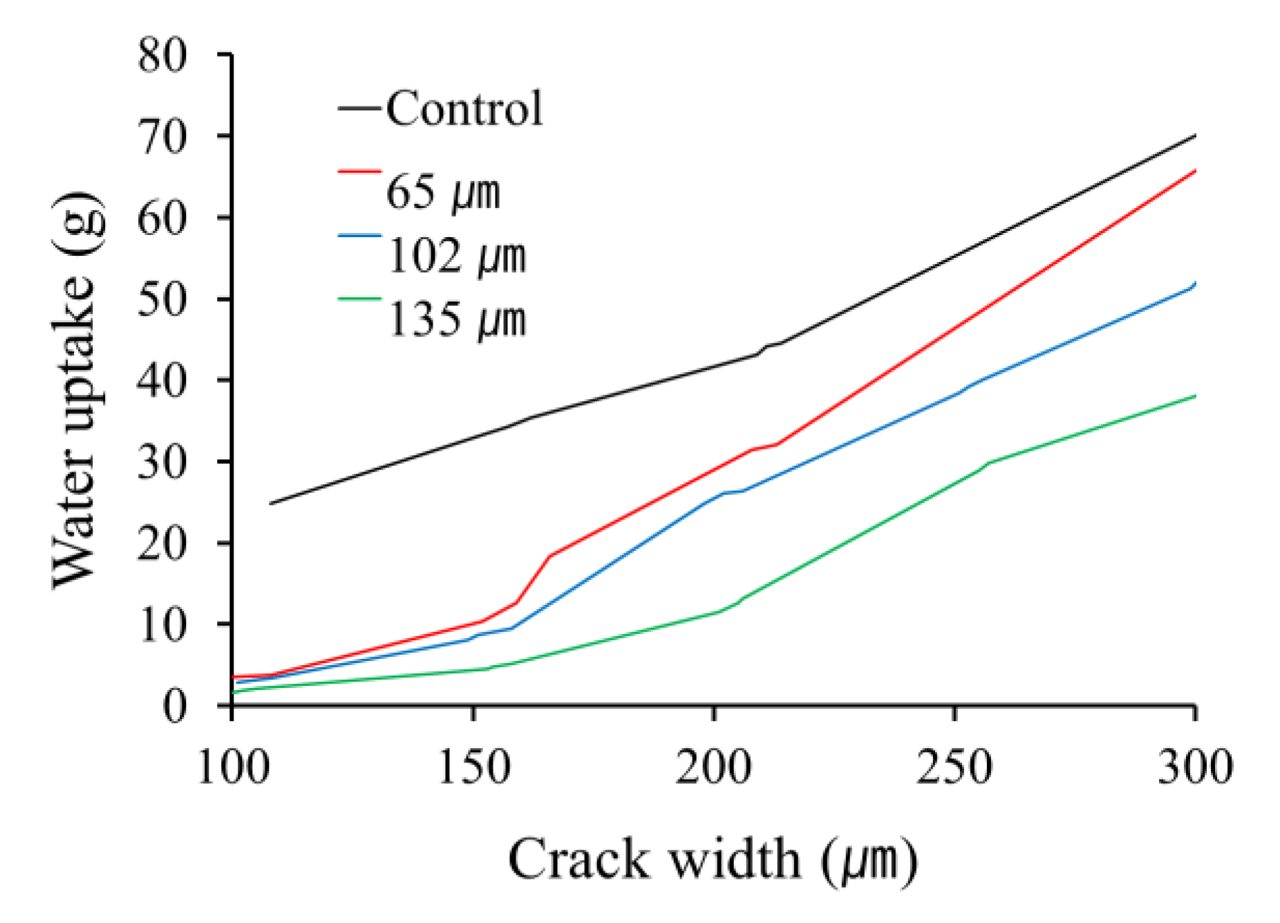
3.2.2. Water Sorptivity According to Damage Width and Microcapsule Size

The water sorptivity of the coated specimens according to damage width and microcapsule size is shown in Figure 4 and Figure 5. The microcapsules with mean diameter of 65, 102 or 135 µm were used and capsule-loading was 20 wt%. The undercoating thickness was controlled to be 50 µm. The water uptake amount increased with increasing scratch width for each mean microcapsule diameter (Figure 4a). This is probably due to less efficient sealing of wider scratch by limited amount of healing agent. On the other hand, for each scratch width, the water uptake amount decreased with increasing capsule size (Figure 4a). It is considered that the healing agent from larger capsules can more effectively flows out and seals the scratch. Figure 4b shows the healing efficiency of the self-healing coatings that was estimated based on the data of Figure 4a. When capsule mean diameter was 65 µm, healing efficiency drastically decreased with increasing scratch width. However, in the case of a 135-µm capsule mean diameter, healing efficiency slowly decreased with increasing scratch width. It should be noted that a 300-µm-wide scratch self-healed in 76% healing efficiency when microcapsule mean diameter was 135 µm.
Figure 5 shows the effect of crack width and capsule size on water uptake of the self-healing coating specimens. As the crack width increased, water uptake increased. Water uptake decreased with increasing capsule size, which is similar to the case of scratch healing described above. For a 200-µm-wide crack, the self-healing coating containing 135 µmmicrocapsules roughly showed 70% healing efficiency. For a 150-µm-wide crack, all the self-healing coatings showed healing efficiency of 70% or higher. Considering the results in Figure 4 and Figure 5, for given conditions of capsule-loading (20wt%) and undercoating thickness (50 µm), the best self-healing performance was obtained at mean capsule diameter of 135 µm.

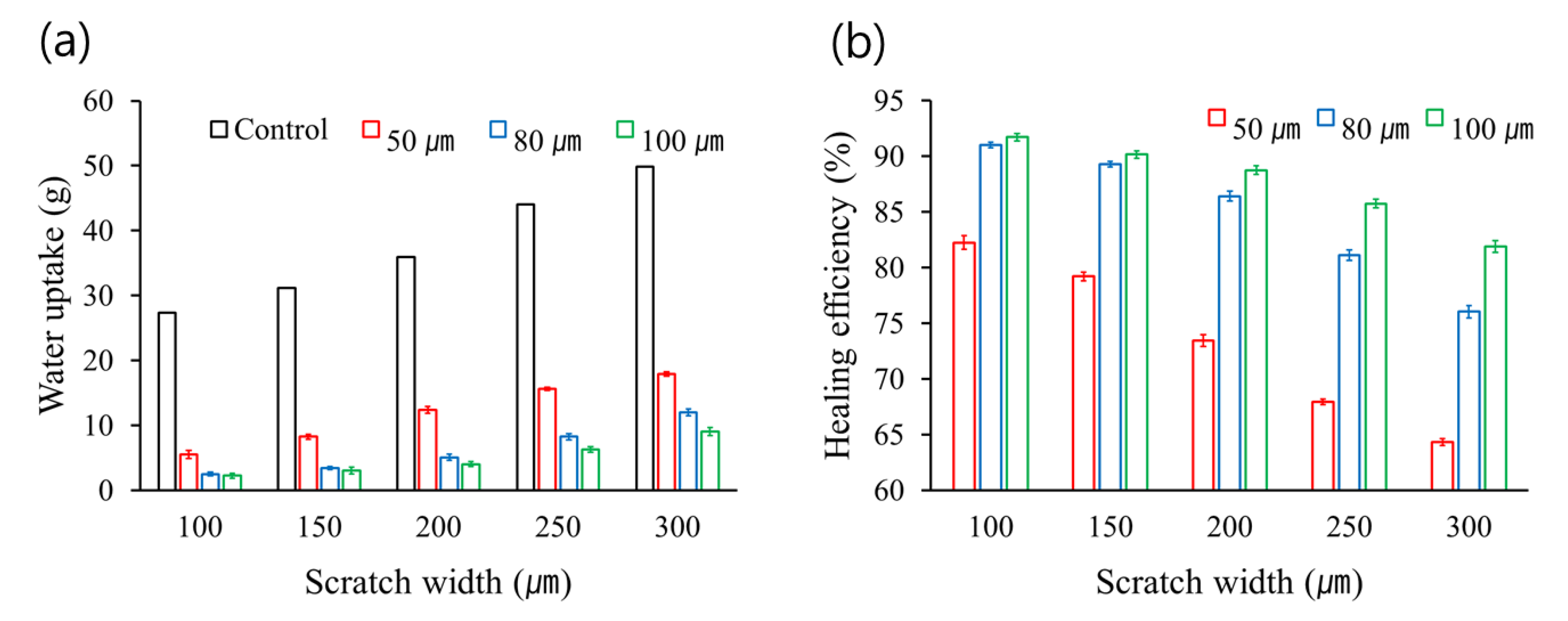
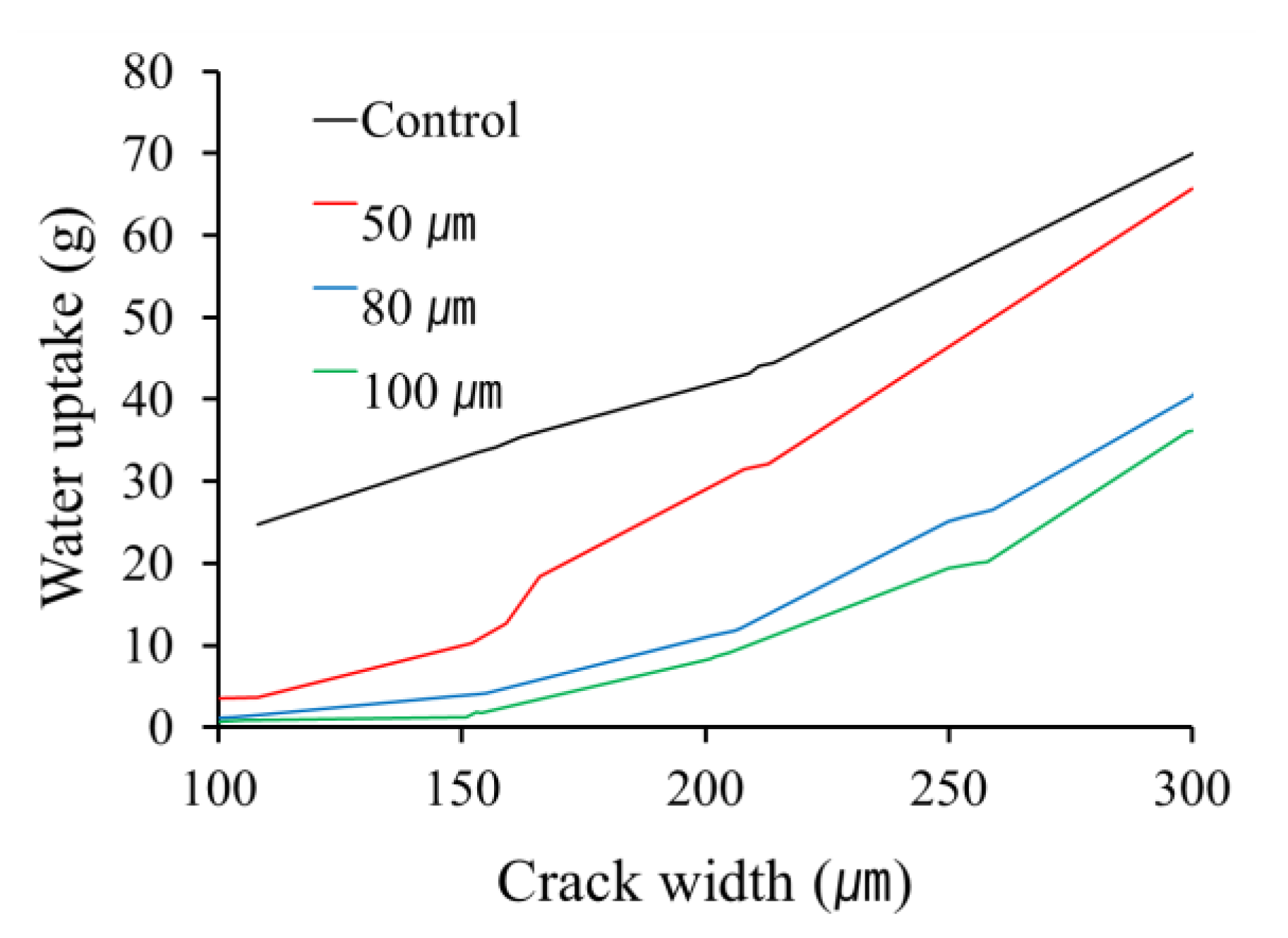
3.2.3. Water Sorptivity According to Damage Width and Undercoating Thickness

The results of the water sorptivity test according to damage width and undercoating thickness are shown in Figure 6 and Figure 7. The undercoating thickness was controlled to 50, 80 or 100 µm. Capsule-loading of the self-healing coatings was 20 wt% and mean microcapsule diameter was 65 µm. The water uptake amount increased with increasing scratch width for each undercoating thickness (Figure 6a). For each scratch width, the water uptake amount decreased with increasing the undercoating thickness. As the undercoating thickness increases, the number of vertically distributed microcapsules would increase, leading to releasing more amount of healing agent from microcapsules. It is considered that more effective sealing of scratched region occurs by increased amount of healing agent, despite of increase in damage volume. Figure 6b shows the healing efficiency of the self-healing coatings that was estimated based on the data of Figure 6a. The self-healing coatings having 80- or 100-µm-thick undercoating showed much higher healing efficiency compared to the self-healing coating based on 50-µm-thick undercoating. It should be noted that a 300-µm-wide scratch self-healed in 82% healing efficiency when undercoating thickness was 100 µm despite of a low capsule-loading of 20 wt%.
Figure 7 shows the effect of crack width and undercoating thickness on water uptake of the self-healing coating specimens. As crack width increased, water uptake increased. Water uptake decreased with increasing undercoating thickness. For a 200-µm-wide crack, the self-healing coating with a 100-µm-thick undercoating showed around 80% healing efficiency. From the results in Figure 6 and Figure 7, the best self-healing performance was achieved with a 100-µm-thick undercoating under the conditions of capsule-loading of 20 wt% and mean capsule diameter of 65 µm.

3.3. Other Performances of the Self-Healing Coating with Damage Width of 300 µm

Form the results in Figure 2, Figure 3, Figure 4, Figure 5, Figure 6 and Figure 7, each parameter that gave the highest healing efficiency was chosen and the combination of the parameters was used for other performance tests. An undercoating formulation with 40-wt%-capsule-loading was prepared using microcapsules having a mean diameter of 135 µm. The coating formulation was applied on mortar specimens to prepare 100-µm-thick undercoating. The top-coating formulation was applied on the undercoating to prepare self-healing coating. Control-coating specimens were prepared in a similar fashion without using microcapsules. Accelerated carbonation test, chloride ion penetration test and SEM study were conducted for the control and self-healing specimens after generation of a 300-µm-wide scratch in the coatings.
The carbonation depth of mortar with damaged control coating was measured to be 1.4 mm. In contrast, the carbonation of mortar with self-healing coating was measured to be 0.3 mm in depth. On the other hand, the chloride ion penetration of scratched control coating was measured to be 6164 coulombs, while that of the scratched self-healing coating was measured to be 474 coulombs. These results indicate that the self-healing coating specimens repaired the 300-µm-wide scratch, resulting in effective protection of the substrate mortar from carbonation and chloride ion penetration.
For SEM study, CRC board was used as substrate instead of mortar. The control and self-healing coatings were scratched using a scratcher with a 300-µm-thickness and left for 48 h at room temperature to induce self-healing. From the SEM image, the scratched region remained unfilled in the control coating (Figure 8a), while the healing agent filled the scratched region in the self-healing coating (Figure 8b).

4. Conclusions

The effect of capsule-loading, capsule size and coating thickness on healing efficiency was investigated by water sorptivity test for the self-healing coating with damage width of 100–300 µm. The increasing rate of water uptake according to increasing crack width was much higher than that according to increasing scratch width. It was considered that crack was generated in both the mortar and the coating, leading to the formation of larger damage volume than scratch. In addition, water probably can penetrate more easily through crack of the mortar. Healing efficiency of the self-healing coating decreased with increasing damage width because the amount of the healing agent released from microcapsules was limited. Healing efficiency increased with increasing capsule-loading, which was due to that the amount of healing agent released from microcapsules would increase with increasing capsule-loading. Water uptake amount decreased with increasing capsule size, which may be attributed to that the healing agent from larger capsules can more effectively flows out and seals the scratch. As the undercoating thickness increases, the number of vertically distributed microcapsules would increase, leading to more effective sealing of damaged region. Healing efficiency of 76% or higher was achieved using the self-healing coating with a 300-µm-wide scratch. The self-healing coating with a 200-µm-wide crack showed healing efficiency of 70% or higher. Form the results in Figure 2, Figure 3, Figure 4, Figure 5, Figure 6 and Figure 7, each parameter that gave the highest healing efficiency was chosen: 40-wt%-capsule-loading, mean capsule diameter of 135 µm and 100-µm thickness of undercoating. Under the combination of conditions, the self-healing coating having a 300-µm-wide scratch showed effective protection of the substrate mortar from carbonation and chloride ion penetration, which was supported by SEM study.

Author Contributions

Conceptualization, C.-M.C. and D.-M.K.; methodology, D.-M.K.; formal analysis, J.L., H.-J.P., and J.-Y.C.; investigation, D.-M.K., S.-W.J., S.-H.L., and K.-N.N.; writing—original draft preparation, C.-M.C. and D.-M.K.; writing—review & editing, C.-M.C.; supervision, C.-M.C. All authors have read and agreed to the published version of the manuscript.

Funding

This research was supported by a grant (19SCIP-B112346-05) from the Construction Technology Research Program funded by the Ministry of Land, Infrastructure and Transport of the Korean government.

Acknowledgments

The undercoating and top-coating formulations (Wrapping Coat®) were kindly donated from Samjooncnc Co. (Pocheon, Korea).

Conflicts of Interest

The authors declare no conflict of interest.

References

  1. Wei, H.; Wang, Y.; Guo, J.; Shen, N.Z.; Jiang, D.; Zhang, X.; Yan, X.; Zhu, J.; Wang, Q.; Shao, L.; et al. Advanced micro/nanocapsules for self-healing smart anticorrosion coatings. J. Mater. Chem. A 2015, 3, 469–480. [Google Scholar] [CrossRef]
  2. Samadzadeh, M.; Boura, S.H.; Peikan, M.; Kasiriha, S.M.; Ashrafi, A. A review on self-healing coatings based on micro/nanocapsules. Prog. Org. Coat. 2010, 68, 159–164. [Google Scholar] [CrossRef]
  3. Shchukin, D.G. Container-based multifunctional self-healing polymer coatings. Polym. Chem. 2013, 4, 4871–4877. [Google Scholar] [CrossRef] [Green Version]
  4. Hia, L.L.; Vahedi, V.; Pasbakhsh, P. Self-Healing Polymer Composites: Prospects, Challenges, and Applications. Polym. Rev. 2016, 56, 225–261. [Google Scholar] [CrossRef]
  5. Blaiszik, B.J.; Kramer, S.L.B.; Olugebefola, S.C.; Moore, J.S.; Sottos, N.R.; White, S.R. Self-Healing Polymers and Composites. Annu. Rev. Mater. Res. 2010, 40, 179–211. [Google Scholar] [CrossRef]
  6. An, S.; Lee, M.W.; Yarin, A.L.; Yoon, S.S. A review on corrosion-protective extrinsic self-healing: Comparison of microcapsule-based systems and those based on core-shell vascular networks. Chem. Eng. J. 2018, 2018. 344, 206–220. [Google Scholar] [CrossRef]
  7. Song, Y.-K.; Chung, C.-M. Repeatable self-healing of a microcapsule-type protective coating. Polym. Chem. 2013, 4, 4940–4947. [Google Scholar] [CrossRef]
  8. Song, Y.-K.; Jo, Y.-H.; Lim, Y.-J.; Cho, S.-Y.; Yu, H.C.; Ryu, B.-C.; Lee, S.-I.; Chung, C.-M. Sunlight-induced self-healing of a microcapsule-type protective coating. ACS Appl. Mater. Interfaces 2013, 5, 1378–1384. [Google Scholar] [CrossRef] [PubMed]
  9. Guo, W.; Jia, Y.; Tian, K.; Xu, Z.; Jiao, J.; Li, R.; Wu, Y.; Cao, L.; Wang, H. UV-triggered self-healing of a single robust SiO2 microcapsule based on cationic polymerization for potential application in aerospace coatings. ACS Appl. Mater. Interfaces 2016, 8, 21046–21054. [Google Scholar] [CrossRef] [PubMed]
  10. Kim, D.-M.; Song, I.-H.; Choi, J.-Y.; Jin, S.-W.; Nam, K.-N.; Chung, C.-M. Self-healing coatings based on linseed-oil-loaded microcapsule for protection of cementitious materials. Coatings 2018, 8, 404. [Google Scholar] [CrossRef] [Green Version]
  11. Boura, S.H.; Peikari, M.; Ashrafi, A.; Samadzadeh, M. Self-healing ability and adhesion strength of capsule embedded coatings-micro and nano sized capsules containing linseed oil. Prog. Org. Coat. 2012, 75, 292–300. [Google Scholar] [CrossRef]
  12. Wang, H.; Zhou, Q. Evaluation and failure analysis of linseed oil encapsulated self-healing anticorrosive coating. Prog. Org. Coat. 2018, 118, 108–115. [Google Scholar] [CrossRef]
  13. Behzadnasab, M.; Mirabedini, S.M.; Esfandeh, M.; Farnood, R.R. Evaluation of corrosion performance of a self-healing epoxy-based coating containing linseed oil-filled microcapsules via electrochemical impedance spectroscopy. Prog. Org. Coat. 2017, 105, 212–224. [Google Scholar] [CrossRef]
  14. Zhao, Y.; Zhang, W.; Liao, L.; Wang, S.; Li, W. Self-healing coatings containing microcapsule. Appl. Surf. Sci. 2012, 258, 1915–1918. [Google Scholar] [CrossRef]
  15. Kim, D.-M.; Yu, H.-C.; Yang, H.-I.; Cho, Y.-J.; Lee, K.-M.; Chung, C.-M. Microcapsule-type self-healing protective coatings for cementitious composites with secondary crack preventing ability. Materials 2017, 10, 114. [Google Scholar] [CrossRef] [PubMed] [Green Version]
  16. Wang, W.; Xu, L.; Sun, H.; Li, X.; Zhao, S.; Zhang, W. Spatial resolution comparison of AC-SECM with SECM and their characterization of self-healing performance of hexamethylene diisocyanate trimer microcapsule coatings. J. Mater. Chem. 2015, 3, 5599–5607. [Google Scholar] [CrossRef]
  17. Attaei, M.; Vale, M.; Shakoor, A.; Kahraman, R.; Montemor, F.; Marques, A.C. Hybrid shell microcapsules containing isophorone diisocyanate with high thermal and chemical stability for autonomous self-healing of epoxy coatings. J. Appl. Polym. Sci. 2020, 137, 48751. [Google Scholar] [CrossRef]
  18. Cui, J.; Li, X.; Pei, Z.; Pei, Y. A long-term stable and environmental friendly self-healing coating with polyaniline/sodium alginate microcapsule structure for corrosion protection of water-delivery pipelines. Chem. Eng. J. 2019, 358, 379–388. [Google Scholar] [CrossRef]
  19. Yamuna, J.; Siva, T.; Kumari, S.S.; Sathiyanarayanan, S. A smart poly(aniline-formaldehyde) microcapsule based self-healing anticorrosive coating. RCS Adv. 2016, 6, 79–86. [Google Scholar] [CrossRef]
Figure 1. Scanning electron microscope (SEM) images of the linseed-oil-loaded microcapsules formed at agitation rates of (a) 1000 rpm, (b) 1500 rpm and (c) 2000 rpm. (d) Size distribution of the microcapsules according to agitation rate.
Coatings 10 00543 g001
Figure 2. (a) Water uptake versus scratch width of the self-healing coatings with capsule-loading of 0 (control), 20 wt%, 30 wt% or 40 wt%; (b) healing efficiency versus scratch width of the self-healing coatings with capsule-loading of 20 wt%, 30 wt% or 40 wt%. Mean diameter of the microcapsules and undercoating thickness were 65 µm and 50 µm, respectively.
Coatings 10 00543 g002
Figure 3. Water uptake versus crack width of the self-healing coatings with capsule-loading of 0 (control), 20 wt%, 30 wt% and 40 wt%. Mean diameter of the microcapsules and undercoating thickness were 65 µm and 50 µm, respectively.
Coatings 10 00543 g003
Figure 4. (a) Water uptake versus scratch width of the control and self-healing coatings containing microcapsules with mean diameter of 65, 102 or 135 µm; (b) Healing efficiency versus scratch width of the self-healing coatings containing microcapsules with mean diameter of 65, 102 or 135 µm. Capsule-loading of the self-healing coating was 20 wt% and undercoating thickness was 50 µm.
Coatings 10 00543 g004
Figure 5. Water uptake versus crack width of the control and self-healing coatings containing microcapsules with mean diameter of 65, 102 or 135 µm. Capsule-loading of the self-healing coating was 20 wt% and undercoating thickness was 50 µm.
Coatings 10 00543 g005
Figure 6. (a) Water uptake versus scratch width of the self-healing coatings having undercoating thickness of 50, 80 or 100 µm; (b) Healing efficiency versus scratch width of the self-healing coatings having undercoating thickness of 50, 80 or 100 µm. Capsule-loading of the self-healing coatings was 20 wt% and mean microcapsule diameter was 65 µm.
Coatings 10 00543 g006
Figure 7. Water uptake versus crack width of the control and self-healing coatings having undercoating thickness of 50, 80 or 100 µm. Capsule-loading of the self-healing coatings was 20 wt% and mean microcapsule diameter was 65 µm.
Coatings 10 00543 g007
Figure 8. SEM images of scratched (a) control and (b) self-healing coatings with 300-µm damage width.
Coatings 10 00543 g008
Table 1. Variable parameters of the coating samples.
Variable ParameterParameter Value
Microcapsule-loading (wt%)20, 30, 40
Mean diameter of microcapsules (µm)65, 102, 135
Undercoating thickness (µm)50, 80, 100
Scratch width (µm)100, 150, 200, 250, 300
Crack width (µm)95–105, 145–155, 195–205, 245–255, 295–305

