Article

Bio-Inspired Facile Synthesis of Graphene-Based Nanocomposites: Elucidation of Antimicrobial and Biofilm Inhibitory Potential against Foodborne Pathogenic Bacteria

by
Abdullah Aljaafari
1,*,
Faheem Ahmed
1 and
Fohad Mabood Husain
2
1
Department of Physics, College of Science, King Faisal University, P.O. Box 400, Al-Ahsa 31982, Saudi Arabia
2
Department of Food Science and Nutrition, King Saud University, Riyadh 11451, Saudi Arabia
*
Author to whom correspondence should be addressed.
Coatings 2020, 10(12), 1171; https://doi.org/10.3390/coatings10121171
Submission received: 25 October 2020 / Revised: 27 November 2020 / Accepted: 27 November 2020 / Published: 29 November 2020
(This article belongs to the Section Coatings for Food Technology and System)

Abstract

:
Herein, a new and simple biogenic method for the preparation of gold nanoparticles (AuNPs) and their reduced graphene oxide based nanocomposites (Au-RGO) by using microwave irradiation method for antimicrobial and biofilm inhibition against foodborne pathogenic bacteria was reported. X-ray diffraction (XRD), Raman, and transmission electron microscopy (TEM) analyses confirmed that the AuNPs with face centered cubic (FCC) structure were indeed anchored onto the RGO sheets. Ultraviolet-Vis (UV-VIS) spectrum showed a shifting and broadening of absorption peaks of AuNPs when attached on the surface of RGO sheets. The effect of sub-inhibitory concentrations of Au-RGO nanocomposites on biofilm formation in five foodborne pathogens was assessed. Au-RGO nanocomposites reduced the formation of biofilm by 75%, 78%, 68%, 80% and 79% in L. monocytogenes, MRSA, E. coli, S. marcescens and P. aeruginosa, respectively. Exopolysaccharides (EPS), a vital component of the biofilm was also inhibited significantly and pre-formed mature biofilms were also reduced considerably. Further, this study demonstrated that the reactive oxygen species (ROS) generation induced in bacterial cells as a result of Au-RGO treatment could be the plausible mechanism for biofilm inhibitory action. The tested concentrations were found non-toxic to human embryonic kidney cell lines (HEK-293). The investigation highlights the broad-spectrum biofilm inhibitory properties of Au-RGO nanocomposites that could be exploited in the food industry to prevent biofilm-based food contamination.

1. Introduction

Foodborne pathogens have become one of the major causes of illness, hospitalization, and mortality across the globe [1]. Transfer of bacteria from contaminated food contact surface to the food products is responsible for the disease outbreaks [1]. One of the major factor accountable for the cross-contamination in the food industry is the formation of biofilms [2]. Biofilms are complex structures comprising of one or more species embedded in an extracellular envelope made up of polysaccharides, protein or exogenous DNA [3]. Biofilm formation occurs by the irreversible attachment of the matrix on food industry equipment’s (conveyer belts, tanks and knives), transport materials, storage surfaces and on biological materials like fruits, vegetables, meat and bones etc. This formation of biofilm in the food industry environments confers the microbes resistance against desiccation, mechanical resistance and protection from disinfectant, antimicrobials and chemicals used in the food industry [4]. Most importantly, the biofilm forming food-associated microbes are also human pathogens and account for food-borne diseases via intoxications or infections putting human health at risk [3]. Currently, conventional disinfectant such as sodium hypochlorite is used to mitigate the effect of biofilm contamination but these chemicals also have limited efficacy for inhibition and removal of resistant biofilms [5]. Thus, there is an unfulfilled requirement to develop agents that not only prevent biofilm formation but also eradicate pre-formed biofilms of antimicrobial resistant pathogenic bacteria.
Among diverse strategies developed to combat the threat of drug-resistant calcitrant biofilm, significant research has focused on the use of nanoparticles as novel antimicrobials [6]. Several metallic nanoparticles of silver and gold, metal oxide nanoparticles such as zinc oxide, iron oxide, tin oxide, titanium oxide, and carbon nanotubes have been reported to possess antimicrobial properties [6,7]. In addition to these, copper, copper oxide and silver oxide have attracted great attention due to their excellent antibacterial properties [8,9,10]. This can be attributed to the small sizes and large surface to volume ratio of the nanomaterials allowing enhanced interaction with the microbes resulting in elevated antimicrobial efficacies [11].
Recently, nanocomposites with antimicrobial properties have also gained enormous attention of the scientific community. In this regard, reduced graphene based nanocomposites have emerged as excellent material with microbicidal action as it possess high specific surface area, exceptional mechanical and electrical properties, non-toxicity, chemical stability and are ideal candidates for tethering metallic nanoparticles [12,13]. Most of the studies were focused on the Ag and RGO based nanocomposites of as exceptional antibacterial and antibiofilm agents with negligible tendency to induce resistance [11,14,15,16]. However, gold nanoparticles (AuNPs) are also known to inhibit the growth of bacteria through various mechanisms and few studies have explored gold nanoparticle decorated RGO (Au-RGO) nanocomposites as antibacterial agents [17,18] but very limited information is available on the biofilm inhibitory action of these nanocomposites.
Various methods for the preparation of AuNPs and their graphene based nanocomposites have been reported earlier [19,20,21,22]. Currently, the focus of the scientific community is to develop antimicrobial nanomaterials through green chemistry approaches, utilizing materials of biological origin and processes that are cost-effective, less time consuming and eco-friendly [23]. Therefore, in the present investigation we have utilized citric acid from lemon extract to fabricate Au-RGO nanocomposites and assessed its antibacterial potential against both Gram-positive and Gram-negative food-associated bacteria. Further, sub-inhibitory concentrations of Au-RGO were used to determine the inhibition of biofilm formation and removal of pre-formed biofilms in test pathogens.

2. Results and Discussion

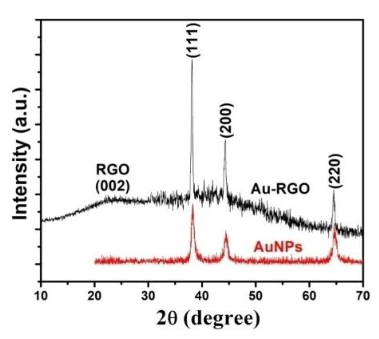
2.1. Structural Properties: XRD

Crystal structure phase determinations of the prepared nanomaterials were studied by using XRD. Figure 1 shows the XRD pattern of AuNPs and Au-RGO nanocomposites which exhibiting the diffraction peaks of both RGO and AuNPs. The peaks positioned at 38.18°, 44.46°, and 64.68° can be indexed to the (111), (200), and (220) reflections of FCC (face centered cubic) structure of metallic gold, respectively [24], while, the peak at 25.04° corresponds to (002) reflection of RGO structure. Additionally, the peak conforming at the (111) is more intense than the other planes suggesting that they are the predominant orientation and the synthesized AuNPs are crystalline in nature. No other phase was detected within the detection limit of XRD. The mean crystalline size of AuNPs was found to be ~8 nm obtained by Debye–Scherrer’s formula [25,26] with (111) plane.

2.2. Optical Properties: UV-VIS

Figure S1 shows the UV-VIS absorption spectra of AuNPs and Au-RGO nanocomposites. It is clear from Figure S1 that a broad peak of surface plasmon resonance at ~520 nm appears which confirms the presence of gold nanoparticles. The broadness of the peak is an accurate indicator of nanoparticle size. As the particle size increases, the peak becomes narrower with a decreased bandwidth and increased band intensity [27]. In the spectrum of Au-RGO nanocomposites, the peak at ~273 nm correspond to the absorption peak of RGO, while the peak related to AuNPs was found to be shifted toward higher wavelength, indicating the slight increase in the size of AuNPs.

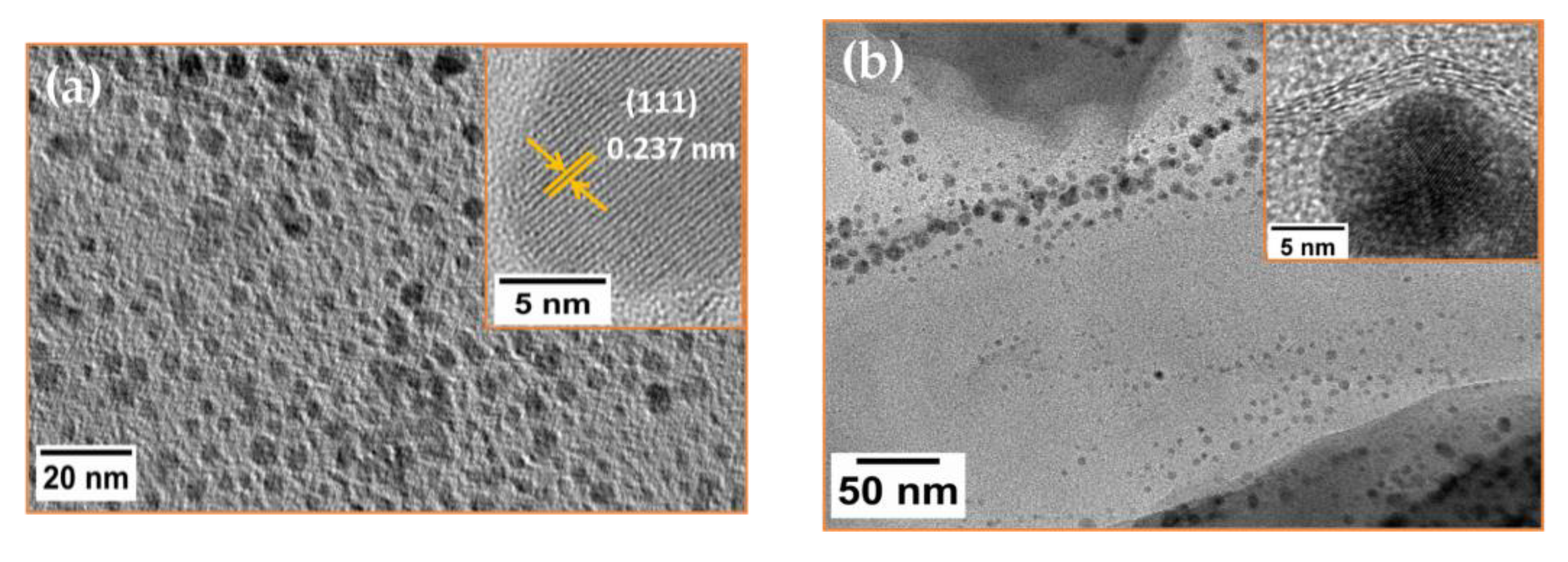
2.3. Morphological Studies: TEM

Morphological features of AuNPs and Au-RGO were studied using TEM micrographs as shown in Figure 2. The TEM image of the synthesized AuNPs is shown in Figure 2a. TEM image exhibits stable, well-dispersed, spherical particles for AuNPs prepared at 40 s irradiation time with cetyltrimethylammonium bromide (CTAB) as binding agent. Interestingly, TEM measurements proved that the obtained AuNPs consists of two populations of nanoparticles: larger particles and smaller particles. For the calculation of particle size, approximately 50 individual nanoparticles were selected randomly from TEM micrographs and found that the larger particles with size range of 8–12 nm in diameter, and small nanoparticles with size range of 2–5 nm (Figure 2a). Structural information of nanomaterials was further analyzed by using high resolution transmission electron microscopy (HRTEM). Inset of Figure 2a shows the HRTEM image of single AuNPs, where clear lattice fringes could be easily seen. The distance between two adjacent planes was found to be 0.237 nm, which is associated with (111) plane of Au structure. Figure 2b depicts the TEM image of Au-RGO nanocomposites prepared with microwave irradiation for 40 s. It can be clearly seen from Figure 2b that AuNPs are attached onto the surface of RGO sheets. The distribution of AuNPs on RGO is uniform without agglomeration, and the particles are distributed all over the surface of RGO sheets.

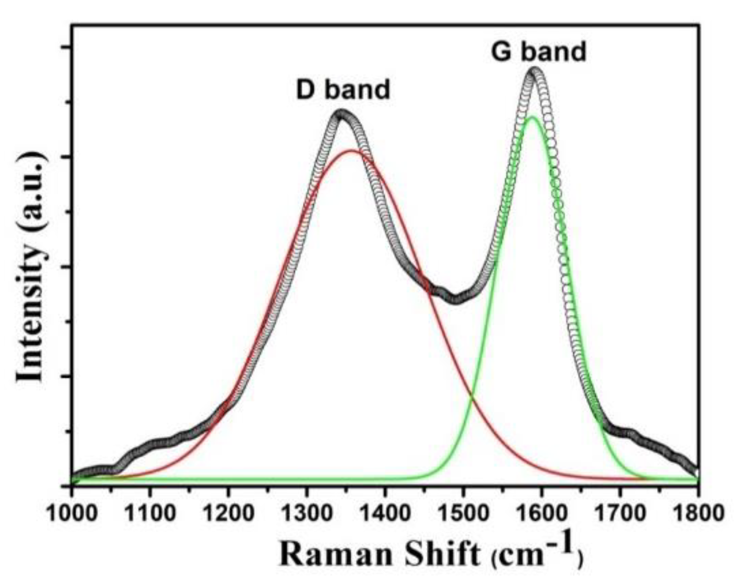
2.4. Raman Spectroscopy

Raman spectroscopy was used for the structural studies of the nanostructures. Figure 3 shows the Raman spectrum of Au-RGO nanocomposites. In the Raman spectrum, two characteristics bands of carbon materials (RGO) were observed; where D band is positioned at 1349 cm−1, and G band at 1601 cm−1, respectively. Raman spectrum can provide adequate information related to the structure of graphene and graphene based materials. It is well known that G band is a result of sp2 bonded carbons present in graphene. The D band is a measurement of “defects” or disruption of sp2 bonds of the carbon and sp3 formation [28]. It is clear from Figure 3 that these defects are observed in the Raman spectrum of Au-RGO nanocomposites, which indicated that in the nanocomposites, GO was successfully reduced.

2.5. Antibacterial Activity: Determination of Minimum Inhibitory Concentration (MICs)

Antibacterial potential of Au-RGO was studied against both Gram-positive and Gram-negative food-borne bacterial pathogens. MIC values of Au-RGO against L. monocytogenes, MRSA, E. coli, S. marcescens and P. aeruginosa was recorded to be 32, 64, 16, 8, and 16 µg/mL, respectively (Table 1). Enhanced antibacterial potential of the graphene nanocomposites is attributed to their ability to bind to non-specifically with the bacteria [29]. These nanocomposites tend to induce sugar and protein leakage from cell membrane leading to bacterial cell disruption [18]. Moreover, AuNPs are known for their antibacterial potential through various mechanisms. It has been reported that AuNPs adhere to the bacterial membrane via electrostatic interactions and disintegrate the cells [30]. AuNPs interfere with translation process in the cells by inhibiting the binding of tRNA with ribosomal subunit, reducing ATP levels in the cells [30]. Further, they also hamper the transcription process by penetrating the cells, causing leakage of cellular content and bind with the DNA. One more mechanism through which these nanoparticles induce cell death is the enhanced intracellular ROS production [31]. We also observed slightly higher MICs for Au-RGO against the Gram-positive bacteria (L. monocytogenes and MRSA) as compared to the Gram-negative bacteria which are possibly due to variations in structure and chemical composition of the cell wall. The presence of multilayered peptidoglycan in the cell wall of Gram positive bacteria might provide better resistance against Au-RGO [32,33].

2.6. Effect of Sub-MICs on the Growth of Test Bacteria

Growth kinetic studies were performed in the presence and absence of sub-MICs (0.25×MIC and 0.5×MIC) of Au-RGO nanocomposites as depicted in Figure S2. Insignificant variation in the growth pattern of all test pathogens was observed upon treatment with sub-inhibitory concentrations of the nanocomposites. Thus, it is concluded that the synthesized Au-RGO nanocomposites do not affect the growth of L. monocytogenes, MRSA, E. coli, S. marcescens and P. aeruginosa at tested sub-MICs.

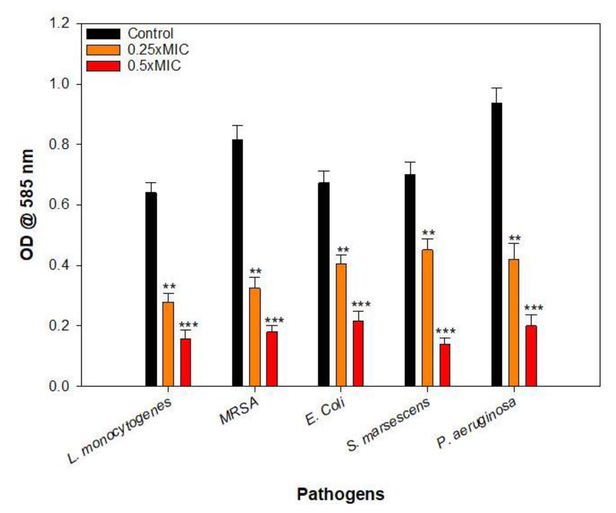
2.7. Inhibition of Biofilm Formation

It is very well documented that biofilm formation can substantially enhance the resistance of bacterial cell against antimicrobials resulting in enhanced persistence of the bacteria in the biofilm matrix.These biofilms are one of the leading causes of cross-contamination of food and food associated materials upon contact with contaminated surfaces [34]. Therefore, we examined the effect of synthesized Au-RGO nanocomposites against five types of biofilm forming food-associated bacterial pathogens. Biofilm formation in all test pathogens was reduced considerably upon treatment with sub-inhibitory concentrations. In the presence of 0.5×MICs of Au-RGO, a decrease of 75%, 78%, 68%, 80% and 79% in the biofilm forming capabilities of L. monocytogenes, MRSA, E. coli, S. marcescens and P. aeruginosa, respectively was observed. Similarly, 0.25×MICs also impeded the biofilm significantly (35%–60%) in all the pathogens as compared to the untreated controls (Figure 4). The results demonstrate that the RGO might be operating as a nano-interface facilitating the interaction of bacteria with AuNPs leading to significant inhibition of biofilm [32,33]. Our results are in agreement with those reported with nanocomposites of graphene oxide and Ag. Statistically significant reduction of biofilm growth in S. mutans and E. cloacae was reported in the presence of RGO-Ag nanocomposites [33].

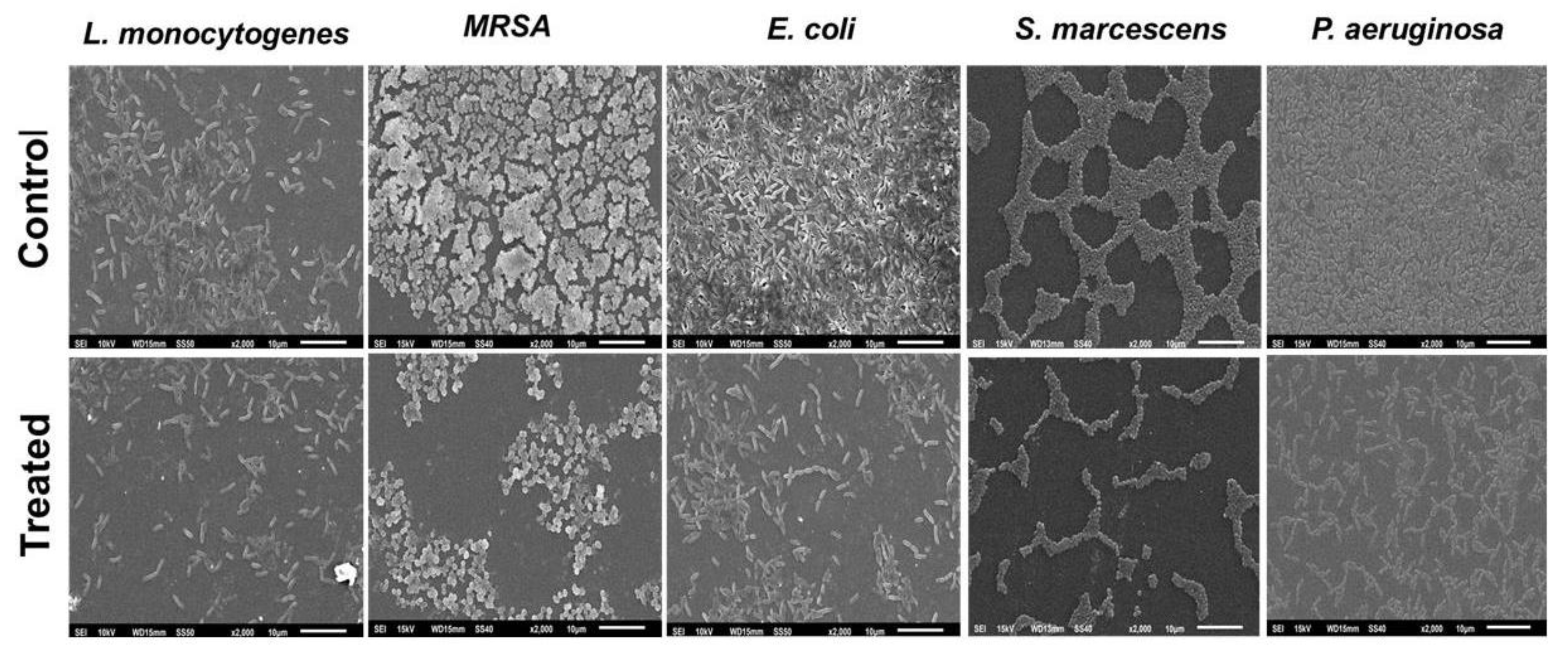
2.8. Microscopic Analysis

Scanning electron microscopic (SEM) analysis of Au-RGO nanocomposites treated and untreated biofilm of test pathogen is depicted in Figure 5. Images of the untreated (control) bacteria show a dense aggregation of cells. The lower panel very clearly displays significantly disturbed biofilm architecture, decreased microcolonies and reduced cell aggregation in all the Au-RGO treated test pathogens. Similar disturbed biofilm architecture and reduced microcolonies in Au-RGO treated cultures was observed with light microscope (Figure S3). SEM images confirm the findings of microtiter plate-based biofilm inhibition assay.

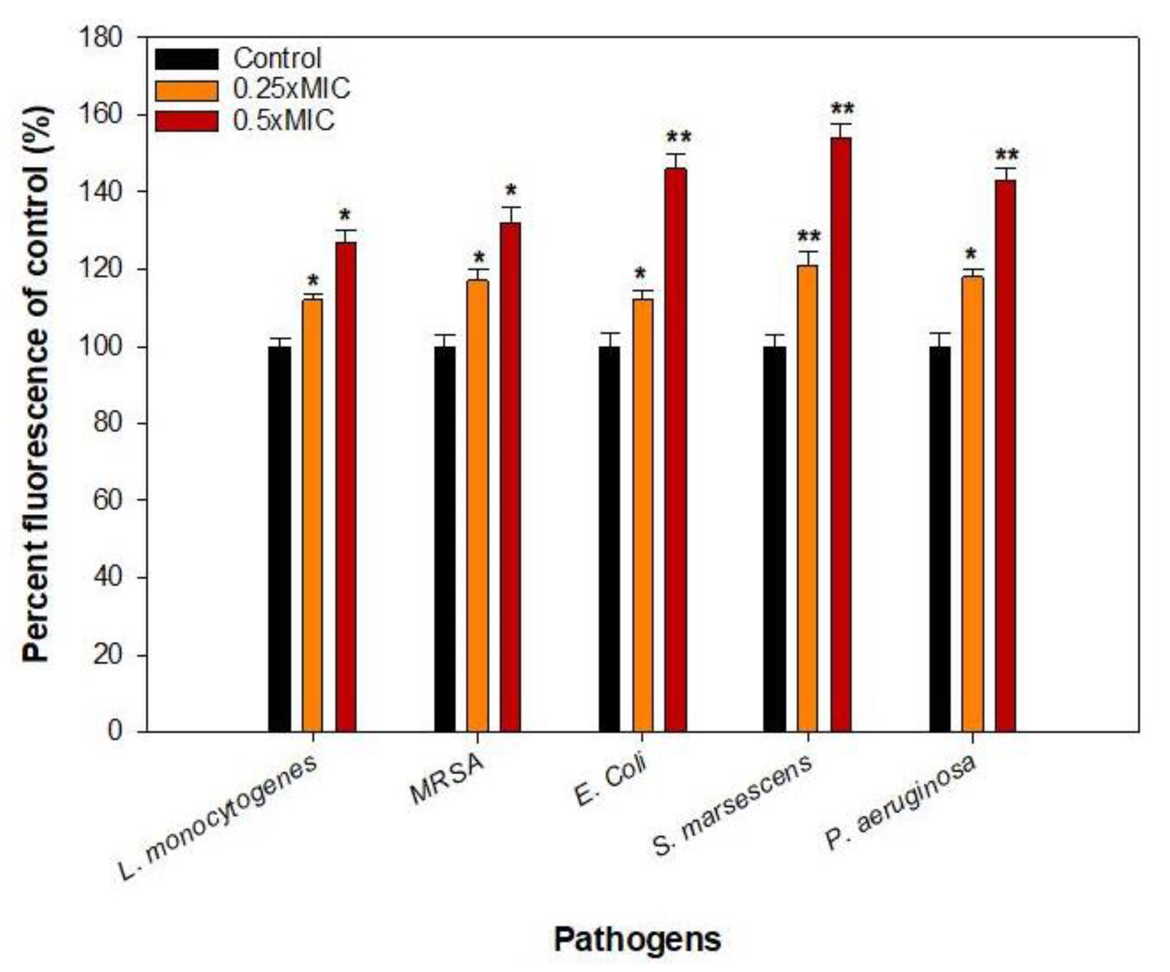
2.9. Effect on the EPS Production

EPS plays a crucial role in the adherence of the bacteria and helps in the maintenance of 3D biofilm structure. Further, it facilitates immune invasion and confers resistance towards antimicrobial agents [35,36]. EPS was extracted and quantified in the presence and absence of sub-MICs of Au-RGO. Synthesized nanocomposites interfered with the EPS production and significant reduction in EPS level ranged 31%–63%, 29%–56%, 54%–72%, 37%–69%, and 43%–81% in L. monocytogenes, MRSA, E. coli, S. marcescens and P. aeruginosa, respectively, in comparison to untreated control (Figure 6). The synthesized nanocomposites clearly impair the EPS production, an essential component of biofilm architecture. This finding is consistent with a previous study conducted with RGO-ZnO nanocomposites that demonstrated impaired EPS production in E. coli, S. marcescens and B. subtilis [37].

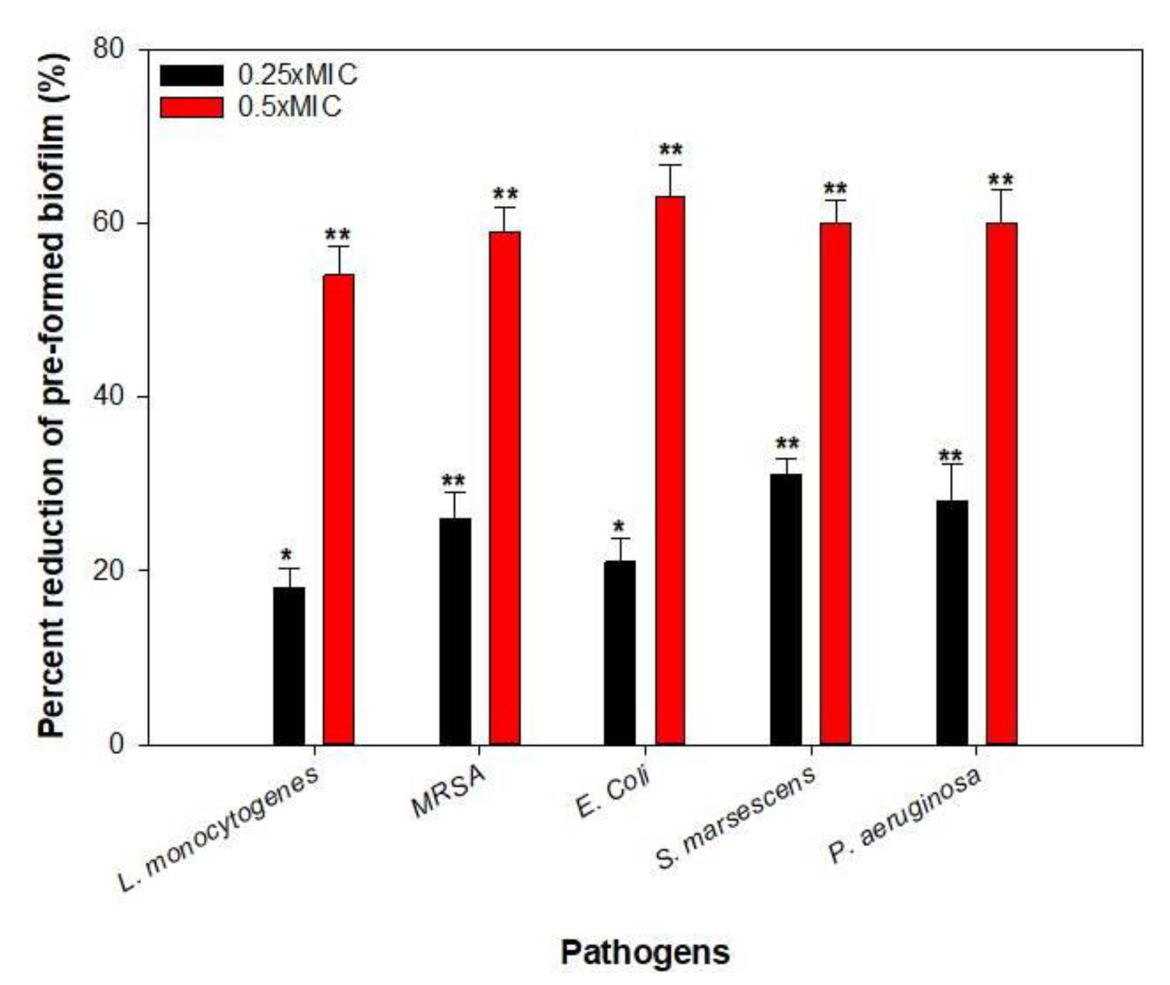
2.10. Disruption of Pre-Formed Biofilms

Bacterial cells inhabiting biofilm are known to be many folds resistant to antimicrobial agents in comparison to their planktonic counterparts. Thus, the removal of pre-formed biofilms becomes very challenging [38]. Hence, the effect of Au-RGO was assessed on the pre-formed mature biofilms of all test pathogens. Sub-inhibitory concentrations (0.25×MIC and 0.5×MIC) tested demonstrated statistically significant (p ≤ 0.05) obliteration of the pre-formed biofilms (Figure 7). At 0.25×MICs Au-RGO reduced pre-formed biofilm by 18%–31% while, at 0.5×MICs, 54%–63% disruption was observed. Concentration dependent effect was recorded and 0.5×MICs were found to be more effective in disrupting mature biofilms. This might probably be the first report demonstrating the destruction of persistent pre-formed biofilms by synthesized nanocomposites of Au-RGO.

2.11. Mode of Biofilm Inhibition by Au-RGO

Growth kinetics studies clearly showed that the tested sub-MICs of Au-RGO did not affect the growth pattern of the test pathogen significantly (Figure S2) and thus, it is envisaged that the reduction in biofilm is not due to the reduced viability of the pathogens.
Further, we evaluated the efficacy of the synthesized nanocomposite in inducing intracellular ROS production in the bacterial cells. Significantly higher levels of ROS were observed upon treatment with sub-MICs of Au-RGO in all test pathogens (Figure 8). An upsurge of 54% was recorded in the intracellular ROS levels in S. marcescens followed by E. coli (46%), P. aeruginosa (43%), MRSA (32%) and L. monocytogenes (27%) in the presence of 0.5×MICs of Au-RGO. Enhanced generation of ROS is one of the chief mechanisms through which gold nanoparticles interfere with the normal functioning of the bacterial cells [31]. Plausibly, interaction of Au-RGO and bacteria induces metabolic imbalance in the bacterial cells that results in enhanced ROS production. The elevated intracellular ROS levels overpower the internal antioxidant system, leading to formation of free radicals causing oxidative stress which eventually accounts for the cell death.

2.12. Cytotoxicity on HEK-293 Cell Line

Cytotoxicity of green synthesized Au-RGO nanocomposites was examined using HEK-293 cell lines. Synthesized nanocomposites did not cause any significant reduction in the viability of the HEK-293 cells at concentrations ranging from 25–400 µg/mL (Figure S4). Significantly decreased viability was observed at concentrations 800–3200 µg/mL but these concentrations are much higher than the one used in the present investigation advocating that the Au-RGO nanocomposites are non-toxic to these normal human cell lines at the sub-MICs used.

3. Material and Methods

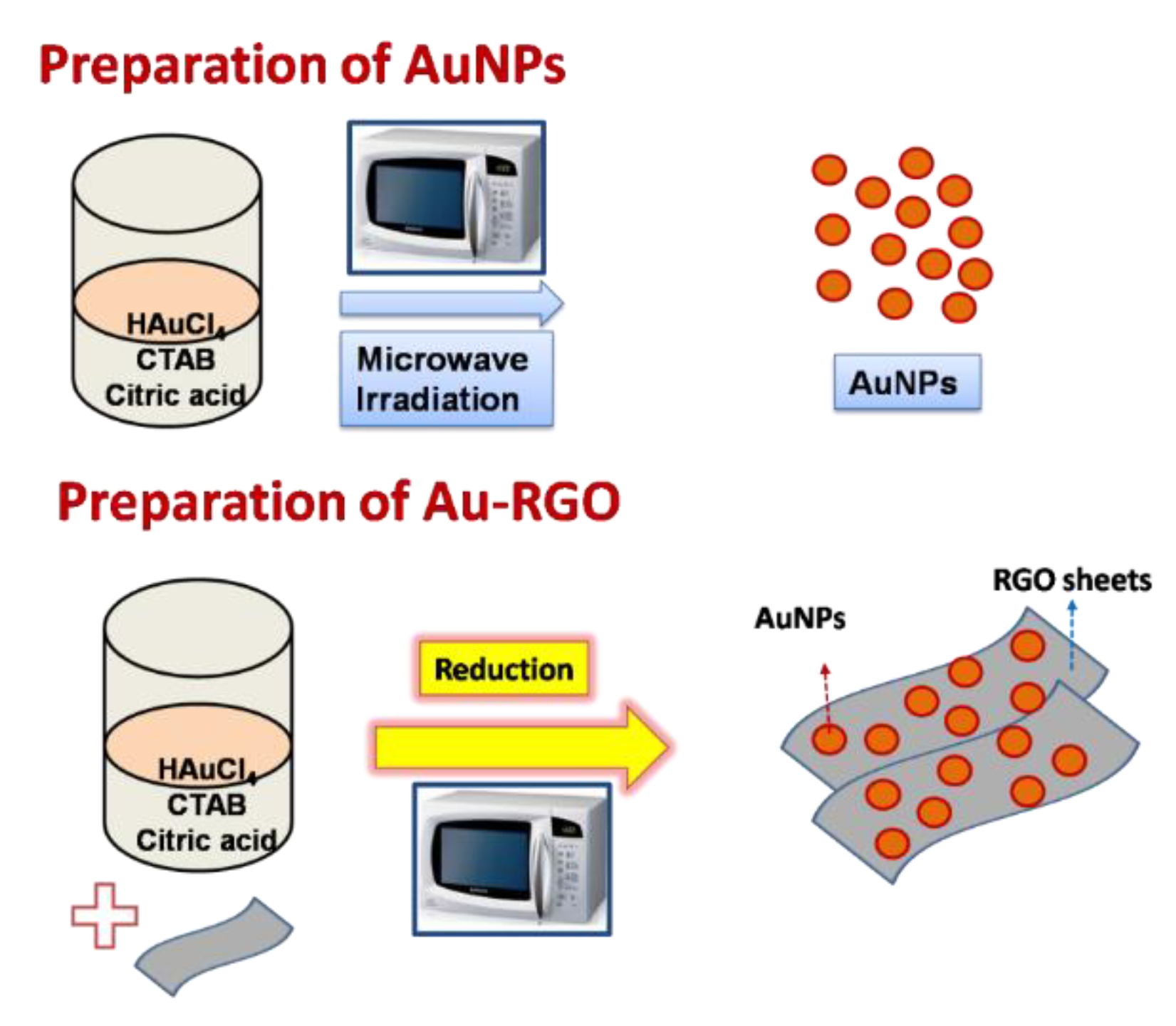
3.1. Preparation of Gold Nanoparticles (AuNPs)

For the preparation of gold nanoparticles, an aqueous solution containing 15 mL of 10 mM HAuCl4 3H2O (chloauric acid; Sigma Aldrich, St. Louis, MO, USA), 0.3 g of citric acid (Sigma Aldrich), and 0.2 g of Cetyl Trimethyl Ammonium bromide (CTAB; Sigma Aldrich, St. Louis, MO, USA) were mixed under continuous stirring. This solution was stirred for 15 s and then transferred to a microwave oven (SAMSUNG, Seoul, Korea, 750 W) for the heating at a power of 100 W for 40 s. After the reaction, the color of the solution was immediately changed (light yellow to shining orange), indicated the successful formation of gold nanoparticles. The as obtained product was used for the characterization.

3.2. Preparation of Graphene Oxide (GO)

Using the Hummers and Offeman method [39], the graphite (Merck, Darmstadt, Germany) was oxidized to synthesize GO. At first, 3.5 g of graphite was added to 100 mL of 98% H2SO4 (Merck) with vigorous stirring. While maintaining a temperature below 20 °C, 10 g of KMNO4 (Merck) was added. After stirring for 2 h at 35 °C, the mixture was transferred to 500 mL of deionized water, and 20 mL of 30% H2SO4 was added to remove excess permanganate. Upon treatment with the peroxide, the suspension turned bright yellow. GO was then purified by filtration via a sintered glass filter. HCl was used to wash the filtrate, which was then washed with hot water to remove the residual sulfate ions yielding a yellowish-brown filter cake. Finally, the resultant GO was dried at 80 °C after repeated washings with hot water.

3.3. Preparation of Reduced Graphene Oxide (RGO)

For the typical synthesis of RGO, 400 mg of GO were suspended in 20 mL of deionized water and sonicated to produce a yellow homogeneous dispersion. 400 µL of hydrazine hydrate (HH; reducing agent; Merck) was added to the mixture, and microwaved (SAMSUNG, 750 W) at 100% power for 30 s cycles (on for 10 s, and stirring for 20 s) for 2 min. This resulted to a gradual change in color of the GO solution from yellow to black, indicating the completion of the chemical reduction of GO. The GO sheets were then collected by centrifugation) (5000 rpm for 15 min) and left to dry at 80 °C overnight.

3.4. Preparation of Au/RGO Nanocomposites

To prepare Au-RGO nanocomposites with 30 wt.% of AuNPs in RGO, a solution of desired HAuCl4 3H2O in RGO was mixed in which 0.30 g of citric acid and 0.20 g of CTAB were added to this solution in a beaker. The solution was stirred for 15 s, and then microwave reaction was carried out for 40 s in a domestic microwave oven (SAMSUNG; 750 W) with 100% power.After the reaction, the product was centrifuged and collected. A schematic representation of the preparation method of AuNPs and Au-RGO nanocomposites is shown in Figure 9.

3.5. Characterizations

The X-ray diffraction (Phillips X’pert; MPD 3040, EA Almelo, The Netherlands) of the material was investigated with Cu radiation (30 kV, 40 mA, Kα radiation (1.54430 A°)). The Raman spectra were measured using a spectrometer (NRS-3100, JASCO, MD, USA) with a wavelength of 532 nm. The morphology and size of the synthesized product were measured by Transmission electron microscopy (TEM) was performed using a JEOL JSM-2100F (JEOL, Corporation Place, Singapore) operated at 200 kV. A drop of the specimen dispersed in ethanol was placed on copper grids and dried for the studies. The average particle size and the size distribution were determined using ‘Image J’ software. Room temperature optical absorption spectra were recorded in the range of 200–800 nm using a UV-VIS spectrophotometer (Agilent-8453, Agilent, CA, USA)) with a quartz cell. Colloidal gold nanoparticles and nanocomposites were added to 5 mL distilled water in a quartz cell. The blank was filled with distilled water solution.

3.6. Strains and Culture Condition

Five food associated bacteria namely, Listeria monocytogenes ATCC 19114, Methicillin resistant Staphylococcus aureus (MRSA) ATCC 43300, E. coli ATCC 35218 (β-lactamase producer), Serratia marcescens ATCC 13880 and Pseudomonas aeruginosa PAO1 were used to evaluate the biofilm inhibitory property of the synthesized nanocomposites. Stock cultures of all the test bacteria were maintained on Nutrient agar under refrigeration and sub-cultured in Tryptic soy broth (TSB) for 24 h.

3.7. Antibacterial Activity

Antibacterial activity of the synthesized Au-RGO nanocomposite was assessed in terms of the minimum inhibitory concentration (MIC) using microbroth dilution assay [40,41].

3.8. Growth Kinetics Studies

Growth kinetics of all the test bacteria was determined at 0.5×MICs of Au-RGO. All bacteria were grown in TSB in the presence and absence of Au-RGO in a Bioscreen C 100 well microtiter plate (Labsystem Oy, Helsinki, Finland). Plates were placed in Bioscreen C and growth was measured as the optical density at 600 nm for 24 h.

3.9. Biofilm Inhibition

Microtiter plate (MTP) assay was employed for the quantitative evaluation of Au-RGO on the biofilm inhibition as described previously [42]. Concisely, overnight grown test bacteria were inoculated in well of MTP and sub-MICs (0.25×MIC and 0.5×MIC) of Au-RGO were added to wells and incubated for 24 h. Wells without Au-RGO treatment were considered as control set. Post incubation, media was decanted, each well was washed thrice and stained with crystal violet (0.1%) for 15 min. Free dye was washed away and dye adhering to the biofilm cells was dissolved in 100 µL ethanol (70%). Optical density of each well was recorded at 585 nm using Multiskan FC (Thermo Scientific, Waltham, MA, USA).

3.10. Microscopic Evaluation of Biofilm Inhibition

Microscopic examination of biofilm architecture was done by forming biofilms on glass coverslips in the presence and absence of sub-MICs (0.5×MIC) of Au-RGO [28]. In short, treated and untreated test bacteria were allowed to grow on glass coverslip placed in 12-well MTP for 24 h. For scanning electron microscopy (SEM), coverslips were washed thoroughly, dehydrated with gradient of ethanol and fixed with glutaraldehyde. Subsequently, the coverslips were sputter coated with gold and observed under JEOL-JSM 6510 LV (JEOL USA Inc., Peabody, MA, USA).

3.11. Effect on Exopolysaccharide Production

All the understudy bacteria were grown in the presence and absence of sub-MICs of Au-RGO. Grown cultures were centrifuged and filtered supernatant was collected for exopolysaccharide extraction. Chilled alcohol (three volumes) was added to the supernatant and incubated overnight at 4 °C to precipitate the EPS [43]. Dubois method [44] was used to quantify the produced EPS.

3.12. Disruption of Pre-Formed Biofilms

Test bacteria were allowed to form biofilm in the wells of 96-well MTP for 24 h. Growth media was aspirated out and wells were supplemented with fresh media. Sub-MICs of Au-RGO were added to all wells except for the control set and incubated again for 24 h. Post incubation, growth media was removed, wells were washed thoroughly and stained with crystal violet. Stained biofilms were evaluated by reading absorbance at 585 nm [45].

3.13. ROS Generation

Fluorescent probe 2,7-dichlorofluorescein diacetate (DCHF-DA) was used to assess the Au-RGO induced ROS production in test pathogens [46]. Briefly, metabolically active bacterial cells were harvested from exponentially growing cultures of test bacteria and centrifuged. Cells were washed and added to fresh TSB to which DCHF-DA was supplemented. Reaction mixture was left for incubation for 30 min at shaking. Then the cells were centrifuged and washed to remove the DCHF-DA. Cells were dispensed into the wells of MTP and each well was supplemented with sub-MICs of Au-RGO. Fluorescence intensity of Au-RGO treated and untreated wells were determined using fluorescence spectrophotometer (JASCO, FP750, JASCO, Tokyo, Japan).

4. Conclusions

In summary, AuNPs and Au-RGO nanocomposites were synthesized successfully using green route with citric acid as reducing agent and CTAB as binding agent by microwave irradiation technique. Synthesized AuNPs were stable, narrow in size distribution with two sized population; ranging from 2–12 nm. TEM images reveal that the stabilization of the gold nanoparticles was controlled by CTAB, which helped in anchoring on the surface of RGO sheets. XRD and Raman measurements revealed the formation of FCC structured AuNPs. Sub-inhibitory concentrations of Au-RGO demonstrated broad-spectrum inhibition of biofilm formation as well as destruction of pre-formed biofilms against both Gram-positive and Gram-negative food-borne pathogens. Au-RGO nanocomposites induced ROS generation was identified as the possible mechanism for the biofilm inhibition. Furthermore, the tested concentrations of Au-RGO nanocomposites were observed to be non-toxic to human embryonic kidney cell lines (HEK-293).Thus, it is envisaged that Au-RGO could be used in the food industry to mitigate the effect of biofilm contamination. Further, these nanocomposites could also be exploited to combat the threat of drug-resistant bacteria.

Supplementary Materials

The following are available online at https://www.mdpi.com/2079-6412/10/12/1171/s1, Figure S1: Room temperature UV-Vis spectra of AuNPsand Au-RGO nanocomposites, Figure S2: Growth curve of test bacteria treated with 0.5×MICs of Au-RGO. (A). L. monocytogenes; (B). MRSA; (C). E. coli; (D). S. marcescens; (E). P. aeruginosa, Figure S3: Inhibition of biofilm by 0.5×MICs of Au-RGO as observed under the light microscope, Figure S4: Effect of different concentration of Au-RGO on the viability of HEK-293 cell lines.

Author Contributions

Conceptualization, A.A. and F.M.H.; data curation, F.A. and F.M.H.; formal analysis, F.A. and F.M.H.; funding acquisition, A.A.; methodology, F.A. and F.M.H.; project administration, A.A.; Validation, F.M.H.; writing—original draft, F.A. and F.M.H.; writing—review & editing, A.A., F.A. and F.M.H. All authors have read and agreed to the published version of the manuscript.

Funding

The authors extend their appreciation to the Deputyship for Research & Innovation, Ministry of Education in Saudi Arabia for funding this research work through the project No. IFT20130.

Conflicts of Interest

The authors declare no conflict of interest.

References

  1. European Food Safety Authority and European Centre for Disease Prevention and Control (EFSA and ECDC). The European Union summary report on trends and sources of zoonoses, zoonotic agents and food-borne outbreaks in 2017. EFSA J. 2018, 16, e05500. [Google Scholar]
  2. De Souza, E.L.; Meira, Q.G.S.; Barbosa, I.D.M.; Athayde, A.J.A.A.; Conceição, M.L.D.; Siqueira Júnior, J.P.D. Biofilm formation by Staphylococcus aureus from food contact surfaces in a meat-based broth and sensitivity to sanitizers. Braz. J. Microbiol. 2014, 45, 67–75. [Google Scholar] [CrossRef] [PubMed] [Green Version]
  3. Galié, S.; García-Gutiérrez, C.; Miguélez, E.M.; Villar, C.J.; Lombó, F. Biofilms in the Food Industry: Health Aspects and Control Methods. Front. Microbiol. 2018, 9, 898. [Google Scholar] [CrossRef] [PubMed]
  4. Flemming, H.-C.; Wingender, H.-C.F.J.; Szewzyk, U.; Steinberg, P.; Rice, S.A.; Kjelleberg, S.A.R.S. Biofilms: An emergent form of bacterial life. Nat. Rev. Genet. 2016, 14, 563–575. [Google Scholar] [CrossRef] [PubMed]
  5. Corcoran, M.; Morris, D.; De Lappe, N.; O’Connor, J.; Lalor, P.; Dockery, P.; Cormican, M. Commonly Used Disinfectants Fail to Eradicate Salmonella enterica Biofilms from Food Contact Surface Materials. Appl. Environ. Microbiol. 2014, 80, 1507–1514. [Google Scholar] [CrossRef] [Green Version]
  6. Husain, F.M.; Khan, M.S.; Siddiqui, S.; Khan, A.; Arshad, M.; Alyousef, A.A.; Rahman, M.; Al-Shabib, N.A.; Ahmad, I. Nanoparticles as New Emerging Antibacterials: Potentials and Limitations. Antibact. Drug Discov. Combat MDR 2019, 561–579. [Google Scholar] [CrossRef]
  7. Yu, Q.; Li, J.; Zhang, Y.; Wang, Y.; Liu, L.; Li, M. Inhibition of gold nanoparticles (AuNPs) on pathogenic biofilm formation and invasion to host cells. Sci. Rep. 2016, 6, 26667. [Google Scholar] [CrossRef]
  8. Benassai, E.; Del Bubba, M.; Ancillotti, C.; Colzi, I.; Gonnelli, C.; Calisi, N.; Salvatici, M.C.; Casalone, E.; Ristori, S. Green and cost-effective synthesis of copper nanoparticles by extracts of non-edible and waste plant materials from Vaccinium species: Characterization and antimicrobial activity. Mater. Sci. Eng. C 2020, C, 111453. [Google Scholar] [CrossRef]
  9. Duffy, L.; Osmond-McLeod, M.J.; Judy, J.; King, T. Investigation into the antibacterial activity of silver, zinc oxide and copper oxide nanoparticles against poultry-relevant isolates of Salmonella and Campylobacter. Food Control 2018, 92, 293–300. [Google Scholar] [CrossRef]
  10. Vithiya, K.; Rajendran, K.; Shampa, S. Antimicrobial Activity of Biosynthesized Silver Oxide Nanoparticles. J. Pure Appl. Microbiol. 2014, 4, 3263–3268. [Google Scholar]
  11. Alsharaeh, E.; Alazzam, S.; Ahmed, F.; Arshi, N.; Al-Hindawi, M.; Sing, G.K. Green Synthesis of Silver Nanoparticles and Their Reduced Graphene Oxide Nanocomposites as Antibacterial Agents: A Bio-inspired Approach. Acta Met. Sin. Engl. Lett. 2016, 30, 45–52. [Google Scholar] [CrossRef] [Green Version]
  12. Liu, L.; Bai, H.; Liu, J.; Sun, D.D. Multifunctional graphene oxide-TiO2-Ag nanocomposites for high performance water disinfection and decontamination under solar irradiation. J. Hazard. Mater. 2013, 261, 214–223. [Google Scholar] [CrossRef] [PubMed]
  13. Kim, H.; Lee, D.; Kim, J.; Kim, T.; Kim, W.J. Photothermally Triggered Cytosolic Drug Delivery via Endosome Disruption Using a Functionalized Reduced Graphene Oxide. ACS Nano 2013, 7, 6735–6746. [Google Scholar] [CrossRef] [PubMed]
  14. Wu, J.; Zhu, B.; Zhao, Y.; Shi, M.; He, X.; Xu, H.; Zhou, Q. One-Step Eco-Friendly Synthesis of Ag-Reduced Graphene Oxide Nanocomposites for Antibiofilm Application. J. Mater. Eng. Perform. 2020, 29, 2551–2559. [Google Scholar] [CrossRef]
  15. Jaworski, S.; Wierzbicki, M.; Sawosz, E.; Jung, A.; Gielerak, G.; Biernat, J.; Jaremek, H.; Łojkowski, W.; Woźniak, B.; Wojnarowicz, J.; et al. Graphene Oxide-Based Nanocomposites Decorated with Silver Nanoparticles as an Antibacterial Agent. Nanoscale Res. Lett. 2018, 13, 116. [Google Scholar] [CrossRef] [PubMed] [Green Version]
  16. Radzig, M.; Nadtochenko, V.A.; Koksharova, O.; Kiwi, J.; Lipasova, V.; Khmel, I. Antibacterial effects of silver nanoparticles on gram-negative bacteria: Influence on the growth and biofilms formation, mechanisms of action. Colloids Surf. B Biointerfaces 2013, 102, 300–306. [Google Scholar] [CrossRef]
  17. Díez-Pascual, A.M. Antibacterial Action of Nanoparticle Loaded Nanocomposites Based on Graphene and Its Derivatives: A Mini-Review. Int. J. Mol. Sci. 2020, 21, 3563. [Google Scholar] [CrossRef]
  18. Hussain, N.; Gogoi, A.; Sarma, R.K.; Sharma, P.; Barras, A.; Boukherroub, R.; Saikia, R.; Sengupta, P.; Das, M.R. Reduced Graphene Oxide Nanosheets Decorated with Au Nanoparticles as an Effective Bactericide: Investigation of Biocompatibility and Leakage of Sugars and Proteins. ChemPlusChem 2014, 79, 1774–1784. [Google Scholar] [CrossRef]
  19. Chuang, M.-K.; Chen, F.-C.; Hsu, C.-S. Gold Nanoparticle-Graphene Oxide Nanocomposites That Enhance the Device Performance of Polymer Solar Cells. J. Nanomater. 2014, 2014, 736879. [Google Scholar] [CrossRef]
  20. Hong, W.; Bai, H.; Xu, Y.; Yao, Z.; Gu, Z.; Shi, G. Preparation of Gold Nanoparticle/Graphene Composites with Controlled Weight Contents and Their Application in Biosensors. J. Phys. Chem. C 2010, 114, 1822–1826. [Google Scholar] [CrossRef]
  21. Da Silva, M.K.; Cesarino, I. Evaluation of a Nanocomposite Based on Reduced Graphene Oxide and Gold Nanoparticles as an Electrochemical Platform for Detection of Sulfamethazine. J. Compos. Sci. 2019, 3, 59. [Google Scholar] [CrossRef] [Green Version]
  22. Szustakiewicz, P.; Kołsut, N.; Leniart, A.; Lewandowski, W. Universal Method for Producing Reduced Graphene Oxide/Gold Nanoparticles Composites with Controlled Density of Grafting and Long-Term Stability. Nanomaterials 2019, 9, 602. [Google Scholar] [CrossRef] [PubMed] [Green Version]
  23. Mohanpuria, P.; Rana, N.K.; Yadav, S.K. Biosynthesis of nanoparticles: Technological concepts and future applications. J. Nanopart. Res. 2008, 10, 507–517. [Google Scholar] [CrossRef]
  24. Aoki, K.; Chen, J.; Yang, N.; Nagasava, H. Charge-Transfer Reactions of Silver Stearate-Coated Nanoparticles in Suspensions. Langmuir 2003, 19, 9904–9909. [Google Scholar] [CrossRef]
  25. Nath, S.S.; Chakdar, D.; Gope, G. Synthesis of CdS and ZnS quantum dots and their applications in electronics. Nanotrends 2007, 2, 3. [Google Scholar]
  26. Nath, S.S.; Chakdar, D.; Gope, G.; Avasthi, D.K. Effect of 100 MeV nickel ions on silica coated ZnS quantum dots. J. Nanoelectron. Optoelectron. 2008, 3, 180–183. [Google Scholar] [CrossRef]
  27. Shahverdi, A.R.; Fakhimi, A.; Shahverdi, H.R.; Minaian, M.S. Synthesis and effect of silver nanoparticles on the antibacterial activity of different antibiotics against Staphylococcus aureus and Escherichia coli. Nanomed. Nanotechnol. Biol. Med. 2007, 3, 168–171. [Google Scholar] [CrossRef]
  28. Fred, F.; Lidorikis, E.; Geim, A.K.; Grigorenko, A.N.; Novoselov, K.S.; Ferrari, A.C. Surface-enhanced Raman spectroscopy of graphene. ACS Nano 2010, 4, 5617–5626. [Google Scholar]
  29. Park, S.; Mohanty, N.; Suk, J.W.; Nagaraja, A.; An, J.; Piner, R.D.; Cai, W.; Dreyer, D.R.; Berry, V.; Ruoff, R.S. Biocompatible, Robust Free-Standing Paper Composed of a TWEEN/Graphene Composite. Adv. Mater. 2010, 22, 1736–1740. [Google Scholar] [CrossRef]
  30. Rai, A.; Prabhune, A.; Perry, C.C. Antibiotic mediated synthesis of gold nanoparticles with potent antimicrobial activity and their application in antimicrobial coatings. J. Mater. Chem. 2010, 20, 6789–6798. [Google Scholar] [CrossRef] [Green Version]
  31. Bindhu, M.; Umadevi, M. Antibacterial activities of green synthesized gold nanoparticles. Mater. Lett. 2014, 120, 122–125. [Google Scholar] [CrossRef]
  32. Tang, J.; Chen, Q.; Xu, L.; Zhang, S.; Feng, L.; Cheng, L.; Xu, H.; Liu, Z.; Peng, R. Graphene Oxide–Silver Nanocomposite As a Highly Effective Antibacterial Agent with Species-Specific Mechanisms. ACS Appl. Mater. Interfaces 2013, 5, 3867–3874. [Google Scholar] [CrossRef] [PubMed]
  33. Kulshrestha, S.; Qayyum, S.; Khan, A.U. Antibiofilm efficacy of green synthesized graphene oxide-silver nanocomposite using Lagerstroemia speciosa floral extract: A comparative study on inhibition of gram-positive and gram-negative biofilms. Microb. Pathog. 2017, 103, 167–177. [Google Scholar] [CrossRef]
  34. Cossu, A.; Si, Y.; Sun, G.; Nitin, N. Antibiofilm effect of poly(vinyl alcohol-co-ethylene) halamine film against Listeria innocua and Escherichia coli O157:H7. Appl. Environ. Microbiol. 2017, 83, e00975-17. [Google Scholar] [CrossRef] [PubMed] [Green Version]
  35. Wu, D.-Q.; Cheng, H.; Duan, Q.; Huang, W. Sodium houttuyfonate inhibits biofilm formation and alginate biosynthesis-associated gene expression in a clinical strain of Pseudomonas aeruginosa in vitro. Exp. Ther. Med. 2015, 10, 753–758. [Google Scholar] [CrossRef] [Green Version]
  36. Kannappan, A.; Santhakumari, S.; Srinivasan, R.; Pandian, S.K.; Ravi, A.V. Hemidesmus indicus, a traditional medicinal plant, targets the adherence of multidrug-resistant pathogens to form biofilms. Biocatal. Agric. Biotechnol. 2019, 21, 101338. [Google Scholar] [CrossRef]
  37. Herzberg, M.; Yang, Y.; Ziemann, E.; Be’Er, A.; Bashouti, M.Y.; Elimelech, M.; Bernstein, R. One-step sonochemical synthesis of a reduced graphene oxide–ZnO nanocomposite with antibacterial and antibiofouling properties. Environ. Sci. Nano 2019, 6, 3080–3090. [Google Scholar] [CrossRef]
  38. Al-Shabib, N.A.; Husain, F.M.; Nadeem, M.; Khan, M.S.; Al-Qurainy, F.; Alyousef, A.A.; Arshad, M.; Khan, A.; Khan, J.M.; Alam, P.; et al. Bio-inspired facile fabrication of silver nanoparticles from in vitro grown shoots of Tamarixnilotica: Explication of its potential in impeding growth and biofilms of Listeria monocytogenes and assessment of wound healing ability. RSC Adv. 2020, 10, 30139–30149. [Google Scholar] [CrossRef]
  39. Mussa, Y.; Ahmed, F.; Arsalan, M.; Alsharaeh, E.H. Two dimensional (2D) reduced graphene oxide (RGO)/hexagonal boron nitride (h-BN) based nanocomposites as anodes for high temperature rechargeable lithium-ion batteries. Sci. Rep. 2020, 10, 1882. [Google Scholar] [CrossRef]
  40. Andrews, J.M. Determination of minimum inhibitory concentrations. J. Antimicrob. Chemother. 2001, 48, 5–16. [Google Scholar] [CrossRef] [Green Version]
  41. Al-Shabib, N.A.; Husain, F.M.; Qais, F.A.; Ahmad, N.; Khan, A.; Alyousef, A.A.; Arshad, M.; Noor, S.; Khan, J.M.; Alam, P.; et al. Phyto-Mediated Synthesis of Porous Titanium Dioxide Nanoparticles From Withaniasomnifera Root Extract: Broad-Spectrum Attenuation of Biofilm and Cytotoxic Properties Against HepG2 Cell Lines. Front. Microbiol. 2020, 11, 1680. [Google Scholar] [CrossRef] [PubMed]
  42. Husain, F.M.; Ansari, A.A.; Khan, A.; Ahmad, N.; Albadri, A.; AlBalawi, T.H. Mitigation of acyl-homoserine lactone (AHL) based bacterial quorum sensing, virulence functions, and biofilm formation by yttrium oxide core/shell nanospheres: Novel approach to combat drug resistance. Sci. Rep. 2019, 9, 18476. [Google Scholar] [CrossRef] [PubMed] [Green Version]
  43. Huston, A.L.; Methe, B.; Deming, J.W. Purification, Characterization, and Sequencing of an Extracellular Cold-Active Aminopeptidase Produced by Marine Psychrophile Colwelliapsychrerythraea Strain 34H. Appl. Environ. Microbiol. 2004, 70, 3321–3328. [Google Scholar] [CrossRef] [PubMed] [Green Version]
  44. Dubois, M.; Gilles, K.A.; Hamilton, J.K.; Rebers, P.A.; Smith, F. Colorimetric Method for Determination of Sugars and Related Substances. Anal. Chem. 1956, 28, 350–356. [Google Scholar] [CrossRef]
  45. Al-Shabib, N.A.; Husain, F.M.; Ahmad, N.; Qais, F.A.; Khana, A.; Khan, A.; Khan, M.S.; Khan, J.M.; Shahzad, S.A.; Ahmad, I. Facile Synthesis of Tin Oxide Hollow Nanoflowers Interfering with Quorum Sensing-Regulated Functions and Bacterial Biofilms. J. Nanomater. 2018, 2018, 6845026. [Google Scholar] [CrossRef] [Green Version]
  46. Al-Shabib, N.A.; Husain, F.M.; Ahmed, F.; Khan, R.A.; Khan, M.S.; Ansari, F.A.; Alam, M.Z.; Ahmed, M.A.; Khan, M.S.; Baig, M.H.; et al. Low Temperature Synthesis of Superparamagnetic Iron Oxide (Fe3O4) Nanoparticles and Their ROS Mediated Inhibition of Biofilm Formed by Food-Associated Bacteria. Front. Microbiol. 2018, 9, 2567. [Google Scholar] [CrossRef] [Green Version]
Figure 1. XRD pattern of AuNPs and Au-RGO nanocomposites.
Coatings 10 01171 g001
Figure 2. (a) TEM image of AuNPs, and inset shows corresponding HRTEM image. (b) TEM image of Au-RGO nanocomposites.
Coatings 10 01171 g002
Figure 3. Room temperature Raman spectrum of Au-RGO nanocomposites.
Coatings 10 01171 g003
Figure 4. Effect of 0.5×MIC of Au-RGO nanocomposites on biofilm formation. ** denotes significance at p ≤ 0.01, and *** denotes significance at p ≤ 0.005.
Coatings 10 01171 g004
Figure 5. Scanning electron microscopic (SEM) images demonstrating inhibition of biofilm formation in test pathogens.
Coatings 10 01171 g005
Figure 6. Effect of Au-RGO on EPS production by test bacteria. * denotes significance at p ≤ 0.05, ** denotes significance at p ≤ 0.01, and *** denotes significance at p ≤ 0.005.
Coatings 10 01171 g006
Figure 7. Disruption of pre-formed biofilm by 0.5×MICs of Au-RGO. * denotes significance at p ≤ 0.05, ** denotes significance at p ≤ 0.01.
Coatings 10 01171 g007
Figure 8. ROS generation inducted in test food bacteria treated with 0.5×MIC of Au-RGO. * denotes significance at p ≤ 0.05, ** denotes significance at p ≤ 0.01.
Coatings 10 01171 g008
Figure 9. Schematic diagram for the synthesis of AuNPs and Au-RGO nanocomposites.
Coatings 10 01171 g009
Table 1. MICs of Au-RGO nanocompositesagainst test foodborne pathogens.
SampleTest Pathogens (MIC Values in µg/mL)
L. monocytogenesMRSA E. coliS. marcescensP. aeruginosa
Au-RGO326416816
Publisher’s Note: MDPI stays neutral with regard to jurisdictional claims in published maps and institutional affiliations.

