Article

Short-Term Lincomycin Exposure Depletion of Murine Microbiota Affects Short-Chain Fatty Acids and Intestinal Morphology and Immunity

1
State Key Laboratory of Animal Nutrition, Institute of Animal Science, Chinese Academy of Agricultural Sciences, Beijing 100193, China
2
College of Animal Science and Technology, Hunan Agricultural University, Changsha 410128, China
*
Authors to whom correspondence should be addressed.
These authors contributed equally to this work.
Antibiotics 2020, 9(12), 907; https://doi.org/10.3390/antibiotics9120907
Submission received: 5 November 2020 / Revised: 7 December 2020 / Accepted: 11 December 2020 / Published: 14 December 2020

Abstract

:
Lincomycin, as one of the most commonly used antibiotics, may cause intestinal injury, enteritis and other side effects, but it remains unknown whether these effects are associated with microbial changes and the effects of different doses of lincomycin on infants. Here, 21-day old mice were exposed to 1 and 5 g/L lincomycin to explore the effects of lincomycin on the gut microbiota, metabolites and inflammation. Compared to the control mice, 1 g/L lincomycin exposure decreased the body weight gain of mice (p < 0.05). Both 1 and 5 g/L lincomycin exposure reduced the diversity and microbial composition of mice (p < 0.05). Furthermore, 1 and 5 g/L lincomycin reduced the relative concentrations of acetate, propionate, butyrate, valerate, isobutyric acid and isovaleric acid in the colon chyme of mice (p < 0.05). In addition, 5 g/L lincomycin exposure reduced the villus height, crypt depth, and relative expression of TLR2, TLR3, TLR4, IL-18, TNF-α, and p65 in the jejunum of mice (p < 0.05), while 1 g/L lincomycin exposure reduced the relative expression of TLR2, TLR3, TNF-α, and p65 (p < 0.05). Collectively, these results highlight the depletion effect of short-term lincomycin exposure on microbiota and the further regulatory effect on intestinal morphology and immunosuppression in infant mice.

1. Introduction

Over the past decades, antibiotics have made a significant contribution to public health and are also widely used in animal husbandry to promote growth, but antibiotic resistance is causing people to rethink and restrict the use of antibiotics. Recently, the effect of antibiotics on human intestinal health and immunity, especially the health of young children, has become a hot topic. Gut microbiota which is abundant in the gastrointestinal tract plays a key role in host health, and is also implicated in the pathogenesis of several diseases (obesity, diabetes, autoimmunity, and malignancies especially colorectal cancer) [1]. It is well-documented that gut microbiota can break down food and promote the utilization of nutrients [2]. Beyond that, the gut microbiota also protects the host by adjusting immunity and inhibiting pathogenic bacteria [3]. They may directly or indirectly produce short-chain fatty acids (SCFAs) [4]. Antibiotics, which work by killing or suppressing pathogens, also disrupt symbiotic bacteria and may induce pseudomembranous colitis. But because of the various types and doses, the antibiotics may have differing effects on microbiota and host [5]. For example, ampicillin, vancomycin, ciprofloxacin, and metronidazole treatment could significantly suppress the production of SCFAs and increased total systemic immunoglobulin E production in mice [6]. Another study showed that enrofloxacin, cephalexin, paromomycin, and clindamycin exposure consumed the colonic microbiota and caused mucosal inflammation in rhesus macaques [7]. However, to achieve the therapeutic effect more quickly, the overuse of antibiotics during infection is common. There are growing concerns that antibiotic treatment may lead to long-term negative health impact [8]. Thus, it is urgent to better understand the complex consequences of commonly used antibiotic treatment.
Lincomycin is commonly used to treat a variety of Gram-positive bacterial infections, including respiratory tract, skin, and soft tissue infections [9]. Due to its low price and no need for a skin test, lincomycin is widely used in clinical practice. Besides, lincomycin is often used as a substitute for penicillin, and its side effects are mainly gastrointestinal reactions. It is also widely used to treat diseases such as diarrhea and fever in animal husbandry. Long-term use of lincomycin has been shown to cause diarrhea, inflammatory bowel disease, and pseudomembranous colitis [10]. These consequences may be related to changes in gut microbiota. However, the mechanisms of these effects remain to be further explored. And there is less attention received on the effects of short-term lincomycin exposure on the infant host’s gut microbiota, intestinal health, and inflammation.
Here, this study aimed to determine the effects of different doses of lincomycin on gut microbiota in infant mice, as well as changes in metabolites, intestinal morphology, and immune function in mice. In the present study, ICR mice were administered 1 and 5 g/L lincomycin for 1 week, respectively, to determine changes in gut microbiota, intestinal morphology, and the immune response to lincomycin.

2. Results

2.1. Antibiotic Exposure Can Decrease Body Weight Gain of Mice

In order to be more in line with the reality in the field of human medicine and animal husbandry, especially in developing countries, we have chosen two different doses of lincomycin to explore their impact on feed and water intake, and body weight in mice. We fed 21-day old mice with 1 g/L lincomycin (LM1) and 5 g/L lincomycin (LM5) in drinking-water for 7 days, and control mice were fed with drinking-water (CON) for 7 days. The results showed that 1 g/L lincomycin significantly reduced the body weight gain of the mice (p < 0.05), but 5 g/L lincomycin had no effects on body weight gain (Figure 1A). The treatments of 1 g/L and 5 g/L lincomycin exposure had no effects on feed intake, water intake, and mice (Figure 1B–D). Altogether, these findings demonstrated that lincomycin had negative effects on the body weight gain of mice.

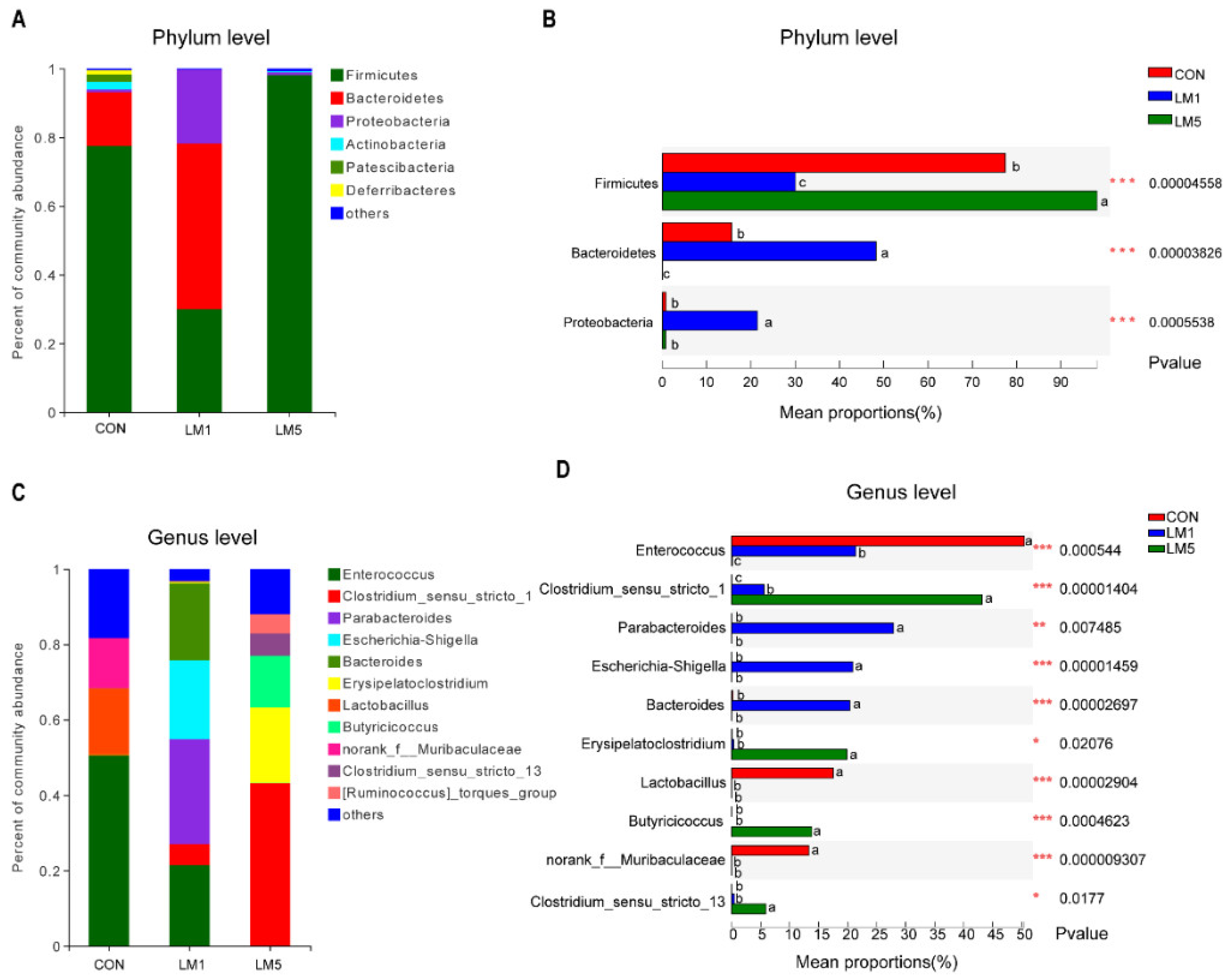
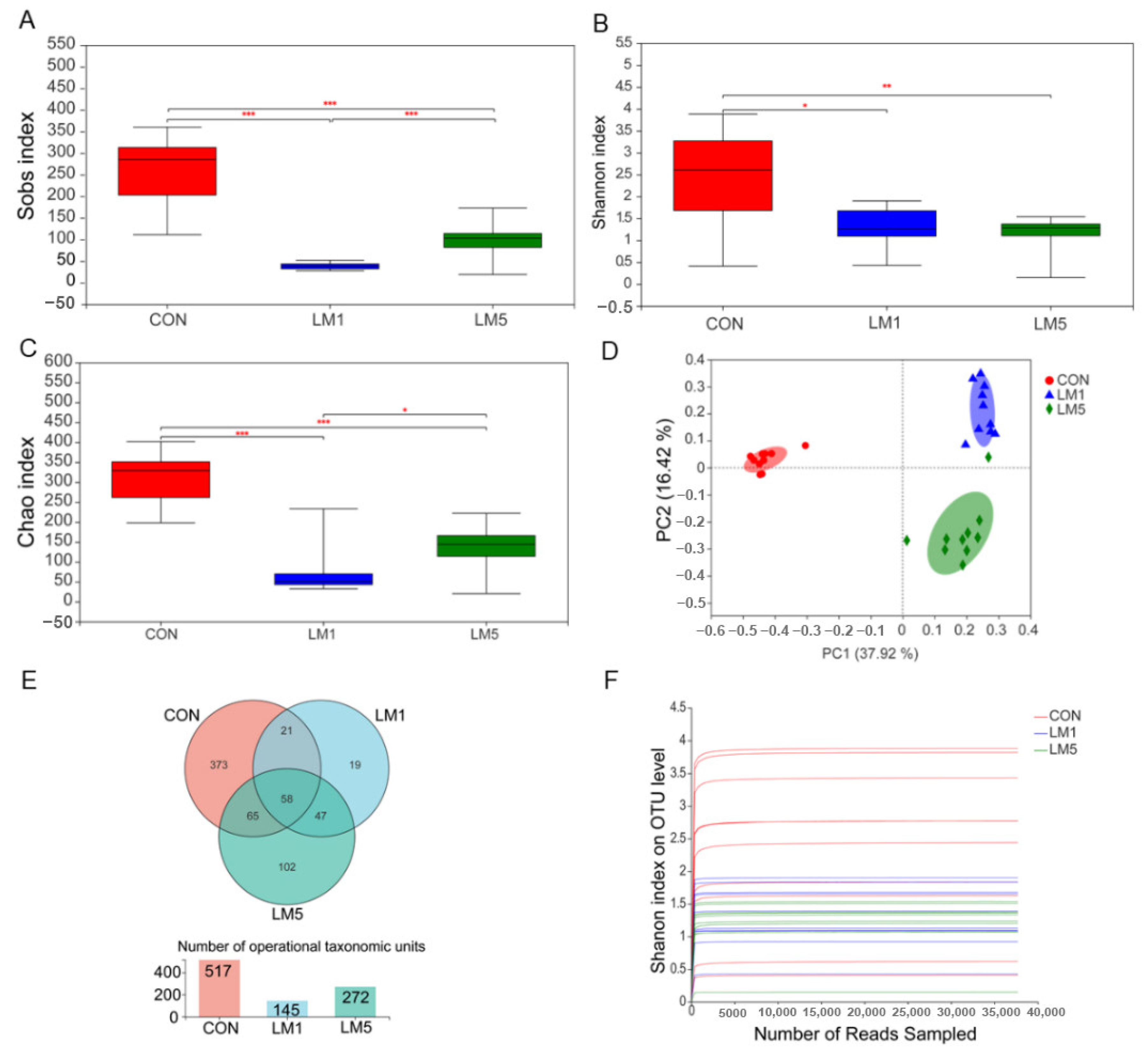
2.2. Antibiotic Treatment Reduces the Amount and Variability of the Gut Microbiota

To determine the impact of lincomycin on gut microbiota, we measured bacterial populations in colon chyme samples for all groups by 16S rRNA gene sequencing. After removing unqualified sequences following the previously reported method, the 30 samples from three groups produced a total of 1,505,868 and an average of 50,195 ± 1242 effective reads. The rarefaction curves indicated that the sequencing was deep enough to capture most of the operational units within our samples (Figure 2F). Principal coordinate analysis (PCoA) based on the unweighted unifrac distance metric is shown in Figure 2D. There was a strong impact of lincomycin on the beta diversity of the gut microbiota in mice. Alpha diversity (Sobs, Shannon, and Chao index) also revealed the gut microbial flora diversity of lincomycin-treated mice was significantly lower compared with control mice (Figure 2A–C, p < 0.01). Furthermore, the Sobs and Chao index in LM5 were greater than that in LM1 (p < 0.05). There were 517, 145, and 272 operational taxonomic units (OTUs) obtained from CON, LM1, and LM5 mice, respectively, of which 58 were common OTUs among the three experimental groups (Figure 2E). Moreover, 373 unique OTUs were found in CON, but only 19 and 102 unique OTUs in LM1 and LM5, respectively.
Notably, the microbial community composition at the phylum level of three groups was shown in Figure 3A. The composition of the gut microbiota of control mice at the phylum level included six major phyla, Firmicutes, Bacteroidetes, Actinobacteria, Proteobacteria, Patescibacteria, and Deferribacteres. The relative abundance of the phyla Firmicutes, Actinobacteria, and Deferribacteres was decreased, while the relative abundance of the phyla Bacteroidetes and Proteobacteria was increased for 1 g/L lincomycin administration group. However, a dramatic decreased in the abundance of the phyla was observed at 5 g/L lincomycin group, and 99% of them were Firmicutes. It is usually considered that a high relative abundance of Proteobacteria and Bacteroidetes are not beneficial to health [11]. Compared to the CON group, the proportion of Bacteroidetes and Proteobacteria was increased in LM1 (p < 0.05, Figure 3B). However, the Bacteroidetes were significantly decreased in LM5 (p < 0.05).
At the genus level, the composition of the gut microbiota in control mice included 3 major genera, Enterococcus, Lactobacillus, and norank_f__Muribaculaceae (Figure 3C). The Enterococcus, Parabacteroides, Escherichia-Shigella, Bacteroides, and Erysipelatoclostridium were the primary genera in LM1. Interestingly, we found Clostridium_sensu_stricto_1, Butyricicoccus, Clostridium_sensu_stricto_13, and Erysipelatoclostridium were abundance in LM5. Next, we compared the major bacterial genera among the three groups (Figure 3D). The abundances of Enterococcus, Lactobacillus, and norank_f__Muribaculaceae were significantly reduced in LM1 and LM5 compared with CON (p < 0.05). However, the relative abundance of Parabacteroides, Escherichia-Shigella, and Bacteroides in LM1 were significantly increased in CON and LM5 (p < 0.05). The relative abundance of Clostridium_sensu_stricto_13, Erysipelatoclostridium, Butyricicoccus, and Clostridium_sensu_stricto_1 in LM5 were greater than CON and LM1 (p < 0.05). All these results showed that lincomycin exposure reduced the number of intestinal microbiota and affected the composition of microbiota in mice, while 1 and 5 g/L lincomycin had different effects on intestinal microbiota in mice.

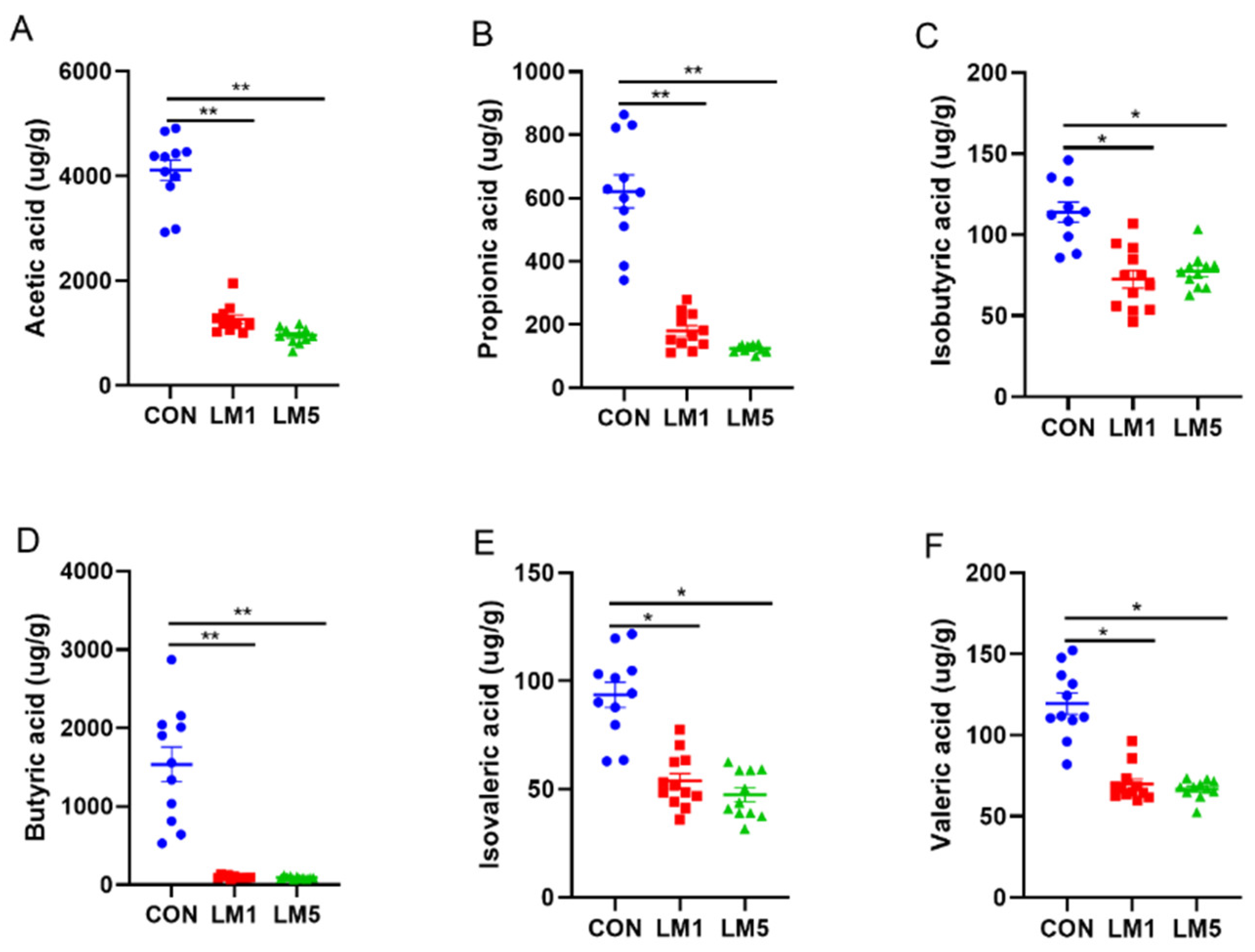
2.3. Effects of Antibiotic on SCFAs Production

To evaluate the effects of lincomycin on the metabolites of the intestinal microbiota, we examined the relative concentration of SCFAs in colon chyme (Figure 4). The results showed that the relative concentration of acetate, propionate, butyrate, valerate, isobutyric acid, and isovaleric acid was dramatically reduced both in LM1 and LM5 compared with CON (p < 0.05). And the relative concentration of acetate, propionate, butyrate, valerate, isobutyric acid, and isovaleric acid were no difference between LM1 and LM5. These results indicated that lincomycin had a negative effect on the concentrations of SCFAs in mice.

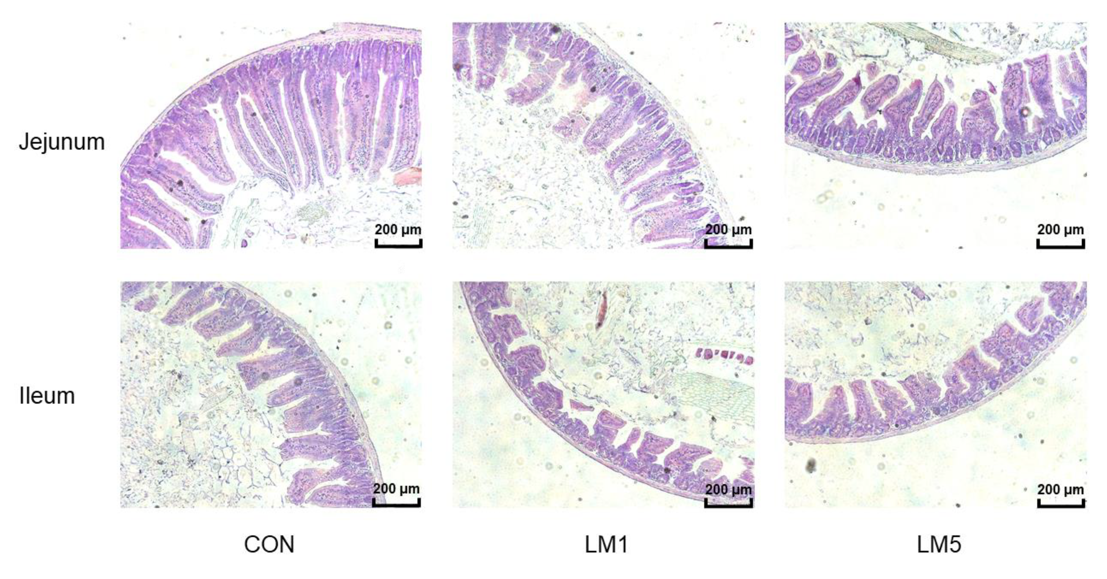
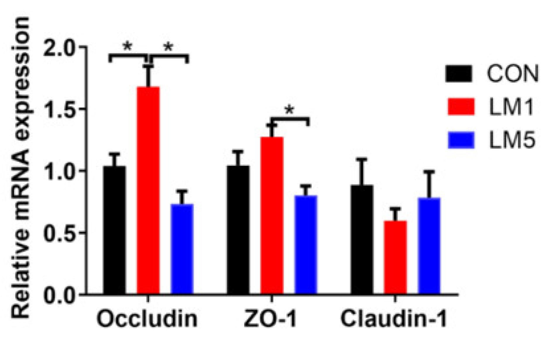
2.4. Antibiotic Exposure Influence Morphology of Jejunum and Ileum and Intestinal Barrier/gut integrity in Mice

The intestinal morphology of mice was presented in Table 1 and Figure 5. The jejunum and ileum of the mice in CON were integral, and composed of slender villi and complete crypts, which include arranged neatly epithelia cell. On the contrary, in LM1 and LM5, the villi were short and small in number, the crypts are irregular and shortened. Besides, there were some shed villi in lumens, and the epithelial cells were irregular and disorganized. In the jejunum and ileum, villus height and crypt depth of the mice in LM5 was reduced compared with CON (p < 0.05). Villus height and crypt depth of the mice in LM1 was below CON and above LM5 statistically, but not to a significant level. The V/C ratio both in jejunum and ileum were similar in three groups. These results declared that lincomycin had negative effects on the intestinal morphology and intestinal barrier of mice, and the effect increases with the dosage.
To evaluate the effects of lincomycin on intestinal barrier/gut integrity, we determined the mRNA expressions of the tight junction components cytosolic scaffold proteins (ZO-1), Occludin, and Claudin-1 in the jejunum (Figure 6). The results showed that the expression of Occludin in LM1 was improved compared with the CON and LM5 (p < 0.05), but there was no difference between LM5 and CON (p > 0.05). Moreover, the expression of ZO-1 in LM1 was greater than LM5 (p < 0.05), but both 1 and 5 g/L lincomycin had no effects on the expression of ZO-1 and Claudin-1 (p > 0.05). These results declared that 1 and 5 g/L lincomycin-induced intestinal dysbiosis may have a different effect on gut permeability.

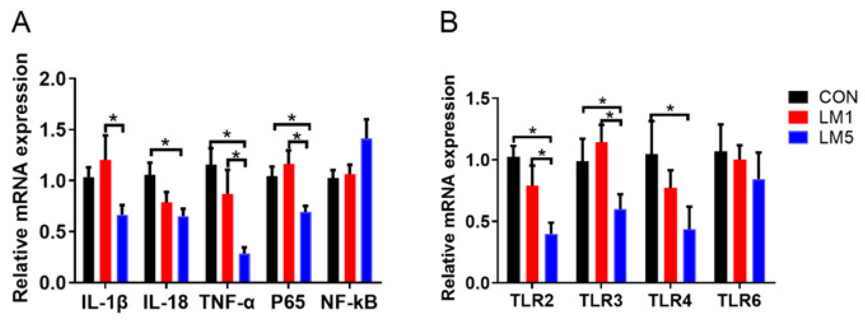
2.5. Effect of Antibiotic on TLRs Expression Patterns and Inflammation of Mice

To examine whether lincomycin induces inflammation in mice, the mRNA levels of the inflammatory cytokine TNF-α, IL-1β, and IL-18 in jejunum were determined (Figure 7A). Compared with CON, the expression of IL-1β, IL-18, and TNF-α in the jejunum was decreased in LM5 (p < 0.05). The expression of IL-1β and TNF-α in LM5 of the jejunum was decreased compared with LM1 (p < 0.05). However, the expression of IL-1β, IL-18, and TNF-α have no difference between LM1 and CON.
In order to investigate the mechanism of lincomycin on the intestinal inflammatory of mice, TLR2, TLR3, TLR4, TLR6, as well as NF-κB signaling pathways were examined. Figure 7B shows different TLR expression in the jejunum of mice treated with lincomycin. The treatment of 5 g/L lincomycin reduced the expression of TLR2, TLR3, and TLR4 in the jejunum (p < 0.05) but had no effects on TLR6 in the jejunum. The treatment of 1 g/L lincomycin treatment had no effects on TLR2, TLR3, TLR4, and TLR6 expression in jejunum compared with CON, while the expression of TLR2 and TLR3 was greater in LM1 than LM5 (p < 0.05). Compared with CON, the expression of P65 in LM1 and LM5 was reduced (Figure 7A, p < 0.05). However, the expression of NF-κB had no differences among three groups (Figure 7A).

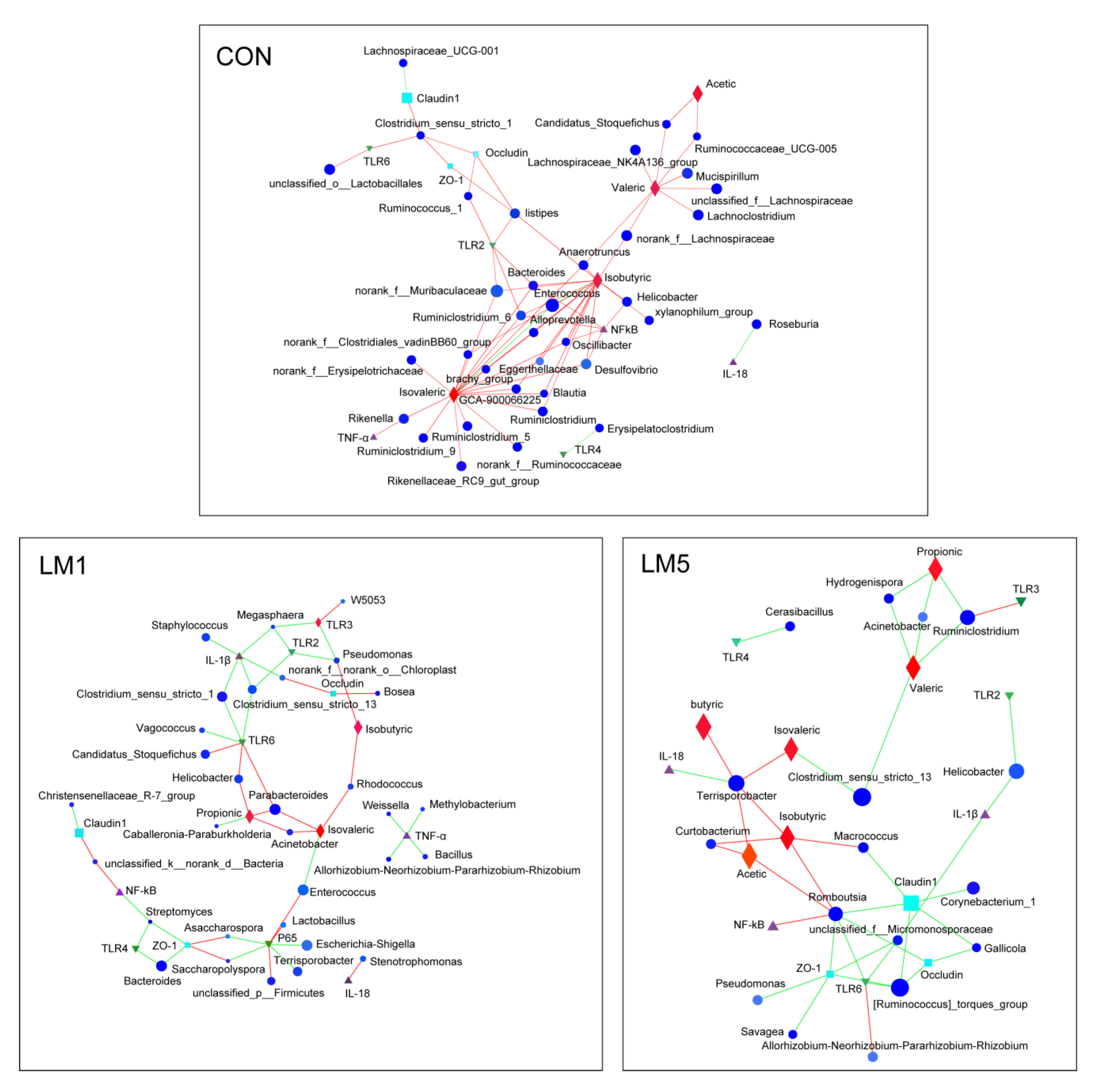
2.6. Antibiotic Modulates the Relevance Network between Relative Expression of Cytokine Contents, Bacterial Members, and Metabolites

To explore the interaction among microbiota, SCFAs, and cytokines in the mice of three groups, these targets were used to construct a network for correlation analysis (Figure 8). The results showed that unique topological patterns were observed among groups. For CON, a large number of bacterial genera formed a positively regular network centered on isobutyric, isovaleric, and valeric acid, suggesting the contribution of plentiful symbiotic bacteria to intestinal homeostasis. However, for LM1 and LM2 incompact and irregular networks emerged, in which the most relationships between microbiota and SCFAs/cytokines were negative, indicating that antibiotics disturbed the intestinal homeostasis in terms of microbiota, tight junctions, immune and metabolism.

3. Discussion

In this study, we found gut microbiota dysbiosis, immunosuppression, and intestinal barrier disruption in mice after a week of antibiotic exposure. Antibiotic exposure has a far-reaching impact on intestinal microbiota [12]. Manuzak et al. [7] reported that enrofloxacin, cephalexin, paromomycin, and clindamycin all disrupted the microbiome of rhesus macaques by reducing abundances of fermentative bacteria and increased abundances of potentially pathogenic bacteria such as Enterobacteriaceae in faeces, and decreasing Helicobacteraceae in the colon. In this study, we found the alpha diversity (Sobs, Shannon, and Chao index) of colon microbiota were significantly reduced by lincomycin exposure, and the microbial population were depleted. Specifically, the abundance of Enterococcus, Lactobacillus, and norank_f__Muribaculaceae were significantly reduced by lincomycin exposure, but more pathogens (Parabacteroides, Escherichia-Shigella, Bacteroides, and Erysipelatoclostridium) obviously grew in lincomycin treated mice. Enterococcus and Lactobacillus were thought to regulate intestinal flora and host immune function [13]. Parabacteroides have been reported to associate with abdominal infection [14]. While Escherichia and Bacteroides have been confirmed to induce chronic infection, IBD, and even colorectal cancer [15,16]. These results suggest that antibiotics disrupted intestinal microbiota and may produce complex responses.
In the present study, the abundance and number of microbiotas dramatically decreased in mice that exposure to 1 and 5 g/L lincomycin, and resulted in many adverse effects. In terms of weight, compared with CON, 1 g/L lincomycin exposure reduced the body weight gain of the mice, but 5 g/L lincomycin had no effects on the body weight gain of the mice. It takes energy to ferment pathogens in the gut, and it triggers an inflammatory response that the host needs energy to fight. Thus, these results seem to be due to the energy loss caused by lincomycin-induced decreased fermentation of hindgut microbiota in LM1. However, immunosuppression caused by high doses of lincomycin may reduce energy loss in LM5. Moreover, differences in nutrient absorption due to intestinal permeability and intestinal wall thickness may also affect body weight. In addition, these results may also be attributed to the lower level of Firmicutes and high levels of Bacteroidetes, which were thought to be associated with obesity [17]. In obese human subjects, fewer Bacteroidetes and more Firmicutes were characteristic of the intestinal microbiota [18]. A recent study indicated that Bacteroidetes had a positive correlation with LDL-and HDL-cholesterol levels, whereas Firmicutes had a negative correlation with total cholesterol, LDL-and HDL-cholesterol [19]. These results demonstrated that alternation of lipid metabolism may be another way in which gut microbes influence weight change. In the current experiment, the Firmicutes was reduced and the Bacteroidetes was improved in LM1, a low proportion of Firmicutes/Bacteroidetes caused a reduction of weight gain. However, a high level of Firmicutes and exhausted Bacteroidetes were observed in LM5, thus the body weight gain was unchanged. The specific mechanism of this result deserves further investigation.
SCFA is an important metabolite in the intestinal microbial environment and plays an important role in reducing inflammation and promoting intestinal health [20]. They are also closely involved in immune, anti-tumor, and anti-inflammatory activities [21]. In this study, we investigated gut microbial metabolites and found lincomycin exposure reduced the concentration of acetate, propionate, butyrate, valerate, isobutyric acid, and isovaleric acid in the colon. These results may be since lincomycin killed many SCFA-producing bacteria in the intestinal and led to the reduction of SCFA production which has also been seen in the study of Romick et al. [22]. For instance, Lactobacillus, which is depleted in LM1 and LM5, is acetic-producing bacteria. Furthermore, Bacteroides, Butyricicoccus, Clostridium, Ruminococcus, and Ruminococcus have been reported as SCFA-producing bacteria [23,24]. The relative abundance of Bacteroides in LM1 was more abundant than LM5 and CON, the abundance of Butyricicoccus and Ruminococcus were less than LM5, while the concentration of SCFA in LM1 and LM5 were similar in this study. These results may be due to a significant decrease of main SCFA-producing bacteria in Firmicutes and Bacteroidetes, which results in a low level of SCFAs concentration both in LM1 and LM5. In the correlation network analysis, we found that the abundance of Rikenella, Ruminococcacea, Blautia, and Ruminiclostridium were positively correlated with the isovaleric content, the abundance of Anaerotruncus, Helicobacter, Bacteroides, Enterococcus, and Desuifovibrio were positively correlated with the isobutyric content, and the abundance of Lachnoclostridium, Mucispirillum, Lachnospiraceae, and Candidatus_Stoquefichus were positively correlated with valeric content. However, in LM1 and LM5, we found the abundance of these microbiota were decreased. Meanwhile, the increased abundance of Hydrogenispora, Acinetobacter, and Ruminiciostridium in LM5 were observed to negatively correlate with propionic and valeric acid content. Therefore, the variation of these microbiota caused by antibiotic may be accompanied by a decrease in propionic, isovaleric, isobutyric, and valeric acid content. All of these results illustrated that antibiotic-induced microbial degradation may affect the production of SCFAs.
Intestinal symbiotic microbiota plays a key role in maintaining healthy intestinal barriers, which are very vital for resistance to infection by pathogenic microorganisms in the lumen [25]. Bifidobacterium has been reported to benefit intestinal homeostasis via competing for nutrients and adhesion sites with pathogens [26]. Antibiotics-induced symbiotic bacteria loss may reduce epithelial exposure with bacteria, followed by disturbing intestinal barrier function and integrity [27]. The downregulation of villus height and crypt depth in the jejunal and ileum (Table 1) suggested that lincomycin exposure may decrease the integrity of the mucous layer, which represented a vulnerable gut environment. It was reported that SCFAs, the substrate for the energy metabolism of intestinal epithelial cells, regulated the functional integrity of intestinal epithelial cells [28,29]. For instance, butyric acid has been found to maintain intestinal integrity by promoting epithelial energy metabolism [30]. Therefore, the low SCFAs in LM1 and LM5 may also contribute to reshaping intestinal morphology. The effects of lincomycin on the mRNA expression of various intestinal tight junction proteins which including occludin, claudin-1, and ZO-1 were also investigated in this study. We found the expression of occludin and ZO-1 in LM1 were greater than LM5. The network analysis showed that the abundance of Macrococcus, Corynebecterium-1, Romboutsia, and Gallicola were negatively affected claudin-1 and occludin expression, and the abundance of Romboutsia, Psoudomonas, Savages, and Micromonosporacese were negatively affected ZO-1 expression in LM5. In addition, Li et al. [31] have reported that the reduced Firmicutes/Bacteroidetes ratio was along with augmented tight junction proteins in mice. Thus, the lincomycin-induced variation of Firmicutes and Bacteroidetes in LM1 could be closely associated with the intestinal tight junction barrier in this study. Increased mRNA levels of ZO-1 and occludin in LM1 indicated the reduced intestinal permeability, which may hinder nutrient absorption in the short term. This may also be the reason why the body weight gain of mice in LM1 was less than CON and LM5. The results described above indicate that antibiotic-induced microbiota and SCFAs reduction may disrupt intestinal morphology and barriers.
Toll-like receptors (TLRs) are pattern recognition receptors (PRRs) that mediate the interaction between microbiota and the host, and very vital for innating immune responses [32,33]. In this experiment, lincomycin exposure consumed vast microbiota in the colon and changed the composition of the gut microbiota. Meanwhile, decreased expressions of TLR2, TLR3, and TLR4 were observed after lincomycin exposure, indicating that the host-microbiota interactions might be also altered. Some symbiotic bacteria such as Lactobacillus, Bifidobacterium and Enterococcus have been reported to improve intestinal mucosal inflammation by regulating TLRs expression [34]. Thus, a possible reason for the decreased expressions of TLRs might be that the depletion of intestinal symbiotic microbiota caused by antibiotics reduces the stimulation of TLRs by symbiotic bacteria. These results were similar to the previous study that the combination of antibiotics ampicillin, vancomycin, neomycin, and metronidazole induced intestinal microbiota depletion decreased the TLR4 expression of the intestinal mucosa in mice [35]. Furrie et al. [33] have reported that Enterococcus was positively correlated with TLR4 expression. The depletion of Enterococcus in LM5 may be responsible for the reduction of TLR4 in this study. Furthermore, TLR2 and TLR4 protein has been shown only in the crypts of mouse colon [36]. Therefore, lincomycin-induced crypts damage in LM5 may be another reason to reduce the expression of TLR2 and TLR4. TLR3 expresses at the luminal surface, and the expression may be enhanced by the interaction of new epithelial cells with gram-positive commensal organisms [33]. In this study, 5 g/L lincomycin treatment consumed gram-positive commensal organisms, leading to the decrease of TLR3 expression. These results suggest that microbial depletion caused by antibiotics may affect TLR-related immune responses.
In general, TLRs play immunomodulatory role via activating NF-κB to initiate the release of inflammatory cytokines such as TNF-α and IL-1β through myeloid differentiation factor 88 (MyD88) dependent/independent pathway [37,38]. As communicators between immune cells, cytokines were crucial parts of the immune system [39]. Their changes were associated with host inflammatory responses. In this study, with the decrease of TLR2 and TLR4 expression, the expression of TNF-α, IL-1β, and P65 was decreased in LM5. This result was similar to the study of Jena et al. [40] who reported that the ileal expression of gene IL-1β and IL-6 were decreased in a polymixin B-treated mouse model. Previous studies have shown that gut microbiota interacts with the immune system by releasing cytokines [5]. For instance, TNF-α production may be related to Odoribacter splanchnic and the genus Bilophila [41]. The above results of this study may be related to the abundance of Clostridium, which induces Treg activity and inflammation [42]. In addition, in LM5, we also observed a decrease of IL-18 expression in the jejunum which was vital in maintaining intestinal homeostasis. Meanwhile, the presence of Terrisorobacter and its negative correlation with IL-18 were also found in LM5. On the one hand, the reduction of microbial diversity caused by antibiotics reduces the stimulation of microorganisms to the host and the interaction of microbial-related molecular patterns with the host in the short term, thus reducing the inflammatory response. On the other hand, microbial metabolites had a strong regulation on cytokine formation [43]. Thus, the decrease of cytokines such as IL-1β, TNF-α, and IL-18 expression in this study may be associated with the drastic reduction of acetate, propionate, butyrate, valerate, isobutyric acid, and isovaleric acid. These results suggest that antibiotics-induced microbial depletion may cause cytokine changes.
Lincomycin and related antibiotics, such as clindamycin, are widely used as prophylactic treatment in gastrointestinal operations, including colorectal resection for colorectal cancer. The reduction of beneficial SCFA along with the down-regulation of TLRs, which are crucial factors for the physiology of epithelial healing, combined with the alterations in gut barrier integrity and specific microbiota alterations (reduction of Lactobacillus and proliferation of Enterobacteriaceae) are well-known to be involved in the pathogenesis of anastomotic leakage [44]. Thus, these results could be valuable regarding the possible effect of perioperative antibiotics in anastomotic complications after bowel resection.

4. Materials and Methods

4.1. Ethics Statement

The experimental protocol was reviewed and approved by the Institutional Animal Care and Use Committee of the Institute of Animal Science at the Chinese Academy of Agricultural Science (IAS2020-85).

4.2. Mice and Antibiotics Treatment

In the present study, thirty 21-day-old female ICR mice purchased from School of Medicine, Peking University (Beijing, China) were housed in the animal facility of the Chinese Academy of Agricultural Sciences under a controlled condition of the lighting cycle with light from 8:30 a.m. to 8:30 p.m., the temperature at 20 ± 5 °C. After 3 days of acclimatization, mice were randomly divided into three groups (CON, LM1, and LM5, respectively) of 10 mice each. The mice in LM1 and LM5 were administered with 1 and 5 g/L lincomycin in drinking water for 1 week, respectively. Control mice received drinking water for 1 week. All mice had free access to food and drinking water, and water containers and feed were changed once three days to supply fresh antibiotics and feed. At the end of the experiment (week 1), body weight, food intake, and water intake were measured for mice, and the jejunum, ileum, colon, and colonic chyme were excised. All samples were either fixed in formalin or snap-frozen and stored at −80 °C until processing.

4.3. Cytokine Genes Expression

Total RNA was extracted using TRIzol reagent (Ambion, UT, USA). Total RNA was reversely transcribed into cDNA using a reverse transcription kit at 37 °C for 15 min, 85 °C 5 s. All primers were designed on the National Center for Biotechnology Information website (Table 2). The House-keeping gene was GADPH to normalize target gene levels. Quantitative real-time RT-PCR (qRT-PCR) was performed using TB Green Premix Ex Taq (TaKaRa, Kusatsu, Japan). The gene of tight junction protein (Occludin, ZO-1, and Claudin-1), pro-inflammatory cytokines (TNF-α, IL-1β, and IL-18), TLR (TLR2, TLR3, TLR4, and TLR6), NF-κB, and P65 were analyzed. The primers used for q-PCR were purchased from Sangon Biotech (Shanghai, China) and shown in Table 2. All results of the target genes were normalized to the housekeeping gene GAPDH as an endogenous control. Reactions were run on an Applied Biosystems 7500 system (Applied Biosystems, Life Technologies, Waltham, MA, USA) using the following protocol: 94 °C for the 30 s, 40 cycles of 5 s at 94 °C, 15 s at 58 °C, 10 s at 72 °C. Results were analyzed using 2−∆∆Ct methods.

4.4. DNA Extraction, 16S rRNA Gene Amplification and Sequencing

The E.Z.N.A.® soil DNA Kit (Omega Bio-tek, Norcross, GA, USA) was used to extract microbial community genomic DNA on the basis of the manufacturer’s instructions. According to the method previously described, the hypervariable region V3-V4 of the bacterial 16S rRNA gene was amplified with primer pairs 338F (5’-ACTCCTACGGGAGGCAGCAG-3’) and 806R (5’-GGACTACHVGGGT WTCTAAT-3’) using an ABI Gene Amp® 9700 PCR thermocycler (ABI, CA, USA) [45]. The meta-genomic sequencing was performed based on Illumina platform using Miseq PE300. The raw reads were deposited into the NCBI Sequence Read Archive (SRA) database (Accession Number: PRJNA665786).

4.5. Sequence Processing and Analysis

The raw sequences were processed using Majorbio I-Sanger Cloud Platform (www.i-sanger.com, Majorbio, Shanghai, China), and chimeric sequences were removed. Operational taxonomic units (OTUs) with a 97% similarity cutoff were clustered using UPARSE (version 7.1, http://drive5.com/uparse/). The taxonomy of each OTU representative sequences were matched to the 16S rRNA database (Silva SSU128) with a confidence threshold of 0.7 using RDP Classifier (http://rdp.cme.msu.edu/). Mothur b.1.30.1 [46] was used to perform refraction analysis, alpha-diversity analysis, and unweighted principal coordinate analysis (PCoA). Sobs, Shannon and Chao indices were employed to evaluate community diversity. For each group, a network based on spearman’s coefficients was established to depict the correlations among cytokine contents, the relative abundances of bacterial genera, and SCFA concentrations in colon.

4.6. The Relative Concentration of SCFAs

The colon chyme samples, approximately 0.1 g of each mouse, were suspended in 1 mL of ddH2O in a 1.5 mL screw-capped tube for SCFAs composition analysis. The gas chromatography method was used to determine the composition of SCFAs as Wu et al. [47].

4.7. Tissue Sample and Intestinal Morphology

Jejunum and ileal segments were immersed and fixed with formalin for 24 h to observe intestinal morphology, and then were cut into 5 μm-thick sections stained with hematoxylin and eosin (H&E) [48]. The villous height and crypt depth of each sample were measured and the villous height/crypt depth ratio was calculated.

4.8. Statistical Analysis

For the data of growth performance, intestinal morphology, SCFAs concentration, and mRNA expression, one-way ANOVA with multiple comparisons using Fisher LSD test was performed (SAS 9.4, Institute, Cary, NC, USA), accepting statistically significant at the level of p-value < 0.05.

5. Conclusions

In conclusion, this study indicated that a week of lincomycin exposure decreased the concentrations of SCFA, altered intestinal morphology, and reduced the elevated expression of TLRs and inflammation-related cytokines in infant mice via alterations of the gut microbiota. Furthermore, these effects differed for different doses of lincomycin. Due to the increased relative abundance of pathogenic bacteria and decreased relative abundance of beneficial bacteria, this might cause subsequent long-term negative impacts of lincomycin on host intestinal health in mice. The absence of follow-up evaluation of the restoration of gut microbiota is one major limitation of this study. Thus, the subsequent long-term evaluation of the restoration of gut microbiota worth be worth study in the future.

Author Contributions

Conceptualization, S.Z., L.C., J.Y., and H.Z.; data curation, S.Z., H.H. and R.Z.; formal analysis, S.Z. and R.Z.; funding acquisition, L.C., J.Y., and H.Z.; investigation, S.Z., H.H., and R.Z.; methodology, S.Z., B.Y., and L.C.; resources, L.C., and H.Z.; software, S.Z.; supervision, L.C. and H.Z.; validation, S.Z., R.Z., L.C., and H.Z.; writing—original draft, S.Z.; writing—review and editing, S.Z., R.Z., L.C., J.Y., and H.Z. All authors revised and approved the final manuscript. All authors have read and agreed to the published version of the manuscript.

Funding

The authors are grateful for the financial support from National Natural Science Foundation of China (3170130194), Science and Technology Innovation project of The Chinese Academy of Agricultural Sciences (CAAS-ZDRW202006-02), Open project of State Key Laboratory of Animal Nutrition (2004DA125184F1912).

Conflicts of Interest

The authors declare no conflict of interest.

References

  1. Honda, K.; Littman, D.R. The microbiota in adaptive immune homeostasis and disease. Nature 2016, 535, 75–84. [Google Scholar] [CrossRef] [PubMed]
  2. Yamanaka, M.; Iwai, H.; Saito, M.; Yamauchi, C.; Nomura, T. Influence of intestinal microbes on digestion and absorption of nutrients in diet and nitrogen retention in germfree, gnotobiotic and conventional mice. 1. Protein and fat digestion and nitrogen retention in germfree and conventional mice. Jpn. J. Zootech. Sci. 1972, 43, 272–283. [Google Scholar] [CrossRef]
  3. Qayed, M.; Horan, J.T. The Role of Intestinal Microbiota in Graft versus Host Disease. Mini-Rev. Med. Chem. 2016, 16, 193–199. [Google Scholar] [CrossRef]
  4. Natarajan, N.; Pluznick, J.L. From microbe to man: The role of microbial short chain fatty acid metabolites in host cell biology. Am. J. Physiol. Cell Physiol. 2014, 307, C979–C985. [Google Scholar] [CrossRef] [PubMed] [Green Version]
  5. Sun, L.; Zhang, X.; Zhang, Y.; Zheng, K.; Xiang, Q.; Chen, N.; Chen, Z.; Zhang, N.; Zhu, J.; He, Q. Antibiotic-Induced Disruption of Gut Microbiota Alters Local Metabolomes and Immune Responses. Front. Cell. Infect. Microbiol. 2019, 9, 99. [Google Scholar] [CrossRef] [PubMed]
  6. Kim, H.J.; Lee, S.H.; Hong, S.J. Antibiotics-Induced Dysbiosis of Intestinal Microbiota Aggravates Atopic Dermatitis in Mice by Altered Short-Chain Fatty Acids. Allergy Asthma Immunol. Res. 2020, 12, 137–148. [Google Scholar] [CrossRef]
  7. Manuzak, J.A.; Zevin, A.S.; Cheu, R.; Richardson, B.; Modesitt, J.; Hensley-McBain, T.; Miller, C.; Gustin, A.T.; Coronado, E.; Gott, T.; et al. Antibiotic-induced microbiome perturbations are associated with significant alterations to colonic mucosal immunity in rhesus macaques. Mucosal Immunol. 2020, 13, 471–480. [Google Scholar] [CrossRef]
  8. Wang, H.; Wang, B.; Zhao, Q.; Zhao, Y.; Fu, C.; Feng, X.; Wang, N.; Su, M.; Tang, C.; Jiang, F.; et al. Antibiotic Body Burden of Chinese School Children: A Multisite Biomonitoring-based Study. Environ. Sci. Technol. 2015, 49, 5070–5079. [Google Scholar] [CrossRef]
  9. Cheng, B.; Jiang, F.; Su, M.L.; Zhou, L.Q.; Zhang, H.; Cao, Z.G.; Liao, X.J.; Xiong, G.H.; Xiao, J.H.; Liu, F.S.; et al. Effects of lincomycin hydrochloride on the neurotoxicity of zebrafish. Ecotoxicol. Environ. Saf. 2020, 201, 110725. [Google Scholar] [CrossRef]
  10. Wang, Q.; Du, W.; Xu, J. Retrospective analysis of adverse reactions of lincomycin. Chin. J. New Drugs Clin. Remedies 2003, 22, 571–574. [Google Scholar]
  11. Shin, N.R.; Whon, T.W.; Bae, J.W. Proteobacteria: Microbial signature of dysbiosis in gut microbiota. Trends Biotechnol. 2015, 33, 496–503. [Google Scholar] [CrossRef]
  12. Nogacka, A.M.; Salazar, N.; Arboleya, S.; Suarez, M.; Fernandez, N.; Solis, G.; de Los Reyes-Gavilan, C.G.; Gueimonde, M. Early microbiota, antibiotics and health. Cell. Mol. Life Sci. 2018, 75, 83–91. [Google Scholar] [CrossRef]
  13. Wang, Y.; Yang, W.; Zhang, G. Effects of Enterococcus faecium on growth performance, intestinal Flora and immune function of weaner piglets. Chin. J. Anim. Nutr. 2013, 25, 1069–1076. [Google Scholar]
  14. Kierzkowska, M.; Majewska, A.; Szymanek-Majchrzak, K.; Sawicka-Grzelak, A.; Mlynarczyk, A.; Mlynarczyk, G. In vitro effect of clindamycin against Bacteroides and Parabacteroides isolates in Poland. J. Glob. Antimicrob. Resist. 2018, 13, 49–52. [Google Scholar] [CrossRef] [PubMed]
  15. Khan, A.A.; Khan, Z.; Malik, A.; Kalam, M.A.; Cash, P.; Ashraf, M.T.; Alshamsan, A. Colorectal cancer-inflammatory bowel disease nexus and felony of Escherichia coli. Life Sci. 2017, 180, 60–67. [Google Scholar] [CrossRef]
  16. Sears, C.L. Enterotoxigenic Bacteroides fragilis: A Rogue among Symbiotes. Clin. Microbiol. Rev. 2009, 22, 349–369. [Google Scholar] [CrossRef] [PubMed] [Green Version]
  17. Cheng, R.; Guo, J.; Pu, F.; Wan, C.; Shi, L.; Li, H.; Yang, Y.; Huang, C.; Li, M.; He, F. Loading ceftriaxone, vancomycin, and Bifidobacteria bifidum TMC3115 to neonatal mice could differently and consequently affect intestinal microbiota and immunity in adulthood. Sci. Rep. 2019, 9, 3254. [Google Scholar] [CrossRef] [PubMed] [Green Version]
  18. Ley, R.E.; Turnbaugh, P.J.; Klein, S.; Gordon, J.I. Microbial ecology—Human gut microbes associated with obesity. Nature 2006, 444, 1022–1023. [Google Scholar] [CrossRef] [PubMed]
  19. Harata, G.; Kumar, H.; He, F.; Miyazawa, K.; Yoda, K.; Kawase, M.; Kubota, A.; Hiramatsu, M.; Rautava, S.; Salminen, S. Probiotics modulate gut microbiota and health status in Japanese cedar pollinosis patients during the pollen season. Eur. J. Nutr. 2017, 56, 2245–2253. [Google Scholar] [CrossRef]
  20. Yao, Y.; Yan, L.J.; Chen, H.; Wu, N.; Wang, W.B.; Wang, D.S. Cyclocarya paliurus polysaccharides alleviate type 2 diabetic symptoms by modulating gut microbiota and short-chain fatty acids. Phytomedicine 2020, 77, 153268. [Google Scholar] [CrossRef]
  21. Zhou, C.; Li, L.Z.; Li, T.M.; Sun, L.H.; Yin, J.H.; Guan, H.D.; Wang, L.C.; Zhu, H.B.; Xu, P.; Fan, X.; et al. SCFAs induce autophagy in intestinal epithelial cells and relieve colitis by stabilizing HIF-1 alpha. J. Mol. Med. JMM 2020, 98, 1189–1202. [Google Scholar] [CrossRef] [PubMed]
  22. Romick-Rosendale, L.E.; Haslam, D.B.; Lane, A.; Denson, L.; Lake, K.; Wilkey, A.; Watanabe, M.; Bauer, S.; Litts, B.; Luebbering, N.; et al. Antibiotic Exposure and Reduced Short Chain Fatty Acid Production after Hematopoietic Stem Cell Transplant. Biol. Blood Marrow Transplant. 2018, 24, 2418–2424. [Google Scholar] [CrossRef] [PubMed] [Green Version]
  23. Rios-Covian, D.; Salazar, N.; Gueimonde, M.; de los Reyes-Gavilan, C.G. Shaping the Metabolism of Intestinal Bacteroides Population through Diet to Improve Human Health. Front. Microbiol. 2017, 8, 376. [Google Scholar] [CrossRef] [PubMed] [Green Version]
  24. Morrison, D.J.; Preston, T. Formation of short chain fatty acids by the gut microbiota and their impact on human metabolism. Gut Microbes 2016, 7, 189–200. [Google Scholar] [CrossRef] [Green Version]
  25. Zhang, C.; Peng, Y.; Mu, C.; Zhu, W. Ileum terminal antibiotic infusion affects jejunal and colonic specific microbial population and immune status in growing pigs. J. Anim. Sci. Biotechnol. 2018, 9, 51. [Google Scholar] [CrossRef]
  26. Ohland, C.L.; MacNaughton, W.K. Probiotic bacteria and intestinal epithelial barrier function. Am. J. Physiol. Gastrointest. Liver Physiol. 2010, 298, G807–G819. [Google Scholar] [CrossRef] [Green Version]
  27. Willing, B.P.; Russell, S.L.; Finlay, B.B. Shifting the balance: Antibiotic effects on host-microbiota mutualism. Nat. Rev. Microbiol. 2011, 9, 233–243. [Google Scholar] [CrossRef]
  28. Kaiko, G.E.; Stappenbeck, T.S. Host-microbe interactions shaping the gastrointestinal environment. Trends Immunol. 2014, 35, 538–548. [Google Scholar] [CrossRef] [Green Version]
  29. Morris, G.; Berk, M.; Carvalho, A.; Caso, J.; Sanz, Y.; Walder, K.; Maes, M. The Role of the Microbial Metabolites Including Tryptophan Catabolites and Short Chain Fatty Acids in the Pathophysiology of Immune-Inflammatory and Neuroimmune Disease. Mol. Neurobiol. 2017, 54, 4432–4451. [Google Scholar] [CrossRef]
  30. Ma, X.; Fan, P.X.; Li, L.S.; Qiao, S.Y.; Zhang, G.L.; Li, D.F. Butyrate promotes the recovering of intestinal wound healing through its positive effect on the tight junctions. J. Anim. Sci. 2012, 90, 266–268. [Google Scholar] [CrossRef] [Green Version]
  31. Li, Y.; Liu, T.; Yan, C.; Xie, R.; Guo, Z.; Wang, S.; Zhang, Y.; Li, Z.; Wang, B.; Cao, H. Diammonium Glycyrrhizinate Protects against Nonalcoholic Fatty Liver Disease in Mice through Modulation of Gut Microbiota and Restoration of Intestinal Barrier. Mol. Pharm. 2018, 15, 3860–3870. [Google Scholar] [CrossRef] [PubMed]
  32. Underhill, D.M.; Ozinsky, A. Toll-like receptors: Key mediators of microbe detection. Curr. Opin. Immunol. 2002, 14, 103–110. [Google Scholar] [CrossRef]
  33. Filippi, C.M. Toll-like receptor activation in immunity vs. tolerance. Front. Immunol. 2015, 6, 146. [Google Scholar] [CrossRef] [PubMed] [Green Version]
  34. Yang, X.; Gao, X.-C.; Liu, J.; Ren, H.-Y. Effect of EPEC endotoxin and bifidobacteria on intestinal barrier function through modulation of toll-like receptor 2 and toll-like receptor 4 expression in intestinal epithelial cell-18. World J. Gastroenterol. 2017, 23, 4744–4751. [Google Scholar] [CrossRef] [PubMed]
  35. Chen, L.W.; Chang, W.J.; Chen, P.H.; Liu, W.C.; Hsu, C.M. TLR ligand decreases mesenteric ischemia and reperfusion injury-induced gut damage through TNF-alpha signaling. Shock 2008, 30, 563–570. [Google Scholar] [CrossRef]
  36. Ortega-Cava, C.F.; Ishihara, S.; Rumi, M.A.K.; Kawashima, K.; Ishimura, N.; Kazumori, H.; Udagawa, J.; Kadowaki, Y.; Kinoshita, Y. Strategic compartmentalization of toll-like receptor 4 in the mouse gut. J. Immunol. 2003, 170, 3977–3985. [Google Scholar] [CrossRef] [Green Version]
  37. Piras, V.; Selvarajoo, K. Beyonc MyD88 and TRIF pathways in Toll-like receptor signaling. Front. Immunol. 2014, 5, 70. [Google Scholar] [CrossRef]
  38. Wang, X.X.; Luo, B.J.; Lu, Y.Y.; Pang, D.G.; Zheng, J.Q.; Mo, J.L.; Huang, H.; Feng, J.F. The triggering receptor expressed by myeloid cells-1 activates TLR4-MyD88-NF-B-dependent signaling to aggravate ventilation-induced lung inflammation and injury in mice. Cell Tissue Res. 2018, 374, 137–148. [Google Scholar] [CrossRef]
  39. Xue, X.; Falcon, D.M. The Role of Immune Cells and Cytokines in Intestinal Wound Healing. Int. J. Mol. Sci. 2019, 20, 97. [Google Scholar] [CrossRef] [Green Version]
  40. Jena, P.K.; Sheng, L.; Liu, H.-X.; Kalanetra, K.M.; Mirsoian, A.; Murphy, W.J.; French, S.W.; Krishnan, V.V.; Mills, D.A.; Wan, Y.-J.Y. Western Diet-Induced Dysbiosis in Farnesoid X Receptor Knockout Mice Causes Persistent Hepatic Inflammation after Antibiotic Treatment. Am. J. Pathol. 2017, 187, 1800–1813. [Google Scholar] [CrossRef] [Green Version]
  41. Schirmer, M.; Smeekens, S.P.; Vlamakis, H.; Jaeger, M.; Oosting, M.; Franzosa, E.A.; ter Horst, R.; Jansen, T.; Jacobs, L.; Bonder, M.J.; et al. Linking the Human Gut Microbiome to Inflammatory Cytokine Production Capacity. Cell 2016, 167, 1125–1136. [Google Scholar] [CrossRef] [PubMed] [Green Version]
  42. Wick, E.C.; Sears, C.L. Bacteroides spp. and diarrhea. Curr. Opin. Infect. Dis. 2010, 23, 470–474. [Google Scholar] [CrossRef] [PubMed] [Green Version]
  43. Nowak, E.C.; de Vries, V.C.; Wasiuk, A.; Ahonen, C.; Bennett, K.A.; Le Mercier, I.; Ha, D.-G.; Noelle, R.J. Tryptophan hydroxylase-1 regulates immune tolerance and inflammation. J. Exp. Med. 2012, 209, 2127–2135. [Google Scholar] [CrossRef] [PubMed] [Green Version]
  44. Koliarakis, I.; Athanasakis, E.; Sgantzos, M.; Mariolis-Sapsakos, T.; Xynos, E.; Chrysos, E.; Souglakos, J.; Tsiaoussis, J. Intestinal Microbiota in Colorectal Cancer Surgery. Cancers 2020, 12, 3011. [Google Scholar] [CrossRef]
  45. Evans, C.C.; LePard, K.J.; Kwak, J.W.; Stancukas, M.C.; Laskowski, S.; Dougherty, J.; Moulton, L.; Glawe, A.; Wang, Y.; Leone, V.; et al. Exercise Prevents Weight Gain and Alters the Gut Microbiota in a Mouse Model of High Fat Diet-Induced Obesity. PLoS ONE 2014, 9, e92193. [Google Scholar] [CrossRef]
  46. Shang, Q.S.; Song, G.R.; Zhang, M.F.; Shi, J.J.; Xu, C.Y.; Hao, J.J.; Li, G.Y.; Yu, G.L. Dietary fucoidan improves metabolic syndrome in association with increased Akkermansia population in the gut microbiota of high-fat diet-fed mice. J. Funct. Foods 2017, 28, 138–146. [Google Scholar] [CrossRef]
  47. Wu, W.; Xie, J.; Zhang, H. Dietary fibers influence the intestinal SCFAs and plasma metabolites profiling in growing pigs. Food Funct. 2016, 7, 4644–4654. [Google Scholar] [CrossRef]
  48. Zhong, G.; Shao, D.; Wang, Q.; Tong, H.; Shi, S. Effects of dietary supplemented of gamma-amino butyric acid on growth performance, blood biochemical indices and intestinal morphology of yellow-feathered broilers exposed to a high temperature environment. Ital. J. Anim. Sci. 2020, 19, 431–438. [Google Scholar] [CrossRef]
Figure 1. Lincomycin reduces growth performance of mice. (A) Body weight gain in each group of mice. (B) Feed intake in each group of mice. (C) Water intake in each group of mice. (D) Water intake/feed intake in each group of mice. Values are means (n 10/diet), with their standard errors represented by vertical bars. Statistical analysis was performed using one-way ANOVA with multiple comparisons by Fisher LSD test. * Means statistically significant (p < 0.05).
Antibiotics 09 00907 g001
Figure 2. Microbial community composition of mice. (A) The alpha-diversity of fecal microbiota in colon chyme of Sobs index. (B) The alpha-diversity of fecal microbiota in colon chyme of Shannon index. (C) The alpha-diversity of fecal microbiota in colon chyme of Chao index. (D) Weighted principal coordinate analysis (PCoA). (E) Venn diagram. (F) Rarefaction curves tended to reach the plateau. * p < 0.05; ** p < 0.01; *** p < 0.001.
Antibiotics 09 00907 g002
Figure 3. Influence of lincomycin treatment on the microbial at the phylum and genus levels in mice. (A) The relative abundance of bacterial at the phylum level. (B) The top three bacterial at the phylum level statistical comparison of the relative abundance in three groups. (C) The relative abundance of bacterial at genus levels. (D) The top ten bacterial at the genus level statistical comparison of the relative abundance in three groups. Less than 1% abundance of the phyla or genus was merged into others. Different letters indicate significant differences between the two groups, while the same letter indicates insignificant differences. * p < 0.05; ** p < 0.01; *** p < 0.001.
Antibiotics 09 00907 g003
Figure 4. Concentrations of short-chain fatty acids (SCFA) in fresh colonic chyme after treated with lincomycin in mice. Scatter plot of SCFA concentration. Statistical analysis was performed using one-way ANOVA with multiple comparisons by Fisher LSD test. * p < 0.05; ** p < 0.01. (A) Acetic acid. (B) Propionic acid. (C) Isobutyric acid. (D) Butyric acid. (E) Isovaleric acid. (F) Valeric acid.
Antibiotics 09 00907 g004
Figure 5. To evaluate the potential intestinal tissue injury caused by antibiotic, the staining profiles by H&E of jejunum and ileum in mice of three groups were displayed (n = 10/group). Scale bars: 100 μm.
Antibiotics 09 00907 g005
Figure 6. Effect of lincomycin on intestinal barrier in mice jejunal, relative expression of ZO-1, Occludin, and Claudin-1 mRNA. Values are means, with their standard errors represented by vertical bars. Statistical analysis was performed using one-way ANOVA with multiple comparisons by Fisher LSD test, * p < 0.05.
Antibiotics 09 00907 g006
Figure 7. Effect of lincomycin on inflammatory cytokine expression in mice jejunal. (A) Relative expression of IL-1β, IL-18, TNF-α, P65, and NF-κB mRNA in the jejunum of mice. (B) Relative expression of TLR2, TLR3, TLR4, and TLR6 mRNA in the jejunum of mice. Values are means, with their standard errors represented by vertical bars. Statistical analysis was performed using one-way ANOVA with multiple comparisons by Fisher LSD test, * p < 0.05.
Antibiotics 09 00907 g007
Figure 8. A network for correlation analysis among the relative abundances of microbiota phylum, the concentrations of SCFAs, and the relative expression of cytokines in the mice of CON, LM1, and LM5, respectively. Only correlations with a partial spearman’s coefficient >0.5 and a p < 0.05 were shown. The different color and shapes of nodes indicates various targets as described in legend, and the size of nodes indicates species abundance. The line color represents correlation, red represents positive correlation and green represents negative correlation.
Antibiotics 09 00907 g008
Table 1. Effects of lincomycin on the morphology of jejunum and ileum in mice.
Jejunum µmIleum µm
GroupVillus HeightCrypt DepthV/CVillus HeightCrypt DepthV/C
CON470.26 a111.02 a4.29209.46 a79.16 a2.63
LM1405.51 ab95.24 ab4.23161.12 ab65.47 ab2.45
LM5310.30 b78.58 b4.02137.75 b55.68 b2.46
SEM21.374.350.1512.123.090.10
p-value0.0060.0070.8970.0480.0040.470
a,b Values in the same column not sharing a common superscript differ significantly (p < 0.05).
Table 2. Primers Used for q-PCR Analysis.
GeneForward Nucleotide Sequence Primers (5′-3′)Reverse Nucleotide Sequence Primers (5′-3′)Product Size (bp)
GADPHACCACAGTCCATGCCATCACTCCACCACCCTGTTGCTGTA172
TLR2AGGTGCCCTGTGCCACCATTCGGAACGAAGTCCCGCTTGT172
TLR3GGTCCCCAGCCTTCAAAGACACGAAGAGGGCGGAAAGGT85
TLR4CTGGTGGCTGTGGAGACAAAATTCCCTGAAAGGCTTGGTC172
TLR6TCATCTCAGCAAACACCGAGTATAGCGCAACCTTATTGAATGTGACCCTCCAGC249
IL-1βTCGCAGCAGCACATCAACAAGAGAGGTCCACGGGAAAGACACAGG97
IL-18AGACCTGGAATCAGACAACTTTTCAGTCATATCCTCGAACACAG117
TNF-αGGACTAGCCAGGAGGGAGAACAGGCCAGTGAGTGAAAGGGACAGAAC103
NF-kBCAAAGACAAAGAGGAAGTGCAAGATGGAATGTAATCCCACCGTA203
P65TCGAGTCTCCATGCAGCTACGGCGGTGGCGATCATCTGTGTCTG93
ZO-1CTGGTGAAGTCTCGGAAAAATGCATCTCTTGCTGCCAAACTATC97
OccludinTGCTTCATCGCTTCCTTAGTAAGGGTTCACTCCCATTATGTACA155
Claudin-1-1AGATACAGTGCAAAGTCTTCGACAGGATGCCAATTACCATCAAG86
Publisher’s Note: MDPI stays neutral with regard to jurisdictional claims in published maps and institutional affiliations.

