Article

Characterization of Two New Multidrug-Resistant Strains of Mycobacterium smegmatis: Tools for Routine In Vitro Screening of Novel Anti-Mycobacterial Agents

1
West African Center for Cell Biology of Infectious Pathogens, Department of Biochemistry, Cell and Molecular Biology, University of Ghana, P. O. Box LG 54, Accra, Ghana
2
Centre for Molecular Microbiology and Infection, Imperial College London, London SW7 2AZ, UK
*
Author to whom correspondence should be addressed.
Antibiotics 2019, 8(1), 4; https://doi.org/10.3390/antibiotics8010004
Submission received: 13 November 2018 / Revised: 7 December 2018 / Accepted: 7 December 2018 / Published: 2 January 2019

Abstract

:
Mycobacterium tuberculosis is a pathogen of global public health concern. This threat is exacerbated by the emergence of multidrug-resistant and extremely-drug-resistant strains of the pathogen. We have obtained two distinct clones of multidrug-resistant Mycobacterium smegmatis after gradual exposure of Mycobacterium smegmatis mc2 155 to increasing concentrations of erythromycin. The resulting resistant strains of Mycobacterium smegmatis exhibited robust viability in the presence of high concentrations of erythromycin and were found to be resistant to a wide range of other antimicrobials. They also displayed a unique growth phenotype in comparison to the parental drug-susceptible Mycobacterium smegmatis mc2 155, and a distinct colony morphology in the presence of cholesterol. We propose that these two multidrug-resistant clones of Mycobacterium smegmatis could be used as model organisms at the inceptive phase of routine in vitro screening of novel antimicrobial agents targeted against multidrug-resistant Mycobacterial tuberculosis.

Graphical Abstract

1. Introduction

Tuberculosis is one of the most successful human infections with an extensive global coverage [1]. It is caused by members of the Mycobacterium tuberculosis complex, following inhalation of aerosols containing the tubercle bacilli [2]. By estimation, one-third of the world’s population has latent tuberculosis infection and this represents a considerable reservoir from which future tuberculosis cases can emanate [3]. Currently, tuberculosis is a major cause of morbidity and death in low- and middle-income countries.
In the clinical setting, drug-resistant Mycobacterium tuberculosis is a serious and persistent threat to global health [4]. Drug resistance in Mycobacterium tuberculosis is largely associated with genetic mutations [5,6]. Resistance to at least rifampicin and isoniazid, also termed multidrug-resistant (MDR) tuberculosis, is on the rise. Over 490,000 MDR tuberculosis cases are recorded yearly, resulting in over 130,000 deaths [7]. Though host genetic factors may play a role, insufficient or incomplete treatment is the most important determinant of the development of MDR tuberculosis [8]. In the global setting, MDR tuberculosis accounts for 3.8% of new cases of the disease and presents in 20% of patients with a history of tuberculosis treatment [9].
Though a number of therapeutic strategies have been employed to treat and eradicate the disease, the emergence of drug-resistant strains has attracted worldwide concern. These strategies include the development of new drugs that target new bacterial proteins, cellular structures, and processes. Routine in vitro screening for novel antimycobacterial compounds would be facilitated by the availability of avirulent mycobacterial strains that are easy to grow and handle. Even though H37Ra is an avirulent strain of Mycobacterium tuberculosis, derived from the virulent H37 parent strain, its slow growth rate hinders the rapid initial screening of antimicrobials to identify candidates that exhibit antimycobacterial activity [10]. An alternative avirulent mycobacterial strain which could be used as a surrogate for Mycobacterium tuberculosis is the Mycobacterium smegmatis mc2 155 strain; Mycobacterium smegmatis is suitable for rapid bioassays, which can be performed in less sophisticated biosafety cabinets. Notably, the diarylquinoline TMC207, which is an anti-tubercular drug, was identified using Mycobacterium smegmatis as a model organism [11]. This study set out to develop extensively-drug-resistant Mycobacterium smegmatis strains to provide a model which could be useful at the initial phase of screening for new antibiotics to combat drug-resistant infections.
Continuous exposure of microbial populations to antibiotics, as employed in this study, is considered the most relevant factor that influences the evolution of drug-resistant strains [12]. Continuous exposure to antibiotics promotes gradual acquisition of resistance by subpopulations of the mycobacterium, leaving their antibiotic susceptible counterparts extinct [13,14]. Interactions among other antibiotics also influence the extent of sensitivity of a microbe to a particular antibiotic. Antibiotic resistance has been shown to impose metabolic and fitness costs that reduces the growth of resistant strains [15,16].
Resistance to an antibiotic has been found to either promote cross-resistance or sensitivity to other antibiotics [17,18]. In the present study, drug-susceptible populations of Mycobacterium smegmatis mc2 155 were driven, under in vitro conditions, to evolve into two distinct erythromycin-resistant strains. The study demonstrates the unique phenotypes exhibited by the two erythromycin-resistant strains, in comparison to the original mycobacterium strain from which they were derived. Collectively, we provide additional data which corroborate our current understanding of the emergence and characteristic phenotypes of drug-resistant strains of mycobacterium following continuous exposure to a drug. The two strains will also serve as valuable drug screening tools for the development of the next generation of anti-mycobacterial agents.

2. Materials and Method

2.1. Bacterial Strains, Media Preparation, and Antibiotic Treatments

Mycobacterium smegmatis mc2 155 was used as a drug-susceptible strain for all experiments. The MDR Mycobacterium smegmatis strains (referred to as erythromycin-resistant Mycobacterium smegmatis A and B) were generated in this study, under in vitro conditions, and were used as drug-resistant strains. Middlebrook 7H10 agar base (Sigma Aldrich) and Middlebrook 7H9 broth base (Sigma Aldrich) were used for preparing agar plates and liquid broth, respectively, according to the manufacturer’s instructions. The M7H10 agar base was supplemented with 0.085% NaCl and 0.5% dextrose and the M7H9 broth base was supplemented with 0.085% NaCl, 0.44% glycerol, and 0.25% Tween 80. All commercially available antibiotics that were used for this study are listed in Tables S2 and S3. The amount or concentration of each antibiotic used for specific assays are indicated in the text of the results.

2.2. Drug Susceptibility Assay

Prior to a drug susceptibility assay, mycobacterial cells were streaked onto fresh M7H10 agar plates and incubated at 30 °C [19] for 48 h. The cells that grew on the agar plates were confirmed to be mycobacteria, via acid-fast staining, and were used for the inoculation of 50 mL of M7H9 broth base. The inoculated broths were incubated at 30 °C for 24 h with shaking (160 rpm). The bacterial cultures that were obtained were diluted to an optical density at 600 nm (OD600) of 0.7 in 50 mL of freshly prepared M7H9 broth base and incubated at 30 °C for 24 h with shaking.
The 24 h, day two cultures were diluted to OD600 of 0.7 and were used for the uniform inoculation (spreading) of M7H10 agar plates. Paper discs containing the indicated amount of antibiotic were placed on the inoculated plates and the plates were incubated at 30 °C for 48 h. The zone of inhibition (in mm) around each antibiotic disc was measured and used to ascertain the susceptibility profile of the organism to the panel of antibiotics.

2.3. Testing Cell Viability via Spot Test Assay

The 24-h, day two cultures were diluted to OD600 of 0.7 in 1 mL of M7H9 broth base containing the indicated concentration of erythromycin. The control for the diluted samples, indicated as untreated, did not contain erythromycin. The diluted mycobacterial cultures (± erythromycin) were incubated at 30 °C for 0 h, 3 h, and 6 h, and an aliquot of 5 μL was spotted on M7H10 agar plates after the indicated duration of incubation. The M7H10 agar plates containing the spots of the bacterial culture were incubated at 30 °C for 48 h.

2.4. Growth Profile of Mycobacterial Cells

The 24-h, day two cultures were diluted to OD600 of 0.01 in 50 mL of M7H9 broth base. The diluted cultures were incubated at 30 °C, with shaking, and the OD600 were measured periodically at the indicated time intervals.

2.5. Colony Morphology Assay

The 24-h, day two cultures were diluted to OD600 of 0.7 and aliquots of 5 μL were spotted on M7H10 agar plates containing 100 mΜ of cholesterol. The corresponding control M710 agar plates did not contain cholesterol. For each M7H10 agar plate, 5 μL of the diluted bacterial cultures were spotted at four positions, which were separated approximately 20 mm from each other. The agar plates containing the spots of the bacterial cultures were incubated at 30 °C for 48 h. Colonies that grew on the agar plates were confirmed to be mycobacterial cells by acid-fast staining.

2.6. Staining of Mycobacterial Cells

A colony of cells that had grown on M7H10 agar plates were spread uniformly on microscope slides. Gram staining and acid-fast staining were performed to confirm that the cells were mycobacteria. Following the Gram staining and acid-fast staining, the cells were visualized using a Bresser Science MPO 401 light microscope equipped with a 100× objective lens. Images were acquired using a camera that was in-built within the microscope.

3. Results

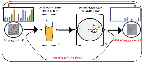
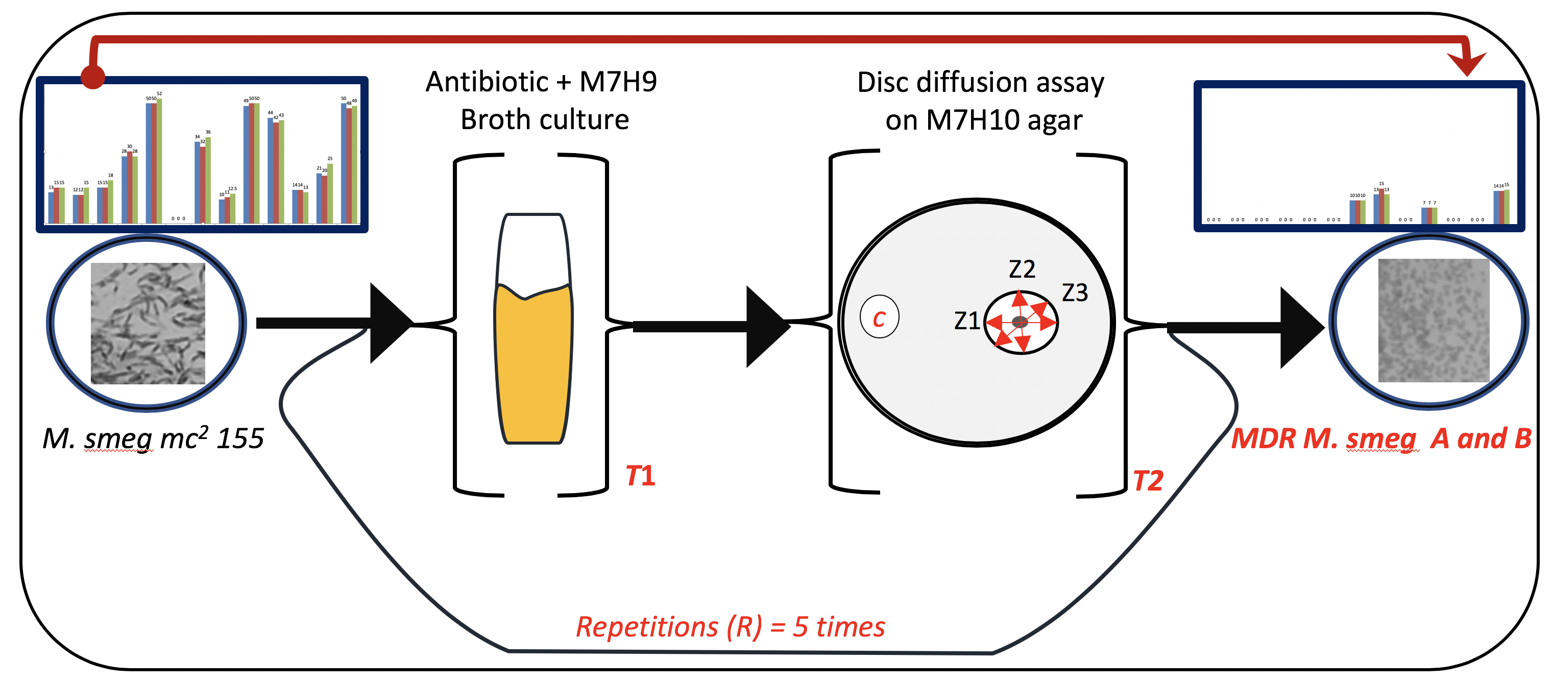
3.1. Exposure of Mycobacteria to Increasing Antibiotic Concentrations Drives Evolution of Drug Resistance

Starting with a single colony of Mycobacterium smegmatis mc2 155, drug-resistant populations of the bacteria were allowed to evolve by systematically exposing the cells to increasing concentrations of either erythromycin, streptomycin, or tetracycline (Figure 1). Prior to each repetition (R), the minimal inhibitory concentrations (MICs) of erythromycin, streptomycin, and tetracycline were determined for the distinct colonies that had grown either within or around the zones of inhibition of these antibiotics (Figure 1). The MIC determination was used to ascertain the extent of the antibiotic resistance acquired by these distinct colonies of Mycobacterium smegmatis. The cells gradually adapted to the systematic exposure to increasing concentrations of each antibiotic, revealing gradual acquisition of resistance (Table 1). Measurement of OD600 at the end of the 2nd, 4th, and 5th steps also confirmed this observation (Table S1). At the end of the 5th step, colonies of Mycobacterium smegmatis were not detected within the zone of inhibition on the M7H10 agar plates containing either streptomycin or tetracycline discs (Table 1). However, distinct colonies of Mycobacterium smegmatis were detected within the zones of inhibition of erythromycin discs on the M7H10 agar plates. These mycobacterial cells were characterized as erythromycin-resistant Mycobacterium smegmatis. Two colonies, isolated by spreading a 10−6 dilution of the erythromycin-resistant Mycobacterium smegmatis culture, showed different susceptibilities to a panel of thirteen commercial antibiotics (Figure 2a,b). These two distinct clones were further characterized as erythromycin-resistant Mycobacterium smegmatis A and B and were continually maintained on M7H10 agar plates (without antibiotics) for the next 6 months, prior to the subsequent assays.
The Mycobacterium smegmatis mc2 155 was susceptible to twelve out of the thirteen panel of commercial antibiotics, while the two clones of erythromycin-resistant Mycobacterium smegmatis (A and B) were both susceptible to only rifampicin, moxifloxacin, and streptomycin (Figure 2b). Notably, clone A, but not clone B, of the erythromycin-resistant Mycobacterium smegmatis strain was also susceptible to tetracycline and chloramphenicol. Hence, the distinct antibiotic susceptibility pattern for each erythromycin-resistant strain was the basis for the characterization of erythromycin-resistant Mycobacterium smegmatis as either clone A or clone B. These observations agree with a previous report that indicated that a single antibiotic treatment may select for resistance against other multiple drugs via a set of mechanisms (collateral resistance) or a single mechanism (cross-resistance) [20]. What is more striking about this data is that the acquisition of broad-spectrum resistance cuts across all classes of antibiotics. This observation is the first to be reported for mycobacteria where exposure to one antibiotic has led to the development of extreme drug resistance (MDR). Thus, these two strains constitute a valuable tool in the continued search for new antibiotic candidates.

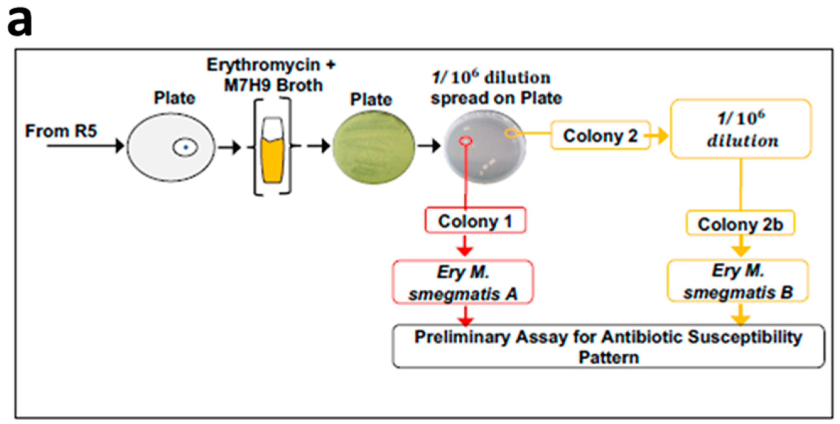
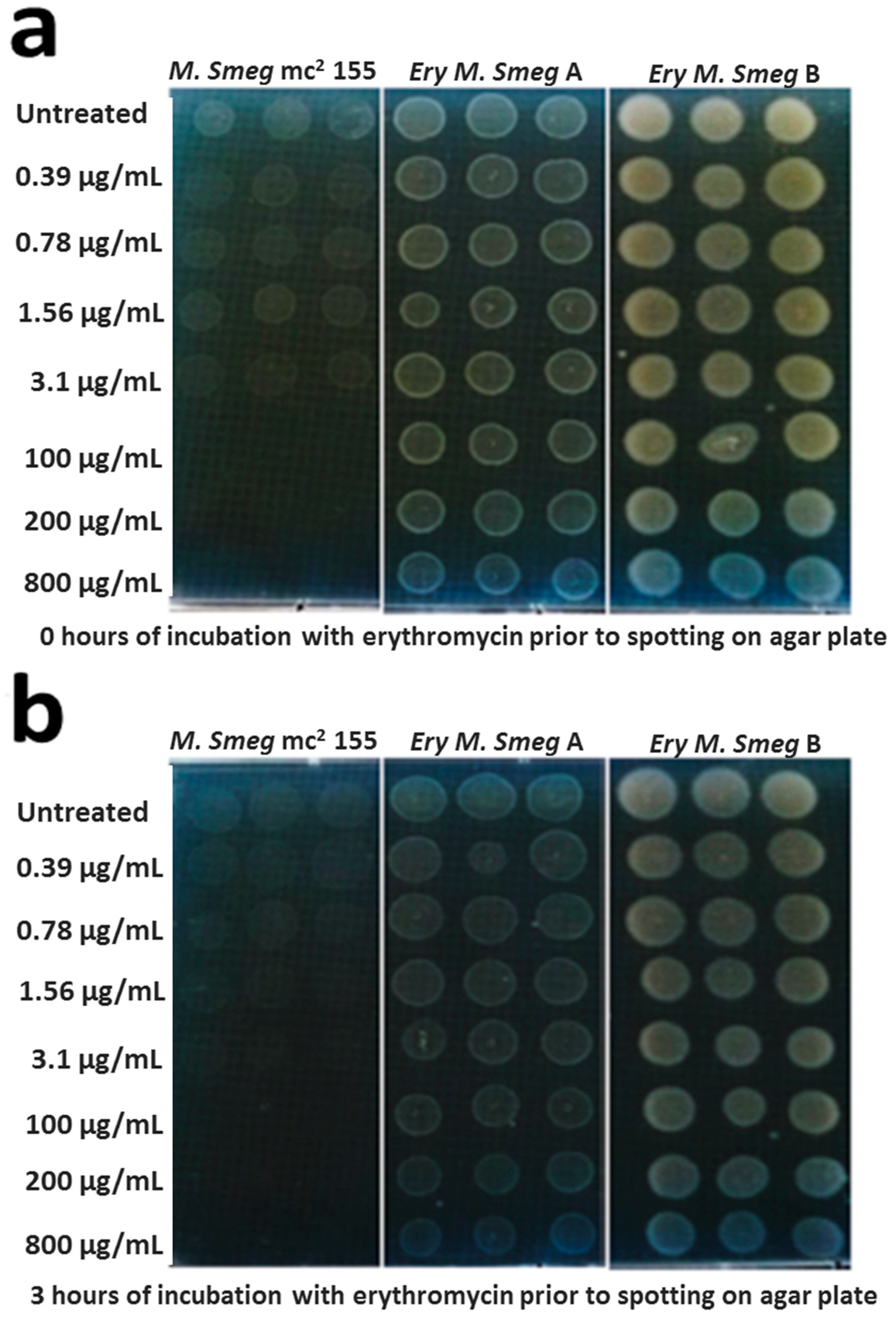
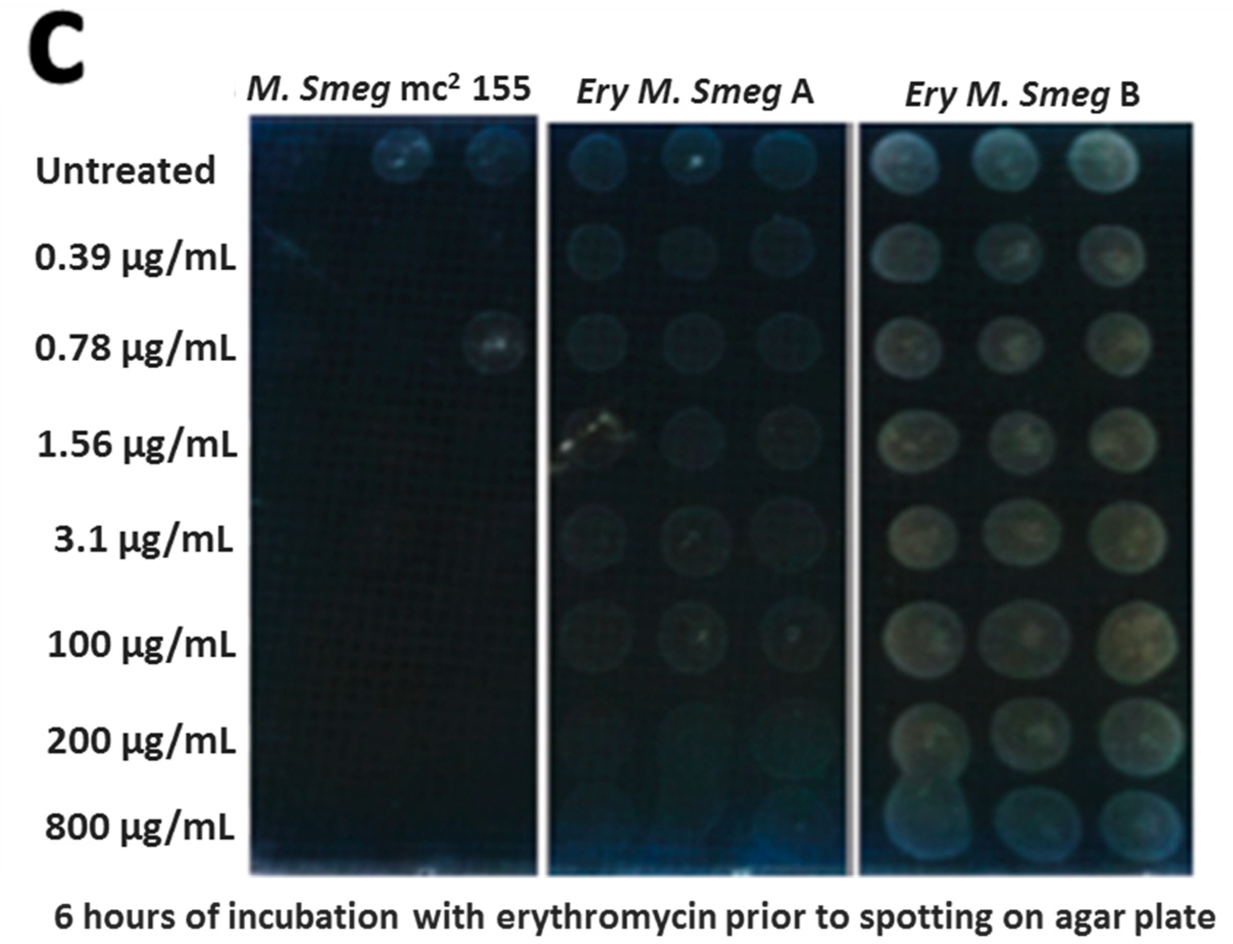
3.2. MDR Mycobacterium Smegmatis is Viable in the Presence of Higher Concentrations of Erythromycin

A spot test assay was used to validate the extent of resistance of the two clones of erythromycin-resistant Mycobacterium smegmatis (clones A and B) relative to the Mycobacterium smegmatis mc2 155 strain. These three mycobacterial strains were exposed to increasing concentrations of erythromycin for 0 h, 3 h, and 6 h, prior to spotting the cells on M7H10 agar plates. The erythromycin-resistant Mycobacterium smegmatis strains were viable at all of the three time points (0 h, 3 h, and 6 h of exposure to erythromycin), and the viability was robust against the low and high concentrations of erythromycin used for the assay (Figure 3). On the contrary, the Mycobacterium smegmatis mc2 155 strain was viable at the low concentrations of erythromycin, whereas growth was undetected at the high concentrations of the antibiotic (Figure 3a,b). Notably, growth was undetected for the Mycobacterium smegmatis mc2 155 strain following 6 h of exposure to the low concentrations of erythromycin (Figure 3c). These observations demonstrate that the two erythromycin-resistant strains exhibit a distinct viability phenotype in the presence of erythromycin, in comparison to the Mycobacterium smegmatis mc2 155 parent strain.

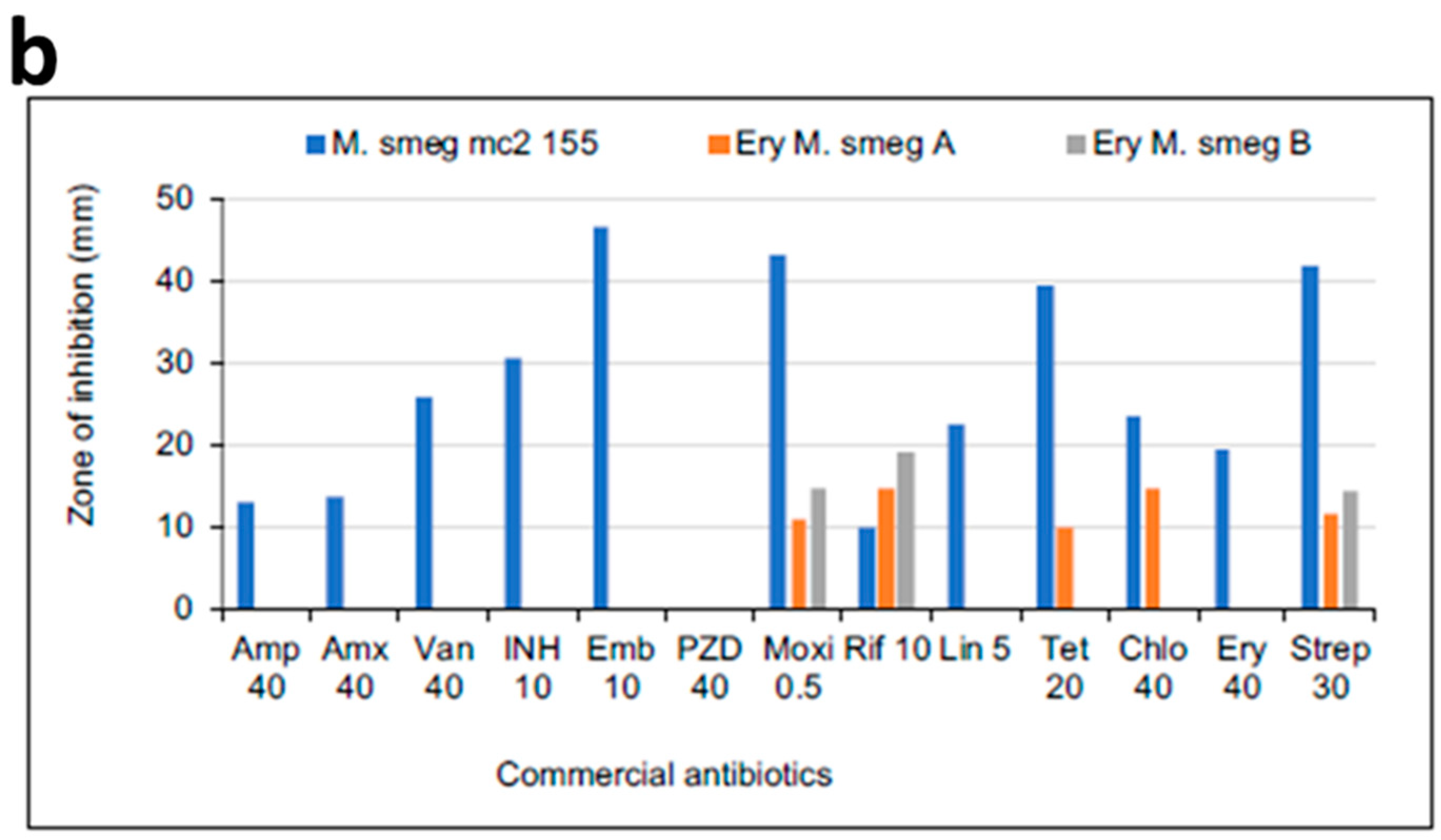
3.3. MICs of Selected Antibiotics against MDR Mycobacterium Smegmatis

The MICs of thirteen commercially-available antibiotics were determined for the Mycobacterium smegmatis mc2 155 strain and the two MDR strains. For each organism, the MIC of an antibiotic of interest was determined by placing four discs, containing decreasing concentrations of the antibiotic, at positions distant from each other on the agar plate. All of the three mycobacterial strains were resistant to the highest concentration of Pyrazinamide (160 µg) and were susceptible to the lowest concentration of moxifloxacin (0.25 µg; Table 2). The two MDR strains were resistant to the highest concentration of isoniazid, ethambutol, linezolid, and erythromycin, while the Mycobacterium smegmatis mc2 155 strain was susceptible to the lowest concentration of these antibiotics. The MIC of vancomycin and streptomycin was 80 µg for the MDR strains; the Mycobacterium smegmatis mc2 155 strain was susceptible to the lowest concentrations of these antibiotics used for the assay. Interestingly, the two MDR strains were susceptible to 20 µg of ampicillin and amoxicillin, even though our earlier observations indicated that these MDR strains were resistant to 40 µg of either of these antibiotics (Figure 2b). Moreover, clone B of the MDR strain was susceptible to 15 µg and 30 µg of tetracycline and chloramphenicol, respectively; an observation that was inconsistent with the data shown in Figure 2b. We proposed that the placement of different classes of antibiotic discs on an agar plate during the test for the drug susceptibility pattern (Figure 2B), as compared to the placement of discs of the same antibiotic for the MIC determination (Table 2), was the basis for the above-mentioned deviations. The antibiotic assay format is the standard widely used for the determination of the antibiotic susceptibility pattern of bacterial isolates. Thus cross-interaction between different classes of antibiotics might be influencing the resistance profiles of the MDR M. smegmatis strains to a sub-collection of the selected commercial antibiotics.

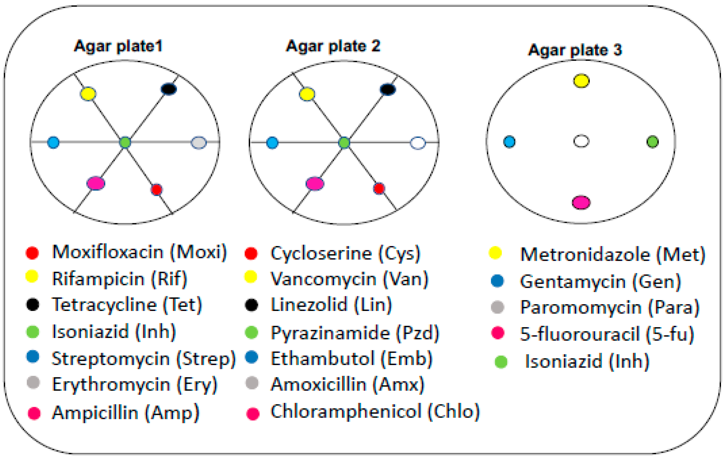
3.4. Effect of Neighboring Antibiotic Discs on the Antimycobacterial Activity of Selected Antibiotics

Using the disc diffusion assay, a number of antibiotics are usually placed at unique positions on an agar plate that has been inoculated with the strain of interest during the test for drug susceptibility patterns (Figure 4). We investigated the effect of neighboring antibiotic discs on the antimycobacterial activity of ampicillin, amoxicillin, linezolid, tetracycline, chloramphenicol, erythromycin, and streptomycin. Ultimately, this experimental approach was anticipated to provide information on specific neighboring antibiotic discs that either enhanced or reduced the antimycobacterial activity of these selected antibiotic discs.
The erythromycin-resistant M. smegmatis A strain was resistant to ampicillin, amoxicillin, and chloramphenicol when each of these selected antibiotics were surrounded by the neighboring antibiotic discs indicated in Table 3. Interestingly, the same concentration of these selected antibiotics (ampicillin, amoxicillin, and chloramphenicol) exhibited antimycobacterial activity against the same resistant strain in the absence of the neighboring antibiotic discs. The opposite effect was detected for streptomycin against the resistant strain when discs of rifampicin, isoniazid, and ampicillin were used as neighbors. These observations were not unique to erythromycin-resistant M. smegmatis A strain; the M. smegmatis mc2 155 strain was highly susceptible to linezolid in the presence of neighboring antibiotic discs, but was resistant to the same concentration of linezolid in the absence of neighboring antibiotic discs. Collectively, these observations demonstrate that the resistance profiles of M. smegmatis mc2 155 and the corresponding MDR strains are influenced by cross-interaction of the different classes of antibiotics. This same dataset demonstrates a subtle phenotypic difference between the two MDR strains.

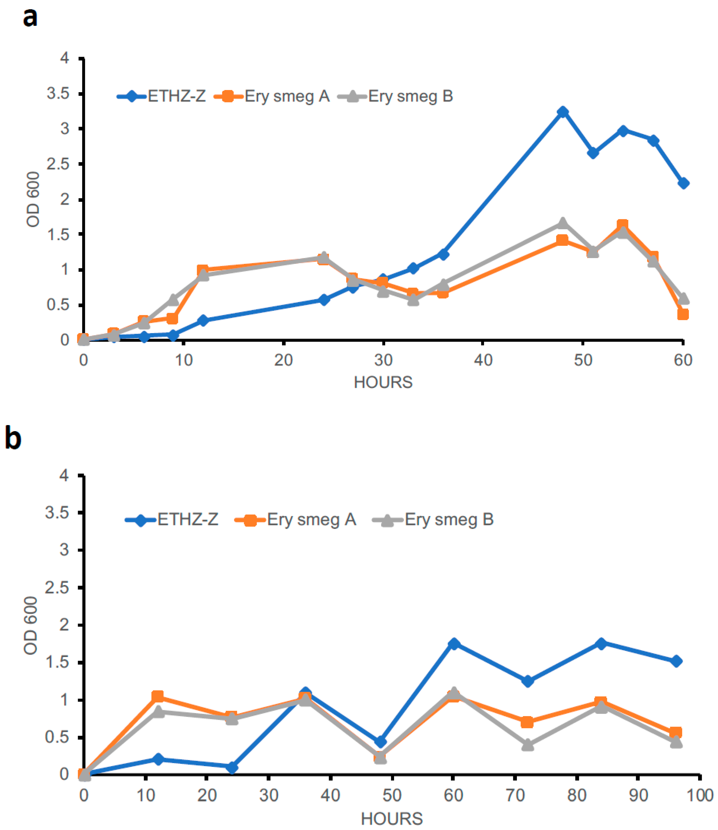
3.5. Evolution of the MDR Phenotype Affects the Growth Profile of Mycobacterium Smegmatis

The growth profiles of the two clones of erythromycin-resistant Mycobacterium smegmatis and the Mycobacterium smegmatis mc2 155 strain were investigated in order to ascertain the effect of the MDR phenotype on growth rate. Single colonies from these three mycobacterial strains were initially grown in M7H9 broth for 24 h at 30 °C with shaking. These mycobacterial cultures were diluted to an OD600 of 0.01 and the growth rate was monitored for 60 h (Figure 5a). The two clones of erythromycin-resistant Mycobacterium smegmatis showed a relatively rapid growth rate during the initial 12 h compared to the Mycobacterium smegmatis mc2 155 strain. Interestingly, the latter strain recuperated from the initial lag phase of growth after 12 h and showed a rapid growth rate, attaining an OD600 of 3.2 by 48 h (Figure 5a). On the contrary, the erythromycin-resistant strains showed a relatively slower growth rate after the initial 12 h. This pattern of growth was confirmed following measurement of OD600 at 12-h intervals for 96 h (Figure 5b), illustrating that the acquisition of MDR phenotype causes a concomitant alteration in the growth profile of Mycobacterium smegmatis.

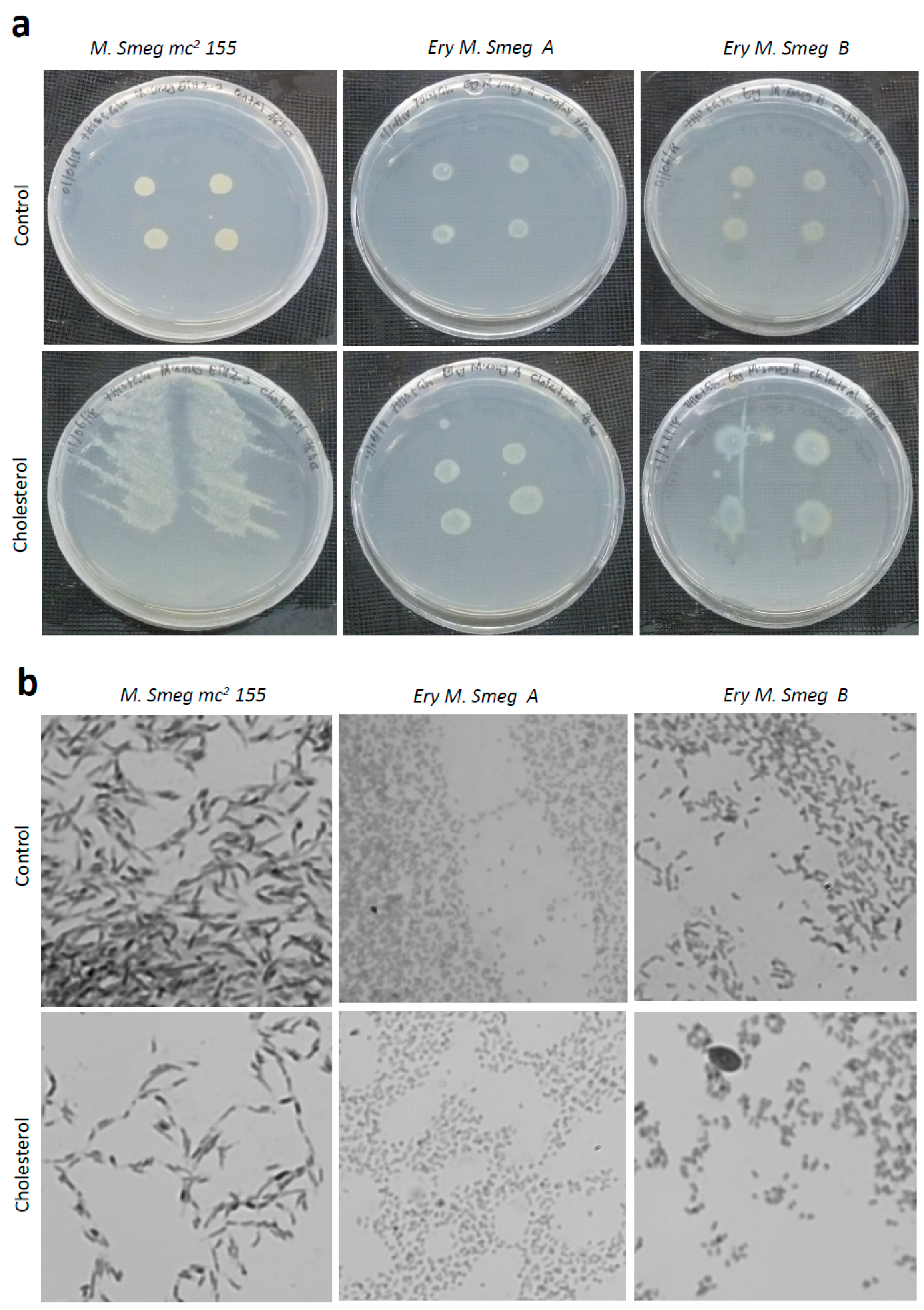
3.6. Cholesterol Enhances the Growth of Colonies of Mycobacterium Smegmatis

It has been reported that Mycobacterium tuberculosis utilizes the cholesterol of its host organism, for virulence and invasion of macrophages, in a mechanism dependent on the modulation of lysosomal calcium levels [21,22]. Cholesterol metabolism in M. tuberculosis is facilitated by the presence of an operon, which consist of genes encoding acrylamine N-acetyltransferase and other associated proteins [23]. Proteins associated with cholesterol metabolism are usually conserved in M. tuberculosis and M. smegmatis and represent a possible target for novel anti-tubercular drugs [24,25]. Even though M. smegmatis is avirulent and does not persist in macrophages, the presence of these conserved proteins makes M. smegmatis a suitable laboratory model for studying the phenotypic effect of cholesterol on mycobacteria. Hence, we investigated the effect of cholesterol on the morphology of Mycobacterium smegmatis mc2 155, as well as the erythromycin-resistant strains.
Colonies of the Mycobacterium smegmatis mc2 155 strain were detected as broad, fluffy-like structures on the cholesterol-containing M7H10 agar plates, whereas the control (no cholesterol) agar plates showed the expected colony phenotype (Figure 6a). The colonies of the two clones of erythromycin-resistant Mycobacterium smegmatis were modestly enlarged on the cholesterol-containing agar plate compared to the colonies on the control agar plates (Figure 6a). Visualization of individual cells within these colonies revealed that all of the three mycobacterial strains tested positive for acid-fast staining (Figure S1). Moreover, individual cells from colonies of the Mycobacterium smegmatis mc2 155 strain had a slender and rod-like morphology (Figure 6b). Even though the erythromycin-resistant strains were also rod-like in shape, they were relatively smaller than the Mycobacterium smegmatis mc2 155 strain. Importantly, the presence of cholesterol had no detectable effect on the morphology of the individual cells (Figure 6b). Hence, cholesterol enhances the spreading of the colonies of Mycobacterium smegmatis across the agar surface, of which we postulate might be an essential feature for tissue invasion by Mycobacterial tuberculosis if the phenotype is conserved across Mycobacterium sp.

4. Discussion

Mycobacterium smegmatis has been reported as a suitable surrogate for Mycobacterium tuberculosis during screening for novel anti-tuberculosis (anti-TB) drugs [26]. However, the emergence of MDR-tuberculosis (MDR-TB) and extensively-drug-resistant tuberculosis (XDR-TB) restrict the suitability of the drug-susceptible Mycobacterium smegmatis for the development of novel drugs against resistant TB strains. In the clinical settings, drug-resistant strains of TB emerge following the exposure of the drug-susceptible strain to mono-drug treatment conditions [27]. This strategy was explored in the present study for isolating two unique MDR strains of Mycobacterium smegmatis, under in vitro conditions. We have demonstrated that the drug-susceptible Mycobacterium smegmatis mc2 155 strain is capable of evolving into distinct clones of MDR strains, following gradual exposure to increasing concentrations of erythromycin. The MDR Mycobacterium smegmatis (referred to as erythromycin-resistant Mycobacterium smegmatis A and B) were robust and viable in the presence of high concentrations of erythromycin. Interestingly, the MDR phenotype affected the growth profile of Mycobacterium smegmatis. This observation is not entirely surprising, as the ability of a drug-susceptible mycobacterium to adapt to increasing concentrations of an antibiotic has been linked to compensatory adaptations, which impacts fitness and metabolic costs [28]. The strategy of switching between broth and plate cultures and the use of antibiotic discs was critical in the isolation of these unique strains.
Antibiotics such as ampicillin and amoxicillin, which did not exhibit activity against the MDR Mycobacterium smegmatis strain, gained their activity in the presence of neighboring antibiotics, and vice versa. These observations illustrate the effect of antibiotic cross-interaction on the dynamic profile of the MDR phenotype. Even though combination chemotherapy has been well established as a robust strategy for eradicating MDR strains [29], our data also suggest that it is capable of suppressing or eliminating the activity of a useful antibiotic. Collectively, these provide insights into the intricate dynamics of drug resistance profiles that are exhibited by pathogenic bacteria, in general.
According to the WHO, a strain of Mycobacterium tuberculosis is classified as MDR if it is resistant to at least the two most potent anti-TB drugs (rifampicin and isoniazid). Extremely-drug-resistant TB strains exhibit additional resistance to isoniazid, a fluoroquinolone, and one of the second-line anti-TB drugs (kanamycin, amikacin, and capreomycin). The two clones of the erythromycin-resistant Mycobacterium smegmatis generated by this study were both susceptible to rifampicin and a fluoroquinolone (moxifloxacin), but were resistant to isoniazid. Thus, the drug resistance profile of the erythromycin-resistant Mycobacterium smegmatis was not entirely consistent with the WHO criteria for the classification of MDR phenotypes of mycobacterium. The susceptibilities to moxifloxacin and streptomycin were markedly reduced in these new MDR strains, which somewhat place them between the classical MDR and XDR mycobacterial strains. Nonetheless, the MDR phenotype exhibited by the two distinct clones of erythromycin-resistant Mycobacterium smegmatis makes the organism a more suitable model organism for the identification of novel bioactive compounds against MDR-TB and XDR-TB, as compared to the Mycobacterium smegmatis mc2 155 strain. Due to the significant genetic disparity between Mycobacterium tuberculosis and Mycobacterium smegmatis, the MDR strains of the latter would be more useful as model organisms at the initial phase of routine chemical library screening procedures. Future studies will be dedicated to the genomic and proteomic characterization of these MDR strains to unravel the mechanisms of resistance. Novel compounds that are selected as potential anti-TB drugs during screening of the chemical library using these MDR Mycobacterium smegmatis strains must undergo extensive characterization to validate their efficacy.

5. Conclusions

Continuous exposure of Mycobacterium smegmatis mc2 155 to erythromycin generated two clones that were extremely resistant to isoniazid, ethambutol and linezolid, while exhibiting reduced susceptibility to other notable anti-TB drugs. The fast growth phenotype of M. smegmatis was maintained in these MDR strains, thereby consolidating their suitability as model organisms for rapid and routine in vitro screening of novel drugs targeted against MDR Mycobacterium tuberculosis.

Supplementary Materials

The following are available online at https://www.mdpi.com/2079-6382/8/1/4/s1, Figure S1: Acid-fast staining of the drug-susceptible and MDR Mycobacterium smegmatis strains following exposure to cholesterol. Scale bar is 5 μm; Table S1: Optical Density of M. smegmatis as a measure of cell survival and proliferation during isolation of the MDR strains; Table S2: Amount of antibiotic in the paper discs used for the disc diffusion assay; Table S3: Amount of antibiotic in the paper discs used as neighbours for the selected antibiotics.

Author Contributions

Conceptualization, P.K.A.; Data curation, P.K.A., P.G., G.B.A., E.J.S.B., M.-S.N.F., I.C. and R.Y.; Formal analysis, P.K.A.; Funding acquisition, P.K.A.; Investigation, P.K.A., V.A., P.G., G.B.A., E.J.S.B., I.C., R.Y. and L.A.; Methodology, P.K.A., V.A., P.G., G.B.A. and E.J.S.B.; Project administration, P.K.A.; Resources, P.K.A.; Supervision, P.K.A.; Validation, P.K.A.; Writing—original draft, P.K.A., V.A., M.-S.N.F. and I.C.; Writing—review & editing, P.K.A., V.A. and B.D.R.

Funding

P.K.A. was supported by equipment grant (P/12/08960 in May 2012) from DAAD (Deutscher Akademischer Austausch Dienst—German Academic Exchange Service) and also funds from a World Bank African Centers of Excellence grant (ACE02-WACCBIP: Awandare) and a DELTAS Africa grant (DEL-15-007: Awandare). V.A., Ethel J. S. Blessie, I.C., R.Y., and L.A. were supported by a WACCBIP–World Bank ACE PhD fellowship (ACE02-WACCBIP: Awandare). The DELTAS Africa Initiative is an independent funding scheme of the African Academy of Sciences (AAS)’s Alliance for Accelerating Excellence in Science in Africa (AESA) and supported by the New Partnership for Africa’s Development Planning and Coordinating Agency (NEPAD Agency), with funding from the Wellcome Trust [107755/Z/15/Z: Awandare) and the UK government. The views expressed in this publication are those of the author(s) and not necessarily those of AAS, NEPAD Agency, Wellcome Trust, or the UK government. P.G. and Mohammed-Sherrif Fuseini were supported by the Department of Biochemistry, Cell and Molecular Biology, University of Ghana as National Service Personnel. P.K.A. and G.B.A. were supported by a grant from the Grand Challenges Canada (grant number: GCC 0237-01).

Acknowledgments

The authors acknowledge the assistance of Dr. Michael Zimmermann of the ETH-Zurich for the supply of the wild type Mycobacterium smegmatis mc2 155 strains.

Conflicts of Interest

The authors declare no conflicts of interest.

References

  1. Kirschner, D.E.; Young, D.; Flynn, J.L. Tuberculosis: Global approaches to a global disease. Curr. Opin. Biotechnol. 2010, 21, 524–531. [Google Scholar] [CrossRef] [PubMed]
  2. Forrellad, M.A.; Klepp, L.I.; Gioffré, A.; Sabio y Garcia, J.; Morbidoni, H.R.; Santangelo, M.d.L.P.; Cataldi, A.A.; Bigi, F. Virulence factors of the Mycobacterium tuberculosis complex. Virulence 2013, 4, 3–66. [Google Scholar] [CrossRef] [PubMed] [Green Version]
  3. Borkowska, D.I.; Napiorkowska, A.M.; Brzezińska, S.A.; Kozińska, M.; Zabost, A.T.; Augustynowicz-Kopeć, E. From Latent Tuberculosis Infection to Tuberculosis. News in Diagnostics (QuantiFERON-Plus). Pol. J. Microbiol. 2017, 66, 5–8. [Google Scholar] [CrossRef] [PubMed]
  4. Pai, M.; Nicol, M.P.; Boehme, C.C. Tuberculosis Diagnostics: State of the Art and Future Directions. Microbiol. Spectr. 2016, 4. [Google Scholar] [CrossRef]
  5. McGrath, M.; Gey van Pittius, N.C.; van Helden, P.D.; Warren, R.M.; Warner, D.F. Mutation rate and the emergence of drug resistance in Mycobacterium tuberculosis. J. Antimicrob. Chemother. 2014, 69, 292–302. [Google Scholar] [CrossRef] [PubMed]
  6. Tadesse, M.; Aragaw, D.; Dimah, B.; Efa, F.; Abdella, K.; Kebede, W.; Abdissa, K.; Abebe, G. Drug resistance-conferring mutations in Mycobacterium tuberculosis from pulmonary tuberculosis patients in Southwest Ethiopia. Int. J. Mycobacteriol. 2016, 5, 185–191. [Google Scholar] [CrossRef] [PubMed]
  7. Yew, W.W.; Lange, C.; Leung, C.C. Treatment of tuberculosis: Update 2010. Eur. Respir. J. 2011, 37, 441–462. [Google Scholar] [CrossRef] [PubMed]
  8. Sharma, S.K.; Mohan, A. Multidrug-resistant tuberculosis. Indian J. Med. Res. 2004, 120, 354–376. [Google Scholar] [CrossRef]
  9. World Health Organization (WHO). Global Tuberculosis Report 2013; WHO: Geneva, Switzerland, 2013. [Google Scholar]
  10. Forbes, L.; Ebsworth-Mojica, K.; DiDone, L.; Li, S.G.; Freundlich, J.S.; Connell, N.; Dunman, P.M.; Krysan, D.J. A High Throughput Screening Assay for Anti-Mycobacterial Small Molecules Based on Adenylate Kinase Release as a Reporter of Cell Lysis. PLoS ONE 2015, 10, e0129234. [Google Scholar] [CrossRef]
  11. Andries, K.; Verhasselt, P.; Guillemont, J.; Gohlmann, H.W.; Neefs, J.M.; Winkler, H.; Van Gestel, J.; Timmerman, P.; Zhu, M.; Lee, E.; et al. A diarylquinoline drug active on the ATP synthase of Mycobacterium tuberculosis. Science 2005, 307, 223–227. [Google Scholar] [CrossRef]
  12. Canton, R.; Morosini, M.I. Emergence and spread of antibiotic resistance following exposure to antibiotics. FEMS Microbiol. Rev. 2011, 35, 977–991. [Google Scholar] [CrossRef] [PubMed] [Green Version]
  13. Levin-Reisman, I.; Ronin, I.; Gefen, O.; Braniss, I.; Shoresh, N.; Balaban, N.Q. Antibiotic tolerance facilitates the evolution of resistance. Science 2017, 355, 826–830. [Google Scholar] [CrossRef] [PubMed]
  14. Baquero, F.; Lemonnier, M. Generational coexistence and ancestor’s inhibition in bacterial populations. FEMS Microbiol. Rev. 2009, 33, 958–967. [Google Scholar] [CrossRef] [PubMed]
  15. Händel, N.; Schuurmans, J.M.; Brul, S.; ter Kuile, B.H. Compensation of the metabolic costs of antibiotic resistance by physiological adaptation in Escherichia coli. Antimicrob. Agents Chemother. 2013. [Google Scholar] [CrossRef] [PubMed]
  16. Zampieri, M.; Enke, T.; Chubukov, V.; Ricci, V.; Piddock, L.; Sauer, U. Metabolic constraints on the evolution of antibiotic resistance. Mol. Syst. Biol. 2017, 13, 917. [Google Scholar] [CrossRef] [PubMed] [Green Version]
  17. Obolski, U.; Stein, G.Y.; Hadany, L. Antibiotic restriction might facilitate the emergence of multi-drug resistance. PLoS Comput. Biol. 2015, 11, e1004340. [Google Scholar] [CrossRef] [PubMed]
  18. Imamovic, L.; Sommer, M.O. Use of collateral sensitivity networks to design drug cycling protocols that avoid resistance development. Sci. Transl. Med. 2013, 5, 204ra132. [Google Scholar] [CrossRef] [PubMed]
  19. Noens, E.E.; Williams, C.; Anandhakrishnan, M.; Poulsen, C.; Ehebauer, M.T.; Wilmanns, M. Improved mycobacterial protein production using a Mycobacterium smegmatis groEL1DeltaC expression strain. BMC Biotechnol. 2011, 11, 27. [Google Scholar] [CrossRef]
  20. Love, W.J.; Zawack, K.A.; Booth, J.G.; Grhn, Y.T.; Lanzas, C. Markov Networks of Collateral Resistance: National Antimicrobial Resistance Monitoring System Surveillance Results from Escherichia coli Isolates, 2004–2012. PLoS Comput. Biol. 2016, 12, e1005160. [Google Scholar] [CrossRef]
  21. Ouellet, H.; Johnston, J.B.; de Montellano, P.R. Cholesterol catabolism as a therapeutic target in Mycobacterium tuberculosis. Trends Microbiol. 2011, 19, 530–539. [Google Scholar] [CrossRef] [Green Version]
  22. Pandey, A.K.; Sassetti, C.M. Mycobacterial persistence requires the utilization of host cholesterol. Proc. Natl. Acad. Sci. USA 2008, 105, 4376–4380. [Google Scholar] [CrossRef] [PubMed] [Green Version]
  23. Ryan, A.; Polycarpou, E.; Lack, N.A.; Evangelopoulos, D.; Sieg, C.; Halman, A.; Bhakta, S.; Eleftheriadou, O.; McHugh, T.D.; Keany, S.; et al. Investigation of the mycobacterial enzyme HsaD as a potential novel target for anti-tubercular agents using a fragment-based drug design approach. Br. J. Pharmacol. 2017, 174, 2209–2224. [Google Scholar] [CrossRef] [PubMed] [Green Version]
  24. Evangelopoulos, D.; Gupta, A.; Lack, N.A.; Maitra, A.; ten Bokum, A.M.; Kendall, S.; Sim, E.; Bhakta, S. Characterisation of a putative AraC transcriptional regulator from Mycobacterium smegmatis. Tuberculosis 2014, 94, 664–671. [Google Scholar] [CrossRef] [PubMed] [Green Version]
  25. Bhakta, S.; Besra, G.S.; Upton, A.M.; Parish, T.; Sholto-Douglas-Vernon, C.; Gibson, K.J.; Knutton, S.; Gordon, S.; DaSilva, R.P.; Anderton, M.C.; et al. Arylamine N-acetyltransferase is required for synthesis of mycolic acids and complex lipids in Mycobacterium bovis BCG and represents a novel drug target. J. Exp. Med. 2004, 199, 1191–1199. [Google Scholar] [CrossRef] [PubMed]
  26. Chaturvedi, V.; Dwivedi, N.; Tripathi, R.P.; Sinha, S. Evaluation of Mycobacterium smegmatis as a possible surrogate screen for selecting molecules active against multi-drug resistant Mycobacterium tuberculosis. J. Gen. Appl. Microbiol. 2007, 53, 333–337. [Google Scholar] [CrossRef] [PubMed]
  27. Kerantzas, C.A.; Jacobs, W.R., Jr. Origins of Combination Therapy for Tuberculosis: Lessons for Future Antimicrobial Development and Application. mBio 2017, 8. [Google Scholar] [CrossRef] [PubMed]
  28. Vogwill, T.; MacLean, R.C. The genetic basis of the fitness costs of antimicrobial resistance: A meta-analysis approach. Evol. Appl. 2015, 8, 284–295. [Google Scholar] [CrossRef] [PubMed]
  29. Blumberg, H.M.; Burman, W.J.; Chaisson, R.E.; Daley, C.L.; Etkind, S.C.; Friedman, L.N.; Fujiwara, P.; Grzemska, M.; Hopewell, P.C.; Iseman, M.D.; et al. American Thoracic Society/Centers for Disease Control and Prevention/Infectious Diseases Society of America: Treatment of tuberculosis. Am. J. Respir. Crit Care Med. 2003, 167, 603–662. [Google Scholar] [CrossRef] [PubMed]
Figure 1. Generating multidrug-resistant (MDR) clones of Mycobacterium smegmatis, in vitro. Single colonies of the Mycobacterium smegmatis mc2 155 strain were transferred into a M7H9 broth, containing a sublethal concentration of an antibiotic (streptomycin, erythromycin, or tetracycline), and incubated at 30 °C for 24 h with shaking. An aliquot of each broth culture was spread, uniformly, onto M7H10 agar plates and two paper discs containing the same amount of antibiotic (streptomycin, erythromycin, or tetracycline) were placed approximately 30 mm apart on the corresponding inoculated agar plate. All the agar plates were incubated at 30 °C for 48 h. Discrete colonies of Mycobacterium smegmatis that grew within the zones of inhibition were re-inoculated into fresh M7H9 broth for determination of the minimum inhibitory concentrations (MICs) of these colonies for each antibiotic. The discrete colonies were also inoculated into fresh M7H9 broth containing an increased concentration of antibiotic for repetition of the entire procedure (5 times). During the repetitions (R), the concentration of antibiotic that was added to M7H9 broth was increased systematically (2×, 4×, 8×, 16×, and 32×) prior to the disc diffusion assay. The initial concentrations of streptomycin, erythromycin, and tetracycline (1×), added to each fresh M7H9 broth, were 30 µg/µL, 60 µg/µL, and 30 µg/µL, respectively. Z1, Z2, and Z3 represent triplicate measurements of each zone of inhibition (in mm).
Antibiotics 08 00004 g001
Figure 2. Isolation of two clones of MDR Mycobacterium smegmatis. (a) Schematic representation of the procedure used for isolating the two clones of erythromycin-resistant Mycobacterium smegmatis. Following the 5th repeat for generating resistant clones of Mycobacterium smegmatis, in vitro, (R5), a 10-fold serial dilution of the bacterial culture was performed. The 10−6 culture dilution was spread on a M7H10 agar plate, from which two distinct colonies of acid-fast bacilli were isolated and identified as erythromycin-resistant Mycobacterium smegmatis A and B. (b) Drug susceptibility pattern of the two clones of the erythromycin-resistant Mycobacterium smegmatis. The definitions of the abbreviated commercial antibiotics and their corresponding amounts in the discs are indicated in Table S2.
Antibiotics 08 00004 g002aAntibiotics 08 00004 g002b
Figure 3. Viability assay of the drug-susceptible and MDR Mycobacterium smegmatis strains following exposure to low and high concentrations of erythromycin. Overnight cultures of these strains were either untreated or exposed to low (0.39 μg/mL; 0.78 μg/mL; 1.56 μg/mL; 3.1 μg/mL) or high (100 μg/mL; 200 μg/mL; 800 μg/mL) concentrations of erythromycin for either 0 h (a), 3 h (b), or 6 h (c). An aliquot of 5 μL of the untreated and treated cultures were spotted onto M7H10 agar plates and incubated at 30 °C for 48 h. For each treatment condition, the three spots on the M7H10 agar plate represent biological replicates. Ery M. smeg A and Ery M. smeg B represent the two erythromycin-resistant clones derived from the Mycobacterium smegmatis mc2 155 strain.
Antibiotics 08 00004 g003aAntibiotics 08 00004 g003b
Figure 4. Schematic representation of the arrangement of antibiotic discs on agar plates. A total of nineteen different commercial antibiotics were placed at unique positions on three M7H10 agar plates, which were inoculated with cultures of either Mycobacterium smegmatis mc2 155 or erythromycin-resistant Mycobacterium smegmatis A. The amount (in μg) of each selected antibiotic-containing disc is indicated in Table S2 and the amount of each antibiotic (in μg) used as a neighbor for the selected antibiotic is indicated in Table S3.
Antibiotics 08 00004 g004
Figure 5. Growth profiles of the drug-susceptible and MDR Mycobacterium smegmatis strains. Overnight cultures of the three strains were diluted to an optical density at 600 nm (OD600) of 0.01 and the growth rate was monitored for 60 h (a) or 96 h (b). For (a), the OD600 was measured every 3 h for 12 h, followed by an intermittent 12-h measurement. For (b), the OD600 was measured every 12 h for 96 h. Ery M. smeg A and Ery M. smeg B represent the two erythromycin-resistant clones derived from the Mycobacterium smegmatis mc2 155 strain.
Antibiotics 08 00004 g005
Figure 6. Effect of cholesterol on the morphology of colonies and individual cells of the drug-susceptible and MDR Mycobacterium smegmatis strains. (a) Colony morphology of the three Mycobacterium smegmatis strains in the absence and presence of cholesterol. Overnight cultures of the three strains were diluted to an OD600 of 0.7, and aliquots of 5 μL were spotted at four positions on M7H10 agar plates containing 100 mM cholesterol, as well as M7H10 agar plates that did not contain cholesterol (control). The plates were incubated for 48 h at 30 °C. (b) Visualization of individual cells from the mycobacterial colonies on the M7H10 agar plates, shown in (a). Ery M. smeg A and Ery M. smeg B represent the two erythromycin-resistant clones derived from the Mycobacterium smegmatis mc2 155 strain.
Antibiotics 08 00004 g006
Table 1. Zone of inhibition.
Zones of Inhibition (mm)
Repetitions (R)Streptomycin (µg/µL)Erythromycin (µg/µL)Tetracycline (µg/µL)
0.116301500.116301500.11630150
-0612152200011.519.589.5182227.5
100101217.5000613.5081115.519
2009813000610008.51114.5
300001800016.524.5106101315
40000170001017.5N/AN/AN/AN/AN/A
5N/AN/AN/AN/AN/A000613N/AN/AN/AN/AN/A
N/A denotes not available.
Table 2. Zones of inhibition.
Antibiotic (×)Antibiotic ConcentrationZones of Inhibition (mm)
M. smeg mc2 155Ery M. smeg AEry M. smeg B
Amp (40 µg)0.5×01210
61114
91714
182120
Amx (40 µg)0.5×61211
91817
131819
101820
Van (40 µg)0.5×1300
2000
2079
21910
Inh (10 µg)0.5×800
2900
3700
4000
Emb (10 µg)0.5×1500
3600
4600
5100
Pzd (40 µg)0.5×000
000
000
000
Moxi (0.5 µg)0.5×301010
361812
402220
472924
Rif (10 µg)0.5×01812
82016
82319
112521
Lin (5 µg)0.5×000
700
800
1000
Tet (30 µg)0.5×46207
40209
422314
542718
Chlo (30 µg)0.5×1490
1897
221510
312213
Ery (40 µg)0.5×800
1000
1000
1500
Strep (20 µg)0.5×1800
2000
3166
3889
The full definitions of the antibiotics are as follows: Ampicillin (Amp), Amoxicillin (Amx), Vancomycin (Van), Isoniazid (Inh), Ethambutol (Emb), Pyrazinamide (Pzd), Moxifloxacin (Moxi), Rifampicin (Rif), Linezolid (Lin), Tetracycline (Tet), Chloramphenicol (Chlo), Erythromycin (Ery), Streptoimycin (Strep).
Table 3. Selected antibiotics.
Selected AntibioticsSelected Antibiotics in the Presence of NeighborsSelected Antibiotics aloneNeighbors
M. smeg mc2 155Ery M. smeg AM. smeg mc2 155Ery M. smeg A
AmpN/A0N/A11Strep, Inh, Moxi
AmxN/A0N/A18Lin, Pzd, Cys
Lin50N/A0N/AVan, Pzd, Amx
TetN/A7N/A20Rif, Inh, Ery
ChloN/A0N/A9Emb, Pzd, Cys
Ery20N/A10N/ATet, Inh, Moxi
Strep4814200Rif, Inh, Amp
Numbers represent zones of inhibition (mm) of the selected antibiotics. The amount of antibiotics (µg) used for the assay are indicated in Tables S2 and S3.

