Article

Antimicrobial Resistance of Clostridioides (Clostridium) difficile in Cambodia

by
Lengsea Eng
1,2,3,
Papanin Putsathit
4,
Su-Chen Lim
4,5,
Jessica M. Chisholm
5,
Deirdre A. Collins
1,4,5,†,
Archie C. A. Clements
1,‡,
Kefyalew Addis Alene
1,3 and
Thomas V. Riley
1,5,6,*
1
School of Population Health, Faculty of Health Sciences, Curtin University, Bentley, WA 6102, Australia
2
Laboratory Department, Calmette Hospital, Phnom Penh 120210, Cambodia
3
Geospatial and Tuberculosis Research, The Kids Research Institute Australia, Nedlands, WA 6009, Australia
4
School of Medical and Health Sciences, Edith Cowan University, Joondalup, WA 6027, Australia
5
School of Biomedical Sciences, The University of Western Australia, Crawley, WA 6009, Australia
6
PathWest Laboratory Medicine, Ellenbrook, WA 6069, Australia
*
Author to whom correspondence should be addressed.
Current address: Antimicrobial Resistance and Infection Control, Health Service Executive, D08 W2A8 Dublin, Ireland.
Current address: School of Biological Sciences, Queen’s University Belfast, Belfast BT7 1NN, UK.
Antibiotics 2025, 14(9), 950; https://doi.org/10.3390/antibiotics14090950
Submission received: 14 August 2025 / Revised: 5 September 2025 / Accepted: 12 September 2025 / Published: 19 September 2025

Abstract

Background/Objectives: Antimicrobial resistance (AMR) remains a major topic of interest in infectious disease management. We studied AMR in Clostridioides difficile isolated in Cambodia. Methods: Agar dilution susceptibility testing was performed according to the CLSI guidelines to determine minimal inhibitory concentrations (MICs) of 10 antimicrobials for 192 isolates of C. difficile from four populations in Cambodia: hospitalised adults, hospitalised children, children from an outpatient department (OPD), and healthy adolescents in the community. Results: Using the CLSI MIC breakpoints for anaerobes and EUCAST breakpoints for C. difficile, all isolates were susceptible to vancomycin, metronidazole, fidaxomicin, and amoxicillin/clavulanic acid, and none were resistant to meropenem. The resistance proportions were for clindamycin, 88% (169/192); tetracycline, 50% (96/192); moxifloxacin, 20% (38/192); and rifaximin, 8% (15/192). Among the 169 clindamycin-resistant isolates, 56.8% (96/169) had an erythromycin MIC of >512 mg/L. Multidrug resistance (MDR) occurred in 20% (39/192) of the isolates, of which 82% (32/39) were non-toxigenic strains. The proportion of MDR varied between collections of isolates from different populations: 28.6% (22/77) in hospitalised adults, 29.8% (14/47) in hospitalised children, 5% (3/59) in OPD children, and none (00/07) in healthy adolescents in the community. Conclusions: C. difficile isolates from Cambodia remained susceptible to antimicrobials used to treat C. difficile infection: vancomycin, metronidazole, and fidaxomicin; however, high proportions of resistance to clindamycin and tetracycline were observed. The high number of MDR strains of C. difficile is a threat to AMR management in Cambodia and a factor contributing to the persistent spread of C. difficile in both hospital and community settings.

1. Introduction

Antimicrobial resistance (AMR) remains an important issue worldwide, and high mortality rates have been reported attributable to and associated with bacterial AMR [1,2]. Clostridioides difficile infection (CDI) is driven by the same factor that drives AMR: antimicrobial use. Thus, in 2013, C. difficile was thought of as a significant AMR pathogen and an “immediate public health threat that requires urgent and aggressive action” by the US CDC [2]. While the treatment of CDI is challenging due to the high number of recurrent infections (13–45%) [3,4], AMR presents additional challenges to CDI patient management. Since C. difficile can colonise humans and animals intermittently and exists in the environment, antimicrobial usage to treat non-C. difficile infection in humans and animals, or to improve animal growth, and other uses in agriculture could potentially induce resistance in C. difficile present in the same setting [5,6].
Fidaxomicin, vancomycin, and rifaximin are the treatments recommended in CDI management [7,8]. Metronidazole is no longer recommended as first-line therapy, although it continues to be used in many countries [9,10] and remains a recommendation as second-line therapy or in the absence of first-line treatment [7,8]. Faecal microbial transplantation and surgery are recommended for recurrent CDI and severe complicated CDI, respectively. Besides these challenges in the management of primary and recurrent CDI, AMR in C. difficile has been reported at varying rates by country, study year, or genotype [11,12,13]. Resistance of C. difficile to metronidazole and vancomycin has been reported at up to 2.7% and 14.3%, respectively [12,14,15,16,17,18,19]; however, much less to fidaxomicin [20,21]. In Asia, metronidazole remains the treatment of choice due to its efficacy and low cost, and the unavailability of other drugs like oral vancomycin and fidaxomicin [9,12]. Resistance to other agents, particularly the macrolide lincosamide streptogramin B (MLSB) group, tetracycline, and moxifloxacin, occurs commonly—up to 90%, 30%, and 44.4%, respectively [11,12,17,18,19]. In addition, C. difficile acquires resistance genes relatively easily from mobile genetic elements (MGEs), often transposons commonly carrying resistance determinants [22,23,24].
AMR in C. difficile has been studied in many countries where CDI is recognised, but not much in low- to middle-income countries like Cambodia. This study aimed to investigate AMR of C. difficile isolates from community and hospital settings in Cambodia.

2. Results

2.1. Antimicrobial Resistance

According to the minimal inhibitory concentration (MIC) breakpoints of the CLSI for anaerobes and EUCAST for C. difficile, all isolates were susceptible to vancomycin, metronidazole, fidaxomicin, and amoxicillin/clavulanic acid (Table 1). Of the 192 isolates, 98% were inhibited by vancomycin at 1 mg/L and metronidazole at 0.25 mg/L, while 96% were inhibited by fidaxomicin at 0.12 mg/L (Figure 1). According to MIC breakpoints of the CLSI for anaerobes, 88% (169/192), 50% (96/192), 20% (38/192), and 8% (15/192) of all isolates were resistant to clindamycin, tetracycline, moxifloxacin, and rifaximin, respectively (Table 1). Both the EUCAST and CLSI guidelines had no interpretive MIC breakpoints for erythromycin; however, erythromycin had a high MIC50/MIC90 of >512 mg/L/>512 mg/L, and 52% of isolates had an MIC > 512 mg/L. Among clindamycin-resistant C. difficile, 56.8% (96/169) also had an erythromycin MIC > 512 mg/L.
Multidrug resistance (MDR), resistance to at least three antimicrobial categories [25], occurred in 20% (39/192) of all isolates, mostly in C. difficile RTs QX011 (n = 22), QX002 (n = 3), QX712 (n = 3), QX710 (n = 2), 046 (n = 2), and one isolate each of seven other RTs. Non-toxigenic strains accounted for 82% (32/39) of MDR strains. The proportions of MDR strains varied greatly between different population groups: 28.6% (22/77) in hospitalised adults and 29.8% (14/47) in hospitalised children versus 5% (3/59) in OPD children and none in healthy adolescents in the community. Resistance to clindamycin, tetracycline, and moxifloxacin occurred in 82% (32/39) of MDR cases.

2.2. Antimicrobial Resistance by Condition

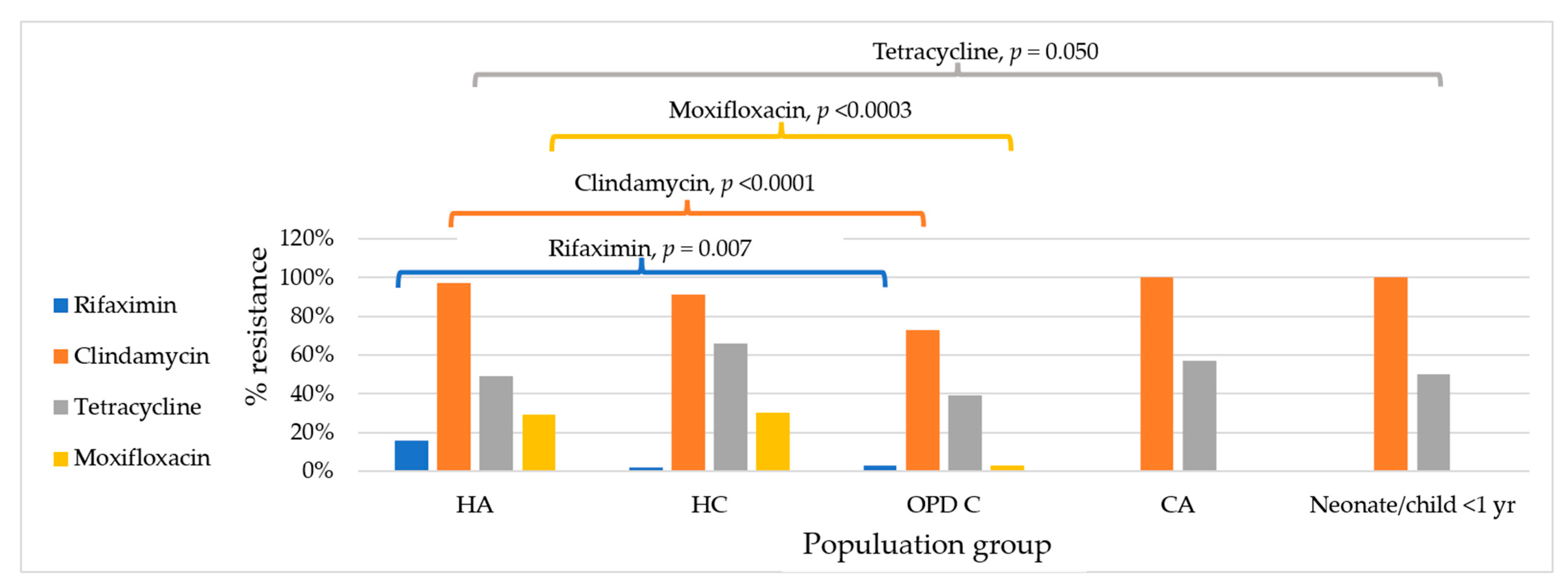
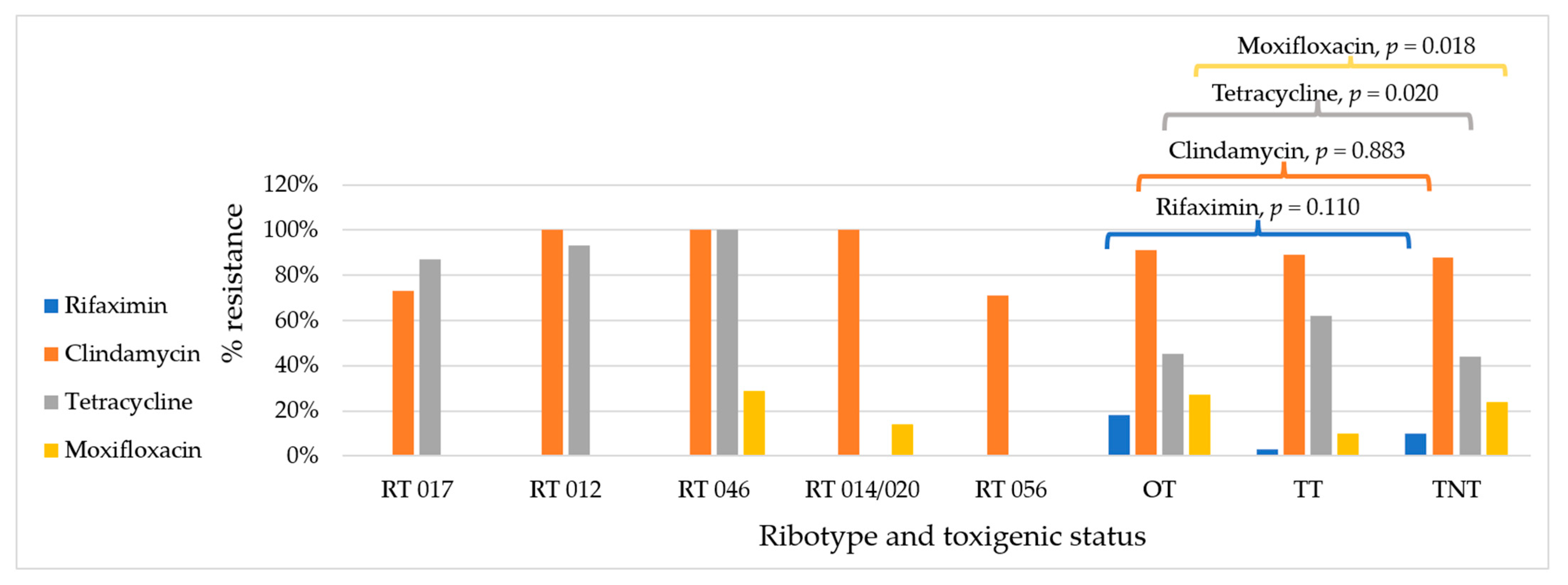
When comparing hospitalised patients to non-hospitalised individuals, C. difficile resistance to clindamycin (95% vs. 74%, p ≤ 0.0001) and moxifloxacin (29% vs. 3%, p ≤ 0.0001) was significantly higher in hospitalised patients than in non-hospitalised individuals (Table 2 and Figure 1). Resistance to rifaximin was more common in hospitalised adults (Figure 2). By residency of cases, up to 10% of isolates from those living outside the capital city were resistant to rifaximin versus 0% in those living in the capital city, while resistance to tetracycline was greater in isolates from those living in the capital city, 68% vs. 46% (p = 0.020) (Table 2). By toxigenic status, non-toxigenic isolates from hospitalised patients were more resistant to moxifloxacin than toxigenic strains (35% vs. 14%, p = 0.017), and toxigenic isolates from non-hospitalised patients were more resistant to tetracycline than non-toxigenic strains (67% vs. 26%, p = 0.001) (Table 3 and Figure 3). Among toxigenic strains, rifaximin resistance (18%, 11/61) was seen only in unknown and uncommon toxigenic strains (Figure 3). While none of RT 046 were susceptible to tetracycline and clindamycin, and none of RT 012 were susceptible to clindamycin, 93% of them were resistant to tetracycline, and only 87% and 73% of RT 017 were resistant to tetracycline and clindamycin, respectively (Figure 3).

3. Discussion

This research aimed to study the antimicrobial susceptibility of C. difficile isolates from Cambodia. While some of the findings were anticipated, the high number of MDR strains of C. difficile was not and is a threat to AMR management in Cambodia. This may be a factor contributing to the persistent spread of C. difficile in both hospital and community settings. The significant difference in clindamycin and moxifloxacin resistance in C. difficile isolates from hospitalised patients versus non-hospitalised individuals strongly suggests antimicrobial selective pressure in hospitals on bacteria residing as commensals or saprophytes and facilitated by higher antimicrobial consumption in hospitalised patients. Another possibility is the transmission of resistance determinants between bacteria pre-existing in hospital settings and C. difficile from the community, brought into hospital settings by the patients.
MDR is defined as resistance to at least three antimicrobial categories [25], and, if more antimicrobials were tested, it is possible that there would be more MDR strains. In the current study, 10 antimicrobial classes were tested, including agents for treatment and CDI-inciting agents. The overall prevalence of MDR (20%, 39/192) in the collection of C. difficile strains from different population groups in Cambodia is comparable to the results in a study conducted in Thailand (21.9%, 23/105) in 2015 [26], but more than twice the prevalence (9.03%, 29/321) in another study from the same hospital in Thailand in 2017–2018 [17]. Noticeably, Vietnam had a similar proportion of MDR strains found in diarrhoeic adults (27.3%, 9/33) [18]. These findings do not directly reflect the different resistance patterns of C. difficile in the three countries due to different study participants; however, similarities in antimicrobial consumption in individual hospitals could be assumed. Although there was a high prevalence of MDR, all the isolates were susceptible to antimicrobials for CDI treatment, with low MICs, except rifaximin (8% were resistant).
C. difficile becomes resistant to rifaximin and rifampin by mutations in RpoB, the β subunit of the RNA polymerase for protein synthesis [27]. Rifaximin-resistant C. difficile in the current study was found only in patients living outside the capital city, and only in unknown and novel toxigenic strains, suggesting that resistance to rifaximin is more likely to occur in community-associated local strains. The prevalence of rifaximin resistance in the current study (8%) was between that found in neighbouring countries, Thailand (10%) [17] and Vietnam (5%) [18], and very similar to North America (7.9–12.4%) [14] and some countries in Europe (0–19.2%) [16,28]. Rifaximin, a derivative of rifampin, is not widely available in Cambodia, but rifampin is a drug used in the first-line treatment of tuberculosis (TB) in the country. Cambodia has endemic TB, with an incidence of 320 per 100,000 population reported in 2022, according to the Tuberculosis Report 2023 by the Ministry of Health of Cambodia [29]. The long-term use of rifampin in patients with TB could be a reason for the high resistance to rifaximin in C. difficile in this country. This is also a reason why patients with TB could acquire C. difficile and develop CDI during treatment of TB, while rifaximin-susceptible cases could be treated with the ongoing rifampin treatment for TB [30,31], and other treatments for CDI besides rifampin might be needed in resistant cases [9].
Tetracycline resistance commonly derives from resistance genes such as tetM, tet40, and tet44 acquired via MGEs, preventing the binding of tetracycline to 16S rRNA [22]. Though MGEs were similar between toxigenic and non-toxigenic strains [24], the significant difference in resistance between toxigenic and non-toxigenic strains in the current study suggests other possible non-genetic determining factors/mechanisms in toxigenic and non-toxigenic C. difficile need to be investigated for resistance to tetracycline. Tetracycline resistance in C. difficile RT 078 driven by selective pressure has been reported as possibly having a role in the spread of this strain, with up to 76.5% possessing the tetM gene [32]. Although there were no C. difficile RT 078 strains isolated in the current study, selective pressure on other strains of C. difficile could induce tetracycline resistance and spread because of tetracycline use in agriculture. In an earlier study of CDI in Cambodia, it was reported that patients might carry C. difficile before being admitted to the hospitals [33]. Those patients were all asymptomatic and all from provinces where agricultural activities were common, suggesting some C. difficile isolates found in hospital settings might be acquired from areas with agricultural activities. Tetracycline is used in humans and animals for disease prevention and treatment and was used for animal growth promotion [6]. Resistance to tetracycline in the current study in Cambodia requires further investigation. The poor knowledge of food producers in Cambodia regarding antimicrobial use in livestock is a potential factor contributing to the increase in AMR in Cambodia in general and particularly in C. difficile, where a lack of awareness of CDI is already a concern [34].
The spread of C. difficile RT 027 in North America [35] and Europe [16,36] in the early 2000s was driven by the high consumption of fluoroquinolones. Resistance to fluoroquinolones derives from point substitutions in GyrA and/or GyrB subunits of DNA gyrase, reducing the binding of antimicrobials to the target [22]. The prevalence of moxifloxacin resistance in the current study (20%) was comparable to that in Thailand (24%) [17] and in the USA (17.5–28.7%) [14], but much less than a previous study in the Asia-Pacific region (44.4%) [12] and in Europe (57.1%) [16]. The reasons for these differences are not known but may relate to variations in fluoroquinolone usage and the diversity of C. difficile genotypes in each country.
Resistance to clindamycin and erythromycin is commonly present in C. difficile from the Asia-Pacific region, compared to other tested antimicrobial classes [12,15]. The resistance mechanism is by methylating 23S rRNA, preventing the binding of antimicrobials, resulting in a high level of resistance to the MLSB drugs, clindamycin and erythromycin. The results from the current study are comparable to previous studies in Asia [12,17,19], suggesting high consumption of MLSB antimicrobials in the region. C. difficile becomes resistant to MLSB by integrating MGEs containing resistance determinants, though unknown mechanisms have been frequently suggested [24]. The resistance derives from the acquisition of erm class genes, more commonly ermB, erm(52), and ermG; non-erm genes, mefH and mefA; and msrD [22]. In earlier studies in Cambodia, individuals in hospital and community settings were frequently colonised by C. difficile asymptomatically, and there was no proper diagnosis [33]. Thus, with high resistance to MLSB drugs, C. difficile might be a silent vector transmitting AMR genes in the region, particularly the erm class genes.
Non-toxigenic C. difficile was resistant to more classes of antimicrobials than toxigenic strains in this study (Figure 3). This emphasises the importance of gaining knowledge about all strains of C. difficile in Asia, not just toxigenic strains causing CDI in the region. Asian strains of C. difficile are largely non-toxigenic or produce only toxin B, such as C. difficile RT 017, which has caused outbreaks worldwide [37,38]. Noticeably, though RT 017 is less resistant to clindamycin and tetracycline (Figure 3), compared to RTs 012 and 046, RT 017 remains predominant in hospital settings. This suggests that resistance to different antimicrobial classes is not the only parameter determining the epidemiology of CDI in each setting.
There is only one paper from Cambodia that described some environmental contamination, and that was contaminated food [39]. Other possible sources and reservoirs of C. difficile in the environment were discussed in three earlier studies from our group [33,40,41]. C. difficile environmental contamination has also been described in studies in neighbouring countries, e.g., Vietnam [42], which is similar to Cambodia. The AMR characteristics of C. difficile in the current study could imply the usage of antimicrobials in different ways in Cambodia, not just for the treatment of infectious diseases. Further investigation into sources/reservoirs of C. difficile in relation to its AMR is needed.
There are some limitations in this study. First, all the strains were from mostly asymptomatic carriers, as described in the previous studies [33,40,41]; thus, the findings from this study only represent the antimicrobial susceptibility of circulating strains in the country and not isolates from cases of CDI. Deciding how these results will impact the treatment for CDI in this country requires further work. Second, the mechanisms of resistance were not investigated, making it difficult to correlate resistance with possible drivers of resistance; however, this topic will be the basis for a future publication. The application of whole-genome sequencing will allow the identification of resistance genes and possibly reflect antimicrobial usage in the country, adding value to AMR surveillance.

4. Materials and Methods

4.1. C. difficile Isolates

In total, 192 isolates were included in this study: 63 isolates from hospitalised adults [33], 47 isolates from hospitalised children [40], 59 isolates from children visiting an outpatient department, 07 isolates from healthy adolescents, 14 isolates from an unpublished pilot study of CDI in hospitalised adults, and two isolates (one from a neonate and one from a child <1 year old) from another unpublished Cambodian study. The majority of isolates, 90% (173/192), were recovered from non-diarrheal, normal, and hard stools.
Of the 192 isolates, 68% (131/192) were non-toxigenic. Among the toxigenic strains, C. difficile ribotypes (RTs) 017 (8%, 15/192) and 012 (7%, 14/192) were the two most common, while RTs 046, 014/020, and 056 were equally the third most common (Table 4). Among the 61 toxigenic strains, C. difficile with the toxin profile A+B+CDT- accounted for 69% (42/61); A-B+CDT-, 29% (18/61); and A+B+CDT+, 2% (1/61).

4.2. MIC Determination

The cryopreserved isolates were recovered on horse blood agar (BA), incubated in an A35 anaerobic chamber (Don Whitley Scientific, Ltd., Shipley, West Yorkshire, UK) at 35 °C for 48 h, with an atmosphere of 80% N2, 10% CO2, and 10% H2 and a 75% relative humidity. A second subculture was conducted to confirm purity before testing. MIC determination was performed using the agar incorporation method according to the CLSI guidelines for anaerobic bacteria (M11-A7) [43]. Antimicrobials for the treatment of CDI, including metronidazole, vancomycin, fidaxomicin, and rifaximin; those with a high risk of inducing CDI, including clindamycin, erythromycin, and moxifloxacin; and those with a variable risk for CDI, including amoxicillin/clavulanic acid, meropenem, and tetracycline, were chosen for testing. The interpretation of rifaximin susceptibility followed O’Connor et al. [27]. Breakpoints for metronidazole, vancomycin, and fidaxomicin were those in the EUCAST guidelines for C. difficile [44] and, for other antimicrobials, the recommendations in the CLSI guidelines were followed [45].

4.3. Statistical Analysis

Summary statistics for categorical variables were reported as proportions. A Chi-squared test was conducted to examine associations between the resistance and ribotype, toxigenic status, host, and setting. A p-value < 0.05 was chosen for statistical significance.

5. Conclusions

To the best of our knowledge, this is the first study of AMR in C. difficile from different population groups in Cambodia. Despite the high number of MDR strains, all C. difficile isolates remained susceptible to CDI treatment drugs, including vancomycin, metronidazole, and fidaxomicin. The resistance patterns of C. difficile necessitate improved infection prevention and control of C. difficile in the country, though AMR is not the only determining factor in the persistence of C. difficile in each setting.

Author Contributions

Conceptualization, L.E., D.A.C., A.C.A.C. and T.V.R.; Methodology, L.E., P.P., S.-C.L., J.M.C. and T.V.R.; Validation, L.E. and T.V.R.; Formal Analysis, L.E.; Investigation and Data Curation, L.E.; Writing—Original Draft Preparation, L.E.; Writing—Review and Editing, L.E., S.-C.L., K.A.A., P.P. and T.V.R.; Visualization, L.E. and T.V.R.; Supervision, T.V.R. Project Administration, T.V.R. and L.E.; Funding Acquisition, T.V.R. All authors have read and agreed to the published version of the manuscript.

Funding

This research received no external funding.

Institutional Review Board Statement

Not applicable.

Informed Consent Statement

Not applicable.

Data Availability Statement

The data presented in this study are available on request from the corresponding author.

Acknowledgments

We would like to acknowledge the help of all Riley laboratory team members during the experimental work. This research is supported by an Australian Government Research Training Program (RTP) Scholarship awarded to L.E.

Conflicts of Interest

All authors declare no conflicts of interest. T.V.R. has received a grant from Roche Diagnostics outside the present work.

Abbreviations

The following abbreviations are used in this manuscript:
AMRAntimicrobial resistance
CDIClostridioides difficile infection
MLSBMacrolide lincosamide streptogramin B
MGEsMobile genetic elements
MICMinimal inhibitory concentration
BABlood agar
RTRibotype
MDRMultidrug resistance
OPDOutpatient department
SSusceptible
IIntermediate
RResistant
TTTotal toxigenic strains
OTOther toxigenic strains
TNTTotal non-toxigenic strains

References

  1. Murray, C.J.; Ikuta, K.S.; Sharara, F.; Swetschinski, L.; Aguilar, G.R.; Gray, A.; Han, C.; Bisignano, C.; Rao, P.; Wool, E.; et al. Global burden of bacterial antimicrobial resistance in 2019: A systematic analysis. Lancet 2022, 399, 629–655. [Google Scholar] [CrossRef]
  2. Centers for Disease Control and Prevention. Antibiotic Resistance Threats in the United States, 2013; Centers for Disease Control and Prevention: Atlanta, GA, USA, 2013.
  3. Cetin, S.; Ugur, M. Clostridioides difficile infections and factors associated with recurrence. Infect. Dis. Clin. Microbiol. 2024, 6, 264–275. [Google Scholar] [CrossRef]
  4. McFarland, L.V.; Elmer, G.W.; Surawicz, C.M. Breaking the cycle: Treatment strategies for 163 cases of recurrent Clostridium difficile disease. Am. J. Gastroenterol. 2002, 97, 1769–1775. [Google Scholar] [CrossRef] [PubMed]
  5. Rodriguez, C.D.; Seyboldt, C.; Rupnik, M. Non-human C. difficile reservoirs and sources: Animals, food, environment. In Updates on Clostridium Difficile in Europe: Advances in Microbiology, Infectious Diseases and Public Health; Mastrantonio, P., Rupnik, M., Eds.; Springer International Publishing: Cham, Switzerland, 2018; Volume 8, pp. 227–243. [Google Scholar]
  6. Cromwell, G.L. Why and how antibiotics are used in swine production. Anim. Biotechnol. 2002, 13, 7–27. [Google Scholar] [CrossRef]
  7. Longhitano, A.; Roder, C.; Blackmore, T.; Campbell, A.; May, M.; Athan, E. Australasian Society of Infectious Diseases updated guidelines for the management of Clostridioides difficile infection in adults and children in Australia and New Zealand. Intern. Med. J. 2025, 55, 503–513. [Google Scholar] [CrossRef] [PubMed]
  8. Johnson, S.; Lavergne, V.; Skinner, A.M.; Gonzales-Luna, A.J.; Garey, K.W.; Kelly, C.P.; Wilcox, M.H. Clinical practice guideline by the Infectious Diseases Society of America (IDSA) and Society for Healthcare Epidemiology of America (SHEA): 2021 focused update guidelines on management of Clostridioides difficile infection in adults. Clin. Infect. Dis. 2021, 73, e1029–e1044. [Google Scholar] [CrossRef] [PubMed]
  9. Imwattana, K.; Putsathit, P.; Leepattarakit, T.; Kiratisin, P.; Riley, T.V. Mild or malign: Clinical characteristics and outcomes of Clostridium difficile infection in Thailand. J. Clin. Microbiol. 2020, 58, e01217-20. [Google Scholar] [CrossRef]
  10. Collins, D.A.; Sohn, K.M.; Wu, Y.; Ouchi, K.; Ishii, Y.; Elliott, B.; Riley, T.V.; Tateda, K. Clostridioides difficile infection in the Asia-Pacific region. Emerg. Microbes Infect. 2020, 9, 42–52. [Google Scholar] [CrossRef]
  11. Zhang, S.; Ma, C.; Zhang, H.; Zhao, C.; Guo, R.; Liu, J.; Wang, J.; Yuan, J.; Jia, K.; Wu, A.; et al. Toxin genotypes, antibiotic resistance and their correlations in Clostridioides difficile isolated from hospitals in Xi’an, China. BMC Microbiol. 2024, 24, 177. [Google Scholar] [CrossRef]
  12. Lew, T.; Putsathit, P.; Sohn, K.M.; Wu, Y.; Ouchi, K.; Ishii, Y.; Tateda, K.; Riley, T.V.; Collins, D.A. Antimicrobial Susceptibilities of Clostridium difficile Isolates from 12 Asia-Pacific Countries in 2014 and 2015. Antimicrob. Agents Chemother. 2020, 64, e00296-20. [Google Scholar] [CrossRef]
  13. Wieczorkiewicz, J.T.; Skinner, A.M.; Cheknis, A.; Petrella, L.A.; Stevens, V.W.; Wright, L.M.; Gerding, D.N.; Johnson, S. Epidemiology of Clostridioides difficile infection at one hospital 10 years after an outbreak of the epidemic C. difficile strain BI/027: Changing strain prevalence, antimicrobial susceptibilities, and patient antibiotic exposures. Antimicrob. Agents Chemother. 2024, T, e00698-24. [Google Scholar] [CrossRef] [PubMed]
  14. Tickler, I.A.; Obradovich, A.E.; Goering, R.V.; Fang, F.C.; Tenover, F.C.; Consortium, H.A.I. Changes in molecular epidemiology and antimicrobial resistance profiles of Clostridioides (Clostridium) difficile strains in the United States between 2011 and 2017. Anaerobe 2019, 60, 102050. [Google Scholar] [CrossRef] [PubMed]
  15. Putsathit, P.; Hong, S.; George, N.; Hemphill, C.; Huntington, P.G.; Korman, T.M.; Kotsanas, D.; Lahra, M.; McDougall, R.; McGlinchey, A.; et al. Antimicrobial resistance surveillance of Clostridioides difficile in Australia, 2015–18. J. Antimicrob. Chemother. 2021, 76, 1815–1821. [Google Scholar] [CrossRef] [PubMed]
  16. Abdrabou, A.M.M.; Ul Habib Bajwa, Z.; Halfmann, A.; Mellmann, A.; Nimmesgern, A.; Margardt, L.; Bischoff, M.; von Muller, L.; Gartner, B.; Berger, F.K. Molecular epidemiology and antimicrobial resistance of Clostridioides difficile in Germany, 2014-2019. Int. J. Med. Microbiol. 2021, 311, 151507. [Google Scholar] [CrossRef]
  17. Imwattana, K.; Putsathit, P.; Knight, D.R.; Kiratisin, P.; Riley, T.V. Molecular characterization of, and antimicrobial resistance in Clostridioides difficile from Thailand, 2017–2018. Microb. Drug Resist. 2021, 27, 1505–1512. [Google Scholar] [CrossRef]
  18. Khun, P.A.; Phi, L.D.; Bui, H.T.T.; Collins, D.A.; Riley, T.V. Clostridioides (Clostridium) difficile in adults with diarrhoea in Vietnam. Anaerobe 2023, 81, 102741. [Google Scholar] [CrossRef]
  19. Khun, P.A.; Phi, L.D.; Pham, P.T.; Thu Nguyen, H.T.; Huyen Vu, Q.T.; Collins, D.A.; Riley, T.V. Clostridioides (Clostridium) difficile in children with diarrhoea in Vietnam. Anaerobe 2022, 74, 102550. [Google Scholar] [CrossRef]
  20. Wolfe, C.; Pagano, P.; Pillar, C.M.; Shinabarger, D.L.; Boulos, R.A. Comparison of the in vitro antibacterial activity of Ramizol, fidaxomicin, vancomycin, and metronidazole against 100 clinical isolates of Clostridium difficile by broth microdilution. Diagn. Microbiol. Infect. Dis. 2018, 92, 250–252. [Google Scholar] [CrossRef]
  21. Freeman, J.; Vernon, J.; Pilling, S.; Morris, K.; Nicolson, S.; Shearman, S.; Clark, E.; Palacios-Fabrega, J.A.; Wilcox, M.; Pan-European Longitudinal Surveillance of Antibiotic Resistance among Prevalent Clostridium difficile Ribotypes’ Study, G. Five-year Pan-European, longitudinal surveillance of Clostridium difficile ribotype prevalence and antimicrobial resistance: The extended ClosER study. Eur. J. Clin. Microbiol. Infect. Dis. 2020, 39, 169–177. [Google Scholar] [CrossRef]
  22. Imwattana, K.; Rodriguez, C.; Riley, T.V.; Knight, D.R. A species-wide genetic atlas of antimicrobial resistance in Clostridioides difficile. Microb. Genom. 2021, 7, 000696. [Google Scholar] [CrossRef]
  23. Knight, D.R.; Elliott, B.; Chang, B.J.; Perkins, T.T.; Riley, T.V. Diversity and evolution in the genome of Clostridium difficile. Clin. Microbiol. Rev. 2015, 28, 721–741. [Google Scholar] [CrossRef] [PubMed]
  24. Imwattana, K.; Kiratisin, P.; Riley, T.V.; Knight, D.R. Genomic basis of antimicrobial resistance in non-toxigenic Clostridium difficile in Southeast Asia. Anaerobe 2020, 66, 102290. [Google Scholar] [CrossRef] [PubMed]
  25. Magiorakos, A.P.; Srinivasan, A.; Carey, R.B.; Carmeli, Y.; Falagas, M.E.; Giske, C.G.; Harbarth, S.; Hindler, J.F.; Kahlmeter, G.; Olsson-Liljequist, B.; et al. Multidrug-resistant, extensively drug-resistant and pandrug-resistant bacteria: An international expert proposal for interim standard definitions for acquired resistance. Clin. Microbiol. Infect. 2012, 18, 268–281. [Google Scholar] [CrossRef] [PubMed]
  26. Putsathit, P.; Maneerattanaporn, M.; Piewngam, P.; Knight, D.R.; Kiratisin, P.; Riley, T.V. Antimicrobial susceptibility of Clostridium difficile isolated in Thailand. Antimicrob. Resist. Infect. Control 2017, 6, 58. [Google Scholar] [CrossRef]
  27. O’Connor, J.R.; Galang, M.A.; Sambol, S.P.; Hecht, D.W.; Vedantam, G.; Gerding, D.N.; Johnson, S. Rifampin and rifaximin resistance in clinical isolates of Clostridium difficile. Antimicrob. Agents Chemother. 2008, 52, 2813–2817. [Google Scholar] [CrossRef]
  28. Jon, J.V.; Mark, H.W.; Jane, F. Antimicrobial resistance progression in the United Kingdom: A temporal comparison of Clostridioides difficile antimicrobial susceptibilities. Anaerobe 2021, 70, 102385. [Google Scholar] [CrossRef]
  29. CENAT. Tuberculosis Report 2023; National Center for Tuberculosis and Leprosy Control: Phnom Penh, Cambodia, 2023. [Google Scholar]
  30. Suh, J.W.; Jeong, Y.J.; Ahn, H.G.; Kim, J.Y.; Sohn, J.W.; Yoon, Y.K. Epidemiologic characteristics and risk factors of Clostridioides difficile infection in patients with active tuberculosis in the Republic of Korea: A nationwide population-based study. J. Hosp. Infect. 2024, 154, 1–8. [Google Scholar] [CrossRef]
  31. Lee, Y.M.; Huh, K.C.; Yoon, S.M.; Jang, B.I.; Shin, J.E.; Koo, H.S.; Jung, Y.; Kim, S.H.; Moon, H.S.; Lee, S.W. Incidence and clinical outcomes of Clostridium difficile infection after treatment with tuberculosis medication. Gut Liver 2016, 10, 250–254. [Google Scholar] [CrossRef]
  32. Dingle, K.E.; Didelot, X.; Quan, T.P.; Eyre, D.W.; Stoesser, N.; Marwick, C.A.; Coia, J.; Brown, D.; Buchanan, S.; Ijaz, U.Z.; et al. A role for tetracycline selection in recent evolution of agriculture-associated Clostridium difficile PCR rbotype 078. mBio 2019, 10, e02790-18. [Google Scholar] [CrossRef]
  33. Eng, L.; Collins, D.A.; Alene, K.A.; Bory, S.; Theng, Y.; Vann, P.; Meng, S.; Limsreng, S.; Clements, A.C.; Riley, T.V. Clostridioides (Clostridium) difficile infection in hospitalised adult patients in Cambodia. Microbiol. Spectr. 2025, 13, e02747-24. [Google Scholar] [CrossRef]
  34. Chea, B.; Kong, S.; Thim, S.; Ban, N.; Seng, S.; Fernandez-Colorado, C.; Kang, K. Knowledge, attitudes, and practices of antimicrobial use and resistance among livestock producers in Cambodia. Open J. Anim. Sci. 2022, 12, 454–466. [Google Scholar] [CrossRef]
  35. Loo, V.G.; Poirier, L.; Miller, M.A.; Oughton, M.; Libman, M.D.; Michaud, S.; Bourgault, A.M.; Nguyen, T.; Frenette, C.; Kelly, M.; et al. A predominantly clonal multi-institutional outbreak of Clostridium difficile-associated diarrhea with high morbidity and mortality. N. Engl. J. Med. 2005, 353, 2442–2449. [Google Scholar] [CrossRef] [PubMed]
  36. Goorhuis, A.; Van der Kooi, T.; Vaessen, N.; Dekker, F.W.; Van den Berg, R.; Harmanus, C.; van den Hof, S.; Notermans, D.W.; Kuijper, E.J. Spread and epidemiology of Clostridium difficile polymerase chain reaction ribotype 027/toxinotype III in the Netherlands. Clin. Infect. Dis. 2007, 45, 695–703. [Google Scholar] [CrossRef] [PubMed]
  37. Imwattana, K.; Knight, D.R.; Kullin, B.; Collins, D.A.; Putsathit, P.; Kiratisin, P.; Riley, T.V. Clostridium difficile ribotype 017—Characterization, evolution and epidemiology of the dominant strain in Asia. Emerg. Microbes Infect. 2019, 8, 796–807. [Google Scholar] [CrossRef] [PubMed]
  38. Riley, T.V.; Collins, D.A.; Karunakaran, R.; Kahar, M.A.; Adnan, A.; Hassan, S.A.; Zainul, N.H.; Rustam, F.R.M.; Wahab, Z.A.; Ramli, R.; et al. High prevalence of toxigenic and nontoxigenic Clostridium difficile strains in Malaysia. J. Clin. Microbiol. 2018, 56, e00170-18. [Google Scholar] [CrossRef]
  39. Rodriguez, C.; Mith, H.; Taminiau, B.; Bouchafa, L.; Van Broeck, J.; Soumillion, K.; Ngyuvula, E.; García-Fuentes, E.; Korsak, N.; Delmée, M.; et al. First isolation of Clostridioides difficile from smoked and dried freshwater fish in Cambodia. Food Control 2021, 124, 107895. [Google Scholar] [CrossRef]
  40. Eng, L.; Alene, K.A.; Collins, D.A.; Lim, S.C.; Srey, V.; Chea, C.; Yohn, S.; Leng, S.; Clements, A.C.A.; Riley, T.V. Clostridioides (Clostridium) difficile in hospitalised children in Cambodia. Anaerobe 2025, 93, 102959. [Google Scholar] [CrossRef]
  41. Eng, L.; Turner, P.; Alene, K.A.; Collins, D.A.; Lim, S.-C.; Tan, P.; Soeng, S.; Hun, D.; Yohn, S.; Vong, S.; et al. Clostridioides (Clostridium) difficile in children and adolescents in the community in Cambodia. Anaerobe 2025, 94, 102982. [Google Scholar] [CrossRef]
  42. Khun, P.A.; Phi, L.D.; Bui, H.T.T.; Bui, N.T.; Vu, Q.T.H.; Trinh, L.D.; Collins, D.A.; Riley, T.V. Environmental contamination with Clostridioides (Clostridium) difficile in Vietnam. J. Appl. Microbiol. 2023, 134, lxad118. [Google Scholar] [CrossRef]
  43. CLSI M11; Methods for Antimicrobial Susceptibility Testing of Anaerobic Bacteria, M11-A7, 7th Edition. Clinical and Laboratory Standards Institute: Wayne, PA, USA, 2007.
  44. EUCAST; The European Society of Clinical Microbiology and Infectious Diseases. Breakpoint Tables for Interpretation of MICs and Zone Diameters, Version 14.0; EUCAST: Växjö, Sweden, 2024. [Google Scholar]
  45. CLSI M100-ED34; 2024 Performance Standards for Antimicrobial Susceptibility Testing, 34th Edition. Clinical and Laboratory Standards Institute: Wayne, PA, USA, 2024.
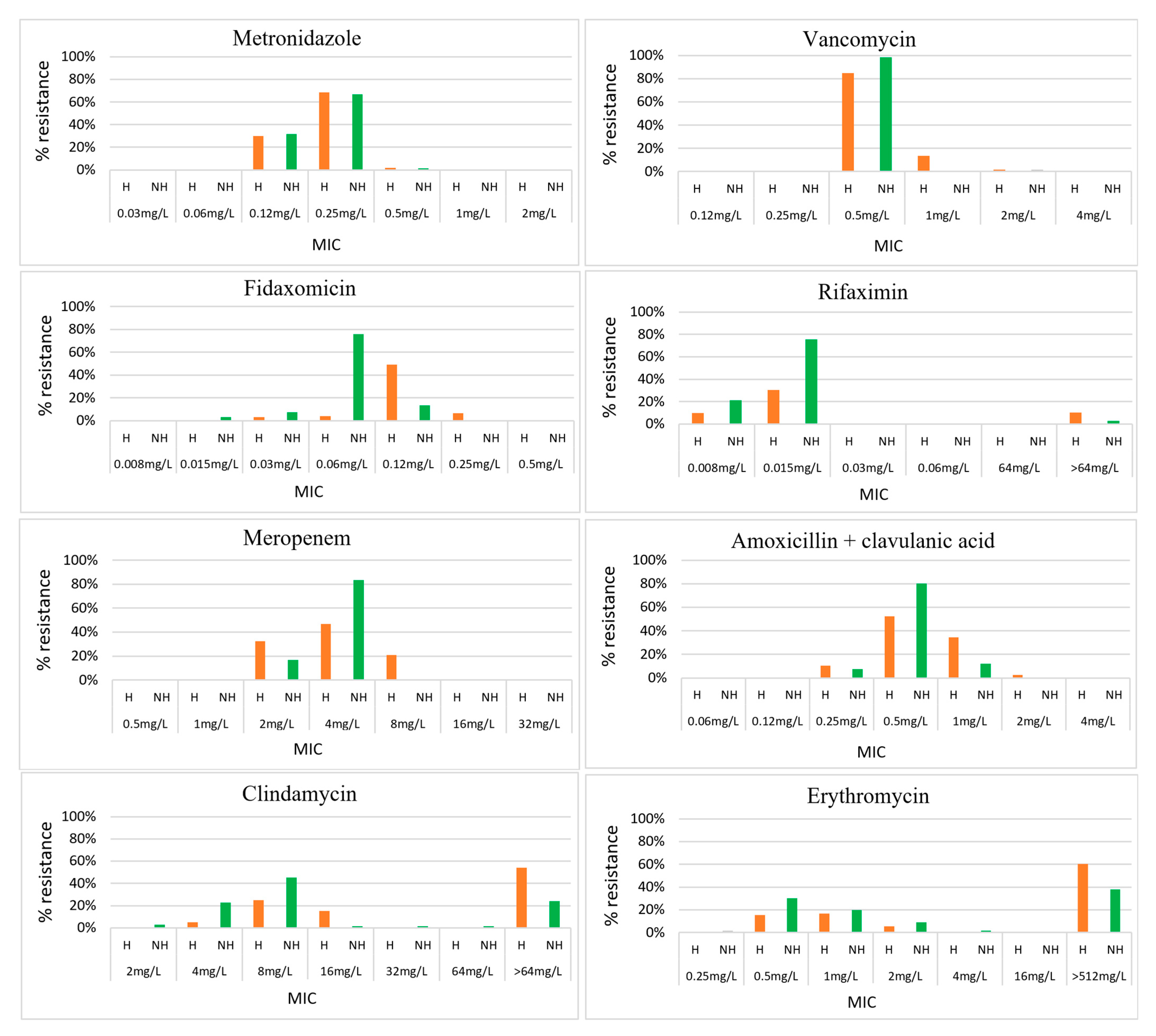
Figure 1. Minimal inhibitory concentrations for 10 antimicrobials against 124 C. difficile isolates from hospitalised patients and 66 C. difficile isolates from non-hospitalised individuals in Cambodia. H: hospitalised patients, in orange; NH: non-hospitalised individuals, in green; MIC: minimum inhibitory concentration.
Antibiotics 14 00950 g001aAntibiotics 14 00950 g001b
Figure 2. Antimicrobial resistance in C. difficile by population group. HA: hospitalised adults; HC: hospitalised children; OPD C: children visiting an outpatient department; CA: healthy adolescents in the community.
Antibiotics 14 00950 g002
Figure 3. Antimicrobial resistance in C. difficile by ribotype and toxigenic status. OT: other toxigenic strains or all toxigenic strains excluding RTs 017, 012, 046, 014/020, and 056 (n = 11); TT: total toxigenic strains or all strains that contained at least one toxin gene or any combination (n = 61); TNT: total non-toxigenic strains or all stains that did not contain any toxin genes (n = 131).
Antibiotics 14 00950 g003
Table 1. Antimicrobial susceptibility of 192 Cambodian strains of C. difficile.
AntimicrobialInterpretive Categories and MIC Breakpoints (mg/L)Result Ranges
(mg/L)
MIC50/MIC90
(mg/L)
Susceptibility % (N = 192)
SIRSIR
Vancomycin ≤2 >20.5–20.5/1100%00
Metronidazole ≤2 >20.12–0.50.25/0.25100%00
Fidaxomicin ≤0.5->0.50.015–0.250.06/0.12100%00
Rifaximin <32 ≥320.0008–>640.015/0.01592% (177)08% (15)
Meropenem≤48≥162 to 84/8.86% (166)14% (26)0
Amoxicillin/clavulanic acid ≤48≥160.25–20.5/1100%00
Clindamycin≤24≥82–>6416/>641% (1)11% (21)88% (169)
Erythromycin- -0.25–>512>512/>512---
Tetracycline≤48≥160.12–648/3248%2% (3)50% (96)
Moxifloxacin≤24≥82–>322/3280%020% (38)
Table 2. Antimicrobial resistance of C. difficile by hospitalisation status and host residency.
AntimicrobialsInterpretive Categories and MIC Breakpoints (mg/L)C. difficile
in Hospitalised
Patients
(n = 124)
C. difficile
in Non-Hospitalised Individuals
(n = 66)
p-ValueC. difficile
in Those Living in the Capital City
(n = 37)
C. difficile
in Those Living Outside the
Capital City
(n = 145)
p-Value
SIRResistant (%)Resistant (%) Resistant (%)Resistant (%)
Vancomycin≤2 >20%0% 0%0%
Metronidazole≤2 >20%0% 0%0%
Fidaxomicin≤0.5->0.50%0% 0%0%
Rifaximin<32 ≥3210%3%0.070%10%
Meropenem≤48≥160%0% 0%0%
Amoxicillin/clavulanic acid ≤48≥160%0% 0%0%
Clindamycin≤24≥895%74%<0.000192%88%0.464
Erythromycin- --- --
Tetracycline≤48≥1655%41%0.06768%46%0.020
Moxifloxacin≤24≥829%3%<0.000124%18%0.378
Table 3. Antimicrobial resistance of C. difficile by toxigenic status.
AntimicrobialsInterpretive Categories and MIC Breakpoints (mg/L)Hospitalised PatientsNon-Hospitalised Individuals
Toxigenic Strains
(n = 36)
Non-Toxigenic Strains
(n = 88)
p-Value Toxigenic Strains
(n = 24)
Non-Toxigenic Strains
(n = 42)
p-Value
SIRResistant (%)Resistant (%) Resistant (%)Resistant (%)
Vancomycin≤2 >20%0% 0%0%
Metronidazole≤2 >20%0% 0%0%
Fidaxomicin≤0.5->0.50%0% 0%0%
Rifaximin<32 ≥323%14%0.0734%2%0.684
Meropenem≤48≥160%0% 0%0%
Amoxicillin/clavulanic acid ≤48≥16 0%0% 0% 0%
Clindamycin≤24≥894%95%0.812 79%71%0.489
Erythromycin- --- --
Tetracycline≤48≥1658%53%0.61767%26%0.001
Moxifloxacin≤24≥814%35%0.0174%2%0.684
Table 4. Number of C. difficile ribotypes in each population group.
RibotypeHospitalised
Adults
Hospitalised
Children
OPD
Children
AdolescentsNeonate and
Children < 1 yr
Total
0175171115
0122723014
046502007
014/020331007
056303107
Other toxigenic strains6122011
Non-toxigenic strains53354201131
Total77475972192
Disclaimer/Publisher’s Note: The statements, opinions and data contained in all publications are solely those of the individual author(s) and contributor(s) and not of MDPI and/or the editor(s). MDPI and/or the editor(s) disclaim responsibility for any injury to people or property resulting from any ideas, methods, instructions or products referred to in the content.