Article

New Insights in blaKPC Gene Mobilization in Pseudomonas aeruginosa: Acquisition of blaKPC-3 and Identification of a New Tn2-like NTE Mobilizing blaKPC-2

by
Deisy Abril
1,
Juan Bravo-Ojeda
2,
Julio-Cesar Garcia
3,*,
Aura Lucia Leal-Castro
2,
Carlos Humberto Saavedra-Trujillo
4,
Johana Madroñero
2,
Rosa-Helena Bustos
3,
Ricaurte Alejandro Marquez-Ortiz
1,
Zayda Lorena Corredor Rozo
1,
Natasha Vanegas Gómez
1,5 and
Javier Escobar-Pérez
1,*
1
Bacterial Molecular Genetics Laboratory-LGMB, Universidad El Bosque, Ak. 9 #131a-02, Bogota 110121, Colombia
2
Facultad de Medicina y Grupo de Investigación en Enfermedades Infecciosas, Universidad Nacional de Colombia, Bogotá 110111, Colombia
3
Evidence-Based Therapeutics Group, Department of Clinical Pharmacology, Faculty of Medicine, Universidad de La Sabana and Clínica Universidad de La Sabana, Campus del Puente del Común, Km. 7, Autopista Norte de Bogotá, Chía 140013, Colombia
4
Facultad de Medicina, Universidad Nacional de Colombia, Bogotá 110111, Colombia
5
The i3 Institute, Faculty of Science University of Technology, Sydney 1168, Australia
*
Authors to whom correspondence should be addressed.
Antibiotics 2025, 14(9), 947; https://doi.org/10.3390/antibiotics14090947
Submission received: 17 March 2025 / Revised: 20 May 2025 / Accepted: 21 May 2025 / Published: 19 September 2025
(This article belongs to the Section Mechanism and Evolution of Antibiotic Resistance)

Abstract

Carbapenem-resistant Pseudomonas aeruginosa is a major cause of healthcare associated infections in hospitalized patients and what is more warring with reduced therapeutic options. The KPC is a powerful enzyme capable of hydrolyzing the carbapenems, described first in Klebsiella pneumoniae and it already has found in P. aeruginosa. Objective: To perform a comparative genomic analysis of two new genetic platforms mobilizing the blaKPC-2 and blaKPC-3 in two ST111 and ST235 pandemic clones of P. aeruginosa in Colombia, South America. Methods: Sixty-six blaKPC-harboring P. aeruginosa isolates were identified and characterized during a prospective study conducted in six high complex hospitals in Colombia. Genetic platforms mobilizing the blaKPC were analyzed. Results: The blaKPC-2 and blaKPC-3 were identified in 24 and 42 isolates, respectively. The blaKPC-2-harboring isolates belonged to ST235 and blaKPC-3 to ST111. The whole genome sequencing indicated that the blaKPC-3 gene was mobilized by the Tn4401b within a 55-kb-size environmental origin plasmid, which, in other isolates, was inserted into the chromosome through a transposition event of ISPa38. Regarding the blaKPC-2 gene, this was mobilized by a new Non-Tn4401 Element (NTE) derived from transposon Tn2 (proposed as variant IIg), which has been transposed into a 43-Kb-size little-studied plasmid related to Klebsiella spp. Conclusions: Our results reveal a new acquisition event of blaKPC in P. aeruginosa, in this case blaKPC-3. Likewise, the pandemic high-risk clones ST111 and ST235 of P. aeruginosa continues to spread blaKPC gene through different mobile genetic elements, jumping of conventional Tn4401b and acquiring new Tn2-derived NTE, which were inserted in diverse plasmids.

Graphical Abstract

1. Introduction

Pseudomonas aeruginosa is one of the most versatile bacteria, which lives mainly in the environment but evolutionarily has acquired and stabilized molecular mechanisms to survive into the human body yielding diverse and potentially mortal infections. According to the World Health Organization (WHO), P. aeruginosa is included in the “critical” category and is an important cause of related infections, mainly in intensive care units (ICU). Infections caused by this microorganism are associated with high morbidity and mortality. Initially, these infections were successfully controlled by the usage of antibiotics, however, P. aeruginosa has achieved to overcome it due to the emergence the different and potent resistance mechanisms such as deregulation of constitutive genes (i.e porins and efflux pump), acquisition of many resistance genes, modification of cell wall, alteration of drug target, degradation/alteration of drug, biofilm formation [1]. One of these powerful mechanisms is the carbapenemases, enzymes capable of degrading carbapenems (and almost all the β-lactam antibiotics), molecules that have been the most effective against P. aeruginosa infections. Unfortunately, the resistance rate to carbapenems has been increasing worldwide, reaching levels up to 93,5% (reported in Ukraine) [2]. Several carbapenemases have been identified in P. aeruginosa such as VIM, IMP, SMP, NDM, OXA-48, GIM, CAM, FIM, AIM, HMB, and KPC [1].
The KPC was first identified in a K. pneumoniae isolate from United States of America in 1996 and since then it has already been reported in countries of the five continents [3]. The KPC enzyme in P. aeruginosa was initially reported from Colombia in 2007 [4], and since then has increased its detection in other countries in America, Asia and Europe [5,6,7,8,9,10,11]. In Colombia, the carbapenem resistance in P. aeruginosa has been increasing during the last years up to 9.1% [2], whereby well-structured surveillance studies are essential to establish the behavior and impact of the arrival of these “new” genes on the infections caused by P. aeruginosa. Dissemination of blaKPC in this species has been associated initially by Tn4401b (mobile genetic element (MGE) most worldwide disseminated in Klebsiella pneumoniae) and NTEKPC-IIc [12]. Over the course of time, the appearance of new genetic arrangements with more remnants of Tn4401b (NTE) have been circulating on different plasmids in both species, some of them in new transposon structures, making the study of mobilization of blaKPC increasingly most important [13,14]. In this way, we conducted a genetic and genome surveillance prospective study of the carbapenem-resistant P. aeruginosa isolates recovered from hospital infections in patients, and we achieved to detect: first, a new plasmid mobilizing the blaKPC-2 into a genetic platform derived from Tn2 transposon circulating within isolates belonging to the high-risk pandemic clone ST235, and second, the blaKPC-3 acquisition by the high-risk pandemic clone ST111 in both plasmid and chromosome (stabilization process) through a transposition event of the insertion sequence ISPa38 (or TnPa38 as it is proposed by this study).

2. Results

2.1. The blaKPC-Harboring P. aeruginosa Isolates Increase Their Frequency in the Colombian Hospital Settings

During the period of study, 128 carbapenem-resistant P. aeruginosa isolates were identified from 121 patients who attended six health institutions in Colombia. The susceptibility profiles of the isolates are showed in Table S2. To highlight, 100 (78.7%) isolates were resistant to the three or more families of antibiotics evaluated (β-lactams, quinolones, aminoglycoside and polymyxin), it significantly reduced the therapeutic options. Considering the importance of carbapenem resistance, 66 (51.6%) and 54 (42.2%) isolates harbored blaKPC and blaVIM, respectively; however, the co-existence of blaKPC and blaVIM was observed in 35 (64.8%) of these, being the dual presence of these carbapenemases in Colombian P. aeruginosa isolates becoming increasingly frequent.

2.2. The Pandemic High-Risk Clones ST111 and ST235 Continue Being the Catcher of the blaKPC Gene

The blaKPC-2 has been the principal variant reported in P. aeruginosa in Colombia and the more frequent in the world, we determined the blaKPC variant of routine in the isolates, hoping that were variant 2; however, surprisingly we found that 42 (63.6%) isolates harbored the variant 3. Now, among the 11 different pulsotypes found by PFGE, the blaKPC-3 gene was associated with pulsotypes a, b, and c; and notably, these all belonged to the ST111, like others reports with this KPC variant in Colombia [15]. The remain eight pulsotypes had the blaKPC-2 gene and belonged to ST235, as has previously been reported for others P. aeruginosa isolates [16].

2.3. The blaKPC Gene Is Being Mobilized for New and Different Genetic Platforms

To determine the genetic platforms mobilizing the blaKPC gene, we used the WGS to establish the chromosome and plasmid sequences of four representative isolates selected on their frequency, blaKPC variant, PFGE pulsotype and ST. The isolates sequenced were 30Pae2 (ST111), 34Pae8 (ST235), 34Pae23 (ST235) and 34Pae36 (ST111). 30Pae2 had a circular chromosome of 7.2 Mbp and harbored the plasmid p30Pae2 of 55.6 Kbp, this isolate had a resistome of 14 genes, 13 of them (blaPDC, blaTEM, blaOXA-395, blaVIM-2, sul1, aac(3)-IIa, aac(6′)-29, aac(6′)Ian, aadA13, aadA6, aph(3″)-lb, aph(3′)-IIb and aph-(6)-Id) were in the chromosome and blaKPC-3 in p30Pae2-KPC. 34Pae8 had a chromosome of 6.8 Kbp and the plasmid p34Pae8-KPC of 42.7 Kbp, from this isolate 10 genes were identified, blaPDC, blaOXA-488, catB7, sul1, aph(3″)-IIb, aadA6, qacEΔ1, fosA were in chromosome, and aac(6′)-29 and blaKPC-2 in p34Pae8-KPC. 34Pae23 had a partial chromosome of 6.8 Kbp and a plasmid p34Pae23 of 42.9 Kbp, this isolate had the same resistance genes as 34Pae8. 34Pae36 had a complete chromosome of 7.4 Kbp and a plasmid p34Pae36, for this isolate 10 genes were identified only in the chromosome: blaPDC, blaOXA-395, blaKPC-3, blaVIM-2, catB7, sul1, aac(6′)-29, aph(3″)-IIb, qacEΔ1 and fosA (Table S3).
In the two isolates belonging to ST235, the blaKPC-2 gene was transported in an almost identical plasmid (99.76% identity), p34Pae8-KPC had a 248 bp deletion, compared to p34Pae23-KPC which had a size of 42,977 bp, corresponding to a genes region of a hypothetical protein and a recombinase (Figure 1), does not belong to any incompatibility group previously reported, does not possess additional resistance genes, and the characteristic Tn4401 was not found.
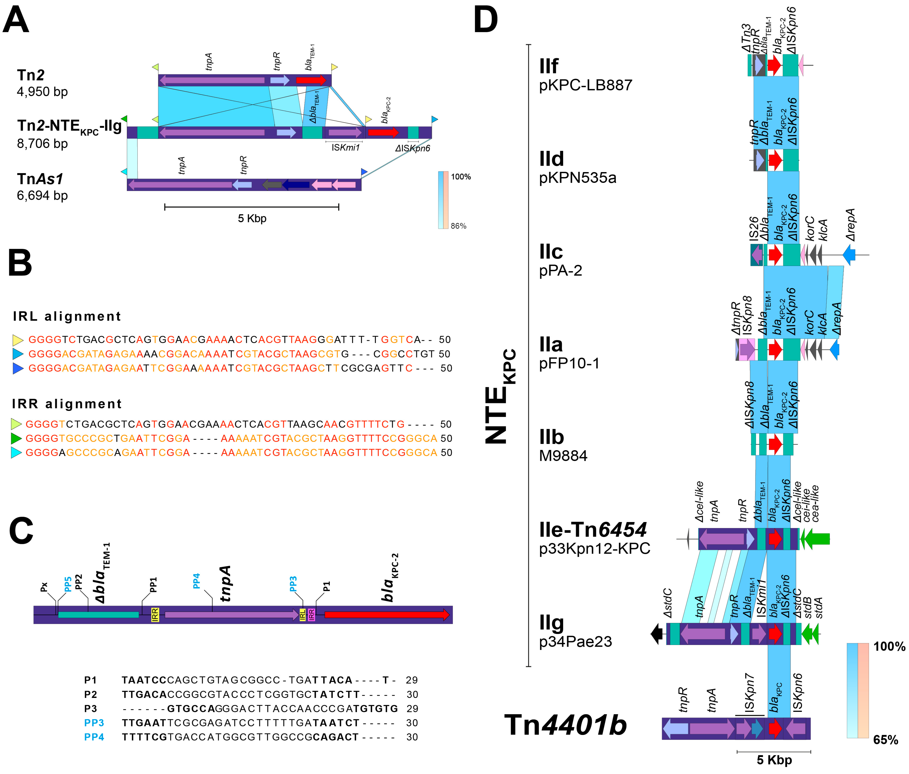
An analysis of the blaKPC-2 gene surroundings showed the presence of a variant of the transposon Tn2 (upstream) with ΔblaTEM gene, which has embedded one copy of an insertion sequence with remarkable similarity (99% of identity) with the recently reported ISKmi1 (Figure 2A). This IS belongs to the IS30 family and was first detected in K. michiganensis, an emerging pathogen first reported in 2013 [17] that posteriorly become carbapenem-resistant due to the acquisition of the blaKPC-3; and also in Enterobacter hormaechei (GenBank accession number NZ_CP049047) [18]. In addition, the ISKmi1 insertion occurred in an independent transposition event (respect with Tn2 transposition) that yielded a 2-bp size target site duplication (TSD), characteristic size duplication of the IS30-related transposases. This insertion also altered the promoter region of the blaKPC gene by introducing a new “-35” promoter element within the right-hand terminal inverted repeat (IRR), as has previously been demonstrated for IS30 elements [19]. This modification could potentially affect transcription of this gene, although further experimental validation is required (Figure 1 and Figure 2B,C).
No additional mobile genetic platforms were identified within the plasmid. Although the Tn2 variant was initially suspected to mediate blaKPC gene mobilization, its left IR was found precisely 74 bp upstream of the gene, suggesting that it may not play a direct role in the transposition of blaKPC.
A deeper analysis of the Tn2 surroundings allowed found traces related with the transposon TnAs1 (11% of coverage and 85.6% of identity), including their IRs, which are located exactly to 893 bp downstream of the Tn2 and 886 bp upstream of blaKPC (Figure 2A). Notably, these IRs have an 86% of identity respect with the IRs of the transposon Tn2 and have identical region at the 3′ site (GTTTTC) and mainly at the 5′ cut recognition site for transposase (GGGG) (Figure 2B).
This plasmid sequence has 99% of identity and 99,8% of query cover with a plasmid unnamed of P. aeruginosa strain HdC (42,750 bp); however, although this plasmid was recently reported, a deep analysis of the mobilization structures was not performed and therefore Tn2-NTEKPC-IIg was not identified [20]. Another interesting feature was found when the plasmid p34Pae23-KPC was compared with a blaKPC-negative plasmid (unnamed) identified within the P. aeruginosa strain AR441 (GenBank accession number CP029092), it seems to show a probable insertion of this TnAs1 IRs-delimited DNA fragment just within the stbC gene (without to affect its open reading frame), and whose transposition would have yielded a 5-bp size TSD (TAGAA) (Figure 3). Taking together these results, we hypothesize that the transposase of the Tn2 could have used the TnAs1 IRs as an alternative route in the recognizing and transposition process of DNA and thus participate in the blaKPC mobilization. Additionally, this Tn2-based TnAs1 IRs-delimited structure corresponds to NTEKPC elements, that, for the ΔblaTEM presence upstream of blaKPC and ΔISKpn6 downstream (distinctive fact for the members of this group) would belong to the group II but being a new isoform. The genetic structure of this new NTEKPC-II is not related to any of the previously reported subgroups (a–f) and we propose that this structure can be described as a new subgroup NTEKPC-IIg (Figure 2D). When looking for NTEKPC-IIg of p34Pae8-KPC (hospital A) and p34Pae23-KPC (hospital D) in all positive blaKPC isolates, it was found in 18 (27.3%) isolates from the five hospitals, suggesting that there was a wide dissemination of this plasmid in the city. Now, a genetic structure related to this NTEKPC-IIg lies within the plasmid pKP18-50-tet(A) (GenBank accession number: MN268581), which was recovered from Klebsiella pneumoniae in 2018 and it seems to be associated to the dissemination of the tet genes in China.
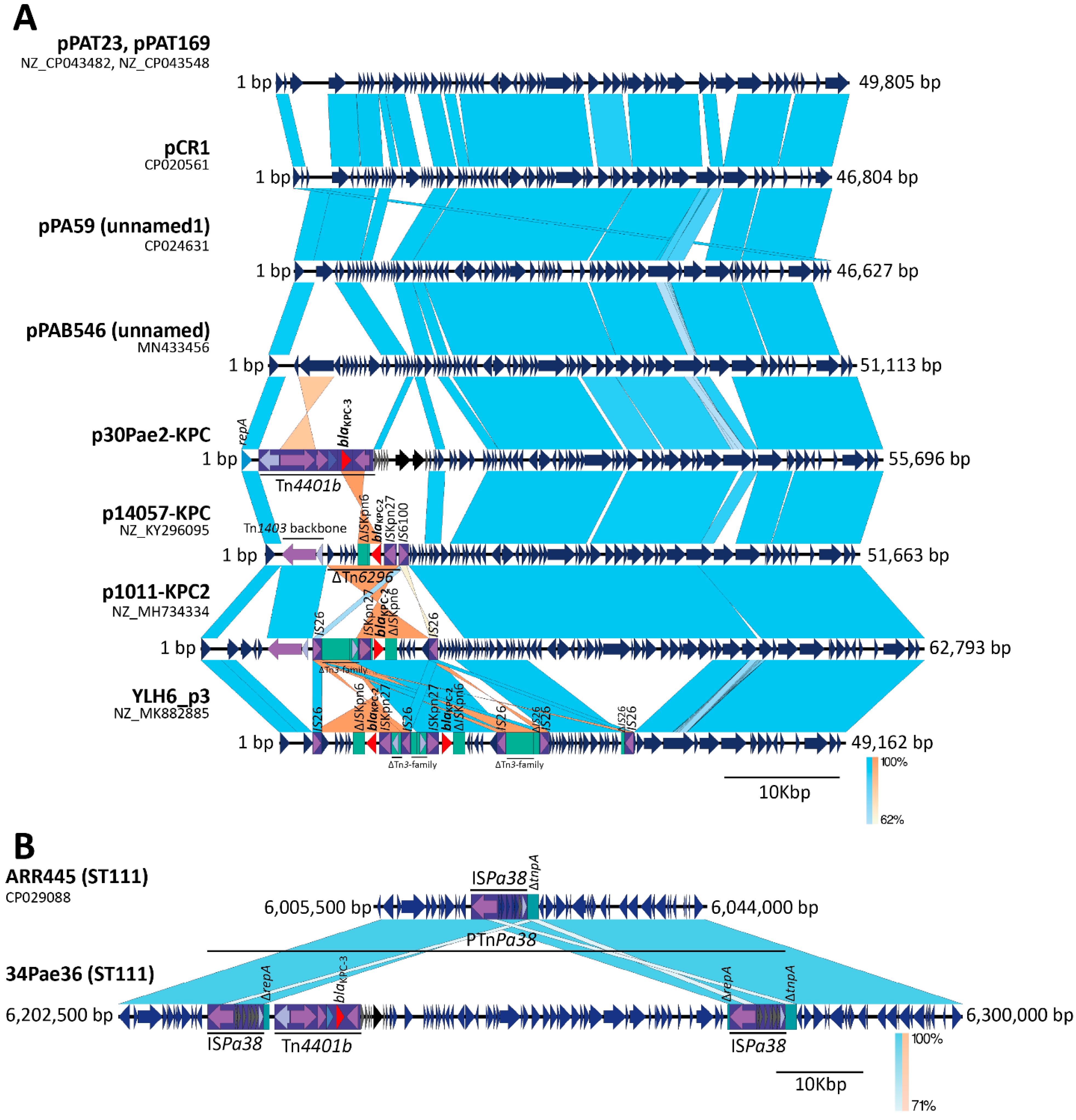
Regarding the ST111 isolates, the sequencing revealed that the blaKPC-3 gene is being mobilized for other platforms. In the case of the isolate 34Pae36, one unique contig, with a size of 7,418,292 bp, was found indicating que the blaKPC-3 was chromosomally located; by contrast, the genome of the isolate 30Pae2 was formed for the chromosome (7213.418 bp) and one plasmid (named as p30Pae2-KPC) with a size of 55,696 bp and where the blaKPC-3 gene was located. This plasmid does not belong to any incompatibility group previously reported. Unlike the blaKPC-2 harboring isolates, the blaKPC-3 was mobilized for the transposon Tn4401 isoform b in both isolates (30Pae2 and 34Pae36). The search for p30Pae2-KPC with Tn4401b in P. aeruginosa isolates harboring blaKPC revealed that this plasmid was found in 33 (50.0%) isolates, of which 32 came from the institution F (where isolate 30Pae2 was recovered) and the remaining isolate from the institution C (where 34Pae36 was also found), so the dissemination of this plasmid seems to be associated to an institutional level than a regional. Interestingly, the insertion of this plasmid in the chromosome region of 34Pae36 was not observed in any other isolate.
A standout fact was that the Tn4401b flanking sequences were not identical, that is, a TSD was not yielded; but these flanking sequences were the same in both structures (plasmid and chromosome), suggesting a possible shared mobile platform. The comparative genomic analysis displayed the insertion of an almost identical plasmid sequence (100% of identity and 96% coverage) into the chromosome (Figure 1). These results show that the chromosomal blaKPC-3 insertion in the isolate 34Pae36 was not produced for the Tn4401b transposition, as it was previously reported by us in other Colombian isolates [16], but for plasmid insertion. Now, what possible genetic mechanism could be involved?

2.4. ISPa38/TnPa38, a New Player Moving Resistance Genes in P. aeruginosa

A detailed analysis of the chromosomal site where one copy of the plasmid p30Pae2-KPC was inserted within the isolate 34Pae36 exhibited the presence of the two directly oriented copies of the ISPa38, abutting the plasmid sequence and forming a pseudo-composite transposon structure (Figure 4B). The ISPa38 has a size of 6455 bp, is bounded by the terminal inverted repeats of 39 bp, and is constituted of eight genes (transposase, resolvase, and six-passenger genes of unknown function) and we achieved to identify a putative res site. It is more appropriate to refer to this structure as a transposon (TnPa38) rather than an IS. The TnpA protein of ISPa38/TnPa38 contains the DDE triad within its catalytic domain and belongs to the Tn3 family or pMULT-4 family (prokaryotic Mutator-Like Transposase) according to the classification proposed by Guerillot et al. [21]. Notably, in the isolate 30Pae2 (that harbored the plasmid p30Pae2-KPC) only one copy of the ISPa38/TnPa38 was found within its chromosome, it was justly inserted in the intergenic region of two genes encoding a hypothetical protein, and a truncated transposase, respectively. This suggests that the chromosomal insertion of the plasmid was accompanied by a duplication of this IS.
To try to decipher the possible transposition events involved in the formation of this structure, we performed an analysis of the ISPa38/TnPa38 flanking sequences (FS). In the 30Pae2, the ISPa38/TnPa38 was flanked by sequences ATGAA and TTCCA (no TSD produced), that is, probably its mobilization was not yielded by a “copy and paste” transposition (Figure 1 and Figure 4B), as it has been described in Tn3 [22]. In this same way, in the 34Pae36, the pseudo-composite transposon structure was also flanked by these two sequences, but interestingly, a TSD was generated in the internal sequences (AGTTC) (Figure 1 and Figure 4B). These genetic transposition prints suggest that the structure within the 34Pae36 chromosome corresponds to a cointegrate (chromosome-plasmid), which was probably yielded by a replicative transposition event of the ISPa38/TnPa38 from chromosome toward plasmid [22].
These results indicate that ISPa38/TnPa38 is involved in the mobilization (and possible stabilization) of the blaKPC gene toward the chromosome of P. aeruginosa. In addition, the formation of this ISPa38/TnPa38-pseudo-composite transposon could have implications in the blaKPC dispersion because it could facilitate the formation of new TU structures, circular structures generated by the resolution of the cointegrate for either mechanism ISPa38/TnPa38-TnpA depend or for homologous recombination (RecA), and the possible mobilization as a composite transposon, as it has been described in Tn3 [22]. However, more studies oriented to establish the functionality and transposition routes used for ISPa38/TnPa38 are necessary.
Then, how could P. aeruginosa have acquired the ISPa38/TnPa38? From our analysis there are several points to highlight, first, the ISPa38/TnPa38 is almost exclusive to P. aeruginosa; when we examined the frequency of this IS within Pseudomonas spp., we found that it has only been described in P. aeruginosa strains (except for the clinical P. putida strain H8234) [23]. Second, the ISPa38/TnPa38 is scarce in P. aeruginosa, from 5678 genomes publicly available from NCBI (last accession November 2021), this IS was only found in 13 genomes. An analysis of the ISPa38/TnPa38 surroundings and FS revealed that this IS has been acquired by at least three independent genetic events, all directly toward the chromosome (Table 1). An acquisition event was produced at the position ~ 6,200,000 (just at the intergenic region of IPC90_06220 and truncated tnpA genes), where the IS is immersed in a genomic island region of 106,730 bp within a probable prophage with some regions genetically related with the filamentous bacteriophage Pf1 (AY324828) with 92.6% of identity and 6% of coverage, and was inserted within a tRNAMet gene copy (Figure 5). Interestingly, this insertion was found exclusively in strains previously reported belonged to ST111 (included the 34Pae36 isolate). Another ISPa38/TnPa38 insertion was located at site 4,250,295 exclusively within a genomic island in the strain AR_0440 (ST357). Finally, a third insertion was produced at the site ~3,000,000 and perhaps due to the acquisition of a pathogenicity island for diverse genetic background (ST175 and ST155), included the well-studied strain DK2 (ST386). No TSD were found in all ISPae38/TnPa38 copies (Table 1).
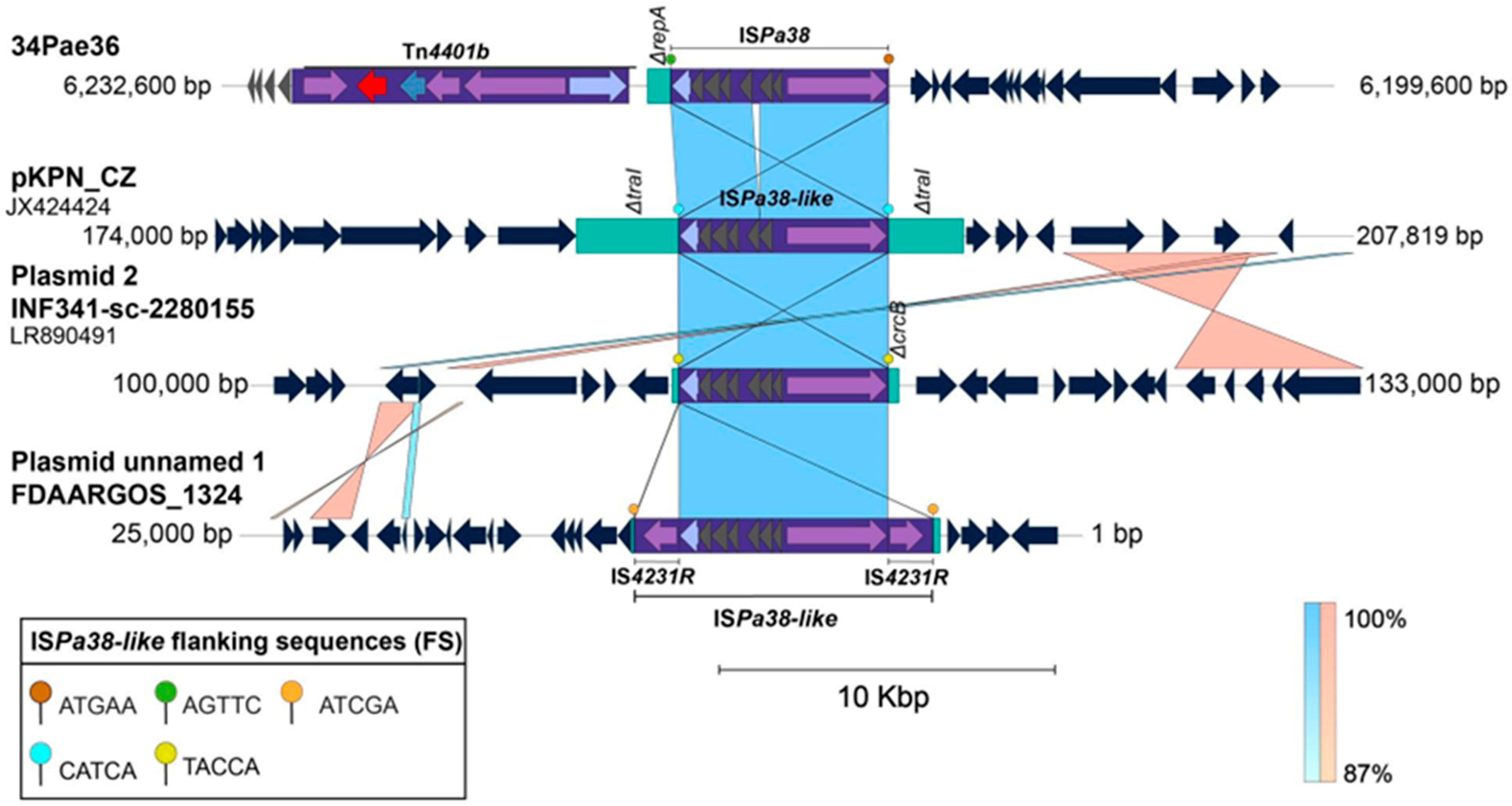
Due to little information about the ISPa38/TnPa38 in P. aeruginosa, we searched IS-related within the Enterobacterales genomes published in NCBI Nucleotide database, and we accomplished to identify a high similar IS (96% of coverage and 99% of identity) within the Klebsiella pneumoniae strains (Figure 6). Specifically, one ISPa38/TnPa38-like copy was located within the plasmids pKPN_CZ, INF341-sc-2280155-plasmid 2, FDAARGOS1306, which have been associated with qnrB genes dissemination in Czech Republic and Australia [16]. By contrast to that found in P. aeruginosa, these ISPa38/TnPa38-like copies are directly bounded by TSDs, all with a size of 5pb (Figure 6).

3. Discussion

The recommendations of the WHO over the last two decades to produce effective antibiotics with fewer side effects have borne fruit. Current guidelines from different scientific societies recommend antibiotics recently introduced to the market in the United States, such as cefiderocol, eravacycline, imipenem-cilastatin relebactam, meropenem vaborbactam, temamycin, and ceftazidime avibactam (CZA) [24,25]. Specifically, in difficult-to-treat resistant-Pseudomonas aeruginosa (DTR-PA), the guidelines include imipenem-cilastatin, relebactam, termomycin, ceftolozane/tazobactam, cefiderocol, and ceftazidime/avibactam. Unfortunately, in Colombia, as in other countries in the region, some of these therapeutic options are only found in major cities, which implies the use of non-recommended antibiotics such as polymyxins, fosfomycin, amikacin, gentamicin, and combination with high dose meropenem or doripenem. Given the limitation of therapeutic schemes for the treatment of infections caused by (DTR PA), it is important to highlight the urgent need to reduce antibiotic pressure in different hospital environments that, through the indiscriminate use of antimicrobials, promote the expression of resistance mechanisms such as those described in this article, added to others such as outer membrane porins (OprD), hyperproduction of AmpC enzymes, upregulation of efflux pumps, and mutations in penicillin-binding protein targets. Additionally, although CZA has shown adequate effectiveness against KPC-2-producing strains, reports of CZA resistance are beginning to appear, such as those that express blaKPC-90, whose genes have mutated from blaKPC [26]. Studies of different mechanisms of resistance, availability, and access to the different available antimicrobials allow the generation of treatment guidelines for Latin American countries in accordance with our reality and force us to make a priority call for the introduction of new therapeutic alternatives in our pharmaceutical market to reduce morbidity and mortality by DTR PA [27].
Since 2008, we have carried out surveillance studies on resistance in P. aeruginosa in Colombia, what have allowed us to decipher the mechanisms adopted by this bacterium to overcome the antibiotic mortal pressure (especially to carbapenem) during these years. First, altering the expression of the constitutive genes such as deregulation of the porins, overexpression of some efflux pump, second, the acquisition of atypical class I integrons, inactivation of the genes encoding porins by IS26 transposition, and third, mobile genetic elements like transposons, and plasmids harboring multiple resistance genes that have allowed the increase in the appearance of carbapenem-resistant and MDR P. aeruginosa isolates, being this last elements in our study, the most frequent (86%) [28].
The population of P. aeruginosa is composed of a small number of widespread clones, being the high-risk MDR clones ST235 and ST111 are the most worldwide observed harboring an arsenal or resistant genes [29,30]. These STs have already been previously reported with blaKPC gene in Colombia [4,16,31,32,33] and other countries such as Brazil and Germany [10,11]. For the first time in the world, the blaKPC gene was acquired by P. aeruginosa in Colombian isolates(4) through the two new and no related plasmids, the pPA2 (ST1006) first plasmid with an NTEKPC in P. aeruginosa) and pCOL-1 (ST308) (GenBank accession numbers: KC609322 and KC609323.). The last one, has a size of the 31 kb and belong to the incompatibility group IncP-6 harboring blaKPC in a Tn4401b. Interestingly, in some surveillance studies we have detected the plasmid pCOL-1 in Klebsiella pneumoniae isolates (where KPC is most frequent) suggesting a possible interspecies transference of this important resistance mechanism [34]. Since then, Tn4401 was the most frequent transposon that have been mobilizing blaKPC (almost all the variant 2) in ST235 and other STs and it has been mobilized in P. aeruginosa isolated from Colombia —even the double transposition of Tn4401 in the ST235 chromosome of this species has been reported— IncHI1 plasmid in Germany and other plasmids not typeable by any known incompatibility group [16,35]. Being these last plasmids found recently harboring NTEKPC in Brazil, Argentina and in this study with the new report of the structure Tn2-NTEKPC-IIg in p34Pae8-KPC and p34Pae23-KPC [10,20].
Although ST235 has been the most widely distributed clone worldwide, in Colombia ST111 harboring the blaVIM gene was first reported —there are reports of the circulation of this carbapenemase gene in P. aeruginosa since 1999 without MLST results— arrived and became the most prevalent in infections produced by carbapenem-resistant P. aeruginosa isolates, as it happened in other countries with this ST and other pandemic clones such as ST175, 233, 277, 357, 654, and 773 [32,36,37,38,39]. ST111 co-harboring blaVIM and blaKPC genes have been reported since 2012, and that continues to be a problem of interest in our country, where overtime reports of this cocirculation have increased due to the increased clinical usage of carbapenems, as evidenced in this study, where 64.8% of the isolates presented this phenomenon for carbapenem-resistance [9,32,33,40,41]. While the dissemination of blaVIM has been attributed in ST111 to its integration as a cassette gene in integrons, blaKPC-2 has been acquired by Tn4401 and now, according to our results, is the MGE associated with P. aeruginosa blaKPC-3 acquisition [9,31,35].
In the present study, our results show that, in the six health institutions, the blaKPC isolates have displaced the blaVIM isolates and are already the most prevalent, suggesting a successful adaption of this resistance mechanism to this non-fermenting bacterium in our country. The blaKPC gene has become the predominant carbapenemase-encoding gene in Colombia, largely due to its efficient dissemination through mobile genetic elements. This increasing frequency of KPC-producing P. aeruginosa isolates can be related with the mobilization of the blaKPC gene toward new plasmid backbones, some of them, with an environmental origin; coming from blaKPC-positive plasmids circulating in Klebsiella pneumoniae, where this gene circulates with very high frequency and within diverse and very variables genetic platforms (including MGE and plasmids), these MGEs could be consulted at https://maphub.net/LGMB/KPC-Pseudomonas-aeruginosa-LGMB (accessed on 20 May 2025) [14,42].
Studies have shown that blaKPC-2 and blaKPC-3 are the most common variants, frequently detected in healthcare-associated infections (HAIs) and occasionally in community-acquired infections [15,43]. Regarding blaKPC-3, our findings were even more interesting, showing that this gene is been mobilized within a little-known plasmid structure, previously detected in isolates of environmental origin in Asia, some of these with blaKPC incorporated, but variant 2, and mobilized for no classical Tn4401 (Figure 2). In contrast, in the case of the plasmid p30Pae2-KPC, the Tn4401b was probably the first element responsible to mobilize the gene, but the insertion into the plasmid was helped maybe for another mobile structure (no TSD was found in Tn4401b). In this case, we also suppose the blaKPC-3 gene has arrived at P. aeruginosa from K. pneumoniae through the shared plasmids, due to that, in Colombia, the blaKPC-3 is the more frequently variant detected in Klebsiella pneumoniae isolates [44,45].
The overuse and misuse of carbapenems in Colombian healthcare settings have exerted strong selective pressure, favoring the emergence and dissemination of blaKPC-producing bacteria and in this case the establishment of blaKPC in P. aeruginosa. Studies have shown that prior exposure to carbapenems is a significant risk factor for infections with carbapenem-resistant Enterobacterales (CRE) [33,46]. Deficiencies in infection control practices have played a pivotal role in the spread of blaKPC-producing bacteria in Colombian healthcare facilities. Horizontal gene transfer and clonal dissemination have been facilitated by inadequate hand hygiene, improper sterilization of medical devices, and insufficient isolation of infected patients [47]. These lapses have allowed blaKPC-producing isolates to thrive in hospital environments, contributing to their dominance over blaVIM-producing isolates. To reduce antibiotic pressure and thus slow the selection for resistance mechanisms in P. aeruginosa clinical isolates and other Enterobacterales species, it is essential that primary strategies can be implemented in hospitals (Table S4) [48,49,50,51,52,53,54,55,56,57,58,59,60,61,62,63,64] and more genomic studies that allowing to know the mobile genetic elements with resistant determinants that are spreading in the institution, that allow a better dissemination control. The implementation of regional genomic surveillance programs has been shown to be effective in tracking the spread of blaKPC-harboring pathogens. For example, a study in three U.S. states demonstrated that genomic surveillance could identify distinct transmission pathways, such as importation versus clonal expansion, and inform targeted interventions [65] The clinical implementation of WGS has been shown to be feasible and effective for identifying transmission events of multidrug-resistant pathogens, including those harboring blaKPC. For example, a study in three Australian hospitals demonstrated that WGS-based surveillance can identify clusters of multidrug-resistant organisms and inform infection control interventions [66].
Standard and contact infection prevention and control practices, including hand hygiene, patient isolation, and environmental disinfection, remain critical for preventing the spread of blaKPC-harboring pathogens. Studies have shown that these practices, when implemented consistently, can reduce the transmission of multidrug-resistant organisms in healthcare settings [67].

4. Materials and Methods

4.1. Source of the Bacterial Isolates, Susceptibility Profiles and Molecular Detection of Carbapenemase Genes

The isolates were collected from an observational, descriptive, prospective, and multicentre surveillance study performed between 2017 and 2020 in six high complexity hospitals (designed as A–F) located in Bogota, the main city of Colombia. During this time, 128 carbapenem-resistant P. aeruginosa isolates were recovered from 121 patients, among those, 66 harbored the blaKPC gene.
The susceptibility profile was determined using the automated systems VITEK® 2 and the breakpoints defined by the Clinical and Laboratory Standards Institute according to the 2020 update. The MIC for meropenem was confirmed by microdilution test in these 30 carbapenem-resistant blaKPC-positive isolates.
The genomic DNA of isolates was extracted with DNeasy UltraClean Microbial Kit (QIAGEN N.V.) and presence of carbapenemases genes as blaGES, blaIMP, blaVIM, blaOXA-48 and blaNDM was determined by PCR [68].

4.2. Genetic Relationship Establishment

Genetic relationship was determined by pulsed-field electrophoresis (PFGE) analysis, following the Centers for Disease Control and Prevention (CDC) protocol with minor modifications [69]. P. aeruginosa isolates were cultivated in BHI broth and incubated at 37 °C with shaking at 200 rpm for 12 h, to an OD625 of 1.5 in TE buffer (100 mM Tris + 100 mM EDTA, pH 8.0). 1% blocks were prepared with SeaKem® Gold Agarose, with150 µL of bacterial suspension and 7.5 µL of proteinase K (20 mg/mL). For lysis, 1 mL of lysis buffer (50 mM Tris, 50 mM EDTA, pH 8.0 + 1% Sarcosyl) with 5 µL of proteinase K (20 mg/mL) was added to the blocks and incubated for 2 h at 55 °C at 175 rpm. Two washes were then performed with sterile deionized distilled water at 55 °C and two washes with TE buffer (10 mM Tris, 1 mM EDTA, pH 8.0). The blocks were stored at 4 °C until use. Restriction was performed with 12 units of the restriction enzyme SpeI (10 U/µL: Promega) in 1X Buffer B for 5 h at 37 °C. Restriction fragments were separated on a 1% SeaKem® Gold Agarose gel in 0.5X TBE (Tris base; boric acid; 0.5M EDTA (pH 8.0); dH2O) buffer using the CHEFFII equipment (BioRad) with the following running conditions: initial time of 6.8 s and final time of 35.4 s for 23 h at an angle of 120° and a voltage of 6.0. The gels were stained with ethidium bromide (10 mg/mL). The image was captured with the ChemiDoc™ MP Imaging System (BioRad) and the dendrogram was obtained using the GelCompar II program (Applied Maths NV). For the analysis, Salmonella serotype Braenderup H9812 was used as a normalizing strain and the pulsotypes were defined according to the criteria of Tenover et al. with a cut-off point greater than 80% similarity [70].
For MLST analysis, fragments of the genes acsA, aroE, guaA, mutL, nuoD, ppsA, and trpE were amplified and sequenced according to the protocol previously reported [71]. The alleles and ST numbers were established from the MLST P. aeruginosa database (available from https://pubmlst.org/organisms/pseudomonas-aeruginosa, accessed on 20 May 2025).

4.3. Whole Genome Sequencing (WGS)

The genome sequencing was performed in four representative P. aeruginosa isolates, which were selected according to its PFGE pulsotype, ST, and the institution where they were recovered. Total DNA was extracted using the UltraClean® Microbial DNA Isolation Kit (QIAGEN N.V). A 20 Kb-enriched SMRTbell™ library was prepared for each DNA sample. Libraries were sequenced using the PacBio RS II platform with the P6-C4 chemistry (one SMRT cell per sample). De novo assembly of subreads was performed with HGAP protocol in the SMRT Analysis v2.3. The contigs obtained were compared with each other for the presence of repeated ends, (suggestive of complete circular structures). Complete sequences were circularized as previously described [72]. Annotation of open reading frames was done in Prokka v1.14.5 [73] and the regions of interest were compared with Protein and Nucleotide NCBI databases using BLAST 2.16.0 and manually curated with ACT [74]. Antibiotic resistance genes were identified using ResFinder and CARD [75]. Genomic pairwise comparisons were plotted using Easyfig [76].

4.4. Comparative Genomics Analysis

A genomic comparative analysis was performed to determine, first, the similarities or differences in the platforms mobilizing the blaKPC gene in the four Colombian P. aeruginosa isolates sequenced, second, comparison respect with the genomes published in the NCBI Nucleotide database, and third, to try to clarify the possible genetic mechanism involved in the blaKPC mobilization.
Genomic and pathogenicity islands (GI and PI, respectively), prophages, insertion sequence (IS) elements and transposons were identified using IslandViewer [77], PHASTER [78], ISfinder (available at https://isfinder.biotoul.fr/, accessed on 20 May 2025) and the Tn Registry database (available at https://transposon.lstmed.ac.uk/tn-registry); respectively. The genetic platforms identified were manually curated throw ACT [21], and pairwise comparisons were visualized and illustrated and EasyFig [76].

4.5. Molecular Identification of blaKPC Mobilization Platforms

Once the sequence of the mobile genetic elements associated with blaKPC gene mobilization was determined, primers were designed to identify the insertion of Tn4401 or NTEKPC in the plasmid of interest, to know its frequency in the population of study. Multiplex PCRs was performed according to conditions described in Table S1.

4.6. Accession Numbers of the Genome Sequences

The chromosome and plasmids sequenced established in this study have been deposited in the Nucleotide database (NCBI) with the accession numbers ON248939, CP095774, CP095775, CP095772, CP095773, and CP095770, which correspond to the sequences p30Pae2-KPC, 34Pae8, p34Pae8-KPC, 34Pae23, p34Pae23-KPC and 34Pae36; respectively.

4.7. Ethics Committee Approbation

This study was revised and approved by the Institutional Ethics Committee of the Universidad Nacional de Colombia (DIB-18-03). In addition, the study was approved by each participant institution. Experiments were performed in accordance with Declaration of Helsinki.

5. Conclusions

The appearance of blaKPC-transporting novelty plasmid backbones suggests that an active dispersion process of this resistance gene is taking place in P. aeruginosa, which is highly been facilitated for the conjunction of several genetic factors such as, first, the interspecies flux of plasmids from Klebsiella pneumoniae towards P. aeruginosa; second, the high transposition of the Tn4401; third, the emergence of new NTEKPC structures, more compact and perhaps with a higher frequency of transposition; and forth, the participation of new transposons and ISs. For another hand, the pandemic high-risk clones ST111 and ST235 continue to demonstrate their genetic advantages with respect to other clones of P. aeruginosa, due to their very high ability to uptake and keep foreign DNA.

Supplementary Materials

The following supporting information can be downloaded at: https://www.mdpi.com/article/10.3390/antibiotics14090947/s1.

Author Contributions

J.-C.G., A.L.L.-C., C.H.S.-T., N.V.G. and J.E.-P. conceived and designed the study; D.A., J.B.-O., J.M., R.-H.B., R.A.M.-O. and Z.L.C.R. performed research; J.-C.G., A.L.L.-C., C.H.S.-T. and R.-H.B. contributed with the active search of the isolates in the health institutions; D.A., J.B.-O., A.L.L.-C., N.V.G. and J.E.-P. analyzed data; and D.A. and J.E.-P. wrote the paper. All authors reviewed the manuscript. All authors have read and agreed to the published version of the manuscript.

Funding

This work was supported by the Ministerio de Ciencia, Tecnologia e Innovacion of Colombia-MinCiencias (Grant number 1308-77758007), Universidad El Bosque (Grant number 605-2018), Universidad de la Sabana (Grant number MED-213-2016), Universidad Nacional de Colombia, and Fundación Santa Fe de Bogotá. The funders had no role in the study design, data collection and interpretation, or the decision to submit the work for publication.

Data Availability Statement

The data presented in this study are available within the article. Raw data supporting this study are available from the corresponding author upon reasonable request.

Acknowledgments

First: we gratefully acknowledge the laboratory staff of the participant institutions for its invaluable support, technical assistance and the follow-up and send of the samples. We also acknowledge the Vicechancellery of Research at Universidad El Bosque for its invaluable support.

Conflicts of Interest

The authors declare no competing interests.

References

  1. Yoon, E.J.; Jeong, S.H. Mobile Carbapenemase Genes in Pseudomonas aeruginosa. Front. Microbiol. 2021, 12, 614058. [Google Scholar] [CrossRef] [PubMed]
  2. Pfizer. Antimicrobial Testing Leadership and Surveillance; Pfizer: New York, NY, USA, 2023. [Google Scholar]
  3. Yigit, H.; Queenan, A.M.; Anderson, G.J.; Domenech-Sanchez, A.; Biddle, J.W.; Steward, C.D.; Alberti, S.; Bush, K.; Tenover, F.C. Novel carbapenem-hydrolyzing β-lactamase, KPC-1, from a carbapenem-resistant strain of Klebsiella pneumoniae. Antimicrob. Agents Chemother. 2001, 45, 1151–1161. [Google Scholar] [CrossRef]
  4. Villegas, M.V.; Lolans, K.; Correa, A.; Kattan, J.N.; Lopez, J.A.; Quinn, J.P. First identification of Pseudomonas aeruginosa isolates producing a KPC-type carbapenem-hydrolyzing β-lactamase. Antimicrob. Agents Chemother. 2007, 51, 1553–1555. [Google Scholar] [CrossRef]
  5. Akpaka, P.E.; Swanston, W.H.; Ihemere, H.N.; Correa, A.; Torres, J.A.; Tafur, J.D.; Montealegre, M.C.; Quinn, J.P.; Villegas, M.V. Emergence of KPC-producing Pseudomonas aeruginosa in Trinidad and Tobago. J. Clin. Microbiol. 2009, 47, 2670–2671. [Google Scholar] [CrossRef]
  6. Poirel, L.; Nordmann, P.; Lagrutta, E.; Cleary, T.; Munoz-Price, L.S. Emergence of KPC-Producing Pseudomonas aeruginosa in the United States. Antimicrob. Agents Chemother. 2010, 54, 3072. [Google Scholar] [CrossRef]
  7. Ge, C.; Wei, Z.; Jiang, Y.; Shen, P.; Yu, Y.; Li, L. Identification of KPC-2-producing Pseudomonas aeruginosa isolates in China. J. Antimicrob. Chemother. 2011, 66, 1184–1186. [Google Scholar] [CrossRef] [PubMed]
  8. Robledo, I.E.; Aquino, E.E.; Vázquez, G.J. Detection of the KPC gene in Escherichia coli, Klebsiella pneumoniae, Pseudomonas aeruginosa, and Acinetobacter baumannii during a PCR-based nosocomial surveillance study in Puerto Rico. Antimicrob. Agents Chemother. 2011, 55, 2968–2970. [Google Scholar] [CrossRef]
  9. Jácome, P.R.L.D.A.; Alves, Ĺ.R.; Cabral, A.B.; Lopes, A.C.S.; Maciel, M.A.V. First report of KPC-producing Pseudomonas aeruginosa in Brazil. Antimicrob. Agents Chemother. 2012, 56, 4990. [Google Scholar] [CrossRef] [PubMed]
  10. de Paula-Petroli, S.B.; Campana, E.H.; Bocchi, M.; Bordinhão, T.; Picão, R.C.; Yamada-Ogatta, S.F.; Carrara-Marroni, E.F. Early detection of a hypervirulent KPC-2-producing Pseudomonas aeruginosa ST235 in Brazil. J. Glob. Antimicrob. Resist. 2018, 12, 153–154. [Google Scholar] [CrossRef]
  11. Hagemann, J.B.; Pfennigwerth, N.; Gatermann, S.G.; von Baum, H.; Essig, A. KPC-2 carbapenemase-producing Pseudomonas aeruginosa reaching Germany. J. Antimicrob. Chemother. 2018, 73, 1812–1814. [Google Scholar] [CrossRef]
  12. Naas, T.; Bonnin, R.A.; Cuzon, G.; Villegas, M.V.; Nordmann, P. Complete sequence of two KPC-harbouring plasmids from Pseudomonas aeruginosa. J. Antimicrob. Chemother. 2013, 68, 1757–1762. [Google Scholar] [CrossRef]
  13. Rada, A.M.; De La Cadena, E.; Agudelo, C.; Capataz, C.; Orozco, N.; Pallares, C.; Dinh, A.Q.; Panesso, D.; Ríos, R.; Diaz, L.; et al. Dynamics of bla KPC-2 Dissemination from Non-CG258 Klebsiella pneumoniaea to Other Enterobacterales via IncN Plasmids in an Area of High Endemicity. Antimicrob. Agents Chemother. 2020, 64, e01743-20. [Google Scholar] [CrossRef]
  14. Abril, D.; Vergara, E.; Palacios, D.; Leal, A.L.; Marquez-Ortiz, R.A.; Madroñero, J.; Rozo, Z.L.C.; De La Rosa, Z.; Nieto, C.A.; Vanegas, N.; et al. Within patient genetic diversity of bla KPC harboring Klebsiella pneumoniaea in a Colombian hospital and identification of a new NTEKPC platform. Sci. Rep. 2021, 11, 21409. [Google Scholar] [CrossRef] [PubMed]
  15. Ibáñez-Prada, E.D.; Bustos, I.G.; Gamboa-Silva, E.; Josa, D.F.; Mendez, L.; Fuentes, Y.V.; Serrano-Mayorga, C.C.; Baron, O.; Ruiz-Cuartas, A.; Silva, E.; et al. Molecular characterization and descriptive analysis of carbapenemase-producing Gram-negative rod infections in Bogota, Colombia. Microbiol. Spectr. 2024, 12, e01714-23. [Google Scholar] [CrossRef]
  16. Abril, D.; Marquez-Ortiz, R.A.; Castro-Cardozo, B.; Moncayo-Ortiz, J.I.; Olarte Escobar, N.M.; Corredor Rozo, Z.L.; Reyes, N.; Tovar, C.; Sánchez, H.F.; Castellanos, J.; et al. Genome plasticity favours double chromosomal Tn4401b-bla KPC-2 transposon insertion in the Pseudomonas aeruginosa ST235 clone. BMC Microbiol. 2019, 19, 45. [Google Scholar] [CrossRef] [PubMed]
  17. Saha, R.; Farrance, C.E.; Verghese, B.; Hong, S.; Donofrio, R.S. Klebsiella michiganensis sp. nov., a new bacterium isolated from a tooth brush holder. Curr. Microbiol. 2013, 66, 72–78. [Google Scholar] [CrossRef]
  18. Zhu, X.; Li, P.; Qian, C.; Liu, H.; Lin, H.; Zhang, X.; Li, Q.; Lu, J.; Lin, X.; Xu, T.; et al. Prevalence of Aminoglycoside Resistance Genes and Molecular Characterization of a Novel Gene, aac(3)-IIg, among Clinical Isolates of the Enterobacter cloacae Complex from a Chinese Teaching Hospital. Antimicrob. Agents Chemother. 2020, 64, e00852-20. [Google Scholar] [CrossRef] [PubMed]
  19. Dalrymple’, B.; Arber, W. Promotion of RNA transcription on the insertion element IS30 of E. coli K12. EMBO J. 1985, 4, 2687–2693. [Google Scholar] [CrossRef]
  20. Cejas, D.; Elena, A.; González-Espinosa, F.E.; Pallecchi, L.; Vay, C.; Rossolini, G.M.; Gutkind, G.; Di Pilato, V.; Radice, M. Characterisation of blaKPC-2–harbouring plasmids recovered from Pseudomonas aeruginosa ST654 and ST235 high-risk clones. J. Glob. Antimicrob. Resist. 2022, 29, 310–312. [Google Scholar] [CrossRef]
  21. Guérillot, R.; Siguier, P.; Gourbeyre, E.; Chandler, M.; Glaser, P. The diversity of prokaryotic DDE transposases of the mutator superfamily, insertion specificity, and association with conjugation machineries. Genome Biol. Evol. 2014, 6, 260–272. [Google Scholar] [CrossRef]
  22. Nicolas, E.; Lambin, M.; Dandoy, D.; Galloy, C.; Nguyen, N.; Oger, C.A.; Halletm, B. The Tn3-family of Replicative Transposons. Microbiol. Spectr. 2015, 3, 693–726. [Google Scholar] [CrossRef] [PubMed]
  23. Molina, L.; Bernal, P.; Udaondo, Z.; Segura, A.; Ramos, J.L. Complete genome sequence of a Pseudomonas putida clinical isolate, strain H8234. Genome Announc. 2013, 1, e00496-13. [Google Scholar] [CrossRef]
  24. Lawandi, A.; Yek, C.; Kadri, S.S. IDSA guidance and ESCMID guidelines: Complementary approaches toward a care standard for MDR Gram-negative infections. Clin. Microbiol. Infect. 2022, 28, 465–469. [Google Scholar] [CrossRef]
  25. Paul, M.; Carrara, E.; Retamar, P.; Tängdén, T.; Bitterman, R.; Bonomo, R.A.; de Waele, J.; Daikos, G.L.; Akova, M.; Harbarth, S.; et al. European Society of Clinical Microbiology and Infectious Diseases (ESCMID) guidelines for the treatment of infections caused by multidrug-resistant Gram-negative bacilli (endorsed by European society of intensive care medicine). Clin. Microbiol. Infect. 2022, 28, 521–547. [Google Scholar] [CrossRef]
  26. Zhu, Y.; Chen, J.; Shen, H.; Chen, Z.; Yang, Q.W.; Zhu, J.; Li, X.; Yang, Q.; Zhao, F.; Ji, J.; et al. Emergence of Ceftazidime-and Avibactam-Resistant Klebsiella pneumoniaea Carbapenemase-Producing Pseudomonas aeruginosa in China. mSystems 2021, 6, e00787-21. [Google Scholar] [CrossRef]
  27. Rubinstein, E.; Lalani, T.; Corey, G.R.; Kanafani, Z.A.; Nannini, E.C.; Rocha, M.G.; Rahav, G.; Niederman, M.S.; Kollef, M.H.; Shorr, A.F.; et al. Telavancin versus vancomycin for hospital-acquired pneumonia due to gram-positive pathogens. Clin. Infect. Dis. 2011, 52, 31–40. [Google Scholar] [CrossRef] [PubMed]
  28. Martinez, E.; Pérez, J.E.; Buelvas, F.; Tovar, C.; Vanegas, N.; Stokes, H.W. Establishment and multi drug resistance evolution of ST235 Pseudomonas aeruginosa strains in the intensive care unit of a Colombian hospital. Res. Microbiol. 2014, 165, 852–856. [Google Scholar] [CrossRef] [PubMed]
  29. del Barrio-Tofiño, E.; López-Causapé, C.; Oliver, A. Pseudomonas aeruginosa epidemic high-risk clones and their association with horizontally-acquired β-lactamases: 2020 update. Int. J. Antimicrob. Agents 2020, 56, 106196. [Google Scholar] [CrossRef]
  30. Pirnay, J.P.; Bilocq, F.; Pot, B.; Cornelis, P.; Zizi, M.; Van Eldere, J.; Deschaght, P.; Vaneechoutte, M.; Jennes, S.; Pitt, T.; et al. Pseudomonas aeruginosa population structure revisited. PLoS ONE 2009, 4, e7740. [Google Scholar] [CrossRef]
  31. Rada, A.M.; De La Cadena, E.; Agudelo, C.A.; Pallares, C.; Restrepo, E.; Correa, A.; Villegas, M.V.; Capataz, C. Genetic Diversity of Multidrug-Resistant Pseudomonas aeruginosa Isolates Carrying blaVIM–2 and blaKPC–2 Genes That Spread on Different Genetic Environment in Colombia. Front. Microbiol. 2021, 12, 663020. [Google Scholar] [CrossRef]
  32. Vanegas, J.M.; Cienfuegos, A.V.; Ocampo, A.M.; López, L.; Del Corral, H.; Roncancio, G.; Sierra, P.; Echeverri-Toro, L.; Ospina, S.; Maldonado, N.; et al. Similar frequencies of Pseudomonas aeruginosa isolates producing KPC and VIM carbapenemases in diverse genetic clones at tertiary-care hospitals in Medellín, Colombia. J. Clin. Microbiol. 2014, 52, 3978–3986. [Google Scholar] [CrossRef] [PubMed]
  33. Pacheco, T.; Bustos-Cruz, R.H.; Abril, D.; Arias, S.; Uribe, L.; Rincón, J.; García, J.-C.; Escobar-Perez, J. Pseudomonas aeruginosa coharboring blaKPC-2 and blaVIM-2 carbapenemase genes. Antibiotics 2019, 8, 98. [Google Scholar] [CrossRef] [PubMed]
  34. Abril, D.; Lesmes-Leon, D.N.; Marquez-Ortiz, R.A.; Leal, A.L.; Tovar-Acero, C.; Corredor Rozo, Z.L.; Gómez, N.V.; Escobar-Perez, J.; Hudson, A.O. Draft genome of the Klebsiella pneumoniaea 24Kpn33 and complete sequence of its pCOL-1, a plasmid related to the bla KPC-2 acquisition in Pseudomonas aeruginosa. Microbiol. Resour. Announc. 2024, 13, e00071-24. [Google Scholar] [CrossRef]
  35. Correa, A.; Del Campo, R.; Perenguez, M.; Blanco, V.M.; Rodríguez-Baños, M.; Perez, F.; Maya, J.J.; Rojas, L.; Cantón, R.; Arias, C.A.; et al. Dissemination of High-Risk Clones of Extensively Drug-Resistant Pseudomonas aeruginosa in Colombia. Antimicrob. Agents Chemother. 2015, 59, 2421–2425. [Google Scholar] [CrossRef]
  36. Woodford, N.; Turton, J.F.; Livermore, D.M. Multiresistant Gram-negative bacteria: The role of high-risk clones in the dissemination of antibiotic resistance. FEMS Microbiol. Rev. 2011, 35, 736–755. [Google Scholar] [CrossRef]
  37. Kos, V.N.; Déraspe, M.; McLaughlin, R.E.; Whiteaker, J.D.; Roy, P.H.; Alm, R.A.; Corbeil, J.; Gardner, H. The resistome of Pseudomonas seudomonas aeruginosa in relationship to phenotypic susceptibility. Antimicrob. Agents Chemother. 2015, 59, 427–436. [Google Scholar] [CrossRef]
  38. Kocsis, B.; Toth, A.; Gulyas, D.; Ligeti, B.; Katona, K.; Rokusz, L.; Szabo, D. Acquired qnrVC1 and bla NDM-1 resistance markers in an international high-risk Pseudomonas aeruginosa ST773 clone. J. Med. Microbiol. 2019, 68, 336–338. [Google Scholar] [CrossRef] [PubMed]
  39. Crespo, M.P.; Woodford, N.; Sinclair, A.; Kaufmann, M.E.; Turton, J.; Glover, J.; Velez, J.D.; CastañeDa, C.R.; Recalde, M.; Livermore, D.M. Outbreak of carbapenem-resistant Pseudomonas aeruginosa producing VIM-8, a novel metallo-β-lactamase, in a tertiary care center in Cali, Colombia. J. Clin. Microbiol. 2004, 42, 5094–5101. [Google Scholar] [CrossRef]
  40. Cai, H.; Zhu, Y.; Hu, D.; Li, Y.; Leptihn, S.; Loh, B.; Hua, X.; Yu, Y. Co-harboring of Novel blaKPC–2 Plasmid and Integrative and Conjugative Element Carrying Tn6203 in Multidrug-Resistant Pseudomonas aeruginosa. Front. Microbiol. 2021, 12, 674974. [Google Scholar] [CrossRef]
  41. Wozniak, A.; Figueroa, C.; Moya-Flores, F.; Guggiana, P.; Castillo, C.; Rivas, L.; Munita, J.M.; García, P.C. A multispecies outbreak of carbapenem-resistant bacteria harboring the bla KPC gene in a non-classical transposon element. BMC Microbiol. 2021, 21, 107. [Google Scholar] [CrossRef]
  42. Forero-Hurtado, D.; Corredor-Rozo, Z.L.; Ruiz-Castellanos, J.S.; Márquez-Ortiz, R.A.; Abril, D.; Vanegas, N.; Lafaurie, G.I.; Chambrone, L.; Escobar-Pérez, J. Worldwide Dissemination of blaKPC Gene by Novel Mobilization Platforms in Pseudomonas aeruginosa: A Systematic Review. Antibiotics 2023, 12, 658. [Google Scholar] [CrossRef]
  43. Contreras-Valero, J.F.; Gualtero-Trujillo, S.M.; Cortés-Fraile, G.C.; Hernández-Garzón, S.; Manrique-Marín, N.; Narváez-Chaves, M.Á.; Valderrama-Beltrán, S.L. Epidemiological and clinical characteristics of patients with carbapenem-resistant Enterobacterales in a university hospital of Colombia: Enzyme coproductions in rise. Heliyon 2024, 10, e33698. [Google Scholar] [CrossRef]
  44. Saavedra, S.Y.; Bernal, J.F.; Montilla-Escudero, E.; Arévalo, S.A.; Prada, D.A.; Valencia, M.F.; Moreno, J.; Hidalgo, A.M.; García-Vega, Á.S.; Abrudan, M.; et al. Complexity of Genomic Epidemiology of Carbapenem-Resistant Klebsiella pneumoniaea Isolates in Colombia Urges the Reinforcement of Whole Genome Sequencing-Based Surveillance Programs. Clin. Infect. Dis. 2021, 73, S290–S299. [Google Scholar] [CrossRef]
  45. Stillwell, T.; Green, M.; Barbadora, K.; Ferrelli, J.G.; Roberts, T.L.; Weissman, S.J.; Nowalk, A. Outbreak of KPC-3 producing carbapenem- resistant Klebsiella pneumoniaea in a US pediatric hospital. J. Pediatr. Infect. Dis. Soc. 2015, 4, 330–338. [Google Scholar] [CrossRef]
  46. García, C.J.C.; Amaya, S.; Briceño, C.W.; Rincón, C.; Pinzón, J. Risk factors for carbapenem-resistant bacterial infection or colonization: A case control study. Acta Colomb. Cuid. Intensivo 2017, 17, 29–35. [Google Scholar] [CrossRef]
  47. Lapp, Z.; Octaria, R.; O’Malley, S.M.; Nguyen, T.N.; Wolford, H.; Crawford, R.; Moore, C.; Vagnone, P.S.; Noel, D.; Duffy, N.; et al. Distinct Origins and Transmission Pathways of blaKPC Enterobacterales across Three U.S. States. J. Clin. Microbiol. 2023, 61, e00259-23. [Google Scholar] [PubMed]
  48. Centers for Disease Control and Prevention (CDC). The Core Elements of Hospital Antibiotic Stewardship Programs; Centers for Disease Control and Prevention (CDC): Atlanta, GA, USA, 2019.
  49. World Health Organization (WHO). Antimicrobial Stewardship Programmes in Health-Care Facilities in Low- and Middle-Income Countries. A WHO Practical Toolkit; World Health Organization (WHO): Geneva, Switzerland, 2019. [Google Scholar]
  50. Timbrook, T.T.; Morton, J.B.; McConeghy, K.W.; Caffrey, A.R.; Mylonakis, E.; LaPlante, K.L. The Effect of Molecular Rapid Diagnostic Testing on Clinical Outcomes in Bloodstream Infections: A Systematic Review and Meta-analysis. Clin. Infect. Dis. 2017, 64, 15–23. [Google Scholar] [CrossRef] [PubMed]
  51. Schuetz, P.; Wirz, Y.; Sager, R.; Christ-Crain, M.; Stolz, D.; Tamm, M.; Bouadma, L.; Luyt, C.E.; Wolff, M.; Chastre, J.; et al. Effect of procalcitonin-guided antibiotic treatment on mortality in acute respiratory infections: A patient level meta-analysis. Lancet Infect. Dis. 2018, 18, 95–107. [Google Scholar] [CrossRef] [PubMed]
  52. World Health Organization (WHO). Guidelines on Core Components of Infection Prevention and Control Programmes at the National and Acute Health Care Facility Level; World Health Organization (WHO): Geneva, Switzerland, 2016. [Google Scholar]
  53. Centers for Disease Control and Prevention (CDC). HAI Prevention and Control for Healthcare; Centers for Disease Control and Prevention (CDC): Atlanta, GA, USA, 2024.
  54. World Health Organization (WHO). Global Action Plan on Antimicrobial Resistance; World Health Organization (WHO): Geneva, Switzerland, 2016. [Google Scholar]
  55. Barlam, T.F.; Cosgrove, S.E.; Abbo, L.M.; MacDougall, C.; Schuetz, A.N.; Septimus, E.J.; Srinivasan, A.; Dellit, T.H.; Falck-Ytter, Y.T.; Fishman, N.O.; et al. Implementing an Antibiotic Stewardship Program: Guidelines by the Infectious Diseases Society of America and the Society for Healthcare Epidemiology of America. Clin. Infect. Dis. 2016, 62, e51–e77. [Google Scholar] [CrossRef]
  56. Tabah, A.; Bassetti, M.; Kollef, M.H.; Zahar, J.R.; Paiva, J.A.; Timsit, J.F.; Roberts, J.A.; Schouten, J.; Giamarellou, H.; Rello, J.; et al. Antimicrobial de-escalation in critically ill patients: A position statement from a task force of the European Society of Intensive Care Medicine (ESICM) and European Society of Clinical Microbiology and Infectious Diseases (ESCMID) Critically Ill Patients Study Group (ESGCIP). Intensive Care Med. 2020, 46, 245–265. [Google Scholar]
  57. Kalil, A.C.; Metersky, M.L.; Klompas, M.; Muscedere, J.; Sweeney, D.A.; Palmer, L.B.; Napolitano, L.M.; O’Grady, N.P.; Bartlett, J.G.; Carratalà, J.; et al. Management of Adults With Hospital-acquired and Ventilator-associated Pneumonia: 2016 Clinical Practice Guidelines by the Infectious Diseases Society of America and the American Thoracic Society. Clin. Infect. Dis. 2016, 63, e61–e111. [Google Scholar] [CrossRef] [PubMed]
  58. Spellberg, B. The New Antibiotic Mantra-”Shorter Is Better”. JAMA Intern. Med. 2016, 176, 1254–1255. [Google Scholar] [CrossRef]
  59. Baur, D.; Gladstone, B.P.; Burkert, F.; Carrara, E.; Foschi, F.; Döbele, S.; Tacconelli, E. Effect of antibiotic stewardship on the incidence of infection and colonisation with antibiotic-resistant bacteria and Clostridium difficile infection: A systematic review and meta-analysis. Lancet Infect. Dis. 2017, 17, 990–1001. [Google Scholar] [CrossRef]
  60. World Health Organization (WHO). Global Antimicrobial Resistance and Use Surveillance System (GLASS) Report: 2022; World Health Organization (WHO): Geneva, Switzerland, 2022. [Google Scholar]
  61. Centers for Disease Control and Prevention (CDC). COVID-19: U.S. Impact on Antimicrobial Resistance, Special Report 2022; Centers for Disease Control and Prevention (CDC): Atlanta, GA, USA, 2022.
  62. McCullough, A.R.; Parekh, S.; Rathbone, J.; Del Mar, C.B.; Hoffmann, T.C. A systematic review of the public’s knowledge and beliefs about antibiotic resistance. J. Antimicrob. Chemother. 2016, 71, 27–33. [Google Scholar] [CrossRef]
  63. World Health Organization (WHO). Antimicrobial Resistance: Fact Sheet on Sustainable Development Goals (SDGs): Health Targets; World Health Organization (WHO): Geneva, Switzerland, 2021. [Google Scholar]
  64. Laxminarayan, R.; Matsoso, P.; Pant, S.; Brower, C.; Røttingen, J.A.; Klugman, K.; Davies, S. Access to effective antimicrobials: A worldwide challenge. Lancet 2016, 387, 168–175. [Google Scholar] [CrossRef]
  65. Forde, B.M.; Bergh, H.; Cuddihy, T.; Hajkowicz, K.; Hurst, T.; Playford, E.G.; Henderson, B.C.; Runnegar, N.; Clark, J.; Jennison, A.V.; et al. Clinical Implementation of Routine Whole-genome Sequencing for Hospital Infection Control of Multi-drug Resistant Pathogens. Clin. Infect. Dis. 2023, 76, E1277–84. [Google Scholar] [CrossRef]
  66. Santos-Marques, C.; Ferreira, H.; Gonçalves Pereira, S. Infection prevention and control strategies against carbapenem resistant Enterobacteriaceae—A systematic review. J. Infect. Prev. 2022, 23, 167–185. [Google Scholar] [CrossRef]
  67. Ji, B.; Ye, W. Prevention and control of hospital-acquired infections with multidrug-resistant organism A review. Medicine 2024, 103, E37018. [Google Scholar] [CrossRef] [PubMed]
  68. Monteiro, J.; Widen, R.H.; Pignatari, A.C.C.; Kubasek, C.; Silbert, S. Rapid detection of carbapenemase genes by multiplex real-time PCR. J. Antimicrob. Chemother. 2012, 67, 906–909. [Google Scholar] [CrossRef] [PubMed]
  69. Centers for Disease Control and Prevention (CDC). Standard Operating Procedure for Pulsenet pfge of Escherichia coli o157:h7, Escherichia coli Non-o157 (stec), Salmonella serotypes, Shigella sonneiand Shigella flexneri Code: PNL05; Centers for Disease Control and Prevention (CDC): Atlanta, GA, USA, 2013.
  70. Tenover, F.C.; Arbeit, R.D.; Goering, R.V.; Mickelsen, P.A.; Murray, B.E.; Persing, D.H.; Swaminathan, B. Guest commentary Interpreting Chromosomal DNA Restriction Patterns Produced by Pulsed-Field Gel Electrophoresis: Criteria for Bacterial Strain Typing. J. Clin. Microbiol. 1995, 33, 2233–2239. [Google Scholar] [CrossRef]
  71. Curran, B.; Jonas, D.; Grundmann, H.; Pitt, T.; Dowson, C.G. Development of a multilocus sequence typing scheme for the opportunistic pathogen Pseudomonas aeruginosa. J. Clin. Microbiol. 2004, 42, 5644–5649. [Google Scholar] [CrossRef] [PubMed]
  72. Marquez-Ortiz, R.A.; Haggerty, L.; Olarte, N.; Duarte, C.; Garza-Ramos, U.; Silva-Sanchez, J.; Castro, B.E.; Sim, E.M.; Beltran, M.; Moncada, M.V.; et al. Genomic epidemiology of NDM-1-encoding plasmids in latin American clinical isolates reveals insights into the evolution of multidrug resistance. Genome Biol. Evol. 2017, 9, 1725–1741. [Google Scholar] [CrossRef]
  73. Seemann, T. Prokka: Rapid prokaryotic genome annotation. Bioinformatics 2014, 30, 2068–2069. [Google Scholar] [CrossRef]
  74. Carver, T.J.; Rutherford, K.M.; Berriman, M.; Rajandream, M.A.; Barrell, B.G.; Parkhill, J. ACT: The Artemis comparison tool. Bioinformatics 2005, 21, 3422–3423. [Google Scholar] [CrossRef]
  75. Alcock, B.P.; Raphenya, A.R.; Lau, T.T.Y.; Tsang, K.K.; Bouchard, M.; Edalatmand, A.; Huynh, W.; Nguyen, A.-L.V.; Cheng, A.A.; Liu, S.; et al. CARD 2020: Antibiotic resistome surveillance with the comprehensive antibiotic resistance database. Nucleic Acids Res. 2020, 48, D517–D525. [Google Scholar] [CrossRef]
  76. Sullivan, M.J.; Petty, N.K.; Beatson, S.A. Easyfig: A genome comparison visualizer. Bioinformatics 2011, 27, 1009–1010. [Google Scholar] [CrossRef]
  77. Bertelli, C.; Laird, M.R.; Williams, K.P.; Lau, B.Y.; Hoad, G.; Winsor, G.L.; Brinkman, F.S.L.; Simon Fraser University Research Computing Group. IslandViewer 4: Expanded prediction of genomic islands for larger-scale datasets. Nucleic Acids Res. 2017, 45, W30–W35. [Google Scholar] [CrossRef] [PubMed]
  78. Arndt, D.; Grant, J.R.; Marcu, A.; Sajed, T.; Pon, A.; Liang, Y.; Wishart, D.S. PHASTER: A better, faster version of the PHAST phage search tool. Nucleic Acids Res. 2016, 44, W16–W21. [Google Scholar] [CrossRef] [PubMed]
Figure 1. Comparison of the blaKPC gene surroundings from four Pseudomonas aeruginosa strains sequenced p30Pae2-KPC (ON248939), 34Pae36 (CP095770), p34Pae23-KPC (CP095773) y p34Pae8-KPC (CP095775). The localization of the new Tn2-based NTEKPC–II element (isoform g) identified in the P. aeruginosa isolates p34Pae23-KPC and p34Pae8-KPC is showed. The shaded area between the sequences delimits the alignment regions with a percentage identity ≥ 84%. The arrows indicate the open reading frames (blue), transposases (purple), resolvases (lilac), and the blaKPC gene (red). The ISs and transposons are highlighted and delimited by a purple rectangle. The flanking sequences (FS) for each mobile genetic element are displayed in the bottom box and are illustrated as color pallets. The graph has a scale line of 10,000 bp. ST: Sequence Typi.ng.
Antibiotics 14 00947 g001
Figure 2. Analysis of the new Tn2-based NTEKPC–II element (isoform g) identified in the Pseudomonas aeruginosa isolate 34Pae8 and 34Pae23, (A) Genomic comparison of Tn2 transposon and TnAs1 with the new Tn2-NTEKPC-IIg. Structure of the new Tn2-based NTEKPC–IIg element and localization of the TnAs1 traces. The flanking sequences (FS) for each mobile genetic element are illustrated as color flags. (B) Multiple alignment of the IRs belonging to Tn2, NTEKPC–IIg, and TnAs1 showing the 5′ and 3′ conserved fragments (i.e., GGGG, TAAG and GTTTTC). (C) Identification of the two potential new promotors (PP3 and PP4) of the blaKPC gene yielded for the upstream ISKmi1 insertion and their comparison with promotor regions of Tn4401: P1 (Tn4401a, Tn4401b, Tn4401f and Tn4401h), P2 (Tn4401a, Tn4401b, Tn4401c, Tn4401e, Tn4401f, Tn4401g, Tn4401h and Tn4401i) y P3 (Tn4401b and Tn4401f) (D) Comparison with other NTEKPC-II elements (GenBank accession numbers: IIa, HQ651092; IIb, JN048641; IIc, KC609322; IId, MH595533; IIe, CP062794, IIf MT569433). The shaded area between the sequences delimits the alignment regions with a percentage identity ≥ 65. The arrows indicate the open reading frames (blue), transposases (purple), resolvases (lilac), and the blaKPC gene (red). The ISs and transposons are highlighted and delimited by a purple rectangle. The graph has a scale line of 5000 bp.
Antibiotics 14 00947 g002
Figure 3. Identification of the putative site insertion of the Tn2-based NTEKPC–IIg element within the plasmid p34Pae23-KPC according to the comparison with the plasmid AR441 (Genbank accession number: CP029092). The shaded area between the sequences delimits the alignment regions with a percentage identity ≥ 72%. The arrows indicate the open reading frames (blue), transposases (purple), resolvases (lilac), and the blaKPC gene (red). The ISs and transposons are highlighted and delimited by a purple rectangle. The graph has a scale line of 5000 bp. The comparative analysis of the plasmid p30Pae2-KPC revealed a genetic relation with seven poorly studied plasmids with and without the blaKPC gene, which have been recovered from different species of Pseudomonas spp. (Figure 4A). To notice, the blaKPC-negative plasmids were isolated from Pseudomonas of environmental origin such as P. putida. It is plausible that these plasmids were circulating in the environmental setting and at some moment, were transferred to strains of P. aeruginosa, where, posteriorly, acquired the blaKPC gene for different routes, first, through NTE structures (plasmids p14057-KPC, p1011-KPC2, and YLH6_p3) and then for conventional Tn4401 transposon via, as happened with the plasmid p30Pae2-KPC.
Antibiotics 14 00947 g003
Figure 4. Comparative genomic analysis of the blaKPC-mobilizing platforms in Pseudomonas aeruginosa strains identified in this study. (A) Comparison of the p30Pae2-KPC with related plasmids publicly available. (B) Comparison of the site insertion for the plasmid p30Pae2-KPC in the isolates 30Pae2 and 34Pae36. Notably, in 30Pae2, one copy of the ISPa38/TnPa38 was present while in the 34Pae36 two copies were found, suggesting the possible participation of this IS in the chromosomal insertion of the plasmid. The shaded area between the sequences delimits the alignment regions with a percentage identity ≥ 62%. The red, purple, lilac, and blue arrows indicate the blaKPC gene, transposases, resolvases, and other open reading frames, respectively. The mobile genetic elements (ISs and transposons) are highlighted and delimited by a purple rectangle. The graph has a scale line of 10,000 bp.
Antibiotics 14 00947 g004
Figure 5. Analysis of the chromosomal ISPa38/TnPa38 insertion site in the genomic island region of Pseudomonas aeruginosa isolate 34Pae36. The localization of the different tRNAs found in this region are showed, highlighting the copy of the tRNAMet gene where the Pf1-like phage was inserted. The P. aeruginosa PAO1 genome was used as reference (NC_002516). The shaded area between the sequences delimits the alignment regions with a percentage identity ≥ 80%. The arrows indicate the open reading frames (blue), transposases (purple), resolvases (lilac), and the blaKPC gene (red). The ISs and transposons are highlighted and delimited by a purple rectangle. The graph has a scale line of 10,000 bp. ST: Sequence Typing.
Antibiotics 14 00947 g005
Figure 6. Comparative genomic analysis of the ISPa38/TnPa38 identified in Pseudomonas aeruginosa strains respect to those ISPa38/TnPa38-like elements found within Klebsiella pneumoniae strains. The results indicate that this IS/transposon seems to be more active in K. pneumoniae. The shaded area between the sequences delimits the alignment regions with a percentage identity ≥ 87%. The arrows indicate the open reading frames (blue), transposases (purple), resolvases (lilac), and the blaKPC gene (red). The ISs and transposons are highlighted and delimited by a purple rectangle. The flanking sequences (FS) and TSDs (Target Site Duplication) for each mobile genetic element are displayed in the bottom box and are illustrated as color pallets. The graph has a scale line of 10,000 bp.
Antibiotics 14 00947 g006
Table 1. Analysis of the ISPa38/TnPa38 acquisition events in 13 Pseudomonas aeruginosa genomes. The comparison of their flanking sequences (each one highlighted with a different background color) indicated that it has arrived at this bacterium in at least three independent acquisition events.
InsertionStrainFlanking SequencesChromosomal PositionInsertion SiteGenetic ElementSequence Type
LeftRight
134Pae36 and 30Pae2ATGAAAGTTC6,273,055–6,279,529Intergenic region of IPC90_06220
and ΔtnpA *
ProphageST111
AGTTCTTCCA6,212,818–6,219,281
AG1ATGAATTCCA6,049,321–6,055,784Intergenic region of IPC90_06220
and ΔtnpA
ProphageST111
AR445ATGAATTCCA6,016,755–6,022,321Intergenic region of IPC90_06220
and ΔtnpA
ProphageST111
Carb01ATGAATTCCA6,373,687–6,381,055Intergenic region of IPC90_06220
and ΔtnpA
ProphageST111
PA38182ATGAATTCCA6,623,388–6,616,919Intergenic region of IPC90_06220
and ΔtnpA
ProphageST111
Pa5486ATGAATTCCA6,080,366–6,086,830Intergenic region of IPC90_06220
and ΔtnpA
ProphageST111
PaAI2ATGAATTCCA6,054,509–6,060,982Intergenic region of IPC90_06220
and ΔtnpA
ProphageST111
Y82 *ATGAATTCCA5,102,848–5,109,262Intergenic region of IPC90_06220
and ΔtnpA
ProphageST111
RIVM-EMC2982ATGAATTCCA1,117,512–1,123,981Intergenic region of IPC90_06220
and ΔtnpA
ProphageST111
2AR_0440 **-AGGTA–4,250,295Intergenic regionGenomic IslandST357
3CF39SAGGTATACCA3,334,102–3,340,563Intergenic regionPathogenicity IslandST175
PcyII-29AGGTATACC–A3,181,009–3,187,470Intergenic regionPathogenicity IslandST175
DK2AGGTATACCA3,066,754–3,073,219Intergenic regionPathogenicity IslandST386
PABCH0AGGTATACCA3,046,061–3,412,525Intergenic regionPathogenicity IslandST155
* ΔtnpA: Truncated transposase; ** ISPa38/TnPa38 is truncated at the 5′ side.
Disclaimer/Publisher’s Note: The statements, opinions and data contained in all publications are solely those of the individual author(s) and contributor(s) and not of MDPI and/or the editor(s). MDPI and/or the editor(s) disclaim responsibility for any injury to people or property resulting from any ideas, methods, instructions or products referred to in the content.