Article

Profiles of Sensitivity to Antibiotics and Heavy Metals in Strains of Pseudomonas mendocina Isolates from Leachate Pond

by
Aura Falco
1,2,
Alejandra Mondragón-Quiguanas
2,3,
Laura Burbano
2,
Miguel Ángel Villaquirán-Muriel
2,
Adriana Correa
2 and
Carlos Aranaga
4,*
1
Departamento de Biología, Facultad de Ciencias, Universidad del Tolima, Ibagué 730006, Colombia
2
Grupo de Investigación en Microbiología, Industria y Ambiente (GIMIA), Facultad de Ciencias Básicas, Universidad Santiago de Cali, Cali 760035, Colombia
3
Laboratorio de Salud Pública Departamental, Secretaria Departamental de Salud del Valle del Cauca, Gobernación del Valle del Cauca, Cali 760045, Colombia
4
Grupo de Investigación en Química y Biotecnología (QUIBIO), Facultad de Ciencias Básicas, Universidad Santiago de Cali, Cali 760035, Colombia
*
Author to whom correspondence should be addressed.
Antibiotics 2025, 14(8), 781; https://doi.org/10.3390/antibiotics14080781 (registering DOI)
Submission received: 15 June 2025 / Revised: 23 July 2025 / Accepted: 31 July 2025 / Published: 1 August 2025
(This article belongs to the Special Issue Antibiotic Resistance: The Role of Aquatic Environments)

Abstract

Background/Objetives: Antimicrobial Resistance (AMR) is a multifaceted issue that the World Health Organization (WHO) identifies as one of the primary threats to global health for humans, animals, and the environment. In Colombia, AMR has been extensively studied at the hospital level; however, there are limited environmental studies, particularly concerning leachates from landfills. The objective of this study was to identify and determine the genetic relationships, as well as the sensitivity profiles to antibiotics and heavy metals, of ten Pseudomonas mendocina isolates from a leachate pond. Methods: Identification was conducted using MALDI-TOF (Matrix-Assisted Laser Desorption/Ionization Time-of-Flight), while genotyping was performed via rep-PCR. Antibiotic susceptibility to β-lactams, aminoglycosides, and quinolones was assessed using the Kirby-Bauer method. Additionally, sensitivity profile to heavy metals was evaluated using the broth microdilution technique. Results: Rep-PCR analysis indicated that 60% (n = 6/10) of the isolates exhibited a clonal relationship. Sensitivity testing revealed that 30% (n = 3/10) of the isolates displayed reduced sensitivity to aminoglycosides and β-lactams. Finally, the broth microdilution showed that 90% (n = 9/10) of the isolates were tolerant to copper sulfate. Conclusions: These results provide evidence that landfill leachates may serve as a potential reservoir for bacteria harboring antimicrobial resistance determinants.

1. Introduction

Pseudomonas mendocina is a Gram-negative bacterium belonging to the Pseudomonadaceae Family [1]. Its natural habitat is soil and water, and it was first isolated in Mendoza, Argentina [2]. Although P. mendocina has traditionally been considered an environmental bacterium, recent evidence suggests that it is an emerging opportunistic pathogen capable of causing serious infections. This is supported by an increasing number of clinical case reports describing its involvement in various human infections [3], including endocarditis [4,5,6,7,8], ocular keratitis [9], meningitis [10], urinary tract infections (UTIs) [11], bacteremia [12,13,14,15], spondylodiscitis [16], and peritonitis in patients undergoing peritoneal dialysis [17]. In addition, it has been reported to be the causative agent of mastitis in cattle [18].
Beyond its pathogenic potential, P. mendocina has also been isolated from various contaminated environments, including residual wastewater [19] and landfill leachates, which are characterized by elevated organic and inorganic pollutant loads [20]. Leachate ponds form due to the percolation of water through solid waste [21], a process that is exacerbated by global population growth and increasing waste generation [22]. These leachates often contain trace levels of antibiotics and heavy metals, posing risks to surface and groundwater quality and, consequently, to human, animal, and environmental health, consistent with the One Health approach [23].
Several studies have documented the presence of antibiotics in landfill leachates. Wu et al. [24] identified sulfonamides, tetracyclines, macrolides, and chloramphenicol in landfills in Shanghai, China. Similarly, You et al. [25] reported fluoroquinolones and β-lactams in landfills across seven megacities in China: Shanghai, Beijing, Guangzhou, Shenzhen, Tianjin, Chongqing, and Nanjing. Threedeach et al. [26] found Escherichia coli isolates from Thailand landfill leachates resistant to aminoglycosides, quinolones, chloramphenicol, sulfonamides, and β-lactams. P. mendocina has also been isolated from effluents of the wastewater treatment plant in Nanjing, China [27], as well as from hospital wastewater in Ghana and India, where multidrug-resistant strains have been identified [28].
In Colombia, two reports have documented antibiotic-resistant bacteria isolated from landfill leachates. One report identified β-lactam-resistant Enterobacterales [29], while the other found isolates of Vibrio metschnikovii and Vibrio injensis that were resistant to both β-lactams and aminoglycosides [30]. These findings confirm that landfill leachates can serve as reservoirs for pathogenic bacteria that are resistant to antibiotics.
In contrast to antibiotics, heavy metals are non-degradable and accumulate in the environment through various anthropogenic activities such as mining, metal processing, and the disposal of electronic waste. These elements also enter landfills through industrial and household waste, including medical and veterinary products that use metals as antimicrobial agents. Common heavy metals found in landfill environments include lead, mercury, cadmium, and chromium, known for their toxicity to humans and animals [31].
The environment contaminated with antibiotics and heavy metals waste exerts evolutionary pressure that favors the selection of antimicrobial-resistant bacteria [32]. From an epidemiological perspective, the situation becomes even more concerning when resistance determinants are located on Mobile Genetic Elements (MGE), such as plasmids or integrons, which facilitate Horizontal Gene Transfer (HGT) [33]. P. mendocina has demonstrated a capacity to tolerate heavy metals and participate in bioremediation processes. Chong et al. [34] conducted a genomic analysis of P. mendocina strain S5.2, which was isolated from soil in France. The authors suggest that this strain may have the potential to tolerate heavy metals, as they detected genes that are part of the mer operon, along with others that encode P-type ATPases, which are known for their roles in ionic homeostasis and biotolerance to heavy metal ions such as Cu2+, Cd2+, Zn2+, and Ag+ [34].
Resistance to heavy metals and antibiotics is often interconnected through shared mechanisms such as efflux pumps, reduced membrane permeability, and co-selection via MGE [35]. The One Health framework emphasizes the interconnectedness of human, animal, and environmental systems. In this context, the World Health Organization (WHO) recognizes antimicrobial resistance (AMR) as one of the most urgent global health threats, projecting potential healthcare costs of up to US$1 trillion annually by 2050 if current trends continue [36].
Although infections caused by P. mendocina remain rare, understanding the dynamics of resistance in emerging environmental species is essential to anticipate and mitigate their potential clinical and economic impact. Therefore, the objective of this study was to identify and determine the genetic relationships, as well as the susceptibility profiles to antibiotics and heavy metals, in P. mendocina isolates from landfill leachates.

2. Results

2.1. Isolation and Identification of Pseudomonas

A total of ten non-lactose fermenting isolates growing on Cetrimide Agar were obtained. All isolates were confirmed as Gram-negative bacilli and identified as P. mendocina using MALDI-TOF (VITEK® MS bioMérieux, Durham, NC, USA), yielding high-confidence scores (≥99.9%).

2.2. Antibiotic Sensitivity Profile in Pseudomonas mendocina Isolates

Among the ten isolates, 20% (n = 2/10) showed reduced sensitivity to aminoglycosides, distributed as follows: L3-12 exhibited diminished sensitivity to amikacin, while L3-3 showed reduced sensitivity to gentamicin (Table 1). Additionally, one isolate, L3-1 (Table 1), demonstrated reduced susceptibility to imipenem. Seventy percent (n = 7/10) of the isolates were sensitive to all antibiotics tested.

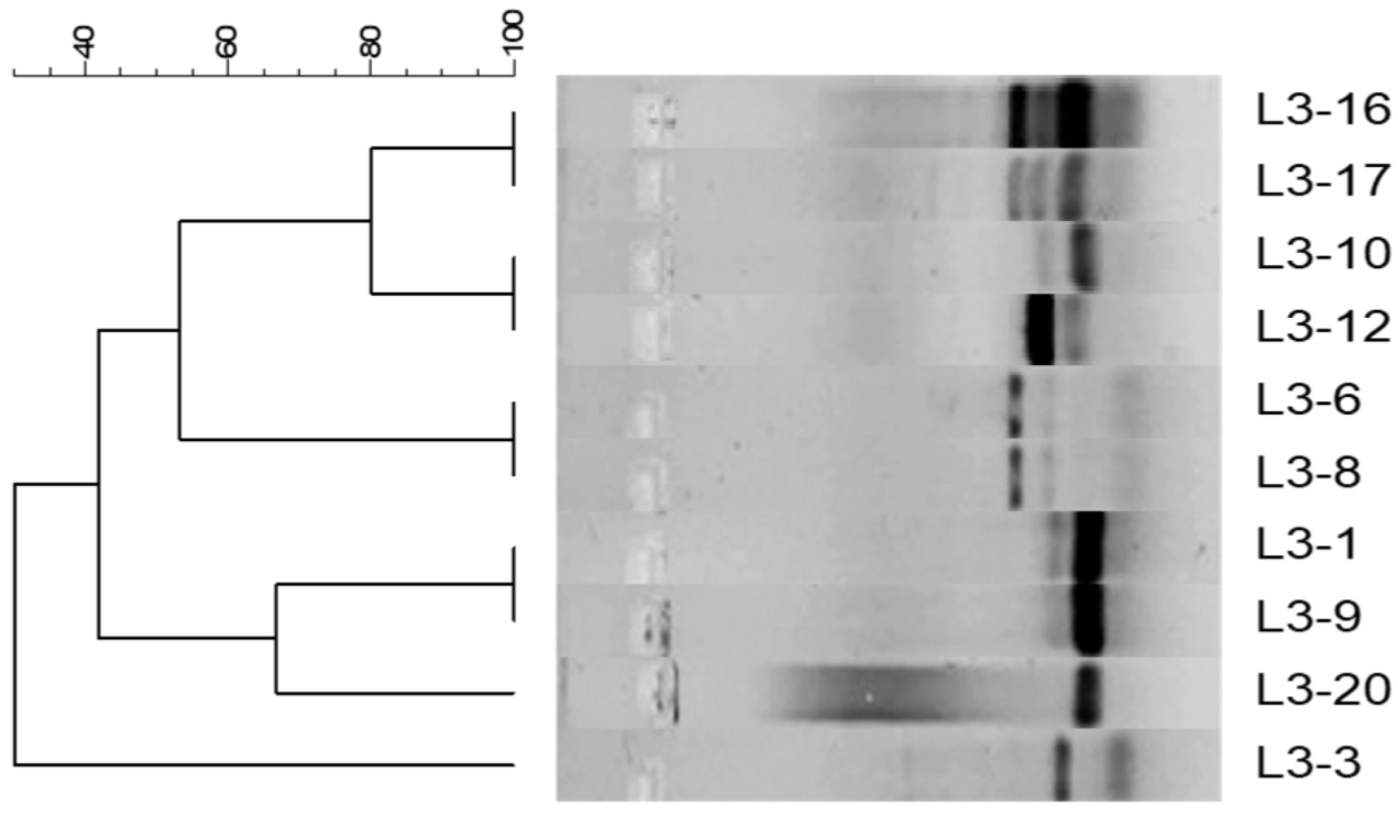
2.3. Genetic Relationship Between Pseudomonas mendocina Isolates

In the analysis of rep-PCR, indistinguishable band patterns can be observed between isolates L3-1 and L3-9, L3-6 and L3-8, and L3-16 and L3-17, indicating that these are clones. Furthermore, L3-20 is closely related to the clones L3-1 and L3-9. Similarly, L3-10 and L3-12 are closely related to L3-16 and L3-17 (Figure 1). Finally, L3-3 shows no relationship with the other isolates (Figure 1).

2.4. Conjugation Assays in Pseudomonas mendocina Isolates with Decreased Antibiotic Sensitivity

Conjugation assays were performed using the following donor strains: L3-12, which exhibited diminished sensitivity to amikacin (Figure 1), and L3-1, which showed reduced sensitivity to imipenem (Table 1). These strains could transfer DNA associated with resistance to the aforementioned antibiotics to the recipient strain E. coli J-53 through the process of conjugation (Table 2). This capability was determined by the growth of transconjugants on Trypticase Soy Agar (TSA) supplemented with rifampicin, amikacin, and imipenem, respectively. In contrast, isolate L3-3 was unable to conjugate with E. coli J-53 under the tested conditions.

2.5. Heavy Metal Susceptibility Profiles of Pseudomonas mendocina Isolates

Minimum Inhibitory Concentrations (MIC) testing of heavy metal salts indicates that 90% (n = 9/10) of the isolates were tolerant to CuSO4, 60% (n = 6/10) to NiSO4, and 40% (n = 4/10) to CdCl2 (Table 3). In addition, 90% (n = 9/10) of the isolates demonstrated tolerance to Pb(NO3)2 comparable to the control strain P. aeruginosa ATCC 27853, while 50% (n = 5/10) were tolerant to ZnSO4 (Table 3). Furthermore, all isolates (n = 10/10) exhibited lower tolerance to CoCl2 compared to the control strain, and none showed tolerance to HgCl2 under the tested conditions (Table 3).

3. Discussion

In this study, ten isolates of non-fermenting Gram-negative bacteria were recovered from landfill leachate and identified as P. mendocina using MALDI-TOF mass spectrometry. To the best of our knowledge, this is the first report of P. mendocina in landfill leachates in Colombia.
The antibiotics tested in this study were selected based on their common use in Colombian healthcare settings for the treatment of Gram-negative infections, including those potentially caused by Pseudomonas spp. Although CLSI guidelines are designed primarily for P. aeruginosa, limited guidance is available for P. mendocina. Therefore, the antibiotics chosen provide a practical and clinically relevant approach for evaluating environmental resistance patterns. According to the results, 20% of the isolates (n = 2/10) exhibited reduced sensitivity to aminoglycosides (amikacin and gentamicin), while 10% (n = 1/10) demonstrated reduced sensitivity to imipenem, a carbapenem (Table 1). These findings align with previous clinical reports. In accordance with the 21 case studies published to date, P. mendocina is capable of causing infections in humans, the most common of which is endocarditis [4,5,6,7,8]. Ioannou et al. [3] reported that, among these 21 cases, resistance to ampicillin was 80%, to cotrimoxazole was 33%, to carbapenems was 10%, and to fourth-generation cephalosporins was 53.3%, while resistance to aminoglycosides was 20%, to quinolones was 33.3%, and to colistin was 6.7%. According to various reports, the most common antibiotics for treating P. mendocina infections are fluoroquinolones and third-generation cephalosporins [3,13,17]. In our study, the isolate with reduced sensitivity to imipenem is noteworthy, as Fang et al. [37] have reported P. mendocina isolates carrying the genes that encode for carbapenemases, NDM-1 and IMP-1, and demonstrated their transferability via plasmid conjugation.
Although the presence of transconjugants in our study suggests HGT of antibiotic resistance determinants, the absence of plasmid extraction or molecular confirmation (e.g., PCR or sequencing) is a limitation. However, the acquisition of reduced susceptibility to β-lactam antibiotics by E. coli J-53, a rifampicin-resistant strain naturally susceptible to β-lactams, supports the hypothesis that resistance genes were mobilized via conjugative plasmids. Moreover, the fact that the transconjugants mirrored the resistant profile of the donor strains further indicates successful horizontal transfer. Future research should include plasmid profiling and molecular characterization to confirm these findings and clarify the underlying mechanisms.
In terms of heavy metal tolerance, 90% (n = 9/10) of isolates were tolerant to CuSO4, 60% (n = 6/10) to NiSO4, and 40% (n = 4/10) to CdCl2 (Table 2). This tolerance is consistent with the metabolic versatility of bacteria belonging to the genus Pseudomonas, which confers an evolutionary advantage in contaminated environments. Consequently, these microorganisms have been proposed as potential agents for the bioremediation of metal-polluted soils. For instance, an isolate of P. mendocina collected from the copper mines of Khetri, in the Jhunjhunu district of Rajasthan, India, demonstrated tolerance to arsenic [38]. Similarly, Carrazca et al. [39] reported arsenic tolerance in P. mendocina originating from mines in Durango, Mexico.
The tolerance to heavy metals is associated with various mechanisms, including biosorption, precipitation of heavy metals, efflux pumps, and the production of chelating ligands and siderophores. In P. mendocina, as in other heavy metal-resistant bacteria [38,39,40], specific genes may be present in the genome that encode proteins responsible for resistance to these metals. These genes can be transferred between bacteria through the process of HGT [40]. However, there are no previous reports of P. mendocina strains exhibiting tolerance to copper sulfate and nickel sulfate; therefore, this study provides the first report of such traits.
When examining resistance to both antibiotics and heavy metals, our results partially align with previous findings. For instance, P. mendocina isolates from the Almendares River in Cuba were reported to be resistant to chromium [41], while others from contaminated soil exhibited tolerance to mercury, lead, and cadmium, and showed potential to resist multiple drugs through efflux pumps [34]. These findings reinforce the role of environmental bacteria as reservoirs of antimicrobial resistance genes, expanding the scope of concern beyond clinical environments.
One of the limitations of this study is the absence of a chemical characterization of the landfill leachate samples. Future studies should include chemical analyses of the leachates to better understand the selective pressures shaping the resistome of environmental bacteria and to strengthen the ecological interpretation of resistance mechanisms.
Although this study demonstrated HGT of antibiotic resistance from P. mendocina to E. coli J-53, no such transfer was observed for heavy metal tolerance. Despite testing donor strains that exhibited phenotypic tolerance to copper, nickel, and cadmium, the recipient strain showed no change in its metal resistance profile after conjugation. This may be due to several factors. First, metal resistance genes may be chromosomally encoded, making them non-transferable by conjugation. Second, even if plasmid-borne, these genes may not be expressed in E. coli due to interspecies regulatory incompatibilities, such as mismatched promoters, transcription factors, or post-transcriptional mechanisms. These barriers to heterologous gene expression across phylogenetically distant species have been documented [42,43]. Therefore, the lack of observable gene transfer does not rule out the potential mobility of heavy metal resistance determinants. Future studies should consider the whole genome or plasmid sequencing to determine the location of resistance genes.
Interestingly, 70% (n = 7/10) of the P. mendocina isolates were phenotypically sensitive to all tested antibiotics; however, several of these exhibited heavy metal tolerances. This is particularly concerning in the context of landfill leachates, which host diverse bacterial populations, including potential pathogens. The presence of mobile resistance genes, even in isolates with low phenotypic resistance, suggests an environmental risk of horizontal gene dissemination. From a One Health perspective, these findings emphasize the importance of monitoring environmental reservoirs of resistance beyond traditional clinical definitions of multidrug resistance.
To further investigate the potential for plasmid-mediated resistance, we performed conjugation experiments with isolates L3-1, L3-3, and L3-12, which showed reduced sensitivity to aminoglycosides and imipenem. This study represents the first report of conjugation between isolates of P. mendocina and E. coli. Such events have significant environmental implications [33], as bacteria can act as reservoirs of resistant genes with the potential to impact environmental, human, and animal health [44,45,46].
Rep-PCR analysis revealed indistinguishable banding profiles between isolates of P. mendocina L3-1 and L3-9, L3-6 and L3-8, and L3-16 and L3-17, indicating that these isolates are clones (Figure 2). Additionally, L3-20 appeared closely related to the clones L3-1 and L3-9, while L3-10 and L3-12 were genetically similar to L3-16 and L3-17 (Figure 2). Despite their high genetic similarity, some of these clones exhibited phenotypic differences in resistance to antibiotics and heavy metals. These differences may be due to the acquisition or loss of MGE, such as plasmids, integrons, and transposons, or point mutations in chromosomal resistance genes [47]. Conversely, other clonal pairs, such as L3-6/L3-8 and L3-16/L3-17, showed concordance at both genotypic and phenotypic levels.
These findings highlight the complexity of resistance dynamics in environmental bacteria and suggest that shared genetic features may result from common selective pressures in similar ecological niches [48]. To date, no previous studies have applied rep-PCR genotyping to P. mendocina. While Pulse Field Gel Electrophoresis (PFGE) remains the gold standard technique for bacterial genotyping, it is more expensive, labor-intensive, and lacks global standardization. Rep-PCR, although less costly, also lacks interlaboratory comparability [49], and no Multi-Locus Sequence Typing (MLST) schemes are currently available for this species. Whole genome sequencing would provide a more robust and comparative analysis, representing a limitation of this study.
In recent years, there has been an increasing number of reports regarding P. mendocina’s impact on human and animal health, indicating that it may be classified as an emergent opportunistic pathogen [50]. Therefore, identification, molecular characterization, and genetic analysis of environmental isolates are essential for understanding the genetic diversity of this species and its potential to disseminate resistance traits. Moreover, deciphering the mechanisms by which these bacteria survive in contaminated environments will be critical for addressing the global challenges of antimicrobial resistance and environmental pollution.

4. Materials and Methods

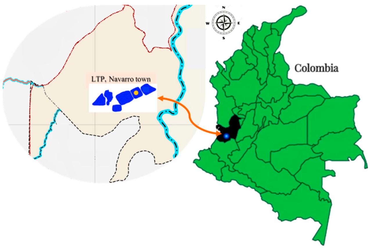
4.1. Sampling and Collection Site of Leachate Sample

This is a descriptive study employing purposive and intentional sampling, along with selective and differential cultivation methods, to promote the growth of presumptive Pseudomonas spp. Leachate samples were collected from Lagoon 3 of a Treatment Plant Landfill (TPL) in the Municipality of Navarro (3°23′13.8″ N, 76°29′7.5″ W), Valle del Cauca, Colombia. The TPL was established in 2008 following the closure of a municipal landfill used for solid waste disposal. It consists of eight lagoons; five are treated using physicochemical methods, while the remaining three, including Lagoon 3, remain untreated (Figure 2).

4.2. Isolation and Identification of Pseudomonas

A total of 300 mL of leachate was collected from Lagoon 3 and filtered in three 100 mL aliquots using cellulose ester membrane (0.45 µm pore membrane; Merck Millipore, Darmstadt, Germany) with vacuum filtration equipment. The membranes were then placed on MacConkey and Cetrimide agar plates (Merck Millipore, Darmstadt, Germany) and incubated at 37 °C for 18–20 h. Colonies with presumptive phenotype of Pseudomonas were subcultured on Trypticase Soy Agar (Merck Millipore, Darmstadt, Germany) for purification. The presumed colonies of Pseudomonas were identified using MALDI-TOF MS (VITEK® MS bioMérieux, Durham, NC, USA), following the manufacturer’s protocol.

4.3. Determination of Antibiotic Susceptibility Profiles in P. mendocina Isolates

Antibiotic susceptibility was determined using the Kirby-Bauer disk diffusion method, in accordance with the Clinical and Laboratory Standards Institute guidelines [51]. Isolates were first subcultured on Mueller-Hinton (MH) agar (bioMérieux, Durham, NC, USA) and suspended in a saline solution (SS, 0.85%) to a turbidity equivalent to 0.5 McFarland. The bacterial suspension was then plated en masse onto MH agar plates using a sterile swab. Antibiotic discs were placed on the agar surface, and plates were incubated at 37 °C for 18 to 20 h.
The antibiotics tested included: Ceftazidime (30 µg), Cefotaxime (30 µg), Ciprofloxacin (5 µg), Ertapenem (10 µg), Imipenem (10 µg), Amikacin (30 µg), and Gentamicin (10 µg) (Oxoid, Basingstoke, UK). The antibiotics represent three major antimicrobial classes, such as β-lactams, fluoroquinolones, and aminoglycosides, and were selected based on their frequent use in Colombia for treating Gram-negative infections. Inhibition zones were measured with a vernier caliper and interpreted according to the following M-100 CLSI guidelines [51]. As no species-specific breakpoints exist for P. mendocina, results were interpreted based on P. aeruginosa standards. The strain P. aeruginosa ATCC 27853 was used as the quality control strain.

4.4. Sensitivity Profile of P. mendocina Isolates to Heavy Metals

The microdilution method was used to evaluate tolerance to heavy metals. Trypticase Soy Broth (TSB; Merck Millipore, Darmstadt, Germany) was supplemented with the following salts: lead nitrate (12.5 mM), cobalt chloride (0.625 mM), zinc sulfate (12.5 mM), nickel sulfate (6.25 mM), copper sulfate (6.25 mM), cadmium chloride (6.25 mM), and mercury (II) chloride (6.25 mM).
Each isolate was grown overnight in 2 mL of TSB at 37 °C. The cellular density was adjusted to 4–6 × 10⁶ cells/mL, and 0.1 mL of this suspension was inoculated into 96-well microplates. An equal volume (0.1 mL) of TSB supplemented with the respective metal salt was added to the wells. Controls include (i) a sterile control (medium + metal only), (ii) a medium control (medium without bacteria or metal), and (iii) a growth control (medium + bacteria, without metal). Microplates were incubated at 37 °C for 24 h. Afterward, 30 μL of resazurin (0.04%) was added to each well and incubated at 37 °C for an additional 4 h. The Minimum Inhibitory Concentration (MIC) was defined as the lowest visible concentration of metal that prevented a color change from blue to pink, indicating inhibited metabolic activity. Due to the absence of established concentrations of universally accepted metals for defining tolerance or resistance, the MIC was defined as the lowest concentration of metal that inhibited visible growth after an incubation period of 72 h at the optimal growth temperature. Pseudomonas aeruginosa ATCC 27853 was used as the control strain, as no control was available for the species P. mendocina.

4.5. DNA Extraction of P. mendocina Isolates

DNA was extracted from P. mendocina isolates using a boiling lysis method. A single colony from TSA was resuspended in 100 μL of sterile distilled water and heated at 100 °C for 10 min. Lysates were centrifuged at 13,000 rpm for 5 min, and the supernatant was used as a DNA template for PCR. The quality of extracted DNA was verified by PCR amplification of the gene encoding the 16S ribosomal RNA subunit using universal primers U1 (5′-CCAGCAGCCGCGGTAATACG-3′) and U2 (5′-ATCGG(C/T)TACCTTGTTACGACTTC-3′) [52].

4.6. Amplification by PCR of Repetitive Extragenic Palindromic Sequences (Rep-PCR)

The genetic relationships among P. mendocina isolates were determined using polymerase chain reaction (PCR) of repetitive extragenic palindromic sequences (rep-PCR). Primers REP1 (5′-IIIGCGCCGICATCAGGC-3′) and REP2 (5′-ACGTCTTATCAGGCCTAC-3′) [53] were used at 0.5 µM each. The PCR mix included 10% DNA template, 100 µL of 2X PCR master mix (CorpoGen, Bogotá, Colombia, OP-TBM-00006) for a final 1X concentration, and ultrapure water to adjust the volume.
PCR was performed in a thermocycler (Eppendorf Mastercycler personal, Z316391, Hamburg, Germany) using the following conditions: initial denaturation at 95 °C for 5 min, followed by 30 cycles of denaturation at 94 °C for 1 min, annealing at 45 °C for 1 min, and extension at 72 °C for 8 min, with a final extension at 72 °C for 10 min [53]. As a negative control for the reaction, the volume of DNA was replaced with sterile water.
The criteria for interpreting the pattern generated by rep-PCR were established by Tenover et al. [54], who defined four categories of genetic relationships and epidemiological significance: (a) Indistinguishable—isolates exhibiting identical band patterns can be considered clones; (b) Closely related—band patterns that differ by two or three bands; (c) Possibly related—patterns showing four to six different bands; and (d) Unrelated—patterns differing by more than six bands. Band profiles were analyzed using Bionumerics software, version 7.5 (Applied Maths NV/Inc., Sint-Martens-Latem, Belgium), employing Dice’s coefficient and the Unweighted Pair Group Method (UPGM) with optimization and tolerance set to 1% to create the dendrogram. The positions of the bands were normalized using the 100 bp Ladder molecular weight marker (New England Biolabs® Inc., Ipswich, MA, USA) as an external reference standard.

4.7. Horizontal Gene Transfer via Conjugation

Conjugation assays were performed to evaluate the horizontal transfer of antibiotic resistance genes. Donor strains P. mendocina L3-1, L3-3, and L3-12 (OD600nm 0.2), which showed reduced sensitivity to aminoglycosides and β-lactams (Table 1), were mixed with recipient strain E. coli J-53 (F met pro RifR) [55] (OD600nm 0.1) at a 1:10 donor-to-recipient ratio in TSB. The mixture was incubated at 37 °C for 24 h. Post-incubation, conjugation mixtures and serial dilutions (in 0.85% SS) were plated on TSA supplemented with rifampicin (25 μg/mL) and one of the following: amikacin (100 μg/mL), gentamicin (7 μg/mL), or imipenem (1 μg/mL), to select and quantify transconjugants. Plates were incubated at 37 °C for 18-20 h, after which we assessed the growth of transconjugants. As positive control, E. coli 1646 carrying a conjugative plasmid with blaKPC-2 gene (conferring resistance to β-lactams) was included.
The frequency of transfer (FT) was calculated as follows:
FT = Titre of transconjugants Titre of donnors

5. Conclusions

This study demonstrates that leachates serve as a potential reservoir for P. mendocina, some of which exhibit reduced sensitivity to antibiotics and tolerance to heavy metals. This is the first report in Colombia to characterize P. mendocina isolates from leachates in terms of their antibiotic susceptibility, metal tolerance, and genetic features. Although the overall frequency of resistance, the presence of genetically related strains harboring conjugative plasmids highlights the potential for HGT. These findings underscore the need to monitor microbial communities in waste environments, as they may contribute to the environmental dissemination of resistance genes. The results may inform future surveillance strategies and environmental policies aimed at mitigating the spread of antimicrobial resistance.

Author Contributions

Conceptualization, C.A., A.C. and A.F.; methodology, A.M.-Q., L.B., M.Á.V.-M., C.A., A.C. and A.F.; formal analysis, A.M.-Q., L.B., M.Á.V.-M., C.A., A.C. and A.F.; investigation, A.M.-Q., L.B., M.Á.V.-M., C.A., A.C. and A.F.; resources, C.A., A.C. and A.F.; data curation, C.A. and A.F.; writing—original draft preparation, A.M.-Q. and A.F.; writing—review and editing, C.A. and A.F.; supervision, C.A., A.C. and A.F.; project administration: C.A., A.C. and A.F.; funding acquisition, C.A., A.C. and A.F. All authors have read and agreed to the published version of the manuscript.

Funding

This research was funded by the Dirección General de Investigaciones, Universidad Santiago de Cali, Colombia, under grants numbers 934-621119-E04 and 934-621121-3047 and under call No. 06-2023 (Jóvenes investigadores santiaguinos) and No. 01-2025.

Institutional Review Board Statement

Not applicable.

Informed Consent Statement

Not applicable.

Data Availability Statement

The data used to support the findings of this study are included within the article and are available from the corresponding author upon request.

Acknowledgments

The authors thank Doris Rosero and the Empresa de Servicio Público de Aseo de Cali en Liquidación (EMSIRVA E.S.P.) for authorizing the entry into the landfill leachates plan.

Conflicts of Interest

The authors declare no conflicts of interest. The funders had no role in the design of the study; in the collection, analysis, or interpretation of data; in the writing of the manuscript; or in the decision to publish the results.

References

  1. Ruiz-Roldán, L.; de Toro, M.; Sáenz, Y. The Whole Genome Analysis of Environmental Pseudomonas mendocina Strains: Virulence Mechanisms and Phylogeny. Genes 2021, 12, 115. [Google Scholar] [CrossRef]
  2. Palleroni, N.J.; Doudoroff, M.; Stanier, R.Y.; Solánes, R.E.; Mandel, M. Taxonomy of the aerobic pseudomonads: The properties of the Pseudomonas stutzeri group. J. Gen. Microbiol. 1970, 60, 215–231. [Google Scholar] [CrossRef]
  3. Ioannou, P.; Vougiouklakis, G. A Systematic Review of Human Infections by Pseudomonas mendocina. Trop. Med. Infect. Dis. 2020, 5, 71. [Google Scholar] [CrossRef]
  4. Rapsinski, G.J.; Makadia, J.; Bhanot, N.; Min, Z. Pseudomonas mendocina native valve infective endocarditis: A case report. J. Med. Case Rep. 2016, 10, 275. [Google Scholar] [CrossRef]
  5. Aragone, M.R.; Maurizi, D.M.; Clara, L.O.; Navarro Estrada, J.L.; Ascione, A. Pseudomonas mendocina, an environmental bacterium isolated from a patient with human infective endocarditis. J. Clin. Microbiol. 1992, 30, 1583–1584. [Google Scholar] [CrossRef]
  6. Johansen, H.K.; Kjeldsen, K.; Høiby, N. Pseudomonas mendocina as a cause of chronic infective endocarditis in patients with situs inversus. Clin. Microbiol. Infect. Off. Publ. Eur. Soc. Clin. Microbiol. Infect. Dis. 2001, 7, 650–652. [Google Scholar] [CrossRef]
  7. Mert, A.; Yilmaz, M.; Ozaras, R.; Kocak, F.; Dagsali, S. Native valve endocarditis due to Pseudomonas mendocina in a patient with mental retardation and a review of literature. Scand. J. Infect. Dis. 2007, 39, 615–616. [Google Scholar] [CrossRef] [PubMed]
  8. Suel, P.; Martin, P.; Berthelot, G.; Robaday, S.; Etienne, M.; Chibani, A. Une endocardite à Pseudomonas mendocina [A case of Pseudomonas mendocina endocarditis]. Med. Mal. Infect. 2011, 41, 109–110. [Google Scholar] [CrossRef] [PubMed]
  9. Thamizhselvi, S.; Pooja, A.; Prajna, L.; Rameshkumar, G.; Prajna, N.V.; Karpagam, R. Incidence, Clinical Profile, and Management of Keratitis Caused by Uncommon Species of Pseudomonas at a Tertiary Eye Care Center. Cornea 2023, 42, 359–364. [Google Scholar] [CrossRef]
  10. Huang, C.R.; Lien, C.Y.; Tsai, W.C.; Lai, W.A.; Hsu, C.W.; Tsai, N.W.; Chang, C.C.; Lu, C.H.; Chien, C.C.; Chang, W.N. The clinical characteristics of adult bacterial meningitis caused by non-Pseudomonas (Ps.) aeruginosa Pseudomonas species: A clinical comparison with Ps. aeruginosa meningitis. Kaohsiung J. Med. Sci. 2018, 34, 49–55. [Google Scholar] [CrossRef] [PubMed]
  11. Vo, T.; Maisuradze, N.; Maglakelidze, D.; Kalra, T.; McFarlane, I.M. Pseudomonas mendocina Urinary Tract Infection: A Case Report and Literature Review. Cureus 2022, 14, e23583. [Google Scholar] [CrossRef]
  12. Nseir, W.; Taha, H.; Abid, A.; Khateeb, J. Pseudomonas mendocina sepsis in a healthy man. Isr. Med. Assoc. J. IMAJ 2011, 13, 375–376. [Google Scholar]
  13. Gani, M.; Rao, S.; Miller, M.; Scoular, S. Pseudomonas mendocina Bacteremia: A Case Study and Review of Literature. Am. J. Case Rep. 2019, 20, 453–458. [Google Scholar] [CrossRef]
  14. Goldberg, M.E.; Blyth, M.; Swiatlo, E. Pseudomonas mendocina Bacteremia in a Hemodialysis Patient With a Central Venous Catheter. Cureus 2020, 12, e10853. [Google Scholar] [CrossRef] [PubMed]
  15. Ezeokoli, E.U.; Polat, M.U.; Ogundipe, O.; Szela, J. A Case of Pseudomonas mendocina Bacteremia in an Elderly Man With Bilateral Leg Lesions. Cureus 2021, 13, e17777. [Google Scholar] [CrossRef] [PubMed]
  16. Chi, C.Y.; Lai, C.H.; Fung, C.P.; Wang, J.H. Pseudomonas mendocina spondylodiscitis: A case report and literature review. Scand. J. Infect. Dis. 2005, 37, 950–953. [Google Scholar] [CrossRef] [PubMed]
  17. Jerónimo, T.M.; Guedes, A.M.; Stieglmair, S.; Guerreiro, R.; Laranjo, C.; Bernardo, I.; Neves, P.L. Pseudomonas mendocina: The first case of peritonitis on peritoneal dialysis. Nefrol. Publicación Of. Soc. Española Nefrol. 2017, 37, 647–649. [Google Scholar] [CrossRef]
  18. Bhatt, V.D.; Ahir, V.B.; Koringa, P.G.; Jakhesara, S.J.; Rank, D.N.; Nauriyal, D.S.; Kunjadia, A.P.; Joshi, C.G. Milk microbiome signatures of subclinical mastitis-affected cattle analysed by shotgun sequencing. J. Appl. Microbiol. 2012, 112, 639–650. [Google Scholar] [CrossRef]
  19. Lee, S.A.; Wrona, L.J.; Cahoon, A.B.; Crigler, J.; Eiteman, M.A.; Altman, E. Isolation and Characterization of Bacteria That Use Furans as the Sole Carbon Source. Appl. Biochem. Biotechnol. 2016, 178, 76–90. [Google Scholar] [CrossRef]
  20. Roy, D.; Azaïs, A.; Benkaraache, S.; Drogui, P.; Tyagi, R. Composting leachate: Characterization, treatment, and future perspectives. Rev. Environ. Sci. Biotechnol. 2018, 17, 323–349. [Google Scholar] [CrossRef]
  21. Del Moro, G.; Prieto-Rodríguez, L.; De Sanctis, M.; Di Iaconi, C.; Malato, S.; Mascolo, G. Landfill leachate treatment: Comparison of standalone electrochemical degradation and combined with a novel biofilter. Chem. Eng. J. 2016, 288, 87–98. [Google Scholar] [CrossRef]
  22. Morales, J.P. Análisis Hidrológico Del Lixiviado Generado En El Relleno Sanitario Del Cantón Gonzalo Pizarro, Ecuador. Rev. Cienc. UNEMI 2022, 15, 24–33. [Google Scholar]
  23. Moody, C.M.; Townsend, T.G. A comparison of landfill leachates based on waste composition. Waste Manag. 2017, 63, 267–274. [Google Scholar] [CrossRef]
  24. Wu, D.; Huang, Z.; Yang, K.; Graham, D.; Xie, B. Relationships between antibiotics and antibiotic resistance gene levels in municipal solid waste leachates in Shanghai, China. Environ. Sci. Technol. 2015, 49, 4122–4128. [Google Scholar] [CrossRef]
  25. You, X.; Wu, D.; Wei, H.; Xie, B.; Lu, J. Fluoroquinolones and β-lactam antibiotics and antibiotic resistance genes in autumn leachates of seven major municipal solid waste landfills in China. Environ. Int. 2018, 113, 162–169. [Google Scholar] [CrossRef] [PubMed]
  26. Threedeach, S.; Chiemchaisri, W.; Watanabe, T.; Chiemchaisri, C.; Honda, R.; Yamamoto, K. Antibiotic resistance of Escherichia coli in leachates from municipal solid waste landfills: Comparison between semi-aerobic and anaerobic operations. Bioresour. Technol. 2012, 113, 253–258. [Google Scholar] [CrossRef] [PubMed]
  27. Zhang, W.; Yan, C.; Shen, J.; Wei, R.; Gao, Y.; Miao, A.; Xiao, L.; Yang, L. Characterization of Aerobic Denitrifying Bacterium Pseudomonas mendocina Strain GL6 and Its Potential Application in Wastewater Treatment Plant Effluent. Int. J. Environ. Res. Public Health 2019, 16, 364. [Google Scholar] [CrossRef]
  28. Kunhikannan, S.; Thomas, C.J.; Sumana, M.N.; Franks, A.E.; Kumar, S.; Nagarathna, S.; Petrovski, S.; Shindler, A.E. Exploring the antibiogram of soil isolates from an indian hospital precinct: Link to antibiotic usage. BMC Res. Notes 2023, 16, 173. [Google Scholar] [CrossRef]
  29. Mondragón-Quiguanas, A.; Villaquirán-Muriel, M.Á.; Rivera, S.P.; Rosero-García, D.; Aranaga, C.; Correa, A.; Falco, A. β-Lactam-Resistant Enterobacterales Isolated from Landfill Leachates. Pathogens 2022, 11, 1077. [Google Scholar] [CrossRef]
  30. Falco, A.; Villaquirán-Muriel, M.Á.; Gallo Pérez, J.D.; Mondragón-Quiguanas, A.; Aranaga, C.; Correa, A. Identification of Vibrio metschnikovii and Vibrio injensis Isolated from Leachate Ponds: Characterization of Their Antibiotic Resistance and Virulence-Associated Genes. Antibiotics 2023, 12, 1571. [Google Scholar] [CrossRef]
  31. Toba, F.; Falco, A.; Aranaga, C.; Alonso, G. Caracterización de bacterias aisladas en un reservorio de agua en Venezuela. In Una Aproximación a la Multirresistencia Bacteriana en Ambientes Naturales; Editorial Universidad Santiago de Cali: Cali, Colombia, 2018; pp. 93–120. [Google Scholar]
  32. Su, Y.; Wang, J.; Huang, Z.; Xie, B. On-site removal of antibiotics and antibiotic resistance genes from leachate by aged refuse bioreactor: Effects of microbial community and operational parameters. Chemosphere 2017, 178, 486–495. [Google Scholar] [CrossRef]
  33. Gorovtsov, A.V.; Sazykin, I.S.; Sazykina, M.A. The influence of heavy metals, polyaromatic hydrocarbons, and polychlorinated biphenyls pollution on the development of antibiotic resistance in soils. Environ. Sci. Pollut. Res. Int. 2018, 25, 9283–9292. [Google Scholar] [CrossRef]
  34. Chong, T.M.; Yin, W.F.; Mondy, S.; Grandclément, C.; Dessaux, Y.; Chan, K.G. Heavy-metal resistance of a France vineyard soil bacterium, Pseudomonas mendocina strain S5.2, revealed by whole-genome sequencing. J. Bacteriol. 2012, 194, 6366. [Google Scholar] [CrossRef] [PubMed]
  35. Ji, X.; Shen, Q.; Liu, F.; Ma, J.; Xu, G.; Wang, Y.; Wu, M. Antibiotic resistance gene abundances associated with antibiotics and heavy metals in animal manures and agricultural soils adjacent to feedlots in Shanghai, China. J. Hazard. Mater. 2012, 235–236, 178–185. [Google Scholar] [CrossRef]
  36. A Health Perspective on the Role of the Environment in One Health; WHO Regional Office for Europe: Copenhagen, Denmark, 2022. Available online: https://iris.who.int/bitstream/handle/10665/354574/WHO-EURO-2022-5290-45054-64214-eng.pdf (accessed on 18 May 2025).
  37. Fang, J.; Liu, Q.; Yang, J.; Kang, X.; Mei, Y.; Liu, J.; Wang, G.; Xiang, T. Functional Portrait and Genomic Feature of Carbapenem-Resistant Pseudomonas mendocina Harboring blaNDM-1 and blaIMP-1 in China. Foodborne Pathog. Dis. 2023, 20, 502–508. [Google Scholar] [CrossRef] [PubMed]
  38. Mishra, S.; Kumar, S.; Verma, S.K. Arsenic Resistance Mechanisms in Pseudomonas mendocina SMSKVR-3 Strain Isolated from Khetri Copper Mines, Rajasthan, India. Curr. Microbiol. 2022, 79, 69. [Google Scholar] [CrossRef]
  39. Miranda-Carrazco, A.; Vigueras-Cortés, J.M.; Villa-Tanaca, L.; Hernández-Rodríguez, C. Cyanotrophic and arsenic oxidizing activities of Pseudomonas mendocina P6115 isolated from mine tailings containing high cyanide concentration. Arch. Microbiol. 2018, 200, 1037–1048. [Google Scholar] [CrossRef]
  40. Gullberg, E.; Albrecht, L.M.; Karlsson, C.; Sandegren, L.; Andersson, D.I. Selection of a multidrug resistance plasmid by sublethal levels of antibiotics and heavy metals. mBio 2014, 5, e01918-14. [Google Scholar] [CrossRef]
  41. Carballo, M.; Heydrich, M.; Rojas, N.; Salgado, I.; Romeu, B.; Manzano, A.; Larrea, J.; Dominguez, O.; Martínez, A.; Sánchez, M.; et al. Impact of microbial and chemical pollution in Cuban freshwater ecosystems: Strategies for environmental recovery. Biotecnol. Apl. 2011, 28, 276–279. [Google Scholar]
  42. Klümper, U.; Riber, L.; Dechesne, A.; Sannazzarro, A.; Hansen, L.H.; Sørensen, S.J.; Smets, B.F. Broad host range plasmids can invade an unexpectedly diverse fraction of a soil bacterial community. ISME J. 2015, 9, 934–945. [Google Scholar] [CrossRef]
  43. Rankin, D.J.; Rocha, E.P.C.; Brown, S.P. What traits are carried on mobile genetic elements, and why? Heredity 2011, 106, 1–10. [Google Scholar] [CrossRef]
  44. Medina-Pizzali, M.L.; Hartinger, S.M.; Salmon-Mulanovich, G.; Larson, A.; Riveros, M.; Mäusezahl, D. Antimicrobial Resistance in Rural Settings in Latin America: A Scoping Review with a One Health Lens. Int. J. Environ. Res. Public Health 2021, 18, 9837. [Google Scholar] [CrossRef]
  45. Brisola, M.C.; Crecencio, R.B.; Bitner, D.S.; Frigo, A.; Rampazzo, L.; Stefani, L.M.; Faria, G.A. Escherichia coli used as a biomarker of antimicrobial resistance in pig farms of Southern Brazil. Sci. Total Environ. 2019, 647, 362–368. [Google Scholar] [CrossRef] [PubMed]
  46. Gambero, M.L.; Blarasin, M.; Bettera, S.; Albo, J.G. Tracing contamination sources through phenotypic characterization of Escherichia coli isolates from surface water and groundwater in an agro-ecosystem. Hydrol. Sci. J. 2018, 63, 1150–1161. [Google Scholar] [CrossRef]
  47. Vilchez, G.; Alonso, G. Alcances y limitaciones de los métodos de epidemiología molecular basados en el análisis de ácidos nucleicos. Rev. Soc. Ven. Microbiol. 2009, 29, 6–12. [Google Scholar]
  48. Samreen Ahmad, I.; Malak, H.A.; Abulreesh, H.H. Environmental antimicrobial resistance and its drivers: A potential threat to public health. J. Glob. Antimicrob. Resist. 2021, 27, 101–111. [Google Scholar] [CrossRef]
  49. Strandén, A.; Frei, R.; Widmer, A.F. Molecular typing of methicillin-resistant Staphylococcus aureus: Can PCR replace pulsed-field gel electrophoresis? J. Clin. Microbiol. 2003, 41, 3181–3186. [Google Scholar] [CrossRef]
  50. Aman, M.; Fomda, B.A.; Roohi, S.; Qadri, U.; Wani, S.J.; Majid, U.; Dar, R.A. Pseudomonas mendocina bacteremia: A case study from Indian subcontinent. Indian J. Med. Microbiol. 2025, 55, 100827. [Google Scholar] [CrossRef]
  51. Clinical and Laboratory Standards Institute. Performance Standards for Antimicrobial Susceptibility Testing: 34th Edition Informational Supplement M100-S27; Clinical and Laboratory Standards Institute: Wayne, PA, USA, 2024. [Google Scholar]
  52. Lu, J.J.; Perng, C.L.; Lee, S.Y.; Wan, C.C. Use of PCR with universal primers and restriction endonuclease digestions for detection and identification of common bacterial pathogens in cerebrospinal fluid. J. Clin. Microbiol. 2000, 38, 2076–2080. [Google Scholar] [CrossRef]
  53. Versalovic, J.; Koeuth, T.; Lupski, J.R. Distribution of repetitive DNA sequences in eubacteria and application to fingerprinting of bacterial genomes. Nucleic Acids Res. 1991, 19, 6823–6831. [Google Scholar] [CrossRef] [PubMed]
  54. Tenover, F.C.; Arbeit, R.D.; Goering, R.V.; Mickelsen, P.A.; Murray, B.E.; Persing, D.H.; Swaminathan, B. Interpreting chromosomal DNA restriction patterns produced by pulsed-field gel electrophoresis: Criteria for bacterial strain typing. J. Clin. Microbiol. 1995, 33, 2233–2239. [Google Scholar] [CrossRef] [PubMed]
  55. Yi, H.; Cho, Y.J.; Yong, D.; Chun, J. Genome sequence of Escherichia coli J53, a reference strain for genetic studies. J. Bacteriol. 2012, 194, 3742–3743. [Google Scholar] [CrossRef] [PubMed]
Figure 1. Rep-PCR profiles of ten P. mendocina isolates obtained from a leachate pond in Cali, Colombia. The band patterns were analyzed using Bionumerics (version 7.5, Applied Maths NV/Inc., Sint-Martens-Latem, Belgium), employing the binary coefficient Dice and the Unweighted Pair Group Method (UPGM) with adjustments for tolerance and optimization set at 1% to create the dendrogram. The positions of the bands were normalized using the 100 bp Ladder molecular weight marker (New England Biolabs® Inc., Ipswich, MA, USA) as an external reference standard.
Antibiotics 14 00781 g001
Figure 2. The location of the leachate treatment plant (LTP) in Navarro town, Santiago de Cali municipality (blue dot), Valle del Cauca department Table (black), and the sampling location: Lagoon 3 (yellow dot).
Antibiotics 14 00781 g002
Table 1. Antimicrobial susceptibility profiles of Pseudomonas mendocina.
Kirby-Bauer Test (μg/mL) by Antibiotic/Interpretive Categories
Strain IDAK 1CN 1CAZ 2IMP 2CTX 2ETP 2CIP 3
L3-1≤2 (S)≤1 (S)≤1 (S)16 (I)≤1 (S)≤1 (S)≤0.25 (S)
L3-3≤2 (S)14 (I)≤1 (S)≤0.25 (S)≤1 (S)≤1 (S)≤0.25 (S)
L3-6≤2 (S)2 (S)2 (S)0.5 (S)2 (S)2 (S)≤0.25 (S)
L3-8≤2 (S)≤1 (S)2 (S)≤0.25 (S)2 (S)2 (S)≤0.25 (S)
L3-9≤2 (S)≤1 (S)2 (S)≤0.25 (S)2 (S)2 (S)≤0.25 (S)
L3-10≤2 (S)≤1 (S)2 (S)≤0.5 (S)2 (S)2 (S)≤0.25 (S)
L3-1216 (I)≤1 (S)≤1 (S)≤0.5 (S)≤1 (S)≤1 (S)≤0.25 (S)
L3-16≤2 (S)≤1 (S)≤1 (S)0.5 (S)≤1 (S)≤1 (S)≤0.25 (S)
L3-17≤2 (S)≤1 (S)≤1 (S)0.5 (S)≤1 (S)≤1 (S)≤0.25 (S)
L3-20≤2 (S)≤1 (S)≤1 (S)≤0.25 (S)≤1 (S)≤1 (S)≤0.25 (S)
Abbreviations: R: Susceptible; I: Intermediate; R: Resistant; Ak: Amikacin; CN: Gentamicin; ETP: Ertapenem; CTX: Cefotaxime; CAZ: Ceftazidime; IMP: Imipenem; CIP: Ciprofloxacin; 1 Aminoglycosides; 2 β-lactams; 3 Fluoroquinolones.
Table 2. Donor and Recipient Strain Titers and Transfer Frequency.
Titers of Donor and Recipient Strains and Frequency of Transfer
StrainTransfer Frequency
E. coli J-53 (recipient)Does not apply
E. coli 1646 (donor, control)0.53
P. mendocina L3-1 (donor)6.4 × 10⁴
P. mendocina L3-3 (donor)There were no transconjugants
P. mendocina L3-12 (donor)0.03
Table 3. Minimum Inhibitory Concentration (MIC) of Heavy Metals in P. mendocina Isolates.
Minimum Inhibitory Concentrations for Metals (mM)
StrainPb(NO2)3CoCl2ZnSO4NiSO4CuSO4CdCl2HgCl2
P. aeruginosa ATCC 27853>506.25>12.5>6.250.19>6.256.25
L3-1>502.512.512.50.196.25N/G
L3-3252.512.512.5>3.1251.56N/G
L3-6>502.5>12.512.51.56>6.25N/G
L3-8>502.5>12.56.251.56>6.25N/G
L3-9>502.5>12.56.250.396.25N/G
L3-10>502.56.256.25>3.1256.25N/G
L3-12>502.53.1253.125>3.1256.25N/G
L3-16>5056.12512.5>3.125>6.25N/G
L3-17>505>6.2512.5>3.125>6.25N/G
L3-20>505>6.2512.5>3.1256.25N/G
Abbreviations: MIC, minimum inhibitory concentration; N/G, No growth; Pa, Pseudomonas aeruginosa; Pb(NO2)3, Lead nitrate; CoCl2, Cobalt chloride; ZnSO4, Zinc sulphate; NiSO4, Nickel sulphate; CuSO4, Copper sulphate; CdCl2, Cadmium chloride; HgCl2, Mercuric chloride.
Disclaimer/Publisher’s Note: The statements, opinions and data contained in all publications are solely those of the individual author(s) and contributor(s) and not of MDPI and/or the editor(s). MDPI and/or the editor(s) disclaim responsibility for any injury to people or property resulting from any ideas, methods, instructions or products referred to in the content.

