Article

Cellulose Nanocrystal/Zinc Oxide Bio-Nanocomposite Activity on Planktonic and Biofilm Producing Pan Drug-Resistant Clostridium perfringens Isolated from Chickens and Turkeys

1
Microbiology and Parasitology Department, Faculty of Veterinary Medicine, Badr University in Cairo (BUC), Badr City 11829, Egypt
2
Food Safety, Hygiene and Technology Department, Faculty of Veterinary Medicine, Badr University in Cairo (BUC), Badr City 11829, Egypt
3
Laboratory of Biotechnology, Department of Microbiology, Agricultural Research Center (ARC), Animal Health Research Institute (AHRI), Zagazig 44516, Egypt
4
Department of Biotechnology, Faculty of Bioengineering of Animals Resources, University of Life Sciences “King Mihai I” from Timisoara, 300645 Timisoara, Romania
5
Department of Microbiology, Faculty of Veterinary Medicine, Zagazig University, Zagazig 44511, Egypt
*
Authors to whom correspondence should be addressed.
Antibiotics 2025, 14(6), 575; https://doi.org/10.3390/antibiotics14060575
Submission received: 17 April 2025 / Revised: 23 May 2025 / Accepted: 27 May 2025 / Published: 3 June 2025
(This article belongs to the Special Issue Antimicrobial and Antibiofilm Activity by Natural Compounds)

Abstract

Background/Objectives: Clostridium perfringens is a normal inhabitant of the intestinal tract of poultry, and it has the potential to induce cholangiohepatitis and necrotic enteritis (NE). The poultry industry suffers significant financial losses because of NE, and treatment becomes more challenging due to resistant C. perfringens strains. Methods: The antimicrobial and antibiofilm activities of cellulose nanocrystals/zinc oxide nanocomposite (CNCs/ZnO) were assesses against pan drug-resistant (PDR) C. perfringens isolated from chickens and turkeys using phenotypic and molecular assays. Results: The overall prevalence rate of C. perfringens was 44.8% (43.75% in chickens and 58.33% in turkeys). Interestingly, the antimicrobial susceptibility testing of C. perfringens isolates revealed the alarming PDR (29.9%), extensively drug-resistant (XDR, 54.5%), and multidrug-resistant (MDR, 15.6%) isolates, with multiple antimicrobial resistance (MAR) indices ranging from 0.84 to 1. All PDR C. perfringens isolates could synthesize biofilms; among them, 21.7% were strong biofilm producers. The antimicrobial potentials of CNCs/ZnO against PDR C. perfringens isolates were evaluated by the agar well diffusion and broth microdilution techniques, and the results showed strong antimicrobial activity of the green nanocomposite with inhibition zones’ diameters of 20–40 mm and MIC value of 0.125 µg/mL. Moreover, the nanocomposite exhibited a great antibiofilm effect against the pre-existent biofilms of PDR C. perfringens isolates in a dose-dependent manner [MBIC50 up to 83.43 ± 1.98 for the CNCs/ZnO MBC concentration (0.25 μg/mL)]. The transcript levels of agrB quorum sensing gene and pilA2 type IV pili gene responsible for biofilm formation were determined by the quantitative real time-PCR technique, pre- and post-treatment with the CNCs/ZnO nanocomposite. The expression of both genes downregulated (0.099 ± 0.012–0.454 ± 0.031 and 0.104 ± 0.006–0.403 ± 0.035, respectively) when compared to the non-treated isolates. Conclusions: To the best of our knowledge, this is the first report of CNCs/ZnO nanocomposite’s antimicrobial and antibiofilm activities against PDR C. perfringens isolated from chickens and turkeys.

1. Introduction

One of the most important parts of the agricultural production system is the poultry industry [1]. Even though there is always a chance that broiler chickens can contract infections, particularly those caused by microorganisms typically present in their guts, they can be reduced by improved management and proper information on infectious poultry diseases [2]. There are numerous etiological agents causing enteric diseases in chickens, one of which is Clostridium species [3]. Clostridium perfringens (C. perfringens) is a Gram-positive, anaerobic, spore-forming bacterium that is a natural component of the poultry gut microbiota [4]. Commonly, C. perfringens is present in the intestines of healthy chickens in concentrations of less than 102–104 CFU/g of intestinal content, as opposed to 107–109 CFU/g in diseased birds [5].
The presence of approximately twenty different extracellular enzymes and toxins is the basis for C. perfringens pathogenicity. The microorganism has been classified into seven toxinotypes, namely A to G, based on the presence of major toxin genes: cpa (alpha), cpb (beta), etx (epsilon), iap (iota), cpe (enterotoxin), and netB (necrotic enteritis B-like toxin) [6,7]. Necrotic enteritis (NE) is a bacterial disease caused by strains of C. perfringens [4]. The disease is brought on by an excessive rise in the number of C. perfringens bacteria in the gastrointestinal tract (GIT) in combination with risk factors such as exposure to mycotoxins, diets high in non-starch polysaccharide grains, and coccidia infection [8]. Both clinical and subclinical forms of NE can cause high mortality rates and growth performance failures, respectively. The estimated yearly cost of NE is over 6 billion USD, or 0.05 USD per chick [4]. Control strategies for NE involve the use of antimicrobials [9]. The prolonged and extensive use of antimicrobials in poultry over the past few years has altered the bacterial environment, eliminated susceptible strains, and promoted the persistence and dominance of antimicrobial-resistant bacteria [10,11].
Biofilms can be described as the houses and cities of the bacterial world [12]. They are aggregates of microorganisms in which extracellular polymeric substances (EPS) adhere to a surface, and/or each other’s cells are frequently embedded in a self-produced matrix [13] that undergo stages of accumulation, attachment, maturation, and dispersal [14]. Bacteria can adhere to substrates and encapsulate themselves in a matrix consisting of extracellular DNA (eDNA), proteins, and EPS, when they form biofilms [15,16]. The antibiotic sensitivity of bacterial cells in biofilms is lower than that of their planktonic counterparts [16], thus, increasing the resistance to antimicrobial drugs and reducing the effectiveness of biofilm-related infection treatment. Several investigations have revealed that biofilm-producing bacteria have a resistance potential that is more than 1000 times higher than that of planktonic ones [17,18]. Antibiotic resistance arises from the slower penetration of antimicrobial drugs due to the formation of the biofilm matrix, which in turn leads to the establishment of persistent cells, a resistant phenotype [18]. Another potential mechanism for antibiotic resistance in biofilms is the upregulation of bacterial efflux pumps [19,20].
The emergence of antibiotic-resistant bacterial strains and their transmission to humans can threaten food safety and public health. Therefore, there is an urgent need to explore natural products as alternatives to antibiotics for the poultry industry.
Copper (Cu), zinc (Zn), and selenium (Se) nanometals have strong antibacterial and antibiofilm activities, which makes them viable options for treating bacterial infections, particularly those are resistant to antibiotics. Each of these nanometals exhibits distinct processes and degrees of efficiency, and their effectiveness against a range of bacteria, including both Gram-positive and Gram-negative species, has been investigated [21,22,23,24].
The most prevalent polymer on Earth is cellulose (Cs), which is also a key part of plant cell walls. Nanocellulose, also known as nanostructured cellulose, combines the characteristics of Cs and nanomaterials [25]. Now, turning our attention to ZnO nanoparticles, which are recognized for their stability and antibacterial properties, are useful in food engineering and other nanotechnology applications [26]. By inhibiting the in vitro bacterial growth and concentrating on bacterial toxin manufacture, the dual-target therapy of new cellulose nanocrystals/zinc oxide (CNCs/ZnO) bio-nanocomposites improves bacterial treatment and lowers the emergence of antibiotic resistance [27]. To the best of our knowledge, no studies have evaluated the antimicrobial and antibiofilm activities of CNCs/ZnO nanocomposite against PDR C. perfringens isolated from poultry by phenotypic and molecular assays.

2. Results

2.1. Prevalence of C. Perfringens in Poultry Samples

As shown in Table 1, the overall prevalence rate of C. perfringens was 44.8% (77/172). In broilers, C. perfringens isolates were detected with a percentage of 44.4% (55/124), with a higher existence in the intestine (25/124; 20.2%), followed by the liver (15/124; 12.1%) and spleen (15/124; 12.1%). While in layers, 41.6% (15/36) were positive for C. perfringens with a higher existence in the spleen (6/36; 16.6%) followed by liver (5/36; 13.8%) and intestine (4/36; 11.1%). On the other hand, 7 out of 12 examined turkeys (58.3%) had C. perfringens isolates with a higher existence in the liver (5/12; 41.6%) followed by the intestine (2/12; 16.6%), and no presence of C. perfringens in spleen. Statistical analysis revealed non significant difference in the prevalence of C. perfringens among liver, spleen, and intestine samples isolated from broiler chickens and layers (p = 0.122 and 1, respectively), while there was a statistically significant difference in the prevalence of C. perfringens among liver, spleen, and intestine samples isolated from turkeys (p = 0.046). Additionally, there was no statistically significant difference in the prevalence of C. perfringens among total broiler, layer, and turkey samples (p = 0.608).

2.2. Antibiogram of C. perfringens Isolates

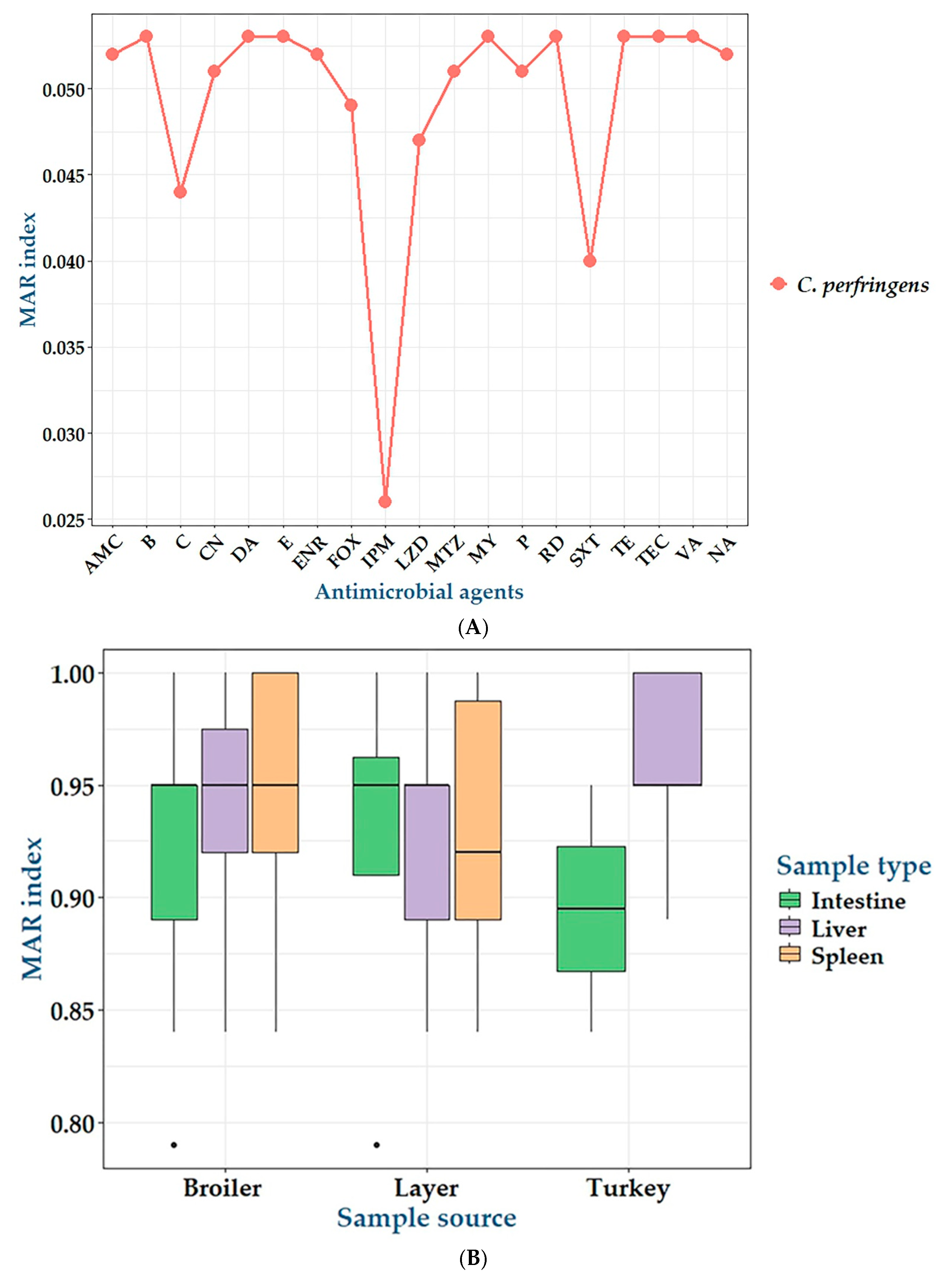
The in vitro antimicrobial susceptibilities of 172 C. perfringens isolates against 19 antimicrobial agents are summarized in Table 2 and Figure 1. The antimicrobial susceptibility testing revealed that C. perfringens isolates exhibited complete resistance to erythromycin, bacitracin, clindamycin, tetracycline, rifampin, vancomycin, teicoplanin, and lincomycin. Moreover, high levels of resistance were detected for amoxycillin-clavulanic acid, nalidixic acid, and ciprofloxacin (99%), gentamicin (97%), penicillin G and metronidazole (96%), cefoxitin (92%), linezolid (90%), chloramphenicol (83%), and sulfamethoxazole trimethoprim (75%). However, a moderate resistance rate was reported for imipenem (53%). Statistical analysis revealed significant variations (p < 0.001) in the antimicrobial susceptibilities of bacterial isolates to all tested antimicrobials except for imipenem (p = 0.374). Interestingly, 29.9% (23/77) of C. perfringens isolates were PDR, 54.5% (42/77) were XDR, while 15.6% (12/77) were MDR (Table 3 and Figure 2) exhibiting multiple antimicrobial resistance (MAR) indices ranged from 0.84 to 1 (Figure 3). There were statistically significant differences (p < 0.05) in the prevalence of various resistance categories among C. perfringens isolates from different origins. Additionally, there were no statistically significant differences in the occurrence of MAR indices (p > 0.05) among C. perfringens isolates obtained from liver, spleen, and intestine samples from broiler chickens, layers, and turkeys. Also, there were no statistically significant differences in the occurrence of MAR indices (p = 1 each) among C. perfringens isolates obtained from broiler, layer, and turkey origins.
The clustering pattern of C. perfringens isolates is displayed in Figure 4. The examined C. perfringens isolates exhibited low diversity based on the antimicrobial resistance profiles. Among the 77 examined isolates, only 15 isolates belonged to various lineages. Two main clusters and five distinct subclusters were observed in our results, and close relatedness was determined between C. perfringens isolates from various sources (Figure 4).

2.3. Biofilm Formation by C. perfringens Isolates

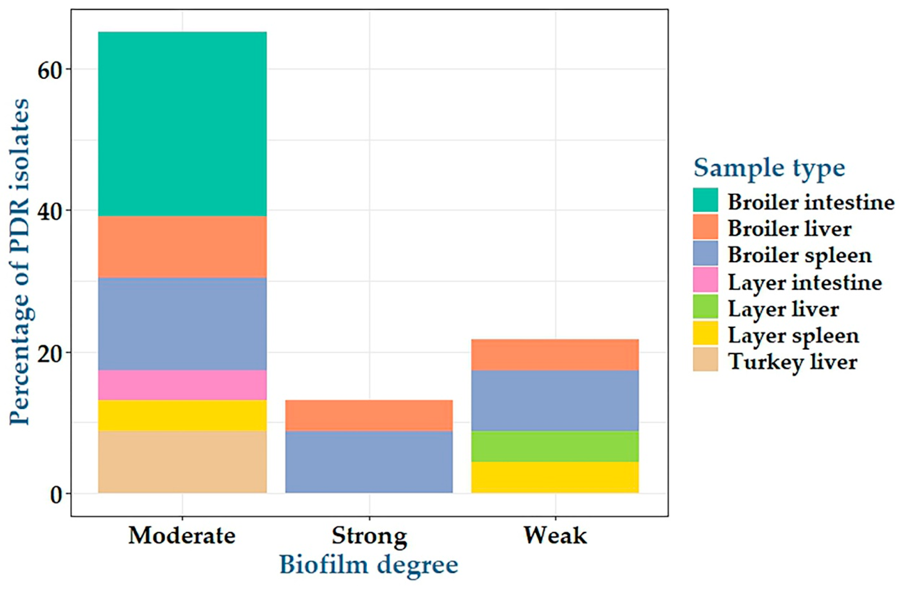
Twenty-three PDR C. perfringens isolates were investigated for biofilm formation using the qualitative Congo red agar and the quantitative microtiter plate assays. The results showed that all tested C. perfringens isolates were biofilm producers; among them, 5, 15 and 3 isolates were weak, moderate, and strong biofilm producers with percentages of 21.7%, 65.2%, and 13.1%, respectively (Table 4, Figure 5, Supplementary Table S1 and Supplementary Figures S1 and S2). Among weak, moderate, and strong biofilm degrees, there were no statistically significant differences (p = 1, 0.166, and 1, respectively) between PDR C. perfringens isolates from various sources.

2.4. Antimicrobial Activity of CNCs/ZnO Bio-Nanocomposite Against PDR C. perfringens Isolates

The antimicrobial potentials of CNCs/ZnO bio-nanocomposite against PDR C. perfringens isolates were evaluated by determining the inhibition zones’ diameters and the minimum inhibitory concentration (MIC) values (Table 4 and Figure 6A). The results revealed that CNCs/ZnO bio-nanocomposite demonstrated marked inhibitory activity against C. perfringens with inhibition zones’ diameters of 36–40 mm at 100% concentration, 28–32 mm at 50% concentration and 20–25 mm at 25% concentration of the nanocomposite with an MIC value of 0.125 µg/mL (Supplementary Figure S3). Statistical analysis displayed significant differences in the antimicrobial activities among various concentrations of CNCs/ZnO bio-nanocomposite against 23 PDR C. perfringens isolates (p < 0.01). CNCs/ZnO bio-nanocomposite at 100% concentration showed the largest zone of inhibition in all tested isolates.

2.5. Antibiofilm Activity of CNCs/ZnO Bio-Nanocomposite Against PDR C. perfringens

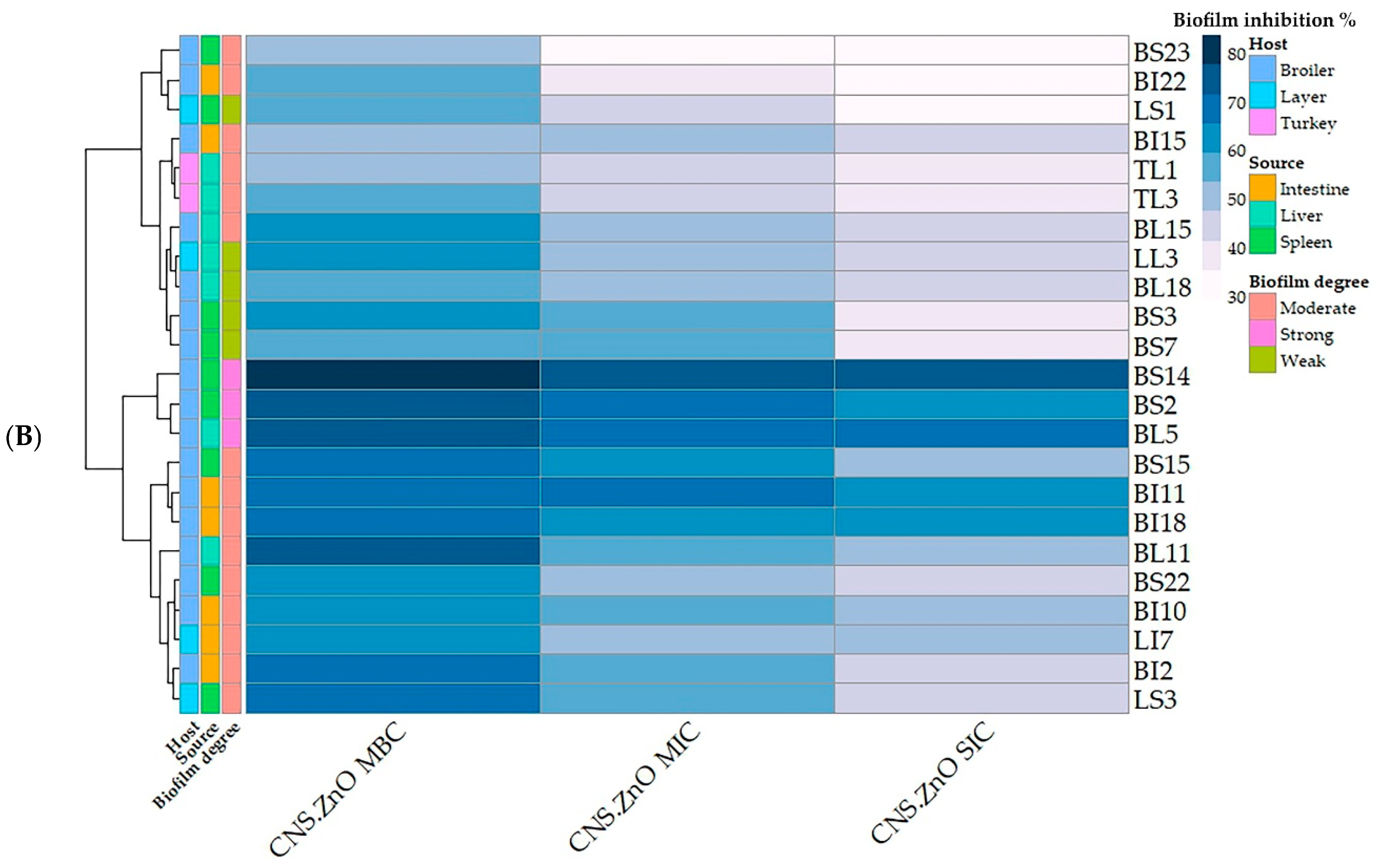
Table 4 and Figure 6 display the antibiofilm activities of CNCs/ZnO bio-nanocomposite at their MBC, MIC, and SIC against 23 PDR C. perfringens isolates. The results showed that CNCs/ZnO exerted great activity against the pre-existent biofilms of PDR C. perfringens isolates in a dose-dependent manner ((MBIC50 up to 83.43 ± 1.98 for the CNS-ZnO MBC concentration (0.25 μg/mL)) (Supplementary Table S1 and Supplementary Figure S4). There were significant differences in the antibiofilm activities among MBC, MIC, and SIC of CNCs/ZnO bio-nanocomposite against the examined PDR C. perfringens isolates (p < 0.05) except isolates of code no. BI18 and BL5. CNCs/ZnO bio-nanocomposite at their MBC showed the highest biofilm inhibition% in 14 tested isolates. CNCs/ZnO bio-nanocomposite at their MIC and SIC showed the highest biofilm inhibition% in 7 tested isolates.

2.6. Expression Analysis of CNCs/ZnO Bio-Nanocomposite Against PDR C. perfringens Using RT-qPCR

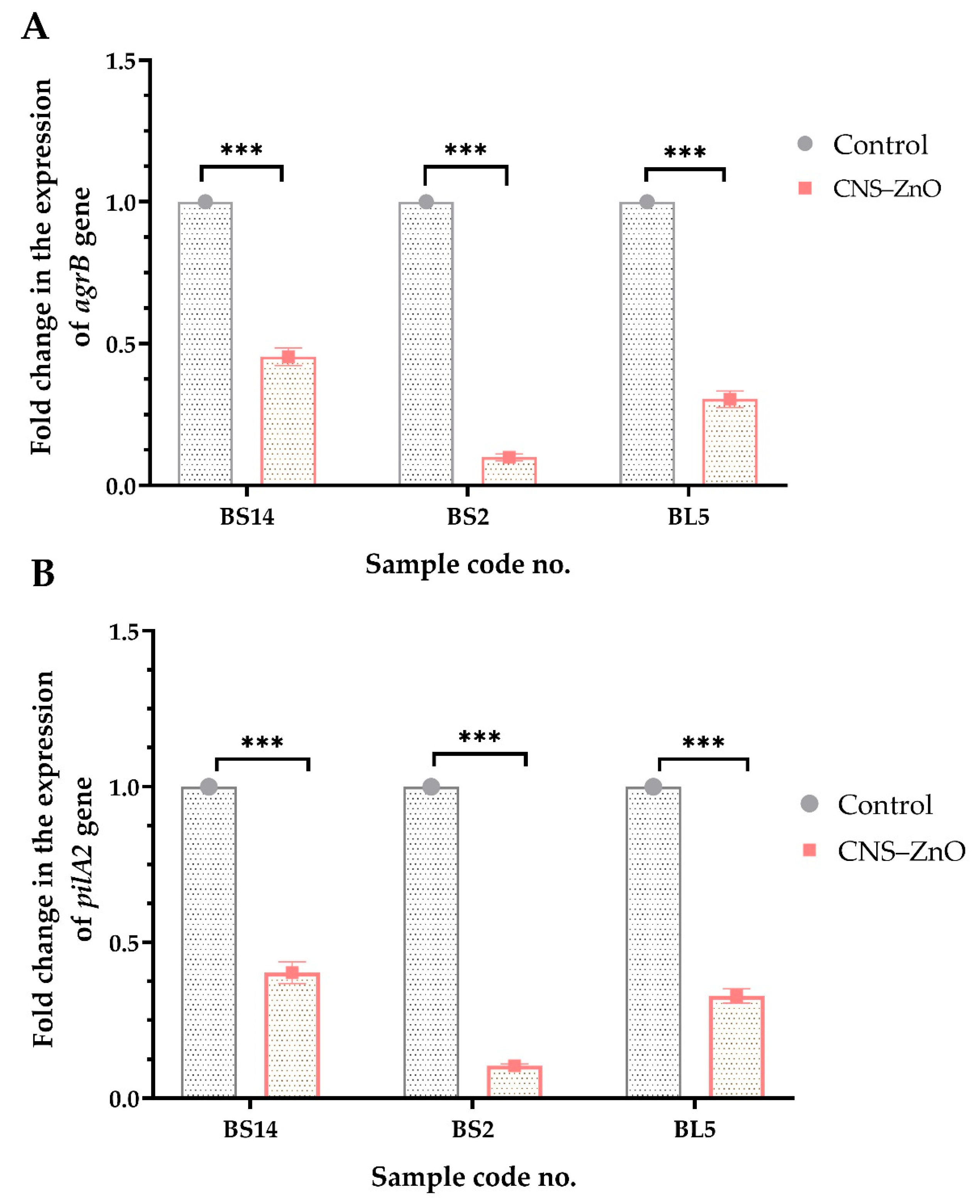
The expression of agrB quorum sensing gene and pilA2 type IV pili biosynthesis gene responsible for biofilm formation were determined by the RT-qPCR technique pre- and post-treatment with the CNCs/ZnO nanocomposite. The results showed that the transcript levels of agrB and pilA2 genes in CNCs/ZnO-treated biofilms decreased (0.099 ± 0.012–0.454 ± 0.031 and 0.104 ± 0.006–0.403 ± 0.035, respectively) when compared to the non-treated isolates. There were statistically significant differences in the transcriptional modulation of agrB and pilA2 genes among CNCs/ZnO treated and control untreated C. perfringens isolates (p < 0.0001) (Figure 7).

3. Discussion

The poultry industry suffers significant financial losses due to NE, and resistant clostridial infections, particularly those caused by the PDR strains, could complicate the therapy [28]. Reducing the use of antibiotics in the animal industry is a significant worldwide concern because of their detrimental effects on public health [29]. This is the first report investigating the in vitro efficacy of the CNCs/ZnO bio-nanocomposite against PDR biofilm-producing C. perfringens accused of NE in Egypt.
Herein, the prevalence of C. perfringens was 44.4% in broilers, which matches with that reported in a previous study in Egypt (46%) [30] with the highest existence in the intestine. In comparison, Gomaa et al. [31] reported that the prevalence rate of C. perfringens in a chicken origin was 38.3% with a higher existence in the liver followed by muscle and intestine. The early colonization of C. perfringens in the digestive tract of poultry, even from hatchery, may be the cause of the high existence of the bacterium in chickens [32]. Also, the bacterium is a normal inhabitant of the chicken’s gut but may proliferate, causing a disease in certain conditions [33]. On the other hand, our result was lower than those reported by Ibrahim and co-authors [34], who isolated C. perfringens from the intestinal samples of diseased birds with a percentage of 80.9%. However, in a previous study [35], C. perfringens was identified in 38.37% of intestinal samples in six Egyptian Governorates. The low isolation rate of C. perfringens in intestinal samples may be attributed to the collection of samples from poultry farms exposed to antimicrobials for therapeutic purposes, which could destroy the gut microbial community in addition to geographical variations [35].
Globally, Zhang et al. [36] could isolate C. perfringens from fresh chicken meat in various outlets (13.6%) and poultry farms (23.1%) in China with lower percentages. Moreover, in Pakistan, Haider’s team [37] isolated C. perfringens from poultry farms with a prevalence rate of 25.37%. Similarly, Rana and coworkers [38] detected C. perfringens in chickens in Bangladesh with a percentage of 34.5%. In contrast, Yadav et al. [39] documented the overall prevalence rate of C. perfringens to be 66.8% and 25.6% among diseased birds with NE and healthy ones, respectively, in India.
In Egypt, NE in turkeys has become a significant economic issue since 2018, resulting in significant losses and raising serious concerns for breeders [40]. In the current study, the prevalence rate of C. perfringens was 58.3% in diseased turkeys. To date, few studies have investigated the prevalence of C. perfringens in turkeys. The occurrence of C. perfringens in this study nearly coincided with the findings of previously published documents [41] (55%) and [40] (41%). The limitations of this study should be acknowledged, including the relatively few numbers of available turkey farms in Egypt for sample collection, which constrained the sample size for this study and reduced the generalisability of the findings.
The variations in prevalence could be ascribed to various isolation techniques, sample selection (number and kind of samples from healthy and/or diseased birds), differences in geographical locations, management of chicken farms (presence or absence of NE predisposing conditions), and varying antibiotic dosing ranges.
Antimicrobials have been used in order to prevent NE and increase broiler production [42]. Antimicrobials are either used for therapeutic or preventative purposes in animal feed [43]. The use of antimicrobials in the poultry industry as growth promoters has contributed to antimicrobial resistance in bacteria [44]. However, this practice has since been banned in a number of countries [28]. The prolonged and extensive use of antimicrobials in poultry over the past few years has altered the bacterial environment, eliminated susceptible strains, and promoted the persistence and dominance of antimicrobial-resistant bacteria [11]. Since MDR strains have emerged as a result of antibiotic abuse, continuing to publish resistance rates is the first step in stopping the spread of antibiotic resistance [45]. In this study, we examined the susceptibilities of 77 C. perfringens isolates to 19 antimicrobials of 17 classes and offered greater insight into the concerning rise in PDR, XDR, and MDR profiles. Interestingly, 29.9%, 54.5%, and 15.6% of C. perfringens isolates were PDR, XDR, and MDR, respectively, with high MAR indices ranged from 0.84 to 1. A previous research [31] demonstrated the first record for the emergence of PDR C. perfringens isolate from a poultry source, while another study conducted on diseased and apparently healthy chickens [46] presented that eight isolates (26.7%) were XDR, one isolate (3.3%) was PDR, and twenty-one isolates (70%) were MDR. The increasing MAR indices suggest that the antimicrobials are being misused without restriction, either to treat clostridial infections in animals and poultry or as growth promoters to enhance growth performance. This promoted the development and dissemination of antibiotic resistance in the common intestinal flora, such as C. perfringens strains [47].
In addition to the ability of C. perfringens to produce toxins, it is able to form biofilms, which makes the bacteria resistant to disinfectants and antibiotics and contributes to its continuing existence in the environment [48,49]. Bacteria can adhere to substrates and encapsulate themselves in a matrix consisting of extracellular DNA (eDNA), proteins, and extracellular polymeric substances (EPS) when they form biofilms [15,16]. Their interactions with exopolysaccharides and nucleic acid constituents aid in the stability of the biofilm matrix, surface colonization, and preservation of the biofilm’s integrity and structure [18,50]. The biofilm layer gives the bacteria the ability to cause a variety of diseases. Thus, it is believed that biofilm-producing bacteria cause 65–80% of infections [51]. In addition to preserving a biofilm’s structural integrity, EPS acts as a barrier against antimicrobial substances, including antibiotics and bleaching chemicals [52].
In this study, all tested PDR C. perfringens isolates turned out to be biofilm producers, which matches a recently published work [53]. This indicates that the presence of slow-growing or persistent cells in biofilms [18] or increased expression of efflux pumps in biofilms [19,20] may be accused of antimicrobial resistance in biofilms.
In Egyptian poultry farms, antimicrobials are routinely administered from first day of life till slaughtering. This widespread and continuous use of antimicrobials creates a significant problem; each time an antimicrobial is used, the bacteria within the chickens’ systems have a higher chance of developing resistance. This resistance occurs because some bacteria possess genes that allow them to survive the antimicrobial effects, and these resistant bacteria then multiply, making future treatments less effective [54]. In this study, CNCs/ZnO nanocomposite showed strong inhibitory activity against C. perfringens isolates (inhibition zone diameters ranged from 36–41 mm for 100% concentration and MIC values ≤ 0.125 μg/mL. Moreover, CNCs/ZnO exerted great activity against the pre-existent biofilms of PDR C. perfringens isolates in a dose-dependent manner with the MBIC50 of up to 83.43 ± 1.98 for the CNCs/ZnO MBC concentration (0.25 μg/mL). Interestingly, there are no published issues regarding CNCs/ZnO nanoparticles’ antibacterial and antibiofilm properties against C. perfringens isolates to compare with the current findings. Significant antibacterial activities have been shown by the CNCs/ZnO nanocomposite against a variety of Gram-positive and Gram-negative bacteria, such as Staphylococcus aureus, Salmonella, and Escherichia coli [27]. The CNCs/ZnO nanocomposite can physically interact with bacterial cell walls, resulting in the destruction of its structure. This contact is facilitated by the nanocomposites’ high surface area and sharp edges, which rupture the membranes and allow cellular contents to leak out [27,55]. Another important mechanism of action of the CNC/ZnO nanocomposite is oxidative stress [55,56]. When ZnO nanoparticles are exposed to light, they produce reactive oxygen species (ROS), which can harm the lipids, proteins, and DNA of bacterial cells. Moreover, the CNC/ZnO nanocomposite can block important bacterial enzymes that are essential for bacterial survival and replication, including dihydrofolate reductase (DHFR) and dihydropteroate synthase (DHPS). This enzymatic inhibition further contributes to the antibacterial properties of the nanocomposites [56,57].
CNC/ZnO bio-nanocomposite has demonstrated antibiofilm activities by interfering with the extracellular polymeric materials that hold biofilms together or penetrating the biofilms and exhibiting antibacterial properties within the biofilm matrix; thus, the bio-nanocomposite can decrease the viability of bacteria entrenched in biofilms [27,58].
In the present study, the expression of agrB quorum sensing and pilA2 type IV pili biosynthesis gene responsible for biofilm formation were inspected by the RT-qPCR technique before and after treatment with the CNCs/ZnO. The relative expression of agrB and pilA2 genes in CNCs/ZnO-treated biofilms was significantly reduced than the non-treated ones. However, similar data were not available for comparison with other authors about the antibiofilm activity of CNCs/ZNO against PDR C. perfringens bacterial pathogens.

4. Materials and Methods

4.1. Samples and Ethical Approval

Poultry samples, including intestines, livers, and spleens, were collected from 172 birds (three samples from each bird) comprising chickens (broilers (n = 124) and layers (n = 36)) and turkeys (n = 12) from various poultry farms in Egypt during the period between November 2022 and June 2023. The birds were clinically examined for any observable clinical signs and gross lesions related to NE. They mostly suffered from diarrhea, low growth rate, and mortality. The samples were transported immediately in an ice box to the bacteriology laboratory for further analysis. The animal study was approved by the Institutional Animal Care and Use Committee, Faculty of Veterinary Medicine, Zagazig University (Approval No ZU-IACUC/2/F/149/2024).

4.2. Bacteriological Analysis and Molecular Identification

Clostridium perfringens were isolated under anaerobic conditions in accordance with the previously defined technique [59]. The samples were enriched for 24 h at 37 °C in cooked meat media (Oxoid, Cambridge, UK). A loopful of enrichment broth culture was plated onto Perfringens Agar Base (Oxoid, Cambridge, UK) provided with TSC supplement (D-cycloserine 200 mg/vial) and egg yolk emulsion then incubated at 37 °C for 24 h. Colonies of presumed C. perfringens isolates were verified by target hemolysis on blood agar, lecithinase activity, motility test, and skim milk coagulation (stormy fermentation) [60]. For molecular confirmation of bacterial isolates, the bacterial DNAs were extracted using a QIAamp DNA Mini kit (Qiagen GmbH, Hilden, Germany) following the manufacturer’s instructions. The 16S rRNA gene of C. perfringens was amplified by conventional PCR using the oligonucleotide primer pair listed in Supplementary Table S2 [61].

4.3. Antimicrobial Susceptibility Testing

Antimicrobial susceptibility testing of C. perfringens isolates against 19 antimicrobial discs (Oxoid, Cambridge, UK) within 17 different categories was carried out adopting the standard disc diffusion protocol following the Clinical and Laboratory Standards Institute guidelines [62]. The antimicrobials included penicillin (penicillin G (P; 1 unit)), penicillin combinations (amoxycillin-clavulanic acid (AMC; 20/10 µg)), cephalosporines (cefoxitin (FOX; 30 µg)), carbapenems (imipenem (IPM; 10 µg)), aminoglycosides (gentamicin (CN; 10 µg)), macrolides (erythromycin (E; 15 µg)), quinolones (nalidixic acid (NA; 30 µg)), fluoroquinolones (enrofloxacin (ENR; 5 µg)), sulfonamides (sulfamethoxazole-trimethoprim (SXT; 23.75/1.25 μg)), amphenicols (chloramphenicol (C; 30 µg)), polypeptides (bacitracin (B; 10 µg)), oxazolidinones (linezolid (LNZ; 30 µg)), lincosamides (clindamycin (DA; 2 µg) and lincomycin (L2; 15 μg)), tetracyclines (tetracycline (TE; 30 µg)), glycopeptides (vancomycin (VA; 30 µg) and teicoplanin (Tec; 30 µg)), nitroimidazole (metronidazole (MET; 5 µg)), and rifamycin (rifampin (RA; 5 µg)). Based on previous reports [31,63] for the majority of antimicrobials and the British Society for Antimicrobial Chemotherapy (BSAC) for penicillin G, imipenem, clindamycin, and metronidazole [64], the interpretive criteria of antimicrobial resistance were established. The multiple antimicrobial resistance (MAR) indices were determined as reported elsewhere [65]. The following drug resistance categories were identified as previously reported [66]: MDR; resistance to three or more classes of antimicrobial agents, XDR; resistance to all classes of antimicrobial agents except two or fewer, and PDR; resistance to all antimicrobial agents. A C. perfringens ATCC 13124 strain was used as quality control.

4.4. Biofilm Growth and Quantification

4.4.1. Qualitative Congo Red Agar Method

Clostridium perfringens isolates were incubated on brain heart infusion agar (BHI; Oxoid, UK) supplemented with 5% (w/v) sucrose and 0.08% (w/v) Congo red dye (Oxoid, Hampshire, UK) for 24 to 48 h at 37 °C. Black colonies with dry crystalline phenotype indicated biofilm formation, while weak or non-biofilm producing isolates formed pink-coloured colonies [67].

4.4.2. Quantitative Microtiter Plate Assay

Biofilm production by C. perfringens isolates was detected using sterile 96-well flat-bottomed polystyrene microtiter plates (Techno Plastic Products, Trasadingen, Switzerland). In brief, 200 μL of 106 CFU/mL bacterial suspension was inoculated in each well and then incubated for 24 h at 37 °C. Negative control wells containing 200 µL of uninoculated Tryptic Soy Broth (TSB; Oxoid, Cambridge, UK) were included. The wells were gently washed using 200 μL of phosphate-buffered saline (PBS) three times, then dried with their sides facing up for 15 min [68]. To stain the biofilm mass, 50 μL of 0.1% (w/v) crystal violet (Oxoid, UK) was added for 30 min; the wells were washed three times with 200 μL of PBS and then air dried upside-down. To solubilize the stain, the wells were finally dissolved in 200 μL of ethanol/acetone solution (80:20, v/v). The optical density (OD) of biofilm mass was measured at 570 nm using a microplate reader (Stat Fax 2100, Awareness Technology, Palm City, FL, USA). The OD cut-off (ODc) was defined as three standard deviations over the mean OD of the negative control. The isolates were categorized into four groups based on their adhesion capabilities: non-biofilm producers (OD ≤ ODc), weak biofilm producers (ODc < OD ≤ 2xODc), moderate biofilm producers (2ODc < OD ≤ 4xODc), and strong biofilm producers (4xODc < OD) [69,70].

4.5. Synthesis of CNCs/ZnO Bio-Nanocomposite

The CNCs/ZnO bio-nanocomposite was synthesized following Dawwam et al. [27] at the Institute of Nanoscience and Nanotechnology, Kafrelsheikh University, Kafr el-Sheikh, Egypt. Briefly, 3.5 g zinc acetate was added to 1% cellulose nanocrystal (CNC) suspension, extracted from Palm sheath fibers. The mixture was stirred (30 min), sonicated (2 h, 80 °C or 35 °C), dialyzed (conductivity ≈ 4 μS·cm⁻1), and dried (60 °C, 6 h). Characterization of the nanocomposite was applied, as illustrated in the previously published work [27].

4.6. Antimicrobial Activities of CNCs/ZnO Against PDR C. perfringens

For the agar well diffusion technique, colonies from an overnight culture of C. perfringens were standardized to 1 × 10⁸ CFU. Each isolate was swabbed twice on a sterile Muller-Hinton agar plate. By using a sterile 1 mL pipette tip, multiple wells were created (approximately 6 mm in diameter), then 100 µL of CNCs/ZnO bio-nanocomposite was inoculated into the wells at different concentrations (10, 50, and 100%). Sterile distilled water was used as a negative control, while the imipenem antibiotic was a positive control. The plates were incubated anaerobically at 37 °C for 24 h, and then zones of growth inhibition were measured to the nearest millimeter. The isolates with inhibition zones diameter ≥ 8 mm were considered susceptible [71].
The minimum inhibitory concentration (MIC) and minimum bactericidal concentration (MBC) of CNCs/ZnO bio-nanocomposite were then determined using the broth microdilution method [72]. Briefly, two-fold serial dilutions of CNCs/ZnO were performed from the stock solution (512 μg/mL), then 100 μL of each dilution was dispended in 96-well culture plates. Thereafter, 100 μL of bacterial suspension of approximately 5 × 10⁵ CFU/mL was added to each well and incubated under anaerobic conditions at 37 °C for 24 h. Ten µL of resazurin dye (stock preparation; 337.5 mg of resazurin powder in 50 mL sterile distilled water) was added to each well; then incubation was done at 37 °C for 2–4 h. The color change is based on bacterial metabolic activity; wells with no bacterial growth remain blue, while wells with growth turn pink [73]. The lowest concentration of CNCs/ZnO exhibiting no growth was considered as the MIC, whereas that which kills 99.9% of bacteria was considered the MBC.

4.7. Antibiofilm Activities of CNCs/ZnO Bio-Nanocomposite Against Biofilm Producing C. perfringens

This assay investigated the ability of CNCs/ZnO bio-nanocomposite to disperse biofilm formation by C. perfringens. Briefly, 100 µL of the antibacterial agent at different concentrations (512–0.25 µg/mL) was added to selected wells of 96-well flat-bottom polystyrene microtiter plate seeded with 100 µL of the C. perfringens suspension (108 cells/mL) and incubated for 24 h at 37 °C to allow biofilm formation. The contents of the wells were aspirated and washed three times with sterile PBS. The extent of biofilm formation was assessed using the crystal violet staining assay as described previously [65,66]. The negative controls (CNCs/ZnO free wells) and the positive controls (C. perfringens biofilm producers) were included. Experiments were carried out in triplicate [74]. The inhibition percentage of biofilm was calculated by the formula: Percentage of biofilm inhibition = (Control OD570 nm − Test OD570 nm)/Control OD570 nm) × 100 [75,76]. Further, the minimal biofilm inhibitory concentration (MBIC) was defined as the minimal antimicrobial concentration showing no color development.

4.8. Gene Expression of Biofilm Biosynthesis Genes by Real-Time Quantitative PCR (RT-qPCR)

Pan drug-resistant strong biofilm producers C. perfringens isolates were separately exposed to the subinhibitory concentration (SIC) of the CNCs/ZnO bio-nanocomposite and then incubated at 37 °C for 24 h. Non-treated C. perfringens biofilms were considered controls. Biofilms were harvested and then gently washed with PBS to remove the planktonic cells. Total RNAs were extracted from biofilms of both treated and untreated C. perfringens isolates using a QIAampRN easy Mini kit (Qiagen, Hilden, Germany) following the manufacturer’s recommendations. Relative expressions of the biofilm biosynthesis gene (pilA2) and its regulator (agrB) were investigated by one-step RT-qPCR using the QuantiTect SYBR Green RT-PCR kit (Qiagen, Germany) in the MX3005p real-time PCR thermal cycler (Stratagene, Lajolla, CA, USA) according to the manufacturer’s guidelines using oligonucleotide primers listed in Supplementary Table S2) [77,78]. The 16S rRNA housekeeping gene was used for normalization [79]. Amplification curves and threshold cycle (ct) values of tested isolates were determined to be compared with the control positive according to the “ΔΔCt” method stated previously [80] using the following ratio: (2 − ΔΔCt).

4.9. Statistical Analysis

The data were analyzed using SPSS version 26 (IBM Corp, Armonk, NY, USA). The Chi-square test was used to study the variations in the prevalence of C. perfringens from different origins and to assess the differences in the antimicrobial resistance patterns of the recovered isolates from various sources. Additionally, one-way ANOVA and Tukey’s post hoc test were used to evaluate the antibiofilm and the antibacterial efficacy of CNCs/ZnO bio-nanocomposite at various concentrations against PDR C. perfringens isolates. All experimental procedures were done in triplicate, and the results were expressed as mean ± standard error of the mean (SEM). The p-values were considered statistically significant if they were less than 0.05. All graphs were generated by R-software version 4.4.3 [81] (https://www.r-project.org/) using ggplot [82], pheatmap [83], and factoextra [84] packages. Moreover, we used an independent samples t-test to test CNCs/ZnO effects on C. perfringens biofilm genes’ expression. All tests were done using SPSS Inc. version 26 (IBM Corp., Armonk, NY, USA). The p-values of less than 0.05 were considered statistically significant. Figures were generated by GraphPad Prism version 8 (San Diego, CA, USA).

5. Conclusions

To the best of our knowledge, this is the first report demonstrating the antimicrobial and antibiofilm activities of CNCs/ZnO nanocomposite against PDR C. perfringens isolates recovered from chickens and turkeys and their pre-existent biofilms, providing a distinct benefit over the conventional antimicrobial agents.

Supplementary Materials

The following supporting information can be downloaded at: https://www.mdpi.com/article/10.3390/antibiotics14060575/s1, Supplementary Figure S1: PDR C. perfringens isolates on Congo red agar media showing strong (A), Moderate (B) and weak (C) biofilm formation. Supplementary Figure S2: Biofilm formation by C. perfringens isolates using the crystal violet microtiter plate biofilm assay. A: An inoculated plate by 23 PDR C. perfringens isolates. B: Stained biofilms of PDR C. perfringens isolates by 0.1% crystal violet. C: C. perfringens biofilms after washing the stain using phosphate buffer saline. Supplementary Figure S3: Antimicrobial activity of CNCs/ZNO against PDR C. perfringens isolates. A: Agar well diffusion of CNCs/ZnO against a C. perfringens isolate. B: Broth microdilution method for determining the minimum inhibitory concentrations of CNCs/ZnO against C. perfringens isolates using Resazurin dye. Supplementary Figure S4: Antibiofilm activity of CNCs/ZnO bio-nanocomposite against 23 PDR C. perfringens isolates. A: An inoculated plate by 23 PDR C. perfringens isolates were then exposed to the nanocomposite. B: Stained biofilms of PDR C. perfringens isolates by 0.1% crystal violet. C: C. perfringens biofilms after washing the stain using phosphate buffer saline. Supplementary Table S1: Biofilm production by PDR C. perfringens isolates and the antibiofilm activities of CNCs/ZnO bio-nanocomposites using the microtiter plate method. Supplementary Table S2: Oligonucleotide primers used in the study.

Author Contributions

Conceptualization, A.A. and N.K.A.E.-A.; methodology, I.A., N.K.A.E.-A. and A.S.E.-D.; validation, I.A., A.A., A.S.E.-D., I.P., M.A. and N.K.A.E.-A.; formal analysis, I.A., A.S.E.-D. and N.K.A.E.-A.; investigation, I.A., A.A., I.P., M.A. and N.K.A.E.-A.; data curation, I.A. and N.K.A.E.-A.; writing-original draft preparation, I.A. and N.K.A.E.-A.; writing-review and editing, I.A., A.A., A.S.E.-D. and N.K.A.E.-A. All authors have read and agreed to the published version of the manuscript.

Funding

This research received no external funding.

Institutional Review Board Statement

The animal study protocol was approved by the Institutional Animal Care and Use Committee, Faculty of Veterinary Medicine, Zagazig University (Approval No ZU-IACUC/2/F/149/2024).

Informed Consent Statement

Written informed consents were acquired from the owners of poultry farms participating in the investigation after a full explanation of the purpose of the study.

Data Availability Statement

The data presented in this study are available upon request from the corresponding author.

Conflicts of Interest

The authors declare no conflicts of interest.

References

  1. Mottet, A.; Tempio, G. Global poultry production: Current state and future outlook and challenges. World’s Poult. Sci. J. 2017, 73, 245–256. [Google Scholar] [CrossRef]
  2. Timbermont, L.; Haesebrouck, F.; Ducatelle, R.; Van Immerseel, F. Necrotic enteritis in broilers: An updated review on the pathogenesis. Avian Pathol. 2011, 40, 341–347. [Google Scholar] [CrossRef] [PubMed]
  3. Cooper, K.K.; Songer, J.G.; Uzal, F.A. Diagnosing clostridial enteric disease in poultry. J. Vet. Diagn. Investig. 2013, 25, 314–327. [Google Scholar] [CrossRef] [PubMed]
  4. Fathima, S.; Hakeem, W.G.A.; Shanmugasundaram, R.; Selvaraj, R.K. Necrotic enteritis in broiler chickens: A review on the pathogen, pathogenesis, and prevention. Microorganisms 2022, 10, 1958. [Google Scholar] [CrossRef]
  5. Gharib-Naseri, K.; Kheravii, S.; Keerqin, C.; Morgan, N.; Swick, R.; Choct, M.; Wu, S.-B. Two different Clostridium perfringens strains produce different levels of necrotic enteritis in broiler chickens. Poult. Sci. 2019, 98, 6422–6432. [Google Scholar] [CrossRef]
  6. Rood, J.I.; Adams, V.; Lacey, J.; Lyras, D.; McClane, B.A.; Melville, S.B.; Moore, R.J.; Popoff, M.R.; Sarker, M.R.; Songer, J.G. Expansion of the Clostridium perfringens toxin-based typing scheme. Anaerobe 2018, 53, 5–10. [Google Scholar] [CrossRef]
  7. Sattar, M.M.K.; Anjum, A.A.; Chang, Y.F.; Yaqub, T.; Aslam, A.; Ali, T. Molecular characterization and toxins optimization of indigenous Clostridium perfringens toxinotype B isolated from lamb dysentery clinical cases. Kafkas Univ. Vet. Fak. Derg. 2023, 29, 79. [Google Scholar]
  8. de Souza, M.; Baptista, A.A.S.; Menck-Costa, M.F.; Justino, L.; da Glória, E.M.; Shimizu, G.D.; Ferraz, C.R.; Verri, W.A.; Van Immerseel, F.; Bracarense, A. Modulation of broiler intestinal changes induced by Clostridium perfringens and deoxynivalenol through probiotic, paraprobiotic, and postbiotic supplementation. Toxins 2024, 16, 46. [Google Scholar] [CrossRef]
  9. Aqeel, M.; Mirani, A.H.; Khoso, P.A.; Sahito, J.K.; Bhutto, A.L.; Leghari, R.A.; Rahimoon, M.M.; Ali, K.; Ali, N. A review on the study of immunomodulators and herbal remedies: A natural approach to treating necrotic enteritis. Pure Appl. Biol. (PAB) 2024, 13, 275–302. [Google Scholar] [CrossRef]
  10. El-Aziz, N.K.A.; Tartor, Y.H.; Gharieb, R.M.A.; Erfan, A.M.; Khalifa, E.; Said, M.A.; Ammar, A.M.; Samir, M. Extensive drug-resistant Salmonella enterica Isolated from poultry and humans: Prevalence and molecular determinants behind the co-resistance to ciprofloxacin and tigecycline. Front. Microbiol. 2021, 12, 738784. [Google Scholar] [CrossRef]
  11. Góchez, D.; Raicek, M.; Pinto Ferreira, J.; Jeannin, M.; Moulin, G.; Erlacher-Vindel, E. OIE annual report on antimicrobial agents intended for use in animals: Methods used. Front. Vet. Sci. 2019, 6, 317. [Google Scholar] [CrossRef] [PubMed]
  12. Ray, S.; Löffler, S.; Richter-Dahlfors, A. High-resolution large-area image analysis deciphers the distribution of Salmonella cells and ECM components in biofilms formed on charged PEDOT: PSS surfaces. Adv. Sci. 2024, 11, 2307322. [Google Scholar] [CrossRef] [PubMed]
  13. Pantaléon, V.; Bouttier, S.; Soavelomandroso, A.P.; Janoir, C.; Candela, T. Biofilms of Clostridium species. Anaerobe 2014, 30, 193–198. [Google Scholar] [CrossRef] [PubMed]
  14. Kelly, S.M.; Lanigan, N.; O’Neill, I.J.; Bottacini, F.; Lugli, G.A.; Viappiani, A.; Turroni, F.; Ventura, M.; van Sinderen, D. Bifidobacterial biofilm formation is a multifactorial adaptive phenomenon in response to bile exposure. Sci. Rep. 2020, 10, 11598. [Google Scholar] [CrossRef]
  15. Yu, S.; Su, T.; Wu, H.; Liu, S.; Wang, D.; Zhao, T.; Jin, Z.; Du, W.; Zhu, M.-J.; Chua, S.L. PslG, a self-produced glycosyl hydrolase, triggers biofilm disassembly by disrupting exopolysaccharide matrix. Cell Res. 2015, 25, 1352–1367. [Google Scholar] [CrossRef]
  16. Czuban, M.; Srinivasan, S.; Yee, N.A.; Agustin, E.; Koliszak, A.; Miller, E.; Khan, I.; Quinones, I.; Noory, H.; Motola, C. Bio-orthogonal chemistry and reloadable biomaterial enable local activation of antibiotic prodrugs and enhance treatments against Staphylococcus aureus infections. ACS Cent. Sci. 2018, 4, 1624–1632. [Google Scholar] [CrossRef]
  17. De Oliveira, A.; Cataneli Pereira, V.; Pinheiro, L.; Moraes Riboli, D.F.; Benini Martins, K.; Ribeiro de Souza da Cunha, M.d.L. Antimicrobial resistance profile of planktonic and biofilm cells of Staphylococcus aureus and coagulase-negative staphylococci. Int. J. Mol. Sci. 2016, 17, 1423. [Google Scholar] [CrossRef]
  18. Li, P.; Yin, R.; Cheng, J.; Lin, J. Bacterial biofilm formation on biomaterials and approaches to its treatment and prevention. Int. J. Mol. Sci. 2023, 24, 11680. [Google Scholar] [CrossRef]
  19. Seukep, A.J.; Mbuntcha, H.G.; Kuete, V.; Chu, Y.; Fan, E.; Guo, M.-Q. What Approaches to thwart bacterial efflux pumps-mediated resistance? Antibiotics 2022, 11, 1287. [Google Scholar] [CrossRef]
  20. Gaurav, A.; Bakht, P.; Saini, M.; Pandey, S.; Pathania, R. Role of bacterial efflux pumps in antibiotic resistance, virulence, and strategies to discover novel efflux pump inhibitors. Microbiology 2023, 169, 001333. [Google Scholar] [CrossRef]
  21. Dovnar, R.; Smotryn, S.; Anufrik, S.; Anuchin, S.; Dovnar, I.; Iaskevich, N. Copper and selenium nanoparticles as a new tool against antibiotic-resistant pathogenic microorganisms. Surg. East. Eur. 2022, 11, 315–328. [Google Scholar]
  22. Hashem, A.H.; Al-Askar, A.A.; Haponiuk, J.; Abd-Elsalam, K.A.; Hasanin, M.S. Biosynthesis, characterization, and antifungal activity of novel trimetallic copper oxide–selenium–zinc oxide nanoparticles against some mucorales fungi. Microorganisms 2023, 11, 1380. [Google Scholar] [CrossRef] [PubMed]
  23. Raja, F.N.; Worthington, T.; Martin, R.A. The antimicrobial efficacy of copper, cobalt, zinc and silver nanoparticles: Alone and in combination. Biomed. Mater. 2023, 18, 045003. [Google Scholar] [CrossRef]
  24. Li, H.; Yang, Z.; Khan, S.A.; Walsh, L.J.; Seneviratne, C.J.; Ziora, Z.M. Characteristics of metallic nanoparticles (especially silver nanoparticles) as anti-biofilm agents. Antibiotics 2024, 13, 819. [Google Scholar] [CrossRef] [PubMed]
  25. Terea, H.; Rebiai, A.; Selloum, D.; Tedjani, M.L. Cellulose/ZnO nanoparticles (CNC/ZnO NPs): Synthesis, characterization, and evaluation of their antibacterial and antifungal activities. Cellulose 2024, 31, 5027–5042. [Google Scholar] [CrossRef]
  26. Kim, I.; Viswanathan, K.; Kasi, G.; Thanakkasaranee, S.; Sadeghi, K.; Seo, J. ZnO nanostructures in active antibacterial food packaging: Preparation methods, antimicrobial mechanisms, safety issues, future prospects, and challenges. Food Rev. Int. 2022, 38, 537–565. [Google Scholar] [CrossRef]
  27. Dawwam, G.E.; Al-Shemy, M.T.; El-Demerdash, A.S. Green synthesis of cellulose nanocrystal/ZnO bio-nanocomposites exerting antibacterial activity and downregulating virulence toxigenic genes of food-poisoning bacteria. Sci. Rep. 2022, 12, 16848. [Google Scholar] [CrossRef]
  28. García-Vela, S.; Martínez-Sancho, A.; Said, L.B.; Torres, C.; Fliss, I. Pathogenicity and antibiotic resistance diversity in Clostridium perfringens isolates from poultry affected by necrotic enteritis in Canada. Pathogens 2023, 12, 905. [Google Scholar] [CrossRef]
  29. Ferri, M.; Ranucci, E.; Romagnoli, P.; Giaccone, V. Antimicrobial resistance: A global emerging threat to public health systems. Crit. Rev. Food Sci. Nutr. 2017, 57, 2857–2876. [Google Scholar] [CrossRef]
  30. Eman, M.; Sharaf, D.M. Comparing the effect of nitazoxanide and tylosin against necrotic enteritis in broilers. J. Adv. Vet. Res. 2024, 14, 8–16. [Google Scholar]
  31. Gomaa, N.H.; El-Aziz, N.K.A.; El-Naenaeey, E.-s.Y.; Abdelaziz, W.S.; Sewid, A.H. Antimicrobial potential of myricetin-coated zinc oxide nanocomposite against drug-resistant Clostridium perfringens. BMC Microbiol. 2023, 23, 79. [Google Scholar] [CrossRef] [PubMed]
  32. Immerseel, F.V.; Buck, J.D.; Pasmans, F.; Huyghebaert, G.; Haesebrouck, F.; Ducatelle, R. Clostridium perfringens in poultry: An emerging threat for animal and public health. Avian Pathol. 2004, 33, 537–549. [Google Scholar] [CrossRef] [PubMed]
  33. Kiu, R.; Hall, L.J. An update on the human and animal enteric pathogen Clostridium perfringens. Emerg. Microbes Infect. 2018, 7, 1–15. [Google Scholar] [CrossRef] [PubMed]
  34. Ibrahim, A.H.; El Khashab, E.; Shakal, M.; Morsy, E.A. Unveiling antibiotic resistance, virulence, and molecular detection of enteric bacterial infections in broilers: A Study in Egypt. Egypt. J. Vet. Sci. 2024, 55, 1537–1551. [Google Scholar] [CrossRef]
  35. Helal, S.S.; Khalaf, N.M.; El Menisy, A.A.; Lebdah, M.A. Clostridium perfringens type A causing necrotic enteritis outbreaks among chickens in Egypt. Zagazig Vet. J. 2019, 47, 398–407. [Google Scholar] [CrossRef]
  36. Zhang, T.; Zhang, W.; Ai, D.; Zhang, R.; Lu, Q.; Luo, Q.; Shao, H. Prevalence and characterization of Clostridium perfringens in broiler chickens and retail chicken meat in central China. Anaerobe 2018, 54, 100–103. [Google Scholar] [CrossRef]
  37. Haider, Z.; Ali, T.; Ullah, A.; Basit, A.; Tahir, H.; Tariq, H.; Ilyas, S.Z.; Hayat, Z.; Rehman, S.-u. Isolation, toxinotyping and antimicrobial susceptibility testing of Clostridium perfringens isolated from Pakistan poultry. Anaerobe 2022, 73, 102499. [Google Scholar] [CrossRef]
  38. Rana, E.A.; Nizami, T.A.; Islam, M.S.; Barua, H.; Islam, M.Z. Phenotypical identification and toxinotyping of Clostridium perfringens isolates from healthy and enteric disease-affected chickens. Vet. Med. Int. 2023, 2023, 2584171. [Google Scholar] [CrossRef]
  39. Yadav, J.P.; Kaur, S.; Dhaka, P.; Vijay, D.; Bedi, J.S. Prevalence, molecular characterization, and antimicrobial resistance profile of Clostridium perfringens from India: A scoping review. Anaerobe 2022, 77, 102639. [Google Scholar] [CrossRef]
  40. El-Gaos, M.; Khalil, M.; Abdelrahman, M.; Ramadan, A. Molecular characterization of Clostridium perfringens isolated from turkeys. Assiut Vet. Med. J. 2020, 66, 103–110. [Google Scholar]
  41. Salem, S.M.; Mustafa, D.I.; Hamed, R.I.; El-Azzouny, M.M.; Anwar, N. Assessment of pathological changes of mixed infection of coccidiosis and necrotic enteritis in turkey. J. Egypt. Vet. Med. Assoc. 2020, 80, 55–84. [Google Scholar]
  42. Yudiarti, T.; Sugiharto, S.; Widiastuti, E.; Wahyuni, H.; Sartono, T.; Nasution, M. Physiological parameters, intestinal microbial population and internal organ weight of broilers supplemented with the fungus monascus purpureus. Adv. Anim. Vet. Sci. 2024, 12, 85–91. [Google Scholar] [CrossRef]
  43. Soromou, L.W.; Leno, P.F.; Kamano, A.; Souare, M.L.; Camara, A.O.D.; Camara, K. Current practices in the veterinary use of antibiotics in poultry laying hens in Friguiagbé (Guinea). J. Drug Deliv. Ther. 2024, 14, 35–40. [Google Scholar] [CrossRef]
  44. Miyakawa, M.E.F.; Casanova, N.A.; Kogut, M.H. How did antibiotic growth promoters increase growth and feed efficiency in poultry? Poult. Sci. 2024, 103, 103278. [Google Scholar] [CrossRef]
  45. Schuetz, A.N. Antimicrobial resistance and susceptibility testing of anaerobic bacteria. Clin. Infect. Dis. 2014, 59, 698–705. [Google Scholar] [CrossRef]
  46. Gharieb, R.; Saad, M.; Abdallah, K.; Khedr, M.; Farag, E.; Abd El-Fattah, A. Insights on toxin genotyping, virulence, antibiogram profiling, biofilm formation and efficacy of disinfectants on biofilms of Clostridium perfringens isolated from poultry, animals and humans. J. Appl. Microbiol. 2021, 130, 819–831. [Google Scholar] [CrossRef]
  47. Slavić, Đ.; Boerlin, P.; Fabri, M.; Klotins, K.C.; Zoethout, J.K.; Weir, P.E.; Bateman, D. Antimicrobial susceptibility of Clostridium perfringens isolates of bovine, chicken, porcine, and turkey origin from Ontario. Can. J. Vet. Res. 2011, 75, 89–97. [Google Scholar]
  48. Semenyuk, E.G.; Laning, M.L.; Foley, J.; Johnston, P.F.; Knight, K.L.; Gerding, D.N.; Driks, A. Spore formation and toxin production in Clostridium difficile biofilms. PLoS ONE 2014, 9, e87757. [Google Scholar] [CrossRef]
  49. Lu, R.; Liu, B.; Wu, L.; Bao, H.; García, P.; Wang, Y.; Zhou, Y.; Zhang, H. A broad-spectrum phage endolysin (LysCP28) able to remove biofilms and inactivate Clostridium perfringens strains. Foods 2023, 12, 411. [Google Scholar] [CrossRef]
  50. Bai, X.; Nakatsu, C.H.; Bhunia, A.K. Bacterial biofilms and their implications in pathogenesis and food safety. Foods 2021, 10, 2117. [Google Scholar] [CrossRef]
  51. Jamal, M.; Ahmad, W.; Andleeb, S.; Jalil, F.; Imran, M.; Nawaz, M.A.; Hussain, T.; Ali, M.; Rafiq, M.; Kamil, M.A. Bacterial biofilm and associated infections. J. Chin. Med. Assoc. 2018, 81, 7–11. [Google Scholar] [CrossRef] [PubMed]
  52. Sheng, Y.; Chen, Z.; Wu, W.; Lu, Y. Engineered organic nanoparticles to combat biofilms. Drug Discov. Today 2023, 28, 103455. [Google Scholar] [CrossRef] [PubMed]
  53. Ahmed, H.A.; El Bayomi, R.M.; Hamed, R.I.; Mohsen, R.A.; El-Gohary, F.A.; Hefny, A.A.; Elkhawaga, E.; Tolba, H.M. Genetic relatedness, antibiotic resistance, and effect of silver nanoparticle on biofilm formation by Clostridium perfringens isolated from chickens, pigeons, camels, and human consumers. Vet. Sci. 2022, 9, 109. [Google Scholar] [CrossRef] [PubMed]
  54. Torky, H.A.; Khalil, S.A.; Elkassas, F.A.; Rezk, M.S.; Tawfik, R.G. Effect of Silver nanoparticles on biofilm formation by Clostridium perfringens isolated from poultry and molecular typing of strains by ERIC-PCR. J. Adv. Vet. Res. 2023, 13, 877–885. [Google Scholar]
  55. Abdalkarim, S.Y.H.; Yu, H.Y.; Song, M.L.; Zhou, Y.; Yao, J.; Ni, Q.Q. In vitro degradation and possible hydrolytic mechanism of PHBV nanocomposites by incorporating cellulose nanocrystal-ZnO nanohybrids. Carbohydr. Polym. 2017, 176, 38–49. [Google Scholar] [CrossRef]
  56. Ikram, M.; Imran, M.; Hayat, S.; Shahzadi, A.; Haider, A.; Naz, S.; Ul-Hamid, A.; Nabgan, W.; Fazal, I.; Ali, S. MoS2/cellulose-doped ZnO nanorods for catalytic, antibacterial and molecular docking studies. Nanoscale Adv. 2022, 4, 211–225. [Google Scholar] [CrossRef]
  57. Worku, L.A.; Tadesse, M.G.; Bachheti, R.K.; Bachheti, A.; Husen, A. Synthesis of carboxylated cellulose nanocrystal/ZnO nanohybrids using Oxytenanthera abyssinica cellulose and zinc nitrate hexahydrate for radical scavenging, photocatalytic, and antibacterial activities. Int. J. Biol. Macromol. 2024, 267, 131228. [Google Scholar] [CrossRef]
  58. Baldelli, A.; Etayash, H.; Oguzlu, H.; Mandal, R.; Jiang, F.; Hancock, R.E.; Pratap-Singh, A. Antimicrobial properties of spray-dried cellulose nanocrystals and metal oxide-based nanoparticles-in-microspheres. Chem. Eng. J. Adv. 2022, 10, 100273. [Google Scholar] [CrossRef]
  59. Smith, L.D.; Holdeman, L.V. The Pathogenic Anaerobic Bacteria; CABI Databases: Springfield, IL, USA, 1968. [Google Scholar]
  60. Quinn, P.; Markey, B.K.; Carter, M.; Donnelly, W.; Leonard, F. Veterinary Microbiology and Microbial Disease; Blackwell Science: Amsterdam, The Netherlands, 2002. [Google Scholar]
  61. Yoo, H.S.; Lee, S.U.; Park, K.Y.; Park, Y.H. Molecular typing and epidemiological survey of prevalence of Clostridium perfringens types by multiplex PCR. J. Clin. Microbiol. 1997, 35, 228–232. [Google Scholar] [CrossRef]
  62. M100-S27; Performance Standards for Antimicrobial Susceptibility Testing: Twenty-Seventh Informational Supplement. CLSI: Malvern, PA, USA, 2017.
  63. Hu, W.-S.; Kim, H.; Koo, O.K. Molecular genotyping, biofilm formation and antibiotic resistance of enterotoxigenic Clostridium perfringens isolated from meat supplied to school cafeterias in South Korea. Anaerobe 2018, 52, 115–121. [Google Scholar] [CrossRef]
  64. Andrews, J. BSAC Methods for Antimicrobial Susceptibility Testing, 12th ed.; British Society for Antimicrobial Chemotherapy: Birmingham, UK, 2013. [Google Scholar]
  65. Tambekar, D.; Dhanorkar, D.; Gulhane, S.; Khandelwal, V.; Dudhane, M. Antibacterial susceptibility of some urinary tract pathogens to commonly used antibiotics. Afr. J. Biotechnol. 2006, 5, 1562–1565. [Google Scholar]
  66. Magiorakos, A.-P.; Srinivasan, A.; Carey, R.B.; Carmeli, Y.; Falagas, M.; Giske, C.G.; Harbarth, S.; Hindler, J.; Kahlmeter, G.; Olsson-Liljequist, B. Multidrug-resistant, extensively drug-resistant and pandrug-resistant bacteria: An international expert proposal for interim standard definitions for acquired resistance. Clin. Microbiol. Infect. 2012, 18, 268–281. [Google Scholar] [CrossRef] [PubMed]
  67. Freeman, D.; Falkiner, F.; Keane, C. New method for detecting slime production by coagulase negative Staphylococci. J. Clin. Pathol. 1989, 42, 872–874. [Google Scholar] [CrossRef] [PubMed]
  68. Tendolkar, P.M.; Baghdayan, A.S.; Gilmore, M.S.; Shankar, N. Enterococcal surface protein, Esp, enhances biofilm formation by Enterococcus faecalis. Infect. Immun. 2004, 72, 6032–6039. [Google Scholar] [CrossRef]
  69. O’Toole, G.A.; Kolter, R. Initiation of biofilm formation in Pseudomonas fluorescens WCS365 proceeds via multiple, convergent signalling pathways: A genetic analysis. Mol. Microbiol. 1998, 28, 449–461. [Google Scholar] [CrossRef]
  70. Stepanović, S.; Vuković, D.; Hola, V.; Bonaventura, G.D.; Djukić, S.; Ćirković, I.; Ruzicka, F. Quantification of biofilm in microtiter plates: Overview of testing conditions and practical recommendations for assessment of biofilm production by Staphylococci. Apmis 2007, 115, 891–899. [Google Scholar] [CrossRef]
  71. Choi, O.; Cho, S.K.; Kim, J.; Park, C.G.; Kim, J. In vitro antibacterial activity and major bioactive components of Cinnamomum verum essential oils against cariogenic bacteria, Streptococcus mutans and Streptococcus sobrinus. Asian Pac. J. Trop. Biomed. 2016, 6, 308–314. [Google Scholar] [CrossRef]
  72. Dalli, M.; Azizi, S.-e.; Benouda, H.; Azghar, A.; Tahri, M.; Bouammali, B.; Maleb, A.; Gseyra, N. Molecular composition and antibacterial effect of five essential oils extracted from Nigella sativa L. seeds against multidrug-resistant bacteria: A Comparative study. Evid.-Based Complement. Altern. Med. 2021, 2021, 6643765. [Google Scholar] [CrossRef]
  73. Teh, C.H.; Nazni, W.A.; Nurulhusna, A.H.; Norazah, A.; Lee, H.L. Determination of antibacterial activity and minimum inhibitory concentration of larval extract of fly via resazurin-based turbidometric assay. BMC Microbiol. 2017, 17, 36. [Google Scholar] [CrossRef]
  74. Raja, A.F.; Ali, F.; Khan, I.A.; Shawl, A.S.; Arora, D.S.; Shah, B.A.; Taneja, S.C. Antistaphylococcal and biofilm inhibitory activities of acetyl-11-keto-β-boswellic acid from Boswellia serrata. BMC Microbiol. 2011, 11, 54. [Google Scholar] [CrossRef]
  75. Kalishwaralal, K.; BarathManiKanth, S.; Pandian, S.R.K.; Deepak, V.; Gurunathan, S. Silver nanoparticles impede the biofilm formation by Pseudomonas aeruginosa and Staphylococcus epidermidis. Colloids Surf. B Biointerfaces 2010, 79, 340–344. [Google Scholar] [CrossRef] [PubMed]
  76. Papa, R.; Selan, L.; Parrilli, E.; Tilotta, M.; Sannino, F.; Feller, G.; Tutino, M.L.; Artini, M. Anti-biofilm activities from marine cold adapted bacteria against Staphylococci and Pseudomonas aeruginosa. Front. Microbiol. 2015, 6, 1333. [Google Scholar] [CrossRef] [PubMed]
  77. Yasugi, M.; Okuzaki, D.; Kuwana, R.; Takamatsu, H.; Fujita, M.; Sarker, M.R.; Miyake, M. Transcriptional profile during deoxycholate-induced sporulation in a Clostridium perfringens isolate causing foodborne illness. Appl. Environ. Microbiol. 2016, 82, 2929–2942. [Google Scholar] [CrossRef] [PubMed]
  78. Soncini, S.R.; Hartman, A.H.; Gallagher, T.M.; Camper, G.J.; Jensen, R.V.; Melville, S.B. Changes in the expression of genes encoding type IV pili-associated proteins are seen when Clostridium perfringens is grown in liquid or on surfaces. BMC Genom. 2020, 21, 45. [Google Scholar] [CrossRef]
  79. Wu, S.-B.; Rodgers, N.; Choct, M. Real-time PCR assay for Clostridium perfringens in broiler chickens in a challenge model of necrotic enteritis. Appl. Environ. Microbiol. 2011, 77, 1135–1139. [Google Scholar] [CrossRef]
  80. Yuan, J.S.; Reed, A.; Chen, F.; Stewart, C.N. Statistical analysis of real-time PCR data. BMC Bioinform. 2006, 7, 85. [Google Scholar] [CrossRef]
  81. Team, R.C. R: A Language and Environment for Statistical Computing; R foundation for Statistical Computing: Vienna, Austria, 2024. [Google Scholar]
  82. Wickham, H.; Chang, W.; Henry, L.; Pedersen, T.L.; Takahashi, K.; Wilke, C.; Woo, K.; Yutani, H.; Dunnington, D.; van den Brand, T. ggplot2: Create Elegant Data Visualisations Using the Grammar of Graphics; The R Foundation: Vienna, Austria, 2007. [Google Scholar]
  83. Kolde, R. Pheatmap: Pretty Heatmaps; The R Foundation: Vienna, Austria, 2010. [Google Scholar]
  84. Kassambara, A.; Mundt, F. Factoextra: Extract and Visualize the Results of Multivariate Data Analyses; The R Foundation: Vienna, Austria, 2016. [Google Scholar]
Figure 1. Hierarchical clustering heatmap showing the overall distribution of the investigated C. perfringens isolates based on the phenotypic antimicrobial resistance pattern. The code numbers on the right of the heatmap refer to the isolates from broiler liver (BL), broiler spleen (BS), broiler intestine (BI), layer liver (LL), layer spleen (LS), layer intestine (LI), turkey intestine (TI), and turkey liver (TL) samples. Different antimicrobial classes, resistance categories, hosts, and sample types are color-coded on the right of the heatmap. P: penicillin G benzylpenicillin, AMC: amoxycillin-clavulanic acid, FOX: cefoxitin, IPM: imipenem, CN: gentamicin, E: erythromycin, NA: nalidixic acid, ENR: enrofloxacin, SXT: sulfamethoxazole-trimethoprim, C: chloramphenicol, B: bacitracin, LZD: linezolid, DA: clindamycin, MY: lincomycin, TE: tetracycline, VA: vancomycin, TEC: teicoplanin, MTZ: metronidazole, RD: rifampin.
Antibiotics 14 00575 g001
Figure 2. Occurrence of MDR, XDR, and PDR categories in C. perfringens isolated from poultry. MDR: multidrug-resistant; XDR: extensive drug-resistant; PDR: pan drug-resistant. Antimicrobial resistance categories are indicated in the colour key; green color originates from the overlapping of both blue and yellow colors, which means that green color contains both MDR and XDR isolates.
Antibiotics 14 00575 g002
Figure 3. (A): Multiple antimicrobial resistance index (MAR index) of the tested antimicrobials against C. perfringens isolates. P: penicillin G benzylpenicillin, AMC: amoxycillin-clavulanic acid, FOX: cefoxitin, IPM: imipenem, CN: gentamicin, E: erythromycin, NA: nalidixic acid, ENR: enrofloxacin, SXT: sulfamethoxazole-trimethoprim, C: chloramphenicol, B: bacitracin, LZD: linezolid, DA: clindamycin, MY: lincomycin, TE: tetracycline, VA: vancomycin, TEC: teicoplanin, MTZ: metronidazole, RD: rifampin. (B): MAR index of the tested C. perfringens isolates belonging to various sources.
Antibiotics 14 00575 g003
Figure 4. Hierarchical clustering dendrogram showing the relatedness of C. perfringens as determined by the antimicrobial resistance profiles. The code numbers refer to the isolates from broiler liver (BL), broiler spleen (BS), broiler intestine (BI), layer liver (LL), layer spleen (LS), layer intestine (LI), turkey intestine (TI), and turkey liver (TL) samples. MDR: multidrug-resistant; XDR: extensive drug-resistant; PDR: pan drug-resistant.
Antibiotics 14 00575 g004
Figure 5. Differences in biofilm production abilities between pan drug-resistant C. perfringens isolates obtained from various sources.
Antibiotics 14 00575 g005
Figure 6. Heatmap showing the antimicrobial (A) and antibiofilm (B) activities of various concentrations of cellulose nanocrystal/ZnO (CNCs/ZnO) bio-nanocomposite against pan drug-resistant C. perfringens isolates. The scale on the right of the heatmap A refers to zone diameter in mm, and in heatmap B, it refers to biofilm inhibition%. The code numbers on the right of the heat map refer to the isolates from broiler liver (BL), broiler spleen (BS), broiler intestine (BI), layer liver (LL), layer spleen (LS), layer intestine (LI), and turkey liver (TL) samples. Different host and sample types are color-coded on the right of the heatmap.
Antibiotics 14 00575 g006aAntibiotics 14 00575 g006b
Figure 7. Fold changes in the expressions of agrB (A) and pilA2 (B) genes among PDR C. perfringens isolates after treatment with cellulose nanocrystal/ZnO bio-nanocomposites (CNCs/ZnO). Values represent the fold changes in comparison with the transcription levels of the control untreated isolates, which were assigned a value of 1. Results were expressed as a means of three independent experiments ± standard error of the mean (SEM). *** p < 0.001. BS: broiler spleen, BL: broiler liver.
Antibiotics 14 00575 g007
Table 1. Prevalence of C. perfringens in poultry samples.
Source (No.)Prevalence of C. perfringens
No. (%) a
p-Value b
Broiler chicken (124)55 (44.35)
Liver15 (12.1)0.122
Spleen15 (12.1)
Intestine25 (20.16)
Layer (36)15 (41.67)
Liver5 (13.89)1
Spleen6 (16.67)
Intestine4 (11.11)
Turkey (12)7 (58.33)
Liver5 (41.67)0.046
Spleen0 (0.0)
Intestine2 (16.67)
p-value c-0.608
Total (172)77 (44.77)-
a The isolation rate was calculated concerning the total number of the examined samples from each source. b p-value among liver, spleen, and intestine isolates from each host. c p-value among total broiler, layer, and turkey isolates.
Table 2. Antibiogram of C. perfringens isolates recovered from poultry samples.
Antimicrobial ClassAMANo. of Resistant
Isolates (%)
MAR Indexp-Value
PenicillinPenicillin G benzylpenicillin74 (96.1)0.051<0.0001 ***
Penicillin combinationsAmoxycillin-clavulanic acid76 (98.7)0.052<0.0001 ***
CephalosporinsCefoxitin71 (92.2)0.049<0.0001 ***
CarbapenemsImipenem39 (50.65)0.0260.374
AminoglycosidesGentamicin75 (97.1)0.051<0.0001 ***
MacrolidesErythromycin77 (100)0.053NA
QuinolonesNalidixic acid76 (98.7)0.052<0.0001 ***
FluoroquinoloneEnrofloxacin76 (98.7)0.052<0.0001 ***
SulfonamidesSulfamethoxazole-trimethoprim58 (75.32)0.040<0.0001 ***
AmphenicolsChloramphenicol64 (83.12)0.044<0.0001 ***
PolypeptidesBacitracin77 (100)0.053NA
OxazolidonesLinezolid69 (89.61)0.047<0.0001 ***
LincosamidesClindamycin77 (100)0.053NA
Lincomycin77 (100)0.053NA
TetracyclinesTetracycline77 (100)0.053NA
GlycopeptidesVancomycin77 (100)0.053NA
Teicoplanin77 (100)0.053NA
NitroimidazoleMetronidazole74 (96.1)0.051<0.0001 ***
AntimycobacterialsRifampin77 (100)0.053NA
AMA: antimicrobial agent; MAR: multiple antimicrobial resistance; NA: non-applicable; *** p < 0.001.
Table 3. Frequency of resistance to various antimicrobial agents and classes in C. perfringens isolates belonging to various sources.
MAR IndexResistance to AMA
(n = 19)
Resistance to AMC
(n = 17)
No. of Resistant Isolates (%)p-ValueTotal No. of Isolates (%) (n = 77)Resistance Category
Broiler (n = 55)Layer (n = 15)Turkey (n = 7)
0.7915132 (3.63)1 (6.67)014 (5.19)MDR
0.8416146 (10.91)2 (13.33)1 (14.29)18 (10.39)
0.89171510 (18.18)3 (20)1 (14.29)114 (18.18)XDR
0.95181620 (36.36)5 (33.33)3 (42.86)128 (36.36)
1191717 (30.91)4 (26.67)2 (28.57)123 (29.87)PDR
MAR: multiple antimicrobial resistance; AMA: antimicrobial agent; AMC: antimicrobial class; MDR: multidrug-resistant; XDR: extensive drug-resistant; PDR: pan drug-resistant.
Table 4. Biofilm production, antimicrobial and antibiofilm activities of cellulose nanocrystal/ZnO bio-nanocomposite against pan drug-resistant C. perfringens isolates.
Code No.PDR Isolate No.SourceOD570Biofilm DegreeCNCs/ZnO Activity
(Zone Diameter, mm)
p-ValueBiofilm Inhibition % atp-Value
100%50%25% MBCMICSIC
LL31Layer liver 0.352Weak38 ± 1.15 a30 ± 0.58 b21 ± 0.58 c<0.0001 ***59.37 ± 1.95 a47.51 ± 1.45 b41.98 ± 1.14 b0.001 **
BI152Broiler intestine 0.542Moderate37 ± 1.73 a28 ± 1.15 b20 ± 1.15 c<0.0001 ***52.95 ± 1.7 a48.70 ± 2.14 a b42.98 ± 1.72 b0.025 *
BI223Broiler intestine 0.449Moderate36 ± 2.31 a28 ± 0.58 b21 ± 1.15 c0.001 **57.68 ± 1.55 a36.08 ± 0.62 b34.07 ± 1.2 b<0.0001 ***
BS234Broiler spleen 0.422Moderate40 ± 1.73 a31 ± 1.15 b22 ± 0.58 c<0.0001 ***53.08 ± 1.78 a30.09 ± 1.15 b29.14 ± 0.66 b<0.0001 ***
TL15Turkey liver 0.416Moderate41 ± 0.58 a32 ± 1.75 b23 ± 1.15 c<0.0001 ***52.40 ± 1.39 a44.71 ± 2.14 b39.90 ± 1.1 b0.004 **
TL36Turkey liver0.410Moderate36 ± 0.58 a28 ± 0.58 b20 ± 0.33 c<0.0001 ***54.39 ± 1.38 a46.34 ± 0.77 b38.29 ± 1.32 c<0.0001 ***
BL187Broiler liver 0.330Weak37 ± 1.15 a29 ± 0.58 b21 ± 0.58 c<0.0001 ***57.57 ± 1.48 a47.57 ± 1.48 b42.42 ± 1.4 b0.001 **
BI118Broiler intestine 0.706Moderate38 ± 1.73 a29 ± 1.15 b20 ± 1.15 c<0.0001 ***70.11 ± 1.15 a66.43 ± 0.83 a60.19 ± 1.73 b0.005 **
BS149Broiler spleen0.851Strong39 ± 0.58 a30 ± 1.15 b22 ± 1.15 c<0.0001 ***83.43 ± 1.98 a76.96 ± 1.71 a b74.38 ± 1.73 b0.03 *
BI1010Broiler intestine 0.608Moderate41 ± 0.58 a31 ± 1.73 b24 ± 0.58 c<0.0001 ***62.66 ± 1.15 a53.94 ± 1.73 b47.36 ± 1.15 c0.001
BL1111Broiler liver 0.551Moderate38 ± 1.73 a30 ± 1.15 b24 ± 1.15 b0.002 **76.04 ± 0.6 a55.17 ± 0.68 b50.63 ± 1.37 c<0.0001 ***
BI212Broiler intestine 0.401Moderate39 ± 1.15 a30 ± 1 b23 ± 1.73 c<0.0001 ***66.58 ± 1.15 a55.11 ± 1.22 b44.38 ± 1.73 c<0.0001 ***
BS313Broiler spleen0.321Weak37 ± 1.73 a28 ± 0.58 b21 ± 1.15 c<0.0001 ***60.74 ± 1.58 a55.76 ± 1.59 a38.94 ± 1.15 b<0.0001 ***
BS2214Broiler spleen0.429Moderate39 ± 1.73 a30 ± 1.15 b22 ± 1.15 c<0.0001 ***63.63 ± 1.73 a51.04 ± 0.6 b46.38 ± 1.73 b<0.0001 ***
BS715Broiler spleen0.290Weak38 ± 1.15 a29 ± 0.58 b21 ± 1.73 b<0.0001 ***55.17 ± 1.15 a53.79 ± 1.73 a39.65 ± 0.95 b<0.0001 ***
BL1516Broiler liver0.473Moderate39 ± 0.58 a29 ± 0.58 b20 ± 0.58 c<0.0001 ***61.94 ± 1.12 a51.37 ± 0.79 b42.91 ± 1.15 c<0.0001 ***
LI717Layer intestine 0.447Moderate39 ± 0.58 a32 ± 1.73 b23 ± 1.15 c<0.0001 ***65.10 ± 1.21 a53.02 ± 1.73 b47.87 ± 1.66 b0.001 **
LS318Layer spleen0.655Moderate39 ± 0.58 a31 ± 0.15 b24 ± 0.58 c<0.0001 ***69.46 ± 1.42 a57.25 ± 1.3 b46.10 ± 0.64 c<0.0001 ***
BS219Broiler spleen0.739Strong40 ± 1.15 a32 ± 1.15 b25 ± 1.15 c<0.0001 ***75.64 ± 1.73 a68.74 ± 1.58 a b63.05 ± 1.73 b0.005 **
BI1820Broiler intestine0.528Moderate40 ± 0.58 a30 ± 1.15 b22 ± 1.15 c<0.0001 ***66.85 ± 1.1262.87 ± 1.6660.60 ± 1.50.086
BL521Broiler liver0.780Strong38 ± 1.15 a31 ± 0.58 b20 ± 0.58 c<0.0001 ***73.07 ± 1.7370.76 ± 1.5967.94 ± 1.70.176
LS122Layer spleen0.280Weak39 ± 0.58 a30 ± 1.73 b23 ± 1.15 c<0.0001 ***53.57 ± 2.06 a42.85 ± 1.65 b33.57 ± 1.48 c0.001 **
BS1523Broiler spleen0.403Moderate38 ± 1.15 a29 ± 1.58 b21 ± 0.58 c<0.0001 ***66.74 ± 1.58 a64.01 ± 1.15 a52.85 ± 1.12 b<0.0001 ***
PDR: pan drug-resistant; OD: optical density; CNCs/ZnO: cellulose nanocrystal/ZnO bio-nanocomposites. Results are expressed as means of triplicate experiment ± standard error of the mean (SEM). a–c Mean values with different superscript letters within the same row represent statistical significance (p > 0.05). * p < 0.05, ** p < 0.01, and *** p < 0.001.
Disclaimer/Publisher’s Note: The statements, opinions and data contained in all publications are solely those of the individual author(s) and contributor(s) and not of MDPI and/or the editor(s). MDPI and/or the editor(s) disclaim responsibility for any injury to people or property resulting from any ideas, methods, instructions or products referred to in the content.