Article

Drug Susceptibility, Siderophore Production, and Genome Analysis of Staphylococcus aureus Clinical Isolates from a University Hospital in Chiang Mai, Thailand

by
Warinda Prommachote
1,2,†,
Manu Deeudom
3,†,
Pimpisid Koonyosying
1,
Phronpawee Srichomphoo
1,
Ratchanee Somnabut
4,
Phadungkiat Khamnoi
4,
Agostino Cilibrizzi
5,
Yuvaraj Ravikumar
6 and
Somdet Srichairatanakool
1,*
1
Department of Biochemistry, Faculty of Medicine, Chiang Mai University, Chiang Mai 50200, Thailand
2
Faculty of Associated Medical Sciences, Walailak University, Nakhonsrithammarat 80160, Thailand
3
Division of Bacteriology, Department of Microbiology, Faculty of Medicine, Chiang Mai University, Chiang Mai 50200, Thailand
4
Clinical Microbiology Laboratory, Maharaj Nakorn Chiang Mai Hospital, Faculty of Medicine, Chiang Mai University, Chiang Mai 50200, Thailand
5
Institute of Pharmaceutical Sciences, King’s College London, London SE1 9NH, UK
6
Department of Biotechnology, Acharya Institute of Technology, Soladevanahalli, Karnataka 560170, India
*
Author to whom correspondence should be addressed.
These authors contributed equally to this work.
Antibiotics 2025, 14(5), 521; https://doi.org/10.3390/antibiotics14050521 (registering DOI)
Submission received: 7 April 2025 / Revised: 12 May 2025 / Accepted: 15 May 2025 / Published: 18 May 2025

Abstract

:
Background/Objective:Staphylococcus aureus produces staphyloferrin A (Sfna) siderophores to sequester host iron during infection and rapid cell proliferation We examined drug susceptibility, siderophore production, and genome sequencing of clinical isolates of S. aureus. Methods: A total of 100 specimens, including pus, sputum, hemoculture, urine, tissue, fluid, and skin scrap specimens, were grown in iron-deprived Luria broth agar. The isolates were investigated for spectral signature using MALDI–TOF/MS, while antibiotic susceptibility and siderophore content were assessed using the chrome azurol S method. Whole genome and partial 16S rRNA DNA sequences were employed, and VITEK/MS revealed specific spectra. Results: Clindamycin, erythromycin, gentamicin, linezolid, moxifloxacin, oxacillin, trimethoprim/sulfamethoxazole, and vancomycin (100%) were the most common antibiotics to which the S. aureus isolates were susceptible. Sfna was not detectable in fluid and skin scrap isolates, which were encoded by sfnaB, sfnaD, and sfnaB/sfnaD genes. However, they were detectable in pus (73.8%), sputum (85.3%), hemoculture (50.0%), and urine (85.7%) isolates. The aureus subspecies, JKD6159, SA268, and MN8, were found to be 72.73% according to genome sequencing. Conclusion: most staphylococci in the isolates, including S. aureus JKD6159, SA268, and MN8, were sensitive to antibiotics and were detected by MALDI–TOF/MS, resulting in the production of Sfna encoded by sfna genes.

1. Introduction

Staphylococcus aureus is an opportunistic Gram-positive cocci commonly present in human skin and mucous membranes of the respiratory tract, causing enterotoxin-induced food poisoning, foodborne gastroenteritis, skin ulcers, lethal sepsis, pneumonia, and septicemia [1,2,3,4]. Staphylococcal infections raise significant public health concerns as they have caused death among many patients. However, antibiotics can be used to treat these infections. Nevertheless, the infections can eventually become resistant to some drugs [5,6], and many of them can become less susceptible to drug treatments, causing methicillin-resistant S. aureus (MRSA), vancomycin-resistant S. aureus (VRSA) [7,8], and community-acquired methicillin-resistant S. aureus (CA–MRSA) [7,9,10]. Interestingly, S. aureus growth from clinical isolates was inhibited by a combination of bacterial siderophores, such as desferrioxamine (DFO), but could be abolished with ferric citrate [11]. In addition, ethylenediamine tetraacetic acid (EDTA) at an equivalent of 1 mM was more potent than DFO in significantly inhibiting S. aureus growth, while 2.5 μM DFO was found to enhance its growth [12].
Catalase (CAT) and coagulase tests have been used to identify the S. aureus species [13]. A matrix-assisted laser desorption ionization–time of flight mass spectrometry (MALDI–TOF/MS) can detect unique biomolecules, intact bacterial signatures [14], and antibiotic-resistant bacteria by accessing the spectral database library of peptide mass fingerprints of a specific microbial [15]. More importantly, the technique can be used to identify all Staphylococcus spp. bacteria and discriminate between them at the genus and species levels [16,17].
Under iron limitations, bacteria produce siderophores to sequester iron from hosts for their rapid proliferation and development [18]. Siderophores are low-molecular-weight molecules encoded by microbial-specific genomes that have a high binding affinity to complexes with ferric ions (Fe3+) with a high stability constant. Subsequently, the chelate exploits specific receptor channels on the cell membrane to traverse the cytoplasm, which is reduced to ferrous ions (Fe2+) through reductase enzymes, and enables bacteria to utilize soluble iron for survival [19]. Indeed, siderophores are required for maximal virulence, ensuring disruptions in the iron homeostasis of the host, increasing the bacterial distribution rate, causing drug resistance [20], and promoting colonization and tissue infection [21,22]. Siderophores, including hydroxamate, catecholate, carboxylate, and various mixed types, can be simply detected using the chrome azurol S (CAS) method [23]. In terms of their benefits, siderophores are used in the treatment of antibiotic-resistant bacteria, the inhibition of pathogen growth, and bacterial classification based on the secretory siderophore type [24].
For iron acquisition, Staphylococcus spp. produce and secrete two hydrophilic hexadentate carboxylate-type siderophores, known as staphyloferrin A (sfna), with a mass-to-charge (m/z) ratio of 479.12, and staphyloferrin B (sfnb), with an m/z value of 447.14 [25,26]. Due to carboxylate moieties, the siderophores favor a low pH environment (pKa values in a range of 3.5–5), and protonation allows the molecule to bind Fe3+ [27,28,29]. In competition, pathogenic S. lugdunensis combats the sfna and sfnb from S. aureus to acquire nonheme and heme iron from the host [30], for which the biosynthesis of sfna is encoded by the sfABCD operon and under the control of the Fe3+ uptake regulator (Fur) [31]. Within the bacterial heme transport system, the ATP synthase-binding cassette (ABC) transporter serves as a specific receptor for sfna, which comprises D–ornithine and two citrate molecules bound together with amide bonds on both sides. Sfna synthesis occurs through a non-ribosomal peptide synthetase-independent pathway [19,32]. Partial sequencing of the 16S ribosomal ribonucleic acid (rRNA) gene serves as a gold standard in the identification of the Staphylococcus species, but it is relatively time-consuming and requires high-quality DNA sequencing [17,33]. Staphylococcus bacteria have many cluster groups based on the 16S rRNA gene sequence [34]; thus, VITEK MALDI–TOF/MS has been recently used for the speciation of S. aureus and its discrimination in clinical isolates. Moreover, specific polymerase chain reaction (PCR) and genome sequencing assays have been used to analyze genetic diversity and virulence factors. Therefore, they have been employed to track the dissemination of Staphylococcus spp. infections [35,36]. It has been hypothesized that siderophores, particularly staphyloferrin, are secreted by Staphylococcus bacteria to chelate and utilize host iron during invasive infections, which can contribute to their antimicrobial activity and virulence. In the present study, we examined Staphylococcus isolates that were recovered from different patients, analyzed the secretory siderophores, evaluated drug susceptibility, and compared the VITEK MS-based species identification results with the data obtained from the 16S rRNA genome sequences.

2. Results

2.1. Specimen Collection and Biochemical Tests

A total of 100 Staphylococcus spp. specimens were isolated from patients (69 male and 31 female), including pus (n = 42), sputum (n = 33), hemoculture (n = 11), urine (n = 9), tissues (n = 3), fluid (n = 1), and skin scrap (n = 1), at the Clinical Microbiology Laboratory, Maharaj Nakorn Chiang Mai Hospital, Faculty of Medicine, Chiang Mai University, Chiang Mai, Thailand, between December 2020 and February 2022 (Table 1 and Supplementary Table S1). In addition, all the isolates showed positive results in CAT and coagulase tests (Table 1 and Supplementary Table S2).

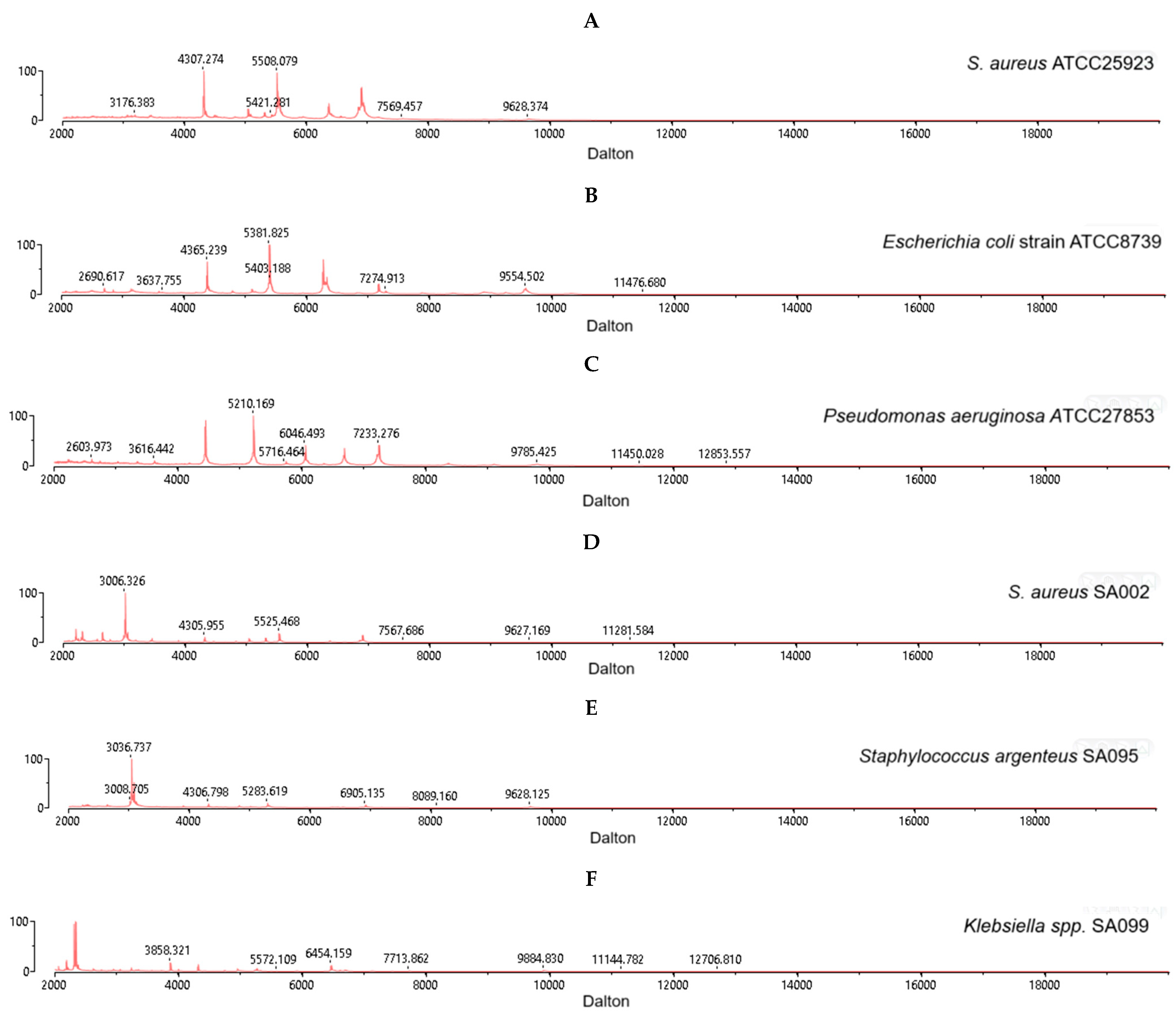
2.2. MALDI–TOF MS Identification of S. aureus

All three reference strains of S. aureus ATCC 25923 (Figure 1A), including Escherichia coli strain ATCC25922 (Figure 1B) and Pseudomonas aeruginosa strain ATCC27853 (Figure 1C), were correctly identified by using the BioMerieux VITEK 2 MALDI–TOF MS system with a high confidence value (99.9%) and m/z values ranging from 2000 to 20,000 daltons. Among a total of 100 clinical isolates, S. aureus isolate SA002 (Figure 1D), S. argenteus isolate SA095 (Figure 1E), and Klebsiella spp. isolate SA099 (Figure 1F) were identified. In addition, our analysis has provided the spectral signatures of S. aureus (n = 94, SA001–SA037, SA039, SA041–SA056, SA058–SA063, SA065–SA094, SA096–SA098, and SA100), S. argenteus (n = 5, SA038, SA040, SA057, SA064, and SA095), and Klebsiella spp. (n = 1, SA099) (Supplementary Table S1 and Figure S1).

2.3. Drug Susceptibility Test for S. aureus Isolates

S. aureus can cause problems related to multidrug resistance (MDR) and virulence in many infections. Therefore, a panel of frontline antibiotics with different modes of action, including trimethoprim/sulfamethoxazole and vancomycin, is being routinely tested to inform antibiotic stewardship. In addition to methicillin, vancomycin has been reported to be resistant to S. aureus infections. Herein, Staphylococcus spp. isolates were subjected to susceptibility testing for these drugs. The MIC values for each antibiotic, including clindamycin, erythromycin, gentamycin, linezolid, moxifloxacin, oxacillin, trimethoprim/sulfamethoxazole, and vancomycin, were obtained from bacterial isolates. The results of the drug susceptibility assessments of one hundred S. aureus isolates are indicated in Table 2 and Supplementary Table S3, in which most of the isolates exhibited a high rate of susceptibility to the tested antibiotics (p < 0.05). For example, the isolates SA003, SA008, SA011, SA014, SA024, SA029, SA043, SA048, SA057, SA062, SA064, SA071, SA081, SA087, SA091, SA096, SA099, and SA100 were resistant to clindamycin and erythromycin. The isolate SA070 showed intermediate resistance to erythromycin. Likewise, the isolates SA002, SA003, and SA081 were resistant to gentamycin. The isolates SA002, SA004, SA011, SA048, SA071, and SA096 were resistant to moxifloxacin. The isolate SA028 was resistant to trimethoprim/sulfamethoxazole. Moreover, the isolates SA002, SA004, SA011, SA029, SA043, SA048, SA057, SA071, SA081, SA087, SA096, and SA099 were detected as MRSA bacteria. The remaining 22 isolates exhibited drug resistance patterns. However, none were resistant to linezolid or vancomycin.

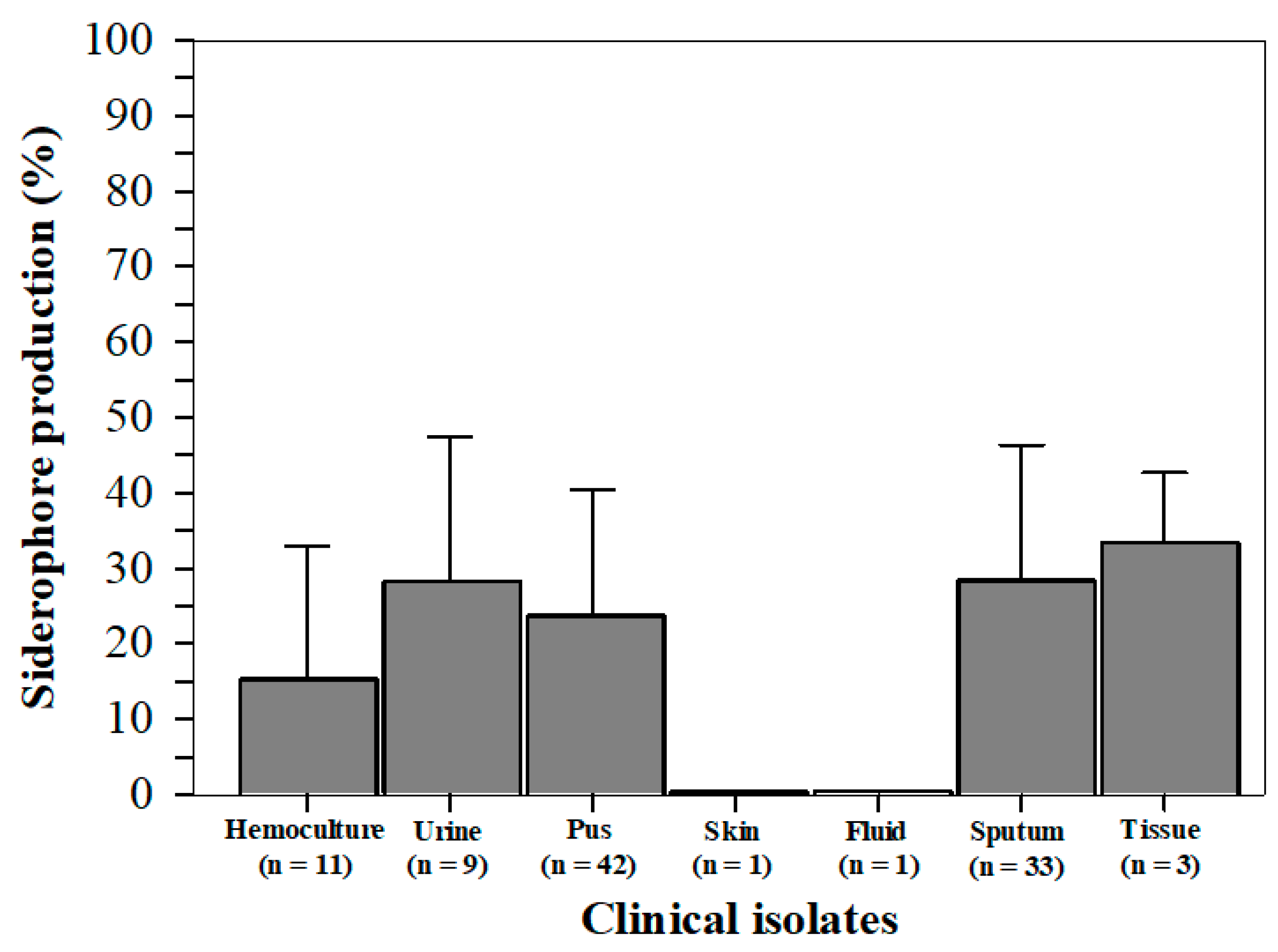
2.4. Siderophore Production from S. aureus Isolates

We tested siderophore activity using a CAS assay. Siderophores were produced chiefly in tissue isolates (33.4 ± 9.3%); moderately (28.3 ± 19.1, 28.3 ± 18.0, and 23.7 ± 16.8%) in urine, sputum, and pus isolates; minutely in hemoculture (15.3 ± 17.8%), but not at all in skin and fluid isolates (Figure 2). The percentage of siderophore production of each isolate is also presented in Table S4.

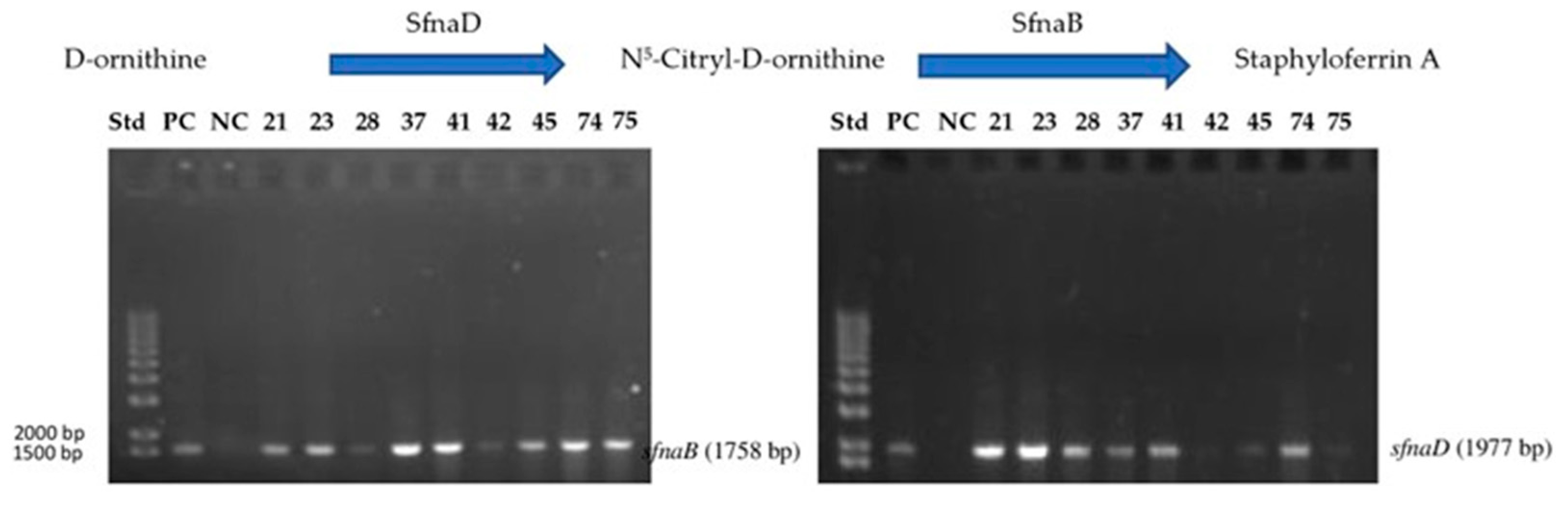
2.5. Genomic DNA and Staphyloferrin A Biosynthesis Gene

From polymerase chain reaction (PCR) analysis, the band for the sfnaB gene was detected in 34 samples, while the band for the sfnaD gene was detected in 18 samples. Here, the two genes were detected in 9 samples and labeled with the following numbers: 21, 23, 28, 37, 41, 42, 45, 74, and 75 (Figure 3).

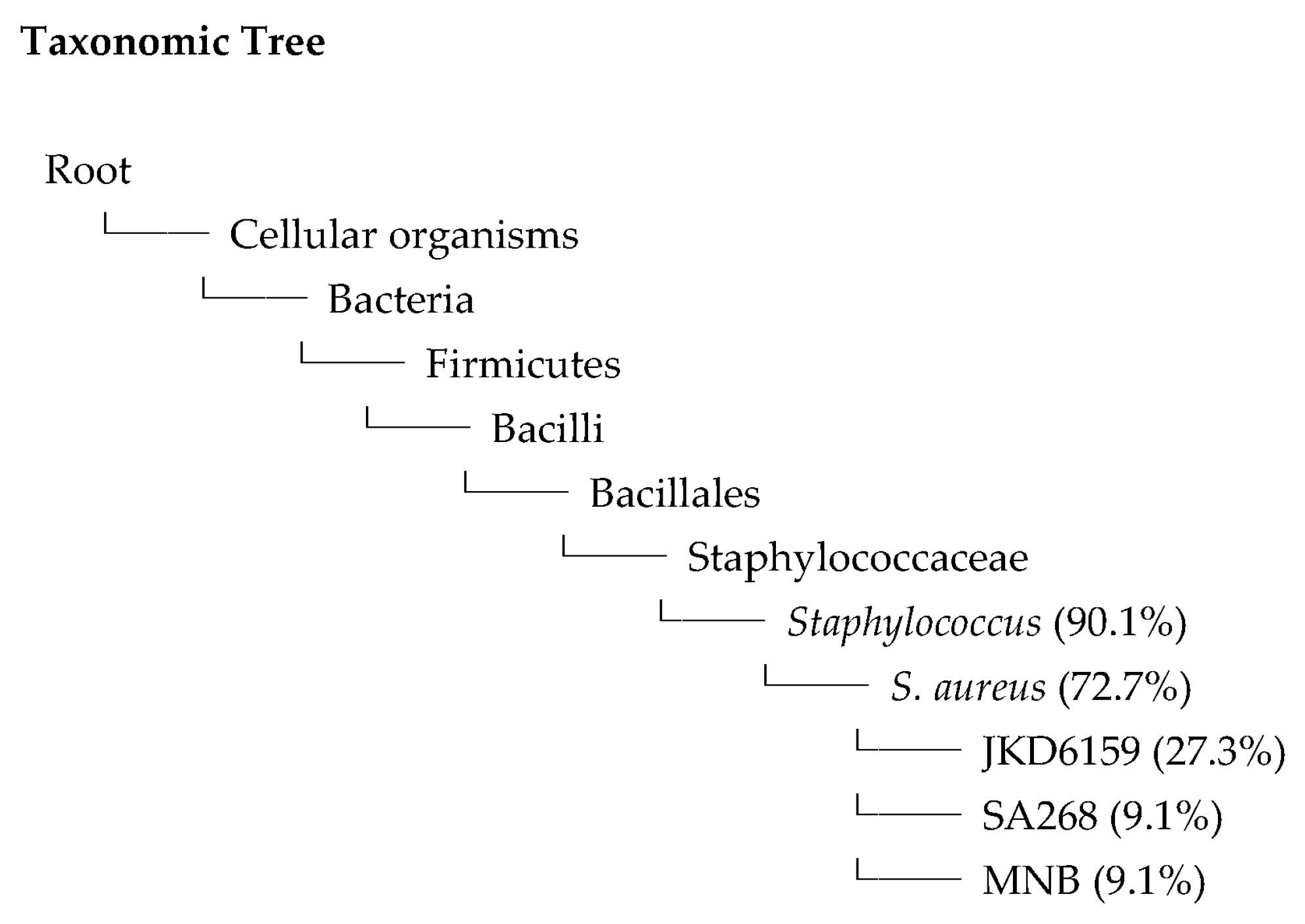
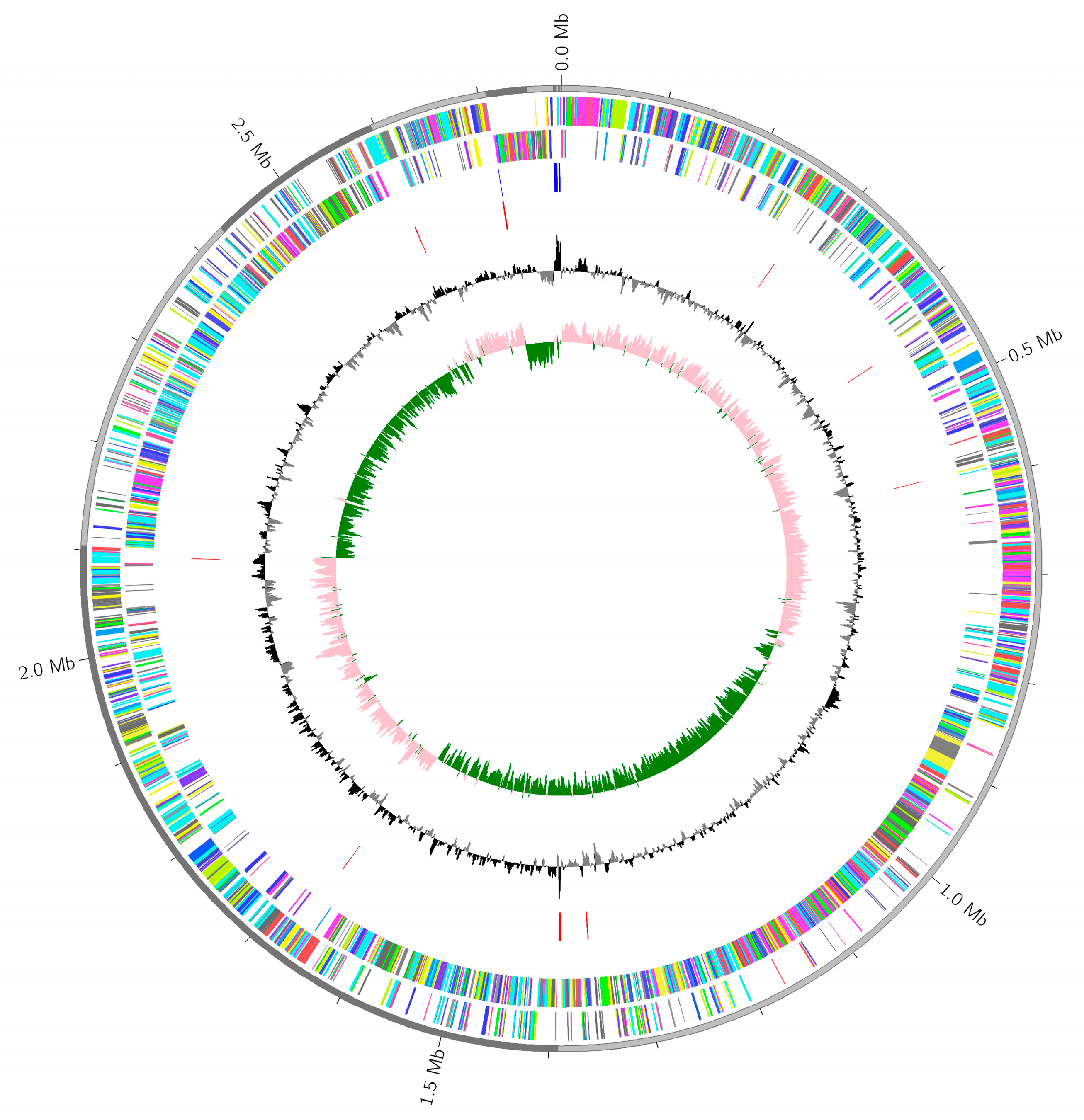
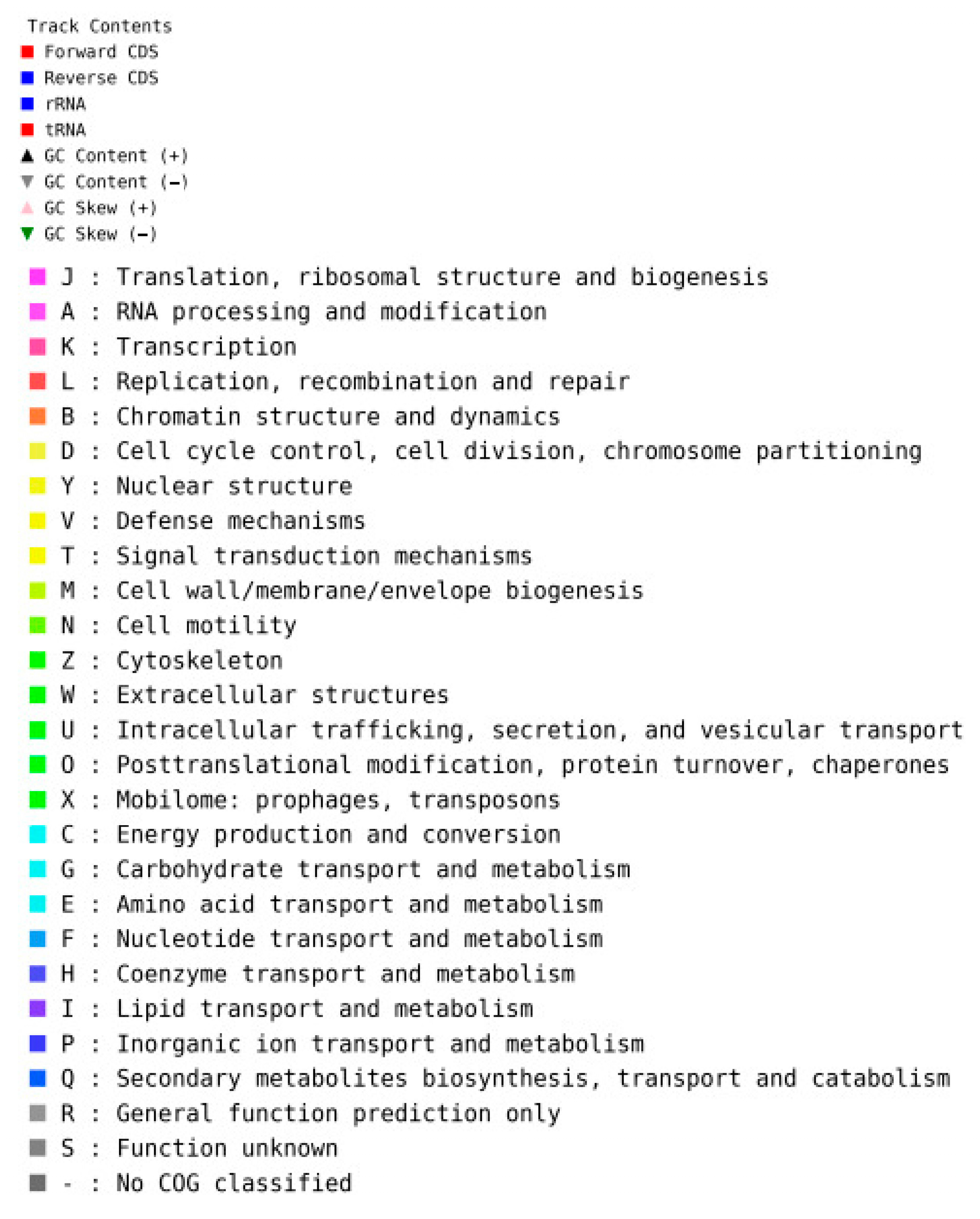
2.6. Whole Genome Sequences

From the phylogenetic analysis shown in Table 3 and Figure 4, S. aureus isolates obtained from the patient’s sputum (isolate SA041) revealed a sequence homology close to S. aureus (72.7%): S. aureus subspeciesJKD6159 (27.3%) to S. aureus SA268 (9.1%) and S. aureus MN8 (9.1%).
Each cluster of the orthologous groups (COG) of proteins is assembled as descendants from the same gene in the ancestral genome. A single genome map has been presented, indicating all assembled contigs achieved from the whole genome sequencing results of S. aureus (Figure 5). The top-edged circle represents all the contigs in the kb scale. The second and third circles show the coding sequencing (CDS) reference genes on the forward and reverse strands that have been represented in various colors according to COG categories. In addition, guanine (G), cytosine (C) content, and GC skews are shown in the last inner circle, respectively.

3. Discussion

Staphylococcus spp. are pathogenic Gram–positive cocci that can be treated by many classes of antibiotics; however, incidences of drug resistance have been reported [7,8]. Under iron limitations, bacteria have produced siderophores for iron acquisition from hosts [18]. These siderophores have also led to maximal virulence, thereby increasing the rate of bacterial distribution and promoting colonization and infection of the tissue [21,22]. In this study, we have included a total of one hundred specimens obtained from male and female patients, of which siderophores were produced mainly in the tissue, moderately in the urine, sputum, and pus, and minutely in the hemoculture but not in the skin and fluid isolates. In addition, drug sensitivity results have shown that most of the S. aureus colonies that were present in clinical isolates were sensitive to the antibiotics used, including clindamycin, erythromycin, gentamycin, linezolid, moxifloxacin, oxacillin, vancomycin, and a combination of trimethoprim and sulfamethoxazole. Nonetheless, Rocha et al. investigated bacterial isolates obtained from uterus and vagina specimens and reported a low level of production of biofilms and siderophores by coagulase-negative MRSA. This suggests the potential influence of certain host-related factors other than bacterial virulence [37].
The worldwide increase in antibiotic resistance has highlighted the need for new potential drugs and iron chelators that can function against bacterial pathogens. A susceptibility test for DFO siderophores was previously developed as a simple diagnostic tool for the identification of S. epidermidis and S. hominis obtained from hemocultures and other fluid specimens, indicating 96.4% efficiency, 97.3% sensitivity, and 91.8% specificity [38,39]. Additionally, two DFO and fosfomycin disc assays reported 99.5% sensitivity and 99.2% positive predictor values for the identification of S. epidermidis and S. hominis among coagulase-negative Staphylococcus spp. isolates [40]. Likewise, DFO-supplemented Columbia agar was adopted for the detection of MRSA with 94% sensitivity and 91% specificity [41]. An investigation of clinical peritoneal dialysis fluid and commensal skin samples demonstrated that S. epidermidis isolates produced the siderophore SA rather than SB, implying that SA production would be a virulence factor for Staphylococcal pathogenesis [42]. Davidov and colleagues reported that enterobactin and salmochelin siderophores effectively inhibited the growth of MRSA isolates; inversely, the low-dose siderophores enhanced bacterial growth [6] suggesting that suitable concentrations could stimulate or suppress S. aureus growth. Interestingly, Chakraborty and colleagues have indicated that macrocyclic polyketides obtained from marine heterotrophic Shewanella algae extract exerted Fe3+-chelating and anti-infective activities against vancomycin-resistant Enterococcus faecalis, MRSA, P. aeruginosa, and K. pneumonia, possibly by combating bacterial siderophores [43]. Consistently, bacteria sensitive to commercially available antibiotics expressed genes (1000–1400 base-pairs) that were determined to be involved in the biosynthesis and production of siderophores (MZ222387 and MZ222388) [44].
In routine microbiological assays, conventional bacteria identification requires a long culturing time of 12–16 h, the separation of the colonies for 24 h, and the identification of the species for 24–48 h by employing coagulase and CAT tests. Herein, we used the VITEK/MS method to confirm the S. aureus species in clinical isolates and compared the results with the two biochemical tests. In molecular analysis, MALDI–TOF/MS can detect proteins and lipids that are released from cells, as well as intact bacterial signatures, illustrating the different MS peaks formed by different peptide and protein m/z values, while indicating particular species and even genotypes within each species [14]. In addition, it can be used to characterize ribosomal protein biomarkers and mutations in the antibiotic resistance of E. coli [45,46]. Despite a fast, convenient, and actionable method, VITEK MALDI–TOF/MS requires an expensive instrument and access to the spectral database library of peptide mass fingerprints of each specific bacteria to provide valuable diagnostic information [15]. Remarkably, this technique can identify Staphylococcus (97.2%), Streptococci (97.8%), Enterococci (100%) [16,47], Cronobacter, and Vibrio isolates [47], both rapidly and correctly.
Production and activity of SA and SB siderophores can be considered a virulence factor for the pathogenesis of coagulase-negative and coagulase-positive staphylococci in environments and clinical isolates. For rapid screening, colorimetric CAS, Arnow and Csaky liquids, and agar methods have been used to detect siderophore production from bacterial colonies and clinical isolates. As a consequence, the S. aureus strain ATCC 6538 and S. epidermidis obtained from human peritoneal dialysate exhibited higher iron utilization system (IUS) activity and grew better than the S. aureus ATCC 25923 strain, suggesting that the IUS activity plays an important role in bacterial growth and pathogenesis [48]. Thus, S. aureus per se adapts to the habitat by increasing the expression of the genes facilitating the acquisition of iron via the iron-binding molecule SB (sfnb) and the heme consumption protein (isd) pathways [49]. P. aeruginosa and S. aureus are the most prevalent respiratory pathogens causing thick mucus and chronic polymicrobial lung infections in cystic fibrosis patients. Accordingly, the supernatant derived from the P. aeruginosa culture reduced the sensitivity of S. aureus growth to a frontline antibiotic vancomycin and protected S. aureus from cell wall-active antibiotics and protein synthesis inhibitors, possibly by the effects of Pseudomonas pyoverdine and pyochelin siderophores [50]. Consistently, pyoverdine exhibited anti-microbial activity and effectively decreased the growth of pathogenic Acinetobacter baumannii, K. pneumoniae, and S. aureus in a concentration-dependent manner [51]. In addition, certain virulence factors, including the staphyloxanthin pigment obtained from S. aureus and the catecholate-type enterobactin siderophores derived from E. coli, are secreted during neutrophil phagocytosis, for which only staphyloxanthin prevented the bacteria from the toxicities of hypochlorous acid and related chloramines [52]. Moreover, a recent study has reported that enterobactin and salmochelin S4 siderophores inhibited the growth of S. aureus, including MRSA, in clinical isolates [6]. Furthermore, the S. aureus FhuD2 gene and protein involved in iron–hydroxamate chelate uptake were upregulated in infected tissues and were required for staphylococcal dissemination and abscess formation [53].
Genome analysis can provide insight into how S. aureus adapts to the environment and infection areas. Biosynthetic gene clusters are consecutive gene subsets that are present in various organisms and employed to produce specialized metabolites, which are mostly the non-ribosomal peptide synthase type and can exhibit antibacterial, anticancer, and iron-chelating activities. PCR-based identification is suitable for slow-growing and non-cultivable bacteria present in specific samples (e.g., cerebrospinal fluid and plasma) and requires nucleic acids obtained from isolated colonies [54,55]. Bacterial phylogenetic analysis is conducted to determine the evolutionary relationships among bacterial species and genera based on their 16S rRNA gene sequences. Based on 16S rRNA identification of staphylococcal genome sequences, S. chromogenes and S. simulans were the most frequent species, and S. aureus was the second most prevalent species associated with clinical mastitis; whereas, S. chromogenes, S. simulans, S. Xylosus, S. haemolyticus, S. cohnii, S. epidermidis, S. capitis, S. sciuri, S. gallinarum, S. warneri, S. equorum, S. saprophyticus, S. succinus, S. arlettae, and S. agnetis were the most common species associated with subclinical mastitis [35]. Consistently, 16S rRNA genes of S. aureus, S. epidermidis, S. caprae, and S. capitis have a close relationship with those observed in human clinical isolates and share a lot of adhesion genes [56,57].
Taken together, the findings suggest that the virulence factor from S. aureus, such as the sfna compound encoded by the sfna gene, is produced to sequester extracellular iron for cell metabolism and proliferation. In terms of its advantages, high-throughput MALDI–TOF MS contributes to a decrease in the amount of time involved with biochemical assays that are used for identifying bacterial species, which can also effectively save on the costs of the tests and lower the mortality rates of patients. Predominantly, the VITEK MS platform is a rapid (<5 min) and accurate (>90%) method of detection for identifying bacterial species by analysis of the molecular spectra of specific biomarkers (e.g., lipids and peptides) obtained from bacterial cell compositions. Nonetheless, the results are limited by intragenomic heterogeneity and certain difficulties associated with the analysis of all isolates, as well as difficulties in distinguishing between related Staphylococcus spp. This study investigated siderophore production in only one hundred staphylococcal isolates of the patients’ specimens, sfna gene expression in nine isolates, and the complete genome sequence in one sputum isolate.

4. Materials and Methods

4.1. Chemicals and Reagents

Trisodium 5–[(3–carboxy–5–methyl–4–oxocyclohexa–2,5–dien–1–ylidene)(2,6–dichloro–3–sulfonatophenyl)methyl]–3–methyl–2–oxidobenzoate or CAS (Product number 199532), ferric chloride hexahydrate (FeCl3·6H2O) (Product number 31232, ≥99% pure), hexadecyltrimethylammonium bromide (HDTMA) (Product number H5882, >98% pure), ethylenediamine–di(o–hydroxyphenylacetic acid (EDDHA) (Product number E4135, 98% pure), and piperazine–N, N′–bis(2–ethanesulfonic acid (PIPES) (Product number P6757) were purchased from Sigma-Aldrich Chemicals Company (Saint Louis, MO, USA). Luria–Bertani (LB) broth (Catalog number AAJ75882A1), Tris–acetate ethylenediamine tetraacetic acid (TAE) (Catalog number AAJ63931K7), Tris–minimal succinate (TMS) broth (Catalog number sc-264476), and agar plates (Catalog number 0701) were obtained from Thermo-Fischer Scientific Inc., Waltham, MA, USA. Antimicrobial drugs, including clindamycin, erythromycin, gentamicin, linezolid, moxifloxacin, oxacillin, trimethoprim/sulfamethoxazole, and vancomycin, were obtained from a pharmacy located at Maharaj Nakorn Chiang Mai Hospital, Faculty of Medicine, Chiang Mai University, Chiang Mai, Thailand.

4.2. Ethics

An expedited review for the accession of the patient and the clinical specimen information was submitted to the Human Ethics Committee of the Faculty of Medicine, Chiang Mai University, Thailand, and acceptance was generously granted by Emeritus Professor Panja Kulapongs, MD., Chairman (Research ID: 0404. Study Code: BIO-2566–0405, Date of Approval: 2 February 2024).

4.3. Institutional Review Board Statement

The protocol for biological material transfer and laboratory investigations was submitted to the Institutional Biosafety Committee (IBC) of the Faculty of Medicine, Chiang Mai University (Reference number: CMUIBC0265001, Date: 10 February 2022). It was then approved by the IBC and authorized by Associate Professor Dr. Sirikarn Lumpakarn, MD., PhD., Chairman (Reference number: CMUIBC02015/2565, Date: 11 August 2022).

4.4. Specimen Collection and Staphylococcus Culturing

Staphylococcus aureus strain ATCC 25923 was the pathogenic indicator strain stored at −80 °C in 40% glycerol. A total of one hundred isolates were recovered from human clinical specimens at Maharaj Nakorn Chiang Mai Hospital, Faculty of Medicine, Chiang Mai University, from December 2020 to February 2022. According to the isolation method described in the United States Food and Drug Administration Bacterial Analytical Manual 2001, all isolates and S. aureus ATCC 25923 reference strains were cultured on LB agar plates overnight and identified for S. aureus bacteria using Gram staining/light microscopy. The selected S. aureus colonies were inoculated into modified iron-deprived LB media, as recommended by Lindsay et al. [58], in which ferric chloride was added to the media to achieve concentrations of 0–2 µM. Afterward, the bacteria suspension was adjusted with the culture medium to reach an optical density of 0.5 McFarland units and incubated at 37 °C in a 5% CO2 incubator (Heraeus HeraCell150, Heraeus Group, Hanau, Germany) via gentle shaking at 150 rpm. Finally, the S. aureus growth was monitored every 24 h by measuring the optical density (OD) value at 600 nm against the culture medium with a double-beam spectrophotometer (Model UV-1900i Plus, Shimadzu Corporation, Kyoto, Japan) [59]. The S. aureus inoculum was identified using the Gram staining/microscopy technique, coagulase test, catalase test, and imaging mass spectrometry, which will be described below. In addition, the S. aureus stocks were maintained in LB broth containing 20% (v/v) glycerol and kept in a deep freezer at −70 °C.

4.5. Biochemical Identification of S. aureus

The coagulase test was performed on overnight bacterial cultures by mixing them with rabbit EDTA plasma. Then, the mixture was incubated at 37 °C for 4–6 h, and clot formation was observed after 4 h, indicating a coagulase-positive reaction by S. aureus. No clot formation meant a coagulase-negative reaction and that the isolate was from some other species of Staphylococcus [33]. In the CAT assay, the LB culture was directly flooded with a few drops of 3% (v/v) hydrogen peroxide, and bubbles were immediately observed.

4.6. MALDI-TOF/MS Analysis of Microbial–Specific Protein

An automated VITEK MS V3.2 machine (bioMérieux SA., Marcy–I’Etoile, Lyon, France) was run by the reagent (Reference number 411071) and operated via MALDI–TOF/MS technology. The process incorporated a comprehensive FDA 510(k) Vitek MS database version 2 for the in vitro diagnosis of bacteria, fungi, and mycobacteria. The machine providing the protein profile of the S. aureus strain, the phenotype-related biomarkers, and the drug-resistant strains was operated according to the prescribed protocol and manufacturer’s instructions [60]. Briefly, S. aureus colonies of the isolates were randomly selected and streaked onto the LB medium, and the E. coli ATCC 8739 calibration strain was streaked onto de Man, Rogosa, and Sharpe (MRS) agar at a pH of 6.8. They were then supplemented with 0.05% (w/v) L-cysteine HCl and then incubated at 37 °C for 24 h. Each colony was then purified by cross-streaking, while a portion of the colony was applied to a spot on a VITEK MS–DS target slide. The VITEK MS–CHCA matrix (a saturated solution of cyano–4–hydroxycinnamic acid in 50% acetonitrile and 2.5% trifluoroacetic acid) (1 µL) was immediately added to the spots and allowed to dry. Finally, the slide was subjected to multiple laser shots inside the VITEK MS instrument, which was operated according to the manufacturer’s instructions and protocol. Via computer operation, VITEK MS Acquisition Station software (Vitek MS version 1.0.0) was used to set up the parameters and record the data according to the manual. For setting up parameters, an m/z range was optimized between 2000 and 20,000, spectra were recorded in the positive linear mode, and 200–500 laser shots were accumulated from each spot. VITEK MS was calibrated with the Escherichia coli strain ATCC8739 for the positive control, and the matrix alone was employed for the negative control. In terms of data acquisition and interpretation, the generated mass spectra displayed three views, while an average of the peaks was processed by the machine computer. The sophisticated spectrum classifier algorithm autonomously recognized the organism by matching the acquired peaks with the reference spectrum of each asserted species to identify S. aureus. A percentage probability (confidential value) was then calculated, and this number represents the similarity of specific peaks between the generated spectrum and the database spectrum. At a confidence score of >99%, the analytical results were expressed as 2300–3000 for highly reliable species identification, 2000–2299 for highly reliable genus identification, 1700–1999 for probable genus identification, and 0–1699 for no reliable identification. When a single unique pattern was not identified, a list of possible bacteria was reported as “low discrimination”, or the species/strain could not be determined within the scope of the database and was reported as “no identification”.

4.7. Drug Susceptibility Testing

Drug susceptibility of all isolates was determined using the Sensititre™ ARIS 2X System (Thermo Fisher Scientific, Waltham, MA, USA) according to the manufacturer’s instructions and in accordance with the Clinical and Laboratory Standards Institute (CLSI) M100 Guidelines (2021) published in: Performance Standards for Antimicrobial Susceptibility Testing, 31st ed., Wayne Carley, Science and Education Publishing, Newark, DE, USA) [61]. Bacterial colonies obtained from the purity plates were prepared to 0.5 McFarland in cation-adjusted Mueller–Hinton (CAMH) broth using a Sensititre nephelometer. The suspension was dispensed into the Sensititre™ standard inoculum tray, while an automated inoculator (Sensititre™ Auto–Inoculator) was used to transfer the suspension into preloaded Sensititre™ susceptibility plates containing dried concentrations of antibiotics. The inoculated microdilution plates were sealed with adhesive covers, incubated at 35 ± 1 °C for 16–24 h under ambient air, and read using the ARIS 2X system, which automatically detected minimum inhibitory concentration (MIC) values by scanning the bacterial growth in each well. Accordingly, the MIC value, which is the lowest concentration that inhibited the visible growth of the bacteria, was obtained for the determination of clindamycin (S ≤ 0.5 µg/mL, I = 1–2 µg/mL, R ≥ 4 µg/mL), erythromycin (S ≤ 0.5 µg/mL, I = 1–4 µg/mL, R ≥ 8 µg/mL), gentamicin, (S ≤ 4 µg/mL, I = 8 µg/mL, R ≥ 16 µg/mL), linezolid (S ≤ 4 µg/mL, R ≥ 8 µg/mL), moxifloxacin (S ≤ 0.5 µg/mL, I = 1 µg/mL, R ≥ 2 µg/mL), oxacillin (S ≤ 2 µg/mL, R ≥ 4 µg/mL), trimethoprim/sulfamethoxazole (S ≤ 2/38 µg/mL, R ≥ 4/76 μg/mL), and vancomycin (S ≤ 2 µg/mL, I = 4–8 µg/mL, R ≥ 16 µg/mL).

4.8. Detection of Siderophore Production by Staphylococcus Isolates

4.8.1. Preparation of Siderophore

S. aureus strains were grown with aeration in TMS broth containing 0.1 μM EDDHA for 40 h at 37 °C. Cells were removed by centrifugation, and supernatants were lyophilized. The dried supernatant was extracted with methanol (one-fifth of the original supernatant volume), passed through Whatman No. 1 filter paper to remove any insoluble materials, and then rotary evaporated. The siderophore sample was solubilized in DI water to 5% of the original supernatant volume and subjected to CAS assay.

4.8.2. Colorimetric CAS Assay

Siderophores can be detected in biological fluids, liquid, and agar media using a colorimetric CAS assay [23,62], in which the chelator sequesters the iron from a blue-colored Fe3+–CAS complex (λmax 630 nm) and decreases the color intensity. In a fresh preparation of the CAS reagent, 6 mL of 10 mM HDTMA solution, 1.5 mL of 1 mM FeCl3 solution previously dissolved in 10 mM HCl, 7.5 mL of 2 mM stock CAS solution, and 4.307 mg of PIPES were mixed thoroughly. The working CAS reagent was then adjusted to a final pH value of 5.6 and stored in the dark in a refrigerator at 4 °C. In the assay, the bacterial supernatant (100 µL each) was first added to each well of a 96-well microplate, and the CAS reagent (100 µL each) was subsequently added. Finally, the mixture was incubated at room temperature for 4 h, and the OD value was measured at 630 nm against the CAS reagent using an ELISA microplate reader (Model: MB580, Zhengzhou Medbes International Trading Limited Company, Henan, China). The percentage of siderophore production unit (SPU) was calculated by using the following Formula (1):
SPU = 100 × (ODR − ODS)/ODR
ODR = optical density of CAS reagent measured at 630 nm, ODS = optical density of CAS treated with the sample measured at 630 nm.

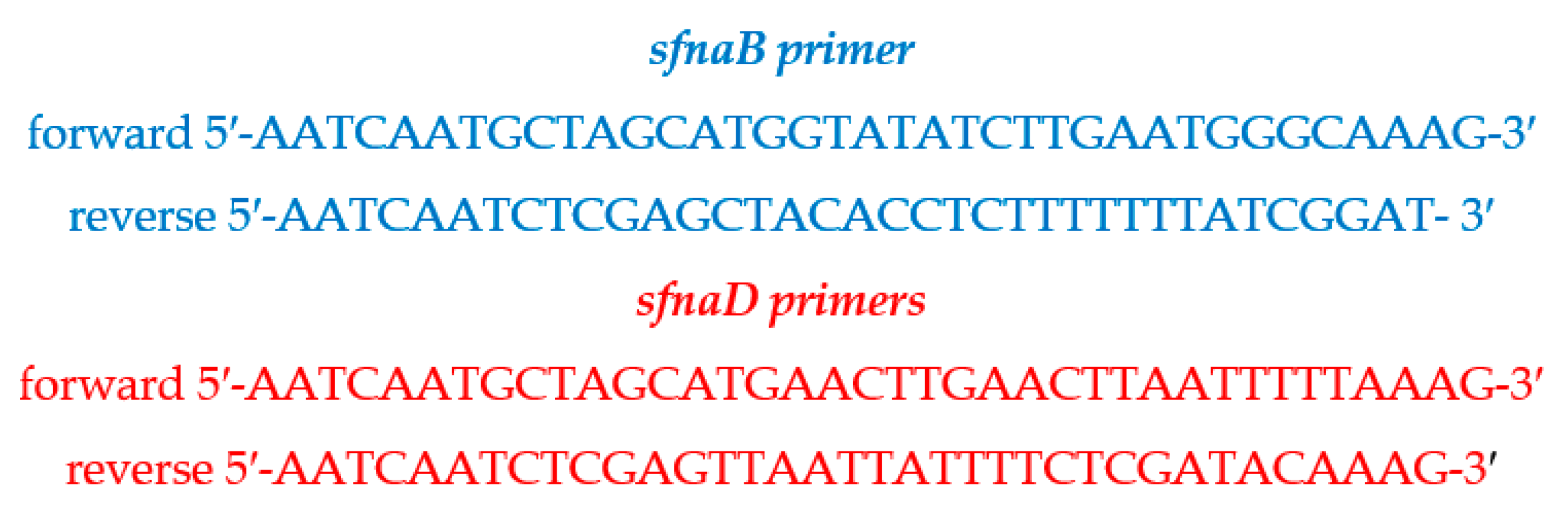
4.9. Identification of sfnaB and sfnaD Genes

4.9.1. Genomic DNA Extraction

Firstly, the S. aureus LB medium (1 mL for each isolate) was centrifuged to sediment the cells, resuspended in 0.5 mL of deionized water (DI), boiled in a water bath at 100 °C for 5 min, shaken vigorously to ensure the disruption of the cell wall, and centrifuged at 13,000× g for 5 min [63]. Then, genomic DNA obtained from the pellet was extracted and purified using DNeasy Blood & Tissue kits (QIAGEN, Valencia, CA, USA) following the manufacturer’s protocol and instructions. The concentration of the purified DNA samples was measured at 260 nm optical density using a NanoDrop 1000 spectrophotometer (NanoDrop Technology, Rockland, DE, USA) and stored at −20 °C until further use [33].

4.9.2. Polymerase Chain Reaction Amplification

A fragment of the gene was amplified using sfnaB and sfnaD primer sets (Figure 6) for PCR and identification of the sfnaB and sfnaD genes [19].
Briefly, a total of 50 μL of the PCR reaction included 25 μL PCR HotStarTaq Master Mix (QIAGEN) and a 25 μL solution containing 200 nM of each primer, 1.5 mM additional MgCl2 (Promega, Madison, WI, USA), and 50 ng template DNA, which were diluted in PCR-grade water. QIAGEN HotStarTaq Master Mix solution containing HotStarTaq DNA polymerase, PCR buffer, and deoxyribonucleoside triphosphates (dNTPs) was diluted to reach a final concentration of 1.5 mM MgCl2 and 200 μM of each dNTP. The reactions were run for 30 cycles, of which each cycle was maintained at 95 °C for 3 min, 55.2 °C for 1 min, and 72 °C for 1 min in a GeneAmp PCR 9700 thermocycler (Applied Biosystems, Foster City, CA, USA) with an initial hot start (94 °C for 3 min) and a final extension step (72 °C for 6 min). The PCR-amplified product was electrophoresed on a 0.8% agarose gel at 100 volts for 30 min using TAE buffer and 1 kb plus DNA ladder markers (InvitrogenTM, Thermo-Fischer Scientific Inc., Waltham, MA, USA). It was then visualized under ultraviolet light exposure after staining [32].

4.9.3. Nucleotide Sequencing and Data Analysis

The confirmed PCR-amplified products were sequenced using ABI BigDye v3.1 dye chemistry and AB 3500 XL automated DNA sequencers (Applied Biosystems, Foster City, CA, USA). To perform DNA sequencing, the PCR products were enzymatically cleaned before cycle sequencing, and 3 μL of ExoSAP–IT (USB Corporation, Cleveland, OH, USA) was added to the amplified PCR products (5 μL each). The mixture was then incubated at 37 °C for 20 min, followed by 80 °C for 15 min on the GeneAmp PCR 9700 thermocycler. The sequencing reactions contained cleaned PCR product (2 μL), BigDye Terminator v3.1 Ready Reaction Mix (1 μL), 5× sequencing buffer (2 μL), forward or reverse-sequencing primer (1.6 pmol), and water to a final volume of 20 μL. The sequencing reactions were subjected to 25 amplification cycles (each cycle was maintained at 96 °C for 30 s, 50 °C for 15 s, and 60 °C for 4 min) and held at 4 °C in a GeneAmp PCR 9700 thermocycler. Afterward, the reactions were cleaned up with Performa DTR Gel Filtration Cartridges (Edge Bio, Gaithersburg, MD, USA) according to the manufacturer’s instructions and protocol. The sequence accuracy of the DNA sequences was confirmed by two-directional sequencing. Multiple alignments of the nucleotide sequences were carried out using BioEdit, ClustalW, and Geneious programs version 7.2 (Informer Computer Systems, Inc., Garden Grove, CA, USA) with manual adjustments [32]. The generated nucleotide sequences of the partial 16S rRNA regions of all the Staphylococcus isolates were accessed and deposited in the GenBank database.

4.10. Whole Genome Sequencing and Phylogenetic Analysis

The putative staphyloferrin A-produced sample SA041 was employed to perform shotgun Illumina sequencing. The next-generation sequencing reads were first removed from primer sequences using Trim Galore version 0.6.7. Using a toolkit, quality control reads were then assembled de novo using a Saint Petersburg Genome Assembler (SPAdes) V3.13.0 and polished using Pilon version 1.23, which had been implemented in a Unicycler 0.5 [64]. Genome completeness was assessed using CheckM V1.2.1. Subsequently, Quast V5.2.0 was used to extract assembly statistics [65]. Genome visualization was performed using a microbial genome circular plotter. Gene calling and annotation were undertaken using Prokka V1.14.5. Accordingly, the predicted amino acid sequences were mapped to different functional categories, e.g., Kyoto Encyclopaedia of Genes and Genomes Orthology, Clusters of Orthologous Groups, and carbohydrate-active enzymes using eggNOG–mapper V–2.1.3 against the EggNog database 5.

4.11. Statistical Analysis

Data were collected and tabulated using Microsoft Excel software (version 16.75; Seattle, WA, USA). Descriptive analysis was performed using the Statistical Package for the Social Sciences Statistics version 21 for Windows (IBM Corporation, Armonk, NY, USA). The Shapiro–Wilk test was used to evaluate the distribution of normal or non-normal data. Quantitative variables were expressed as individual, percentage, and mean ± SD values. One-way analysis of variance with Tukey’s post hoc test for parametric data was used to compare the two groups to determine statistical significance, for which a p-value < 0.05 was considered statistically significant.

5. Conclusions

Staphylococcus-infected clinical isolates were positive for catalase and coagulase tests, of which MALDI–TOF MS signatures indicated 94% S. aureus. Most of the isolates were sensitive to the tested antibiotics. Sfna siderophores encoded by sfnaD, sfnaB, and sfnaD/sfnaB genes were produced chiefly in tissue, urine, sputum, pus, and hemoculture isolates, but not in the skin and fluid isolates. Genome sequence homology was mostly close to S. aureus, followed by subspecies JKD6159, SA268, and MN8. Further studies are needed involving more clinical samples to provide useful virulence profiling and a target for vaccine development against siderophore function during staphylococcal infections. The VITEK MALDI–TOF/MS instrument and the associated learning machines should be applied for rapid and accurate detection of staphyloferrins, enterobactin, salmochelin, pyoverdine, and aerobactin belonging to S. aureus, E. coli, Salmonella typhimurium, P. aeruginosa, and K. pneumoniae, respectively.

Supplementary Materials

The following supporting information can be downloaded at: https://www.mdpi.com/article/10.3390/antibiotics14050521/s1, Table S1: Patients’ information, specimen types, microbiological and biochemical test results. Table S2: Identification of Staphylococcus spp. isolates (n = 100) by microbiological, biochemical, VITEK MS, and rRNA sequencing methods. Table S3: Drug susceptibility test for clinical isolates (n = 100). Table S4: Siderophore activity assayed in the clinical isolates (n = 100), including plus (n = 41), sputum (n =34), hemoculture (n = 11), urine (n = 9), tissue (n = 3), skin (n = 1), and fluid (n = 1). Figure S1: VITEK MS illustrations of Staphylococcus spp. isolates (SA001-SA100).

Author Contributions

Conceptualization, W.P., P.K. (Pimpisid Koonyosying), A.C., M.D. and S.S.; methodology, W.P., P.S., R.S., P.K. (Phadungkiat Khamnoi) and M.D.; formal analysis, W.P., A.C. and Y.R.; investigation, W.P., P.S. and P.K. (Phadungkiat Khamnoi); supervision, P.K. (Pimpisid Koonyosying), M.D. and S.S.; project administration, P.K. (Pimpisid Koonyosying) and Y.R.; funding acquisition, W.P., P.K. (Pimpisid Koonyosying), M.D. and S.S.; writing—original draft preparation, W.P., M.D. and S.S.; writing—review and editing, M.D. and S.S. All authors have read and agreed to the published version of the manuscript.

Funding

This work was supported by the Presidential Ph.D. Scholarship (Number 2563–010, Date: 31 May 2020, CMU Graduate School, Chiang Mai University; Faculty of Medicine Fund, Chiang Mai University (Grant number: 009/2023, Date: 3 October 2023 and the distinguished Professor Dr. Somdet Srichairatanakool, PhD., National Research Council of Thailand (Grant number: N42A670732, Date: 17 May 2024). The Article Processing Charge was funded by the Faculty of Medicine and Research Administration Unit, Chiang Mai University, Thailand.

Institutional Review Board Statement

The study titled “Production of staphyloferrin a siderophore from Staphylococcus aureus and inhibitory effect on skin infection and inflammation in vitro (STUDY CODE: BIO–2566–0405, Research ID: 0405)” was conducted following the rules of the Declaration of Helsinki of 1975, which revised in 2013, and was approved by the Research Ethical Committee (REC) Panel 5 of Faculty of Medicine, Chiang Mai University, Chiang Mai, Thailand, and authorized by Emeritus Professor Panja Kulapongs, MD., the Chairman, on the 2 February 2024. An expedited review for the accession of each patient and the clinical specimen information was submitted to the Human Ethics Committee of the Faculty of Medicine, Chiang Mai University, Thailand, and has been approved by Emeritus Professor Panja Kulapongs, MD., Chairman (Research ID: 0404. Study Code: BIO–2566–0405, Date of Approval: 2 February 2024). The protocol for biological material transfer and laboratory investigations titled “Production of staphyloferrin a siderophore from Staphylococcus aureus and inhibitory effect on skin infection and inflammation (Reference number: CMUIBC0265001, Date: 10 February 2022) was approved by the IBC of the Faculty of Medicine, Chiang Mai University and authorized by Associate Professor Dr. Sirikarn Lumpakarn, MD., PhD., Chairman (Reference number: CMUIBC02015/2565, Date: 11 August 2022).

Informed Consent Statement

Patients’ informed consent was waived by the REC due to the collection of clinical specimens from the Clinical Microbiology Laboratory, Maharaj Nakorn Chiang Mai Hospital, Faculty of Medicine, Chiang Mai University.

Data Availability Statement

All data presented in this study are available from the corresponding author upon reasonable request.

Acknowledgments

We are thankful to Sithichoke Tangphatsornruang, National Center for Genetic Engineering and Biotechnology, National Science and Technology Development Agency, Thailand Science Park, for S. aureus genome analysis and to Thanawat Yasen for the preparation of S. aureus isolates and bacterial MS data analysis.

Conflicts of Interest

The authors declare no conflicts of interest. The funders had no role in the design of this study; in the collection, analysis, or interpretation of the data; in the writing of the manuscript; or in the decision to publish the results.

Abbreviations

The following abbreviations have been used in this manuscript:
ABCATP synthase-binding cassette
CCytosine
CAMHCation-adjusted Mueller–Hinton (broth)
CAMRSACommunity-acquired methicillin-resistant S. aureus
CASChrome azurol S
CATCatalase
CDSCoding sequencing
CLSIClinical and Laboratory Standards Institute
COGCluster of orthologous groups
DFODesferrioxamine
DIDeionized water
DNADeoxyribonucleic acid
dNTPsDeoxyribonucleoside triphosphates
EDDHAEthylenediamine–di(o–hydroxyphenylacetic acid)
EDTAEthylenediamine tetraacetic acid
Fe3+Ferric ion
Fe2+Ferrous ion
GGuanine
HDTMAHexadecyltrimethylammonium bromide
IBCInstitutional Biosafety Committee
IUSIron utilization system
LBLuria–Bertani (broth)
MALDI–TOF/MSMatrix-assisted laser desorption ionization–time of flight mass spectrometry
MICMinimal inhibitory concentration
MRSMan, Rogosa and Sharpe
MRSAMethicillin-resistant S. aureus
m/zMass-to-charge ratio
NCNegative control
NISNon-ribosomal peptide synthetase
ODOptical density
PCPositive control
PCRPolymerase chain reaction
PIPESPiperazine–N, N′–bis(2–ethanesulfonic acid)
rRNARibosomal ribonucleic acid
S. aureusStaphylococcus aureus
SDStandard deviation
sfnaStaphyloferrin A
sfnbStaphyloferrin B
S. epidermidisStaphylococcus epidermidis
S. hominisStaphylococcus hominis
S. hyicusStaphylococcus hyicus
S. lugdunensisStaphylococcus lugdunensis
SPUSiderophore production unit
TAETris–acetate ethylenediamine tetraacetic acid
TMSTris–minimal succinate
T/STrimethoprim/sulfamethoxazole
VRSAVancomycin-resistant S. aureus
v/vVolume by volume
w/vWeight by volume

References

  1. Jones, T.F.; Kellum, M.E.; Porter, S.S.; Bell, M.; Schaffner, W. An outbreak of community-acquired foodborne illness caused by methicillin-resistant Staphylococcus aureus. Emerg. Infect. Dis. 2002, 8, 82–84. [Google Scholar] [CrossRef] [PubMed]
  2. Mihai, M.M.; Holban, A.M.; Giurcaneanu, C.; Popa, L.G.; Buzea, M.; Filipov, M.; Lazar, V.; Chifiriuc, M.C.; Popa, M.I. Identification and phenotypic characterization of the most frequent bacterial etiologies in chronic skin ulcers. Rom. J. Morphol. Embryol. 2014, 55, 1401–1408. [Google Scholar]
  3. Adhikari, R.P.; Karauzum, H.; Sarwar, J.; Abaandou, L.; Mahmoudieh, M.; Boroun, A.R.; Vu, H.; Nguyen, T.; Devi, V.S.; Shulenin, S.; et al. Novel structurally designed vaccine for S. aureus alpha-hemolysin: Protection against bacteremia and pneumonia. PLoS ONE 2012, 7, e38567. [Google Scholar] [CrossRef] [PubMed]
  4. Balaban, N.; Rasooly, A. Staphylococcal enterotoxins. Int. J. Food Microbiol. 2000, 61, 1–10. [Google Scholar] [CrossRef]
  5. McDanel, J.S.; Perencevich, E.N.; Diekema, D.J.; Herwaldt, L.A.; Smith, T.C.; Chrischilles, E.A.; Dawson, J.D.; Jiang, L.; Goto, M.; Schweizer, M.L. Comparative effectiveness of beta-lactams versus vancomycin for treatment of methicillin-susceptible Staphylococcus aureus bloodstream infections among 122 hospitals. Clin. Infect. Dis. 2015, 61, 361–367. [Google Scholar] [CrossRef] [PubMed]
  6. Davidov, Y.; Tejman-Yarden, N.; Robinson, A.; Rahav, G.; Nissan, I. Enterobactin and salmochelin S4 inhibit the growth of Staphylococcus aureus. Front. Cell Infect. Microbiol. 2025, 15, 1456046. [Google Scholar] [CrossRef]
  7. Ippolito, G.; Leone, S.; Lauria, F.N.; Nicastri, E.; Wenzel, R.P. Methicillin-resistant Staphylococcus aureus: The superbug. Int. J. Infect. Dis. 2010, 14 (Suppl. S4), S7–S11. [Google Scholar] [CrossRef]
  8. Turner, N.A.; Sharma-Kuinkel, B.K.; Maskarinec, S.A.; Eichenberger, E.M.; Shah, P.P.; Carugati, M.; Holland, T.L.; Fowler, V.G., Jr. Methicillin-resistant Staphylococcus aureus: An overview of basic and clinical research. Nat. Rev. Microbiol. 2019, 17, 203–218. [Google Scholar] [CrossRef]
  9. Demling, R.H.; Waterhouse, B. The increasing problem of wound bacterial burden and infection in acute and chronic soft-tissue wounds caused by methicillin-resistant Staphylococcus aureus. J. Burns Wounds 2007, 7, e8. [Google Scholar]
  10. Huang, H.; Flynn, N.M.; King, J.H.; Monchaud, C.; Morita, M.; Cohen, S.H. Comparisons of community-associated methicillin-resistant Staphylococcus aureus (MRSA) and hospital-associated MSRA infections in Sacramento, California. J. Clin. Microbiol. 2006, 44, 2423–2427. [Google Scholar] [CrossRef]
  11. Hartzen, S.H.; Frimodt-Moller, N.; Frolund Thomsen, V. The antibacterial activity of a siderophore. 1. In vitro activity of deferoxamine alone and in combination with ascorbic acid on Staphylococcus aureus. APMIS 1989, 97, 419–424. [Google Scholar] [CrossRef]
  12. Al-Azemi, A.; Fielder, M.D.; Abuknesha, R.A.; Price, R.G. Effects of chelating agent and environmental stresses on microbial biofilms: Relevance to clinical microbiology. J. Appl. Microbiol. 2011, 110, 1307–1313. [Google Scholar] [CrossRef]
  13. Bernheimer, A.W.; Avigad, L.S.; Grushoff, P. Lytic effects of staphylococcal alpha-toxin and delta-hemolysin. J. Bacteriol. 1968, 96, 487–491. [Google Scholar] [CrossRef] [PubMed]
  14. Singhal, N.; Kumar, M.; Kanaujia, P.K.; Virdi, J.S. MALDI-TOF mass spectrometry: An emerging technology for microbial identification and diagnosis. Front. Microbiol. 2015, 6, 791. [Google Scholar] [CrossRef]
  15. Biswas, S.; Rolain, J.M. Use of MALDI-TOF mass spectrometry for identification of bacteria that are difficult to culture. J. Microbiol. Methods 2013, 92, 14–24. [Google Scholar] [CrossRef]
  16. Moon, H.W.; Lee, S.H.; Chung, H.S.; Lee, M.; Lee, K. Performance of the Vitek MS matrix-assisted laser desorption ionization time-of-flight mass spectrometry system for identification of Gram-positive cocci routinely isolated in clinical microbiology laboratories. J. Med. Microbiol. 2013, 62, 1301–1306. [Google Scholar] [CrossRef] [PubMed]
  17. Ayeni, F.A.; Andersen, C.; Norskov-Lauritsen, N. Comparison of growth on mannitol salt agar, matrix-assisted laser desorption/ionization time-of-flight mass spectrometry, VITEK((R)) 2 with partial sequencing of 16S rRNA gene for identification of coagulase-negative Staphylococci. Microb. Pathog. 2017, 105, 255–259. [Google Scholar] [CrossRef] [PubMed]
  18. Wallace, D.F. The regulation of iron absorption and homeostasis. Clin. Biochem. Rev. 2016, 37, 51–62. [Google Scholar]
  19. Hammer, N.D.; Skaar, E.P. Molecular mechanisms of Staphylococcus aureus iron acquisition. Annu. Rev. Microbiol. 2011, 65, 129–147. [Google Scholar] [CrossRef]
  20. Sheldon, J.R.; Heinrichs, D.E. Recent developments in understanding the iron acquisition strategies of gram positive pathogens. FEMS Microbiol. Rev. 2015, 39, 592–630. [Google Scholar] [CrossRef]
  21. Challis, G.L. A widely distributed bacterial pathway for siderophore biosynthesis independent of nonribosomal peptide synthetases. Chembiochem 2005, 6, 601–611. [Google Scholar] [CrossRef] [PubMed]
  22. Grigg, J.C.; Cooper, J.D.; Cheung, J.; Heinrichs, D.E.; Murphy, M.E. The Staphylococcus aureus siderophore receptor HtsA undergoes localized conformational changes to enclose staphyloferrin A in an arginine-rich binding pocket. J. Biol. Chem. 2010, 285, 11162–11171. [Google Scholar] [CrossRef] [PubMed]
  23. Schwyn, B.; Neilands, J.B. Universal chemical assay for the detection and determination of siderophores. Anal. Biochem. 1987, 160, 47–56. [Google Scholar] [CrossRef] [PubMed]
  24. Chowdappa, S.; Jagannath, S.; Konappa, N.; Udayashankar, A.C.; Jogaiah, S. Detection and characterization of antibacterial siderophores secreted by endophytic fungi from Cymbidium aloifolium. Biomolecules 2020, 10, 1412. [Google Scholar] [CrossRef]
  25. Perry, W.J.; Spraggins, J.M.; Sheldon, J.R.; Grunenwald, C.M.; Heinrichs, D.E.; Cassat, J.E.; Skaar, E.P.; Caprioli, R.M. Staphylococcus aureus exhibits heterogeneous siderophore production within the vertebrate host. Proc. Natl. Acad. Sci. USA 2019, 116, 21980–21982. [Google Scholar] [CrossRef] [PubMed]
  26. Drechsel, H.; Freund, S.; Nicholson, G.; Haag, H.; Jung, O.; Zahner, H.; Jung, G. Purification and chemical characterization of staphyloferrin B, a hydrophilic siderophore from Staphylococci. Biometals 1993, 6, 185–192. [Google Scholar] [CrossRef]
  27. Beasley, F.C.; Cheung, J.; Heinrichs, D.E. Mutation of L-2,3-diaminopropionic acid synthase genes blocks staphyloferrin B synthesis in Staphylococcus aureus. BMC Microbiol. 2011, 11, 199. [Google Scholar] [CrossRef]
  28. Beasley, F.C.; Vines, E.D.; Grigg, J.C.; Zheng, Q.; Liu, S.; Lajoie, G.A.; Murphy, M.E.; Heinrichs, D.E. Characterization of staphyloferrin A biosynthetic and transport mutants in Staphylococcus aureus. Mol. Microbiol. 2009, 72, 947–963. [Google Scholar] [CrossRef]
  29. Meiwes, J.; Fiedler, H.P.; Haag, H.; Zahner, H.; Konetschny-Rapp, S.; Jung, G. Isolation and characterization of staphyloferrin A, a compound with siderophore activity from Staphylococcus hyicus DSM 20459. FEMS Microbiol. Lett. 1990, 55, 201–205. [Google Scholar] [CrossRef] [PubMed]
  30. Flannagan, R.S.; Brozyna, J.R.; Kumar, B.; Adolf, L.A.; Power, J.J.; Heilbronner, S.; Heinrichs, D.E. In vivo growth of Staphylococcus lugdunensis is facilitated by the concerted function of heme and non-heme iron acquisition mechanisms. J. Biol. Chem. 2022, 298, 101823. [Google Scholar] [CrossRef]
  31. Hannauer, M.; Sheldon, J.R.; Heinrichs, D.E. Involvement of major facilitator superfamily proteins SfaA and SbnD in staphyloferrin secretion in Staphylococcus aureus. FEBS Lett. 2015, 589, 730–737. [Google Scholar] [CrossRef] [PubMed]
  32. Cotton, J.L.; Tao, J.; Balibar, C.J. Identification and characterization of the Staphylococcus aureus gene cluster coding for staphyloferrin A. Biochemistry 2009, 48, 1025–1035. [Google Scholar] [CrossRef] [PubMed]
  33. Sulaiman, I.M.; Banerjee, P.; Hsieh, Y.H.; Miranda, N.; Simpson, S.; Kerdahi, K. Rapid detection of Staphylococcus aureus and related species isolated from food, environment, cosmetics, a medical device, and clinical samples using the VITEK MS microbial identification system. J. AOAC Int. 2018, 101, 1135–1143. [Google Scholar] [CrossRef] [PubMed]
  34. Uchida-Fujii, E.; Niwa, H.; Kinoshita, Y.; Nukada, T. Matrix-assisted laser desorption ionization-time of flight mass spectrometry (MALDI-TOF MS) for identification of bacterial isolates from horses. J. Equine Vet. Sci. 2020, 95, 103276. [Google Scholar] [CrossRef]
  35. Pizauro, L.J.L.; de Almeida, C.C.; Silva, S.R.; MacInnes, J.I.; Kropinski, A.M.; Zafalon, L.F.; de Avila, F.A.; de Mello Varani, A. Genomic comparisons and phylogenetic analysis of mastitis-related Staphylococci with a focus on adhesion, biofilm, and related regulatory genes. Sci. Rep. 2021, 11, 17392. [Google Scholar] [CrossRef]
  36. Meyer, S.; Hernandez-Padilla, A.C.; Fedou, A.L.; Daix, T.; Chainier, D.; Ploy, M.C.; Vignon, P.; Francois, B.; Barraud, O. Longitudinal two-year comparative genomic analysis of respiratory Staphylococcus aureus isolates from intensive care unit mechanically ventilated patients. J. Hosp. Infect. 2024, 154, 37–44. [Google Scholar] [CrossRef]
  37. Rocha, M.F.G.; Paiva, D.D.Q.; Amando, B.R.; Melgarejo, C.M.A.; Freitas, A.S.; Gomes, F.I.F.; Ocadaque, C.J.; Costa, C.L.; Guedes, G.M.M.; Lima-Neto, R.G.; et al. Antimicrobial susceptibility and production of virulence factors by bacteria recovered from bitches with pyometra. Reprod. Domest. Anim. 2022, 57, 1063–1073. [Google Scholar] [CrossRef]
  38. Lindsay, J.A.; Aravena-Roman, M.A.; Riley, T.V. Identification of Staphylococcus epidermidis and Staphylococcus hominis from blood cultures by testing susceptibility to desferrioxamine. Eur. J. Clin. Microbiol. Infect. Dis. 1993, 12, 127–131. [Google Scholar] [CrossRef] [PubMed]
  39. Lindsay, J.A.; Riley, T.V. Susceptibility to desferrioxamine: A new test for the identification of Staphylococcus epidermidis. J. Med. Microbiol. 1991, 35, 45–48. [Google Scholar] [CrossRef]
  40. Antunes, A.L.; Secchi, C.; Reiter, K.C.; Perez, L.R.; de Freitas, A.L.; D’Azevedo, P.A. Feasible identification of Staphylococcus epidermidis using desferrioxamine and fosfomycin disks. APMIS 2008, 116, 16–20. [Google Scholar] [CrossRef]
  41. Temel, A.; Aksoyalp, Z.S. A preliminary study on the effect of deferoxamine on the disruption of bacterial biofilms and antimicrobial resistance. Turk. J. Pharm. Sci. 2024, 21, 267–273. [Google Scholar] [CrossRef] [PubMed]
  42. Lindsay, J.A.; Riley, T.V.; Mee, B.J. Production of siderophore by coagulase-negative staphylococci and its relation to virulence. Eur. J. Clin. Microbiol. Infect. Dis. 1994, 13, 1063–1066. [Google Scholar] [CrossRef]
  43. Chakraborty, K.; Kizhakkekalam, V.K.; Joy, M. Macrocyclic polyketides with siderophore mode of action from marine heterotrophic Shewanella algae: Prospective anti-infective leads attenuate drug-resistant pathogens. J. Appl. Microbiol. 2021, 130, 1552–1570. [Google Scholar] [CrossRef] [PubMed]
  44. Chakraborty, K.; Varghese, C.; Asharaf, S.; Chakraborty, R.D. Antibiotic-active heterotrophic Firmicutes sheltered in seaweeds: Can they add new dimensions to future antimicrobial agents? Arch. Microbiol. 2022, 204, 183. [Google Scholar] [CrossRef]
  45. Amiri-Eliasi, B.; Fenselau, C. Characterization of protein biomarkers desorbed by MALDI from whole fungal cells. Anal. Chem. 2001, 73, 5228–5231. [Google Scholar] [CrossRef] [PubMed]
  46. Pineda, F.J.; Lin, J.S.; Fenselau, C.; Demirev, P.A. Testing the significance of microorganism identification by mass spectrometry and proteome database search. Anal. Chem. 2000, 72, 3739–3744. [Google Scholar] [CrossRef]
  47. Sulaiman, I.M.; Miranda, N.; Hook, W.; Mendoza, J.; Kumfert, Q.; Barnes, T.; Sung, K.; Khan, S.; Nawaz, M.; Banerjee, P.; et al. A single-laboratory performance evaluation of MALDI-TOF MS in rapid identification of Staphylococcus aureus, Cronobacter sakazakii, Vibrio parahaemolyticus, and some closely related bacterial species of public health importance. J. AOAC Int. 2023, 106, 1574–1588. [Google Scholar] [CrossRef]
  48. Chung, J.H.; Park, M.H.; Kim, J.H.; Lim, Y.; Shin, S.H. Growth and siderophore production of Staphylococci in human peritoneal dialysate. J. Korean Med. Sci. 2003, 18, 158–162. [Google Scholar] [CrossRef]
  49. Le Masters, T.; Johnson, S.; Jeraldo, P.R.; Greenwood-Quaintance, K.E.; Cunningham, S.A.; Abdel, M.P.; Chia, N.; Patel, R. Comparative transcriptomic analysis of Staphylococcus aureus associated with periprosthetic joint infection under in vivo and in vitro conditions. J. Mol. Diagn. 2021, 23, 986–999. [Google Scholar] [CrossRef]
  50. Orazi, G.; O’Toole, G.A. Pseudomonas aeruginosa alters Staphylococcus aureus sensitivity to vancomycin in a biofilm model of cystic fibrosis infection. mBio 2017, 8, e00873-17. [Google Scholar] [CrossRef]
  51. Vollenweider, V.; Rehm, K.; Chepkirui, C.; Perez-Berlanga, M.; Polymenidou, M.; Piel, J.; Bigler, L.; Kummerli, R. Antimicrobial activity of iron-depriving pyoverdines against human opportunistic pathogens. eLife 2024, 13, RP92493. [Google Scholar] [CrossRef]
  52. Coker, M.S.; Forbes, L.V.; Plowman-Holmes, M.; Murdoch, D.R.; Winterbourn, C.C.; Kettle, A.J. Interactions of staphyloxanthin and enterobactin with myeloperoxidase and reactive chlorine species. Arch. Biochem. Biophys. 2018, 646, 80–89. [Google Scholar] [CrossRef]
  53. Mishra, R.P.; Mariotti, P.; Fiaschi, L.; Nosari, S.; Maccari, S.; Liberatori, S.; Fontana, M.R.; Pezzicoli, A.; De Falco, M.G.; Falugi, F.; et al. Staphylococcus aureus FhuD2 is involved in the early phase of staphylococcal dissemination and generates protective immunity in mice. J. Infect. Dis. 2012, 206, 1041–1049. [Google Scholar] [CrossRef] [PubMed]
  54. Xu, J.; Millar, B.C.; Moore, J.E.; Murphy, K.; Webb, H.; Fox, A.J.; Cafferkey, M.; Crowe, M.J. Employment of broad-range 16S rRNA PCR to detect aetiological agents of infection from clinical specimens in patients with acute meningitis--rapid separation of 16S rRNA PCR amplicons without the need for cloning. J. Appl. Microbiol. 2003, 94, 197–206. [Google Scholar] [CrossRef] [PubMed]
  55. Wang, C.; Xiong, Y.; Bao, C.; Wei, Y.; Wen, Z.; Cao, X.; Yu, Z.; Deng, X.; Li, G.; Deng, Q. Antibacterial and anti-biofilm activity of radezolid against Staphylococcus aureus clinical isolates from China. Front. Microbiol. 2023, 14, 1131178. [Google Scholar] [CrossRef] [PubMed]
  56. Ronco, T.; Klaas, I.C.; Stegger, M.; Svennesen, L.; Astrup, L.B.; Farre, M.; Pedersen, K. Genomic investigation of Staphylococcus aureus isolates from bulk tank milk and dairy cows with clinical mastitis. Vet. Microbiol. 2018, 215, 35–42. [Google Scholar] [CrossRef]
  57. Watanabe, S.; Aiba, Y.; Tan, X.E.; Li, F.Y.; Boonsiri, T.; Thitiananpakorn, K.; Cui, B.; Sato’o, Y.; Kiga, K.; Sasahara, T.; et al. Complete genome sequencing of three human clinical isolates of Staphylococcus caprae reveals virulence factors similar to those of S. epidermidis and S. capitis. BMC Genom. 2018, 19, 810. [Google Scholar] [CrossRef]
  58. Lindsay, J.A.; Riley, T.V.; Mee, B.J. Staphylococcus aureus but not Staphylococcus epidermidis can acquire iron from transferrin. Microbiology 1995, 141 Pt 1, 197–203. [Google Scholar] [CrossRef]
  59. Kadurugamuwa, J.L.; Anwar, H.; Brown, M.R.; Shand, G.H.; Ward, K.H. Media for study of growth kinetics and envelope properties of iron-deprived bacteria. J. Clin. Microbiol. 1987, 25, 849–855. [Google Scholar] [CrossRef]
  60. Liu, X.; Su, T.; Hsu, Y.S.; Yu, H.; Yang, H.S.; Jiang, L.; Zhao, Z. Rapid identification and discrimination of methicillin-resistant Staphylococcus aureus strains via matrix-assisted laser desorption/ionization time-of-flight mass spectrometry. Rapid Commun. Mass. Spectrom. 2021, 35, e8972. [Google Scholar] [CrossRef]
  61. Clinical and Laboratory Standards Institute. M100 Performance Standards for Antimicrobial Susceptibility Testing, 31st ed.; Clinical and Laboratory Standards Institute: Wayne, MI, USA, 2021. [Google Scholar]
  62. Santos, S.; Neto, I.F.; Machado, M.D.; Soares, H.M.; Soares, E.V. Siderophore production by Bacillus megaterium: Effect of growth phase and cultural conditions. Appl. Biochem. Biotechnol. 2014, 172, 549–560. [Google Scholar] [CrossRef]
  63. Queipo-Ortuno, M.I.; De Dios Colmenero, J.; Macias, M.; Bravo, M.J.; Morata, P. Preparation of bacterial DNA template by boiling and effect of immunoglobulin G as an inhibitor in real-time PCR for serum samples from patients with brucellosis. Clin. Vaccine Immunol. 2008, 15, 293–296. [Google Scholar] [CrossRef] [PubMed]
  64. Patel, R.K.; Jain, M. NGS QC Toolkit: A toolkit for quality control of next generation sequencing data. PLoS ONE 2012, 7, e30619. [Google Scholar] [CrossRef] [PubMed]
  65. Gurevich, A.; Saveliev, V.; Vyahhi, N.; Tesler, G. QUAST: Quality assessment tool for genome assemblies. Bioinformatics 2013, 29, 1072–1075. [Google Scholar] [CrossRef] [PubMed]
Figure 1. Spectra signatures obtained from VITEK 2 MALDI–TOF MS for S. aureus strain ATCC 25923 (A), E. coli strain ATCC 8739 (B), P. aeruginosa strain ATCC 27853 (C), S. aureus isolate SA002 (D), S. argenteus isolate SA095 (E), and Klebsiella spp. isolate SA099 (F).
Antibiotics 14 00521 g001
Figure 2. The percentage of Staphylococcal siderophore activity among hemoculture, urine, pus, skin, fluid, sputum, and tissue isolates (n = 100). Data are shown in individual values and mean ± SD values of two separate triplicate experiments.
Antibiotics 14 00521 g002
Figure 3. Agarose gel electrophoresis of PCR product for detection of sfnaB and sfnaD genes isolated from genomic DNA of S. aureus isolates 21, 23, 28, 37, 41, 42, 45, 74, and 75, including PC and NC samples. Abbreviations: DNA = deoxyribonucleic acid, NC = negative control, PC = positive control, PCR = polymerase chain reaction.
Antibiotics 14 00521 g003
Figure 4. Staphylococcal taxonomic tree based on sfna and 16S rRNA genes. The nucleotide sequences of 16S rRNA obtained from Staphylococci were retrieved from GenBank. Note: relative frequency: the percentage of total sequences that match this taxon; absolute frequency: the number of reads or sequences assigned to that taxon; role: the code indicating the taxonomic rank (e.g., root, domain, genus, species, strain); taxon ID: the National Center for Biotechnology Information taxonomy identifier for that group; taxon, species, strain: the scientific name assigned to that taxon.
Antibiotics 14 00521 g004
Figure 5. Plot results of the Staphylococcus aureus genome.
Antibiotics 14 00521 g005aAntibiotics 14 00521 g005b
Figure 6. Primers used to identify staphyloferrin A subspecies B (sfnaB) and D (sfnaD) genes.
Antibiotics 14 00521 g006
Table 1. Types and biochemical tests of Staphylococcus spp. isolates obtained from patients (n = 100). Data are expressed in absolute and mean ± standard deviation (SD) values.
SpecimenTotal (Female/Male)Age (Years)CATCoagulase
Pus42 (19/23)40.1 ± 21.1+ (42)+ (42)
Sputum33 (8/25)62.5 ± 21.8+ (33)+ (33)
Hemoculture11 (2/9)58.5 ± 17.7+ (11)+ (11)
Urine9 (2/7)63.2 ± 10.3+ (9)+ (9)
Tissue3 (0/3)47.3 ± 9.2+ (3)+ (3)
Fluid1 (0/1)33+ (1)+ (1)
Skin scrap1 (0/1)19+ (1)+ (1)
Abbreviation/symbol: CAT = catalase, + = positive.
Table 2. Antimicrobial susceptibility testing for the isolated colony obtained from different bacterial strains (n = 100). Briefly, bacterial colonies were suspended in cation-adjusted Mueller–Hinton broth, and the bacterial suspension was dispensed on Sensititre™ susceptibility plates containing dried concentrations of the antibiotics. The plates were then incubated at 37 °C overnight, and their drug susceptibility was determined.
IsolatesAntimicrobial Drugs
ClindamycinErythromycinGentamycinLinezolidMoxifloxacinOxacillinTrimethoprim/SulfamethoxazoleVancomycin
Pus35S, 7R34S, 1I, 7R42S42S38S, 4R37S, 5R41S, 1R42S
Sputum28S, 5R28S, 5R32S, 1R33S32S, 1R29S, 4R33S33S
Hemoculture8S,3R8S,3R10S,1R11S11S10S,1R11S11S
Urine7S, 2R7S, 2R8S, 1R9S8S, 1R7S, 2R9S9S
Tissue2S, 1R2S, 1R3S3S3S3S3S3S
Fluid1S1S1S1S1S1S1S1S
Skin scarp1S1S1S1S1S1S1S1S
Abbreviations: I = intermediate, LB = Luria–Bertani (broth), R = resistant, S = sensitive.
Table 3. Relative and absolute frequency, as well as functions of Staphylococcal infections obtained from the complete genome analysis of S. aureus sputum isolate SA041.
FrequencyIdentification
RelativeAbsoluteRoleTaxon IDTaxon, Species and Strain
100%110 R1Root
100%110 R1131567Cellular organism
100%111 D2Bacteria
90.1%100 D11783272Terrabacteria group
90.1%100 P1239Firmicutes
90.1%100 C91061Bacilli
90.1%100 O1385Bacillales
90.1%100 F90964Staphylococcaceae
90.1%102 G1279Staphylococcus
72.7%83 S1280Staphylococcus aureus
27.3%33 S1869816Staphylococcus aureus JKD6159
9.1%11 S11368166Staphylococcus aureus SA268
9.1%11 S1548470Staphylococcus aureus MNB
Abbreviations: C = energy production and conversion, D = cell cycle control, F = nucleotide transport and metabolism, G = carbohydrate transport and metabolism, O = posttranslational modification, P = inorganic ion transport and metabolism, R = general function prediction, S = function unknown.
Disclaimer/Publisher’s Note: The statements, opinions and data contained in all publications are solely those of the individual author(s) and contributor(s) and not of MDPI and/or the editor(s). MDPI and/or the editor(s) disclaim responsibility for any injury to people or property resulting from any ideas, methods, instructions or products referred to in the content.

