Article

Pathogenomic Characterization of Multidrug-Resistant Escherichia coli Strains Carrying Wide Efflux-Associated and Virulence Genes from the Dairy Farm Environment in Xinjiang, China

1
Key Laboratory of New Animal Drug Project Gansu Province, Key Laboratory of Veterinary Pharmaceutical Development, Ministry of Agriculture and Rural Affairs, Lanzhou Institute of Husbandry and Pharmaceutical Sciences of Chinese Academy of Agricultural Sciences, Lanzhou 730050, China
2
Jiangsu Co-Innovation Center for Prevention and Control of Important Animal Infectious Diseases and Zoonoses, College of Veterinary Medicine, Yangzhou University, Yangzhou 225009, China
3
Institute of Microbiology, University of Agriculture, Faisalabad 38000, Pakistan
4
Laboratory of Aquatic Animal Medicine, College of Veterinary Medicine, Chungbuk National University, Cheongju 28644, Republic of Korea
*
Author to whom correspondence should be addressed.
Antibiotics 2025, 14(5), 511; https://doi.org/10.3390/antibiotics14050511
Submission received: 10 February 2025 / Revised: 27 April 2025 / Accepted: 6 May 2025 / Published: 15 May 2025
(This article belongs to the Special Issue Antibiotic Resistance: A One-Health Approach, 2nd Edition)

Abstract

:
Background/Objectives: Livestock species, particularly dairy animals, can serve as important reservoirs of E. coli, carrying antibiotic resistance and virulence genes under constant selective pressure and their spread in the environment. In this study, we performed the pathogenomic analysis of seven multidrug resistant (MDR) E. coli strains carrying efflux-associated and virulence genes from the dairy farm environment in Xinjiang Province, China. Methods: First, we processed the samples using standard microbiological techniques followed by species identification with MALDI-TOF MS. Then, we performed whole genome sequencing (WGS) on the Illumina NovaSeq PE150 platform and conducted pathogenomic analysis using multiple bioinformatics tools. Results: WGS analysis revealed that the E. coli strains harbored diverse antibiotic efflux-associated genes, including conferring resistance to fluoroquinolones, aminoglycosides, aminocoumarins, macrolides, peptides, phosphonic acid, nitroimidazole, tetracyclines, disinfectants/antiseptics, and multidrug resistance. The phylogenetic analysis classified seven E. coli strains into B1 (n = 4), C (n = 2), and F (n = 1) phylogroups. PathogenFinder predicted all E. coli strains as potential human pathogens belonging to distinct serotypes and carrying broad virulence genes (ranging from 12 to 27), including the Shiga toxin-producing gene (stx1, n = 1). However, we found that a few of the virulence genes were associated with prophages and genomic islands in the E. coli strains. Moreover, all E. coli strains carried a diverse bacterial secretion systems and biofilm-associated genes. Conclusions: The present study highlights the need for large-scale genomic surveillance of antibiotic-resistant bacteria in dairy farm environments to identify AMR reservoir spillover and pathogenic risks to humans and design targeted interventions to further stop their spread under a One Health framework.

1. Introduction

Escherichia coli (E. coli) is the most common inhabitant of the intestinal tracts of humans and animals. Most E. coli strains are harmless, but some can cause diseases in humans and animals [1]. Livestock species can serve as key reservoirs of antibiotic-resistant and human pathogenic E. coli, including the Shiga toxin-producing E. coli (STEC) [2]. STEC is a prevalent foodborne human pathogen worldwide, leading to over one million infections and more than 100 fatalities annually [3]. Most E. coli strains are classified into the phylogroups A, B1, B2, and D, differing in phenotypic and genotypic traits, ecological niches, adaptability, and disease-causing potential [4]. Additionally, there are minor phylogroups, such as C, E, F, and the cryptic clade I [5]. The B1 group, notable for its ability to persist in the environment, differs from the others in ecological niches and antibiotic resistance [6]. However, there is limited information about the geographic distribution, host preferences, phenotypic and genotypic characteristics, or disease-causing potential of minor phylogroups [5].
Antimicrobial resistance is a dynamic, multifaceted issue influenced by many diverse factors [7]. Unjustified usage of antimicrobial agents in agricultural and animal settings has contributed greatly to the emergence of multidrug resistance (MDR) traits within animal-derived bacteria [8]. Resistance determinants evolve in environmental niches due to the horizontal transfer of mobile genetic elements (MGEs) across bacterial species, even without direct antimicrobial pressure [9]. E. coli is an important indicator of antimicrobial resistance in various animals because of its role as a common gut commensal and occasional pathogen and its high capacity for acquiring antibiotic resistance genes (ARGs). E. coli can serve as a reservoir, transferring resistance genes to other bacteria [10]. E. coli can acquire antimicrobial resistance because of selective pressure in various environments and hosts [11,12]. E. coli strains are resistant to multiple antibiotics in animals and humans, including tetracyclines, aminoglycosides, phenicols, streptomycin, erythromycin, carbapenems, cephalosporins, sulfonamides, and β-lactams [12]. Antibiotic-resistant E. coli can transfer from livestock to humans and the environment through farm waste disposal and manure applications [13]. The ARGs found in human pathogenic E. coli may be similar to the environmental E. coli of dairy farms, including manure from dairy cows, which may indicate their spread from the environment to clinical pathogens [14].
Shiga toxin virulence markers determine the E. coli strains as STEC. There are two different genes responsible for Shiga toxin production, called stx1 and stx2, which are usually located on lambdoid prophages and reflect the mobile nature of the stx genes. STEC can acquire virulence-related genes by horizontal gene transfer from pathogenic bacteria [15]. Other accessory virulence factors involved in pathogenicity are eae (encoding intimin for enterocyte effacement), tir (translocated intimin receptor), espAB (encoding for needle complex type III secretion system), and hylA or ehxA (encoding hemolysin) [15]. Another virulence factor that plays a role in antibiotic resistance is biofilm formation. Biofilm is the accumulation of bacterial cells on the surface enveloped by a protective extrinsic polymeric matrix [16,17]. Biofilms are significant factors contributing to the long-term survival of E. coli in food processing plants, since they are pervasive and resist disinfection [18].
Furthermore, mobile genetic elements (MGEs), including plasmids, integrons, and others, can be found in different bacteria and play an important role in spreading the virulence and antibiotic resistance genes within and between bacterial pathogens [19,20]. There is a need to characterize pathogenic traits such as virulence genes, serotypes, secretion systems, and biofilm-associated genes of antibiotic-resistant E. coli in dairy farm environments, which was previously a neglected horizon. Dairy farm environments, despite their known role as reservoirs for antibiotic-resistant bacteria, have been understudied compared to clinical or human and pig farm-associated settings in China. The present study was designed to conduct the in-depth genomic analysis of E. coli strains from the dairy farm environment in Xinjiang Province, China, with the primary aim of identifying ARGs, virulence genes, and phylogenetic analysis through whole genome sequencing (WGS) to assess the potential reservoir of ARGs, their pathogenic potential, and relationship with other strains, respectively.

2. Results

2.1. Cluster of Orthologous Genes (COG) Functional Classification of MDR E. coli

The COG functional classification revealed that E. coli strain 18XJ85 carried the highest number of genes (n = 4736), followed by 19XJ31 (n = 4647), 18XJ24 (n = 4572), 18XJ28 (n = 4485), 17XJ28 (n = 4460), 17XJ30 (n = 4455), and 17XJ31 (n = 3951) (Table 1). All of the E. coli strains carried n = 2 RNA processing and modification genes except 18XJ85 (n = 3). Additionally, we found that a large number of genes were involved in the transport and metabolism of various substances, such as nucleic acids, amino acids, carbohydrates, lipids, inorganics, coenzymes, and secondary metabolites. Furthermore, our analysis revealed that numerous functional genes contribute to multiple cellular processes, such as energy production and conversion, cell cycle, transcription, translation, posttranslational modifications, cell wall synthesis, cell membrane synthesis, and various defense mechanisms. Additionally, the present study strains had a large number of unknown function genes (functional class S) and mobilome genes (functional class X) (Table 1). The E. coli strains 18XJ85 and 17XJ31 carried the highest number of unknown function genes (n = 272) and mobilome genes (n = 218), respectively. In contrast, Table 1 presents the detailed distribution of functional genes belonging to each class for every E. coli strain.

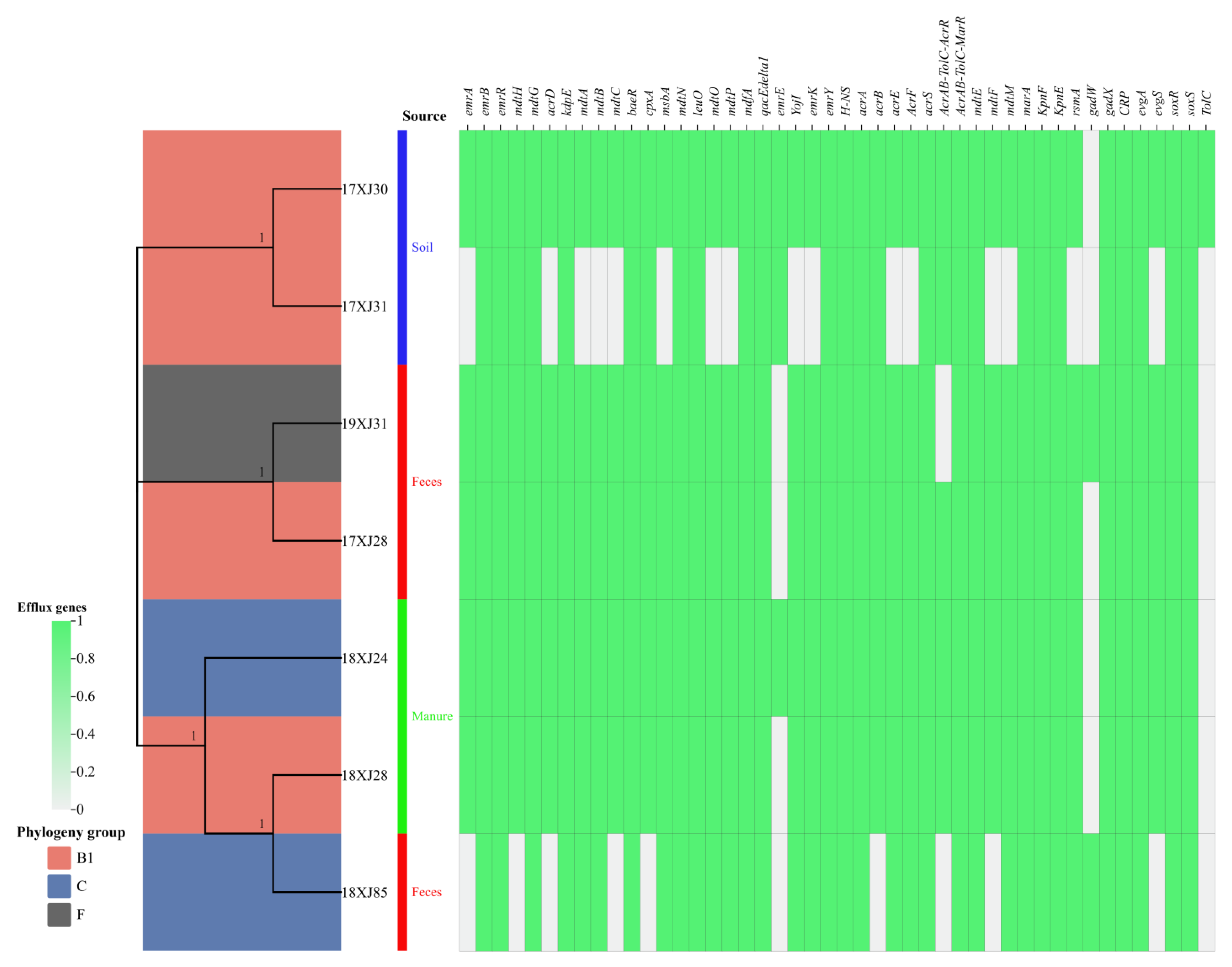
2.2. Phylogenetic Analysis and Distribution of Antibiotic Efflux-Associated Genes

This study recovered seven E. coli strains carrying MDR efflux-associated genes from the dairy farm environment in Xinjiang Province, China [9]. In silico Clermont typing classified the seven E. coli strains isolated from soil (n = 2), feces (n = 3), and manure (n = 2) into three phylogenetic groups: B1 (n = 4), C (n = 2), and F (n = 1) (Figure 1). The 7 strains acquired a total of 46 different efflux-associated genes responsible for resistance to many antibiotic classes. The E. coli strain 17XJ30 was carrying the highest number of efflux-associated genes (n = 45), followed by 18XJ28 (n = 44), 17XJ28, 18XJ28, and 19XJ31 (n = 43 each), 18XJ85 (n = 35), and 17XJ31 (n = 28). This study identified several antibiotic efflux-associated genes, such as emrR, emrB, mdtG, kdpE, baeR, mdtN, leuO, mdfA, qacE-delta1, emrY, H-NS, acrA, acrS, acrAB-TolC-marR, marA, kpnE, kpnF, gadX, CRP, evgA, soxS, and soxR genes, belonging to different antibiotic classes. The distribution of other identified genes was as follows: mdtH, mdtA, mdtB, cpxA, msbA, mdtO, mdtP, YojI, emrK, acrB, acrE, acrF, mdtM, and rsmA in 6/7 strains and emrA, acrD, mdtC, AcrAB-TolC-AcrR, mdtF, and evgS in 5/7 strains, while emrE, gadW, and TolC were identified in 3/7, 2/7, and 1/7 strains, respectively (Figure 1).

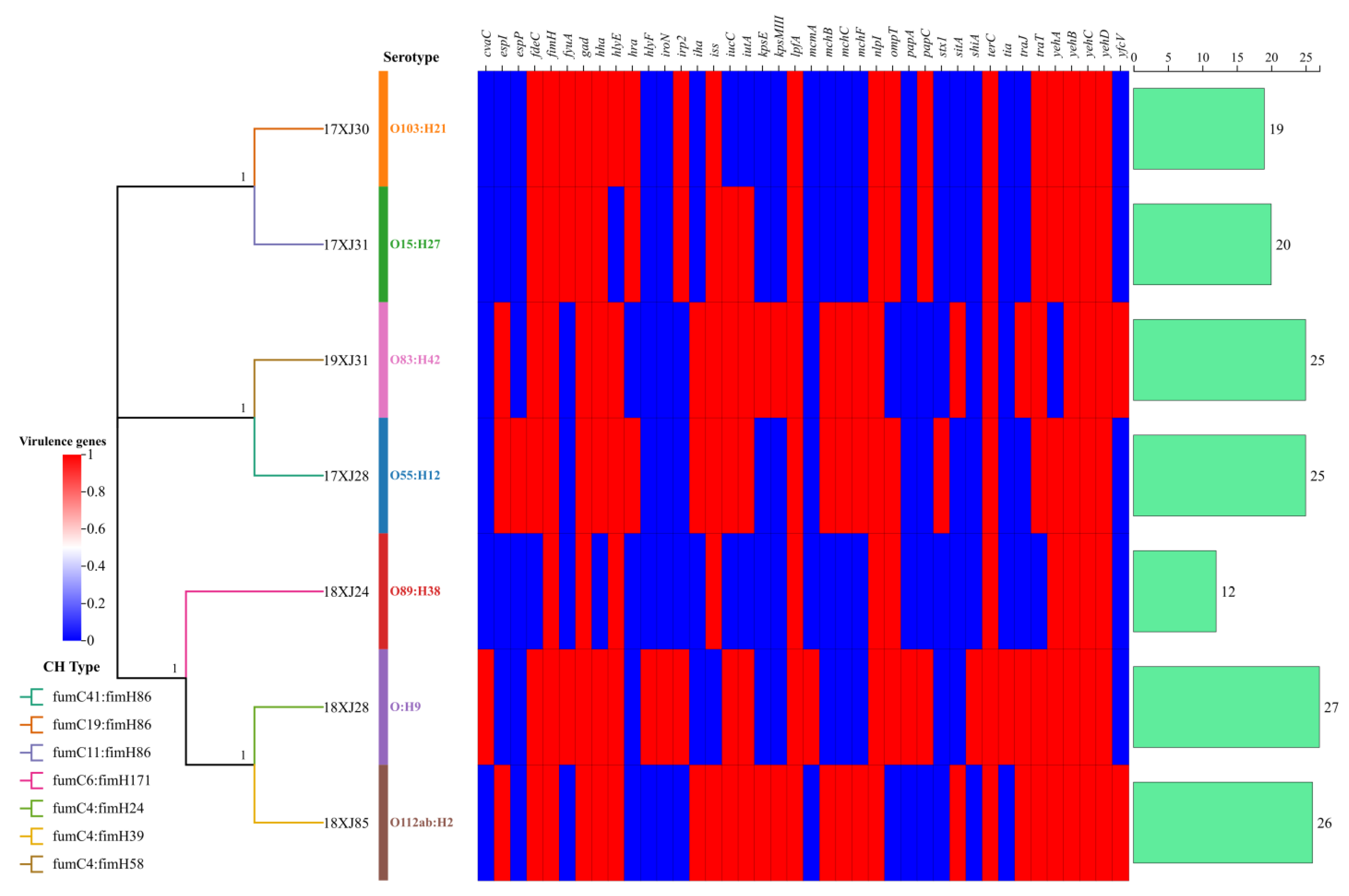
2.3. Serotyping, CH-Typing, and Virulence Determinants Acquired by MDR E. coli

The serotyping and CH typing analysis revealed that the seven E. coli strains belonged to distinct serotypes and CH types carrying diverse virulence genes. The identified serotypes were O55:H12, O103:H21, O15:H27, O89:H38, O novel:H9, O112ab:H42, and O83:H42 (Figure 2). Generally, the seven E. coli strains carried in the range of 12–27 virulence genes. The E. coli strain 18XJ28 was carrying the highest number of virulence genes (n = 27), followed by 18XJ85 (n = 26), 17XJ28 and 19XJ31 (n = 25 each), 17XJ31 (n = 20), 17XJ30 (n = 19), and 18XJ24 (n = 12). Notably, E. coli strain 17XJ28 carries the Shiga toxin-producing gene (stx1) belonging to the O55:H12 serotype and fumC41: fimH86 CH-type (Figure 2). PathogenFinder predicted that all of the E. coli strains are potential human pathogens.

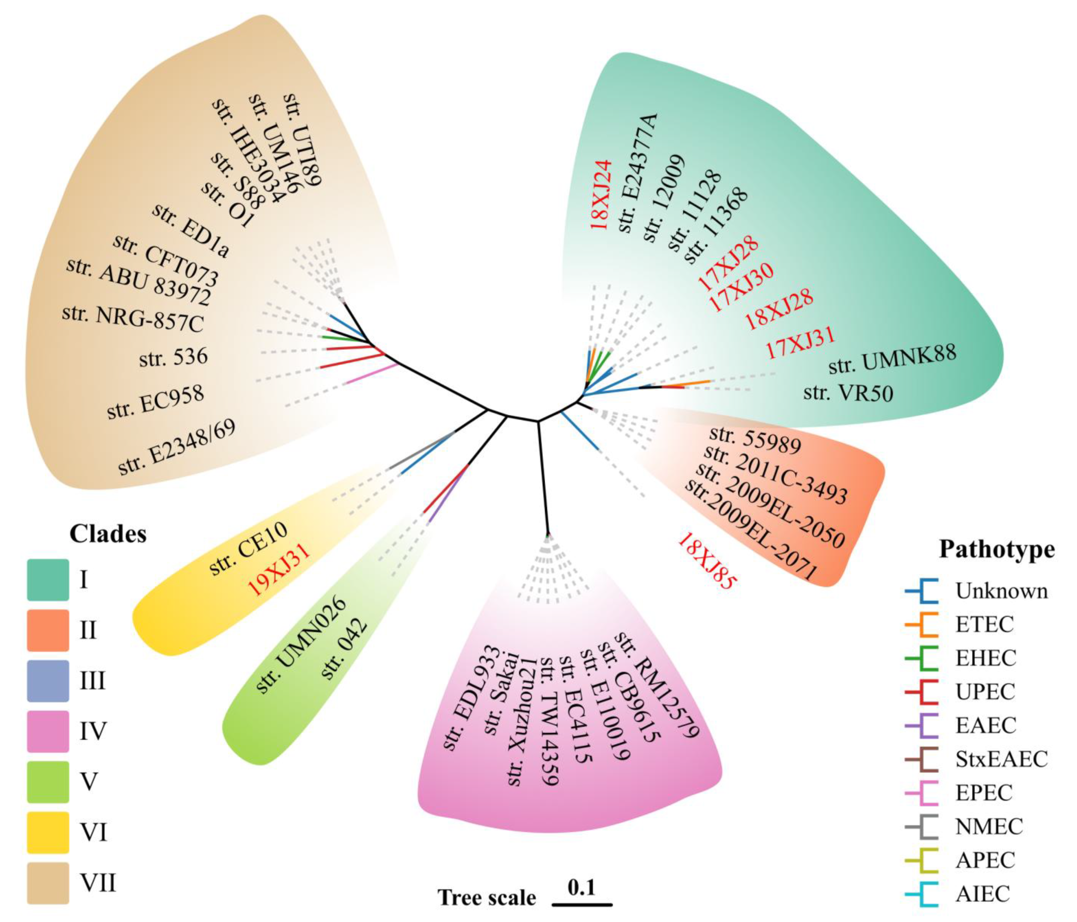
2.4. Phylogenetic Relationship of E. coli Strains with Verified E. coli Pathotypes

Our study performed a phylogenetic analysis of seven E. coli strains with verified E. coli pathotypes to reveal the evolutionary relationship and genetic similarities. The phylogenetic analysis classified all E. coli strains into seven clades (clades I–VII). The clade I carried 11 E. coli strains (five from this study and six other E. coli pathotypes). Clade II was carrying n = 4, clade III n = 1, clade IV n = 8, clade V n = 2, clade VI n = 2, and clade VII n = 12 E. coli strains (Figure 3). The E. coli strain 18XJ85 showed little genetic similarity with other pathotypes included in this study, and we classified it into a separate clade III. Moreover, the E. coli strain 19XJ31 showed increased genetic similarity with the neonatal meningitis-causing E. coli (NMEC) strain CE10 and classified it under clade VI (Figure 3).

2.5. Prophage and Genomic Island-Associated Virulence Genes (VGs)

The prophage analysis of seven E. coli strains revealed that six VGs (stx1, colE5, hha, iha, iss, and ompT) were associated with the prophage genome of E. coli strain 17XJ28, three VGs (hra, iss, and papC) with 17XJ30, three (f17G, f17C, and nlpI) with 17XJ31, one (iss) with 18XJ24 and 18XJ85, one (mcmA) with 18XJ28, and three (iss, nlpI, and terC) with 19XJ31 prophage genome segments. The iss gene was found in 5/7 and nlpI in 2/7 prophage genomes, while other VGs were identified in a single E. coli strain (Table 2). We identified fimH and ompT as genomic island-associated genes in the E. coli strain 18XJ24. The afaA, afaB, afaC, afaD, iha, and kpsE genes were associated with the genomic island of the 19XJ31. However, none of the VG was associated with the genomic island of 17XJ28, 17XJ30, 17XJ31, 18XJ28, and 18XJ85 strains (Table 2).

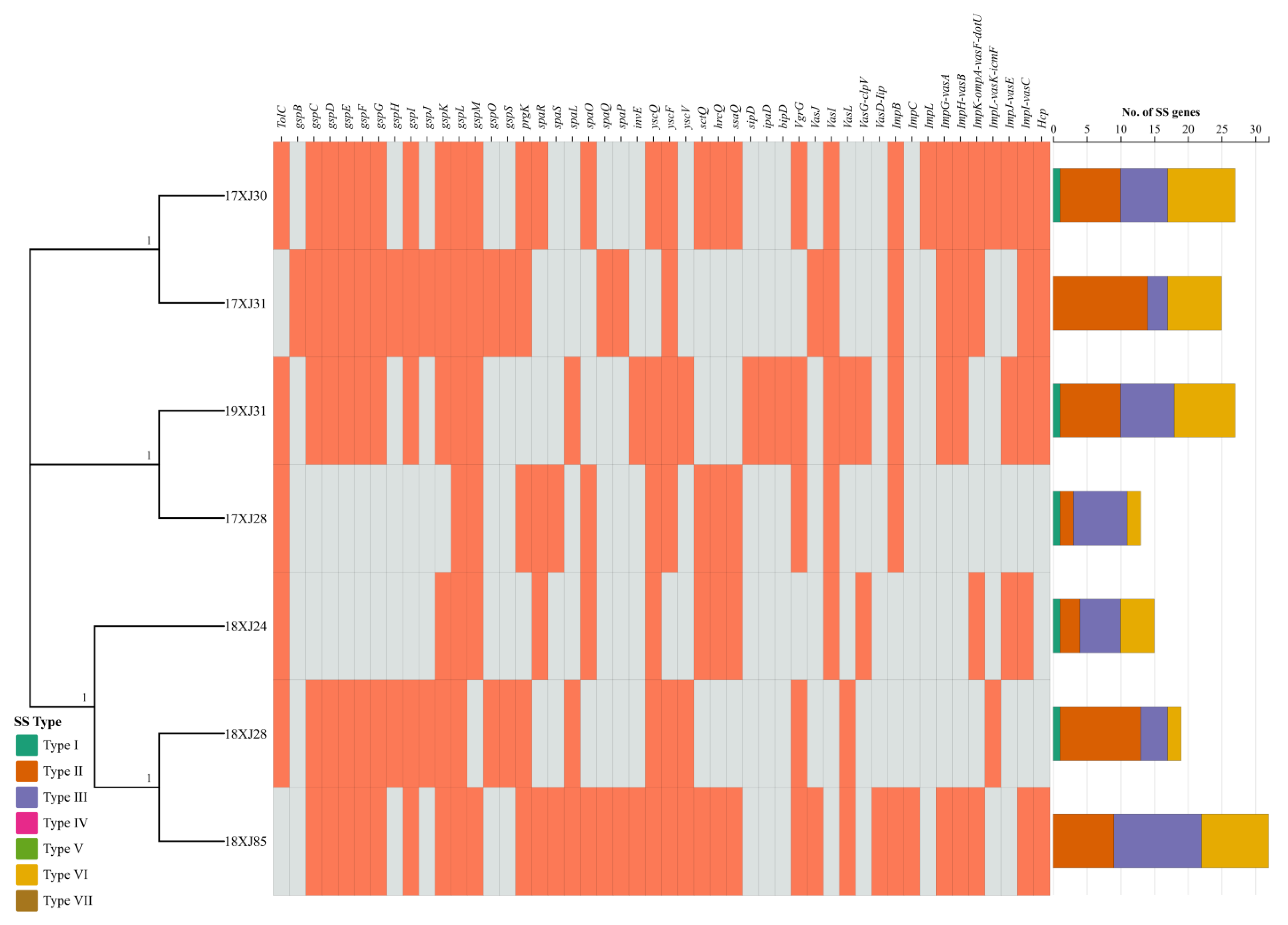
2.6. Bacterial Secretions Systems Related Genes Acquired by MDR E. coli Strains

All of the seven E. coli strains carried at least one of the types 2, 3, and 6 secretion system (SS) genes, while five strains carried type 1 SS genes, except 17XJ31 and 18XJ85. Five E. coli strains carried only one type 1 SS gene. However, type 2, 3, and 6 SS genes were acquired by all E. coli strains, varying from 2–21, 2–9, and 4–13, respectively. The E. coli strain 18XJ85 acquired the highest number (n = 32) of SS genes, followed by 17XJ30 and 19XJ31 (n = 27), 17XJ31 (n = 25), 18XJ28 (n = 19), 18XJ24 (n = 15), and 17XJ28 (n = 13) (Figure 4). We identified the gspL gene (belonging to type 2 SS) in all E. coli strains. In contrast, Figure 4 presents the distribution of other SS genes belonging to different types.

2.7. Biofilm-Associated Genes Acquired by MDR E. coli Strains

All E. coli strains isolated from the dairy environment carried a large set of biofilm genes, including genes activated by environmental signals. Four E. coli strains (17XJ30, 17XJ31, 18XJ28, and 18XJ85) carried a total of n = 48 biofilm genes, while three E. coli strains (17XJ28, 18XJ24, and 19XJ31) carried a total of n = 46 biofilm genes. The four E. coli strains (17XJ30, 17XJ31, 18XJ28, and 18XJ85) carried two additional genes, dksA and ag43 (Table 3). However, none of the E. coli strains carried sRNAs involved in biofilm formation.

3. Discussion

E. coli is a robust organism and survives in all environments, even outside of a host organism. However, a deeper understanding of the diversity and adaptability of these strains in various environments is needed. The present study characterized seven MDR E. coli strains through whole-genome sequencing. In this study, in silico Clermont typing revealed that most of the E. coli strains belonged to phylogroup B1, carrying wide resistance and virulence genes [21]. Another study reported that E. coli strains isolated from milk, farm workers, and environmental settings also carried resistance, virulence, and mobilome genes [21,22]. E. coli belonging to the B1 phylogroup has increased adaptability compared to other groups in different hosts [23,24,25]. Alternatively, it could be due to phylogroup B1’s better survival outside hosts due to its distinctive stress tolerance traits [23]. Rehman et al. [14] also reported the presence of E. coli in manure belonging to the B1 phylogroup. Consistent with our study, E. coli isolated from the animal feces belonging to the B1 and F phylogroups have also been reported [26,27].
Efflux pumps are crucial for E. coli to resist multiple drugs, and their presence or absence significantly impacts the resistance profile of each strain. The efflux-associated genes that mainly contribute to MDR are mdtE, mdtF, mdtM, marA, kpnF, rsmA, gadW gadX, CRP, evgA, evgS, soxR, soxS, tolC, and other antibiotics like fluoroquinolones (emrA, emrB, emrR, and mdtH), phosphonic acid (mdtG and acrD), aminoglycosides (acrD, kdpE, mdtA, mdtB, and mdtC), aminoglycosides and aminocoumarins (baeR and cpxA), nitroimidazoles (msbA), nucleoside and disinfecting agents (mdtN, leuO, mdtO, mdtP, and qac-E-delta), tetracycline, nucleoside and disinfecting agents (mdfA), macrolides (emrE), glycopeptides (YojI), tetracycline (emrK, emrY, and mdfA) [28]. The AcrAB-TolC is an operon that naturally makes E. coli resistant to carbonyl-cyanide m-chlorophenylhydrazone (CCCP) and nalidixic acid and is associated with an MDR efflux pump [29]. Although 18XJ28, 18XJ24, and 17XJ28 strains lack the gadX and TolC genes. However, they have AcrAB-TolC-AcrR and AcrAB-TolC-MarR tripartite efflux systems, which enable them to exhibit MDR phenotype. These settings are known for high microbial diversity and antibiotic pressures, which can drive the selection of resistant strains [29].
The identification of the gadX gene in only two fecal strains in our study suggests the potential role in acid resistance mechanisms among these E. coli strains and is critical for E. coli survival in low-pH environments, such as the stomach. The absence of the gadX gene in the soil and other environmental E. coli strains suggests a decreased Na+ concentration, reduced acid resistance in soil and environment, and downregulated expression of gadE, gadA, gadB, and gadC efflux pumps [30]. This information is crucial for understanding the adaptive strategies of E. coli in surviving extreme gastric acidity before colonizing the intestine [30]. The absence of TolC in all strains except one soil E. coli strain suggests either compensatory mechanisms or niche-specific adaptations in these strains [31]. Strains may utilize TolC-independent pumps, such as EmrAB, MdtABC, which were found in most E. coli strains. Strains 18XJ85 and 19XJ31, collected from feces, have fewer efflux-associated resistance genes compared to others, which may reflect their specific ecological niche or selective pressures in the environment from which they were isolated [32,33]. Continuous monitoring and characterization of bacterial strains are crucial to identify and exploit such vulnerabilities for better treatment strategies.
The CH and serotype analysis provided insights into these strains’ virulence potential and colonization abilities. The fimbrial gene (fimH) associated with the type of adherence system was present in all strains in our study, contributing to their virulence and ability to adhere to host tissues, which is crucial for establishing infections and biofilm formation [1]. The present study identified that Shiga toxin-producing E. coli (STEC) belong to the flagellar H12. E. coli belonging to the O55:H12 serotype have also been identified in environmental, human, and animal samples earlier [34,35,36]. The single STEC strain identified in our study may represent a high-risk outlier or indicate horizontal gene transfer events within the farm environment. In the present study, the 17XJ30 strain isolated from soil belongs to the O103:H21 serotype. O103 is included in one of the “big six” serotypes identified by the FDA and is most frequently associated with foodborne sickness and diarrhea in different countries [37,38]. O103:H21 serotype has also been identified as the verotoxigenic E. coli (VTEC) pathotype from bovine carcass [39]. Brusa et al. [40] identified E. coli belonging to the O103:H21 serotype from cattle slaughterhouses. E. coli strain 17XJ31, isolated from the soil of a dairy environment, belongs to the O15:H27 serotype. Egervärn and Flink have reported that the O15:H27 serotype causes food borne illness [41]. Galarce et al. [42] also reported the O15:H27 serotype from the livestock–food–human interface. Van Overbeek et al. reported that E. coli belonging to the O89:H38 serotype was isolated from manure [43]. E. coli O112ab:H2 serotype was detected in the ileal microbiota of a wild boar suffering from diarrhea and carried hlyE, lpfA, and gad genes [44]. The E. coli O83:H42 serotype is known to be responsible for urinary tract infections [45]. Moreover, this study noted a high prevalence of tia, espI, esp, yeh, and hylE genes in these environmental strains. The yeh fimbrial loci affect gene expression and virulence in enterohemorrhagic E. coli O157 [46]. The higher number of virulence genes acquired by these strains suggests a strong potential for pathogenicity, highlighting the need for monitoring and control strategies for these pathogens to prevent outbreaks.
Prophages and genomic islands frequently play a role in host survival strategies and enhance the genetic diversity of the host genome [47]. Additionally, prophages act as vehicles for the horizontal transfer of antimicrobial resistance and virulence genes [48]. Virulence factor genes in prophages enhance phage infectivity and are often involved in superinfection exclusion, contributing to host virulence. These genes likely benefit the prophage more than the bacteria, reducing selective pressure for the prophage to become defective [49]. The many prophages found in the sequenced genomes support that these mobile genetic elements (MGEs) significantly contribute to the evolution and genetic diversity of E. coli pathotypes [50]. The avian pathogenic E. coli (APEC) strains most frequently carry the increased serum survival (iss) and ompT virulence genes. The iss gene is crucial for resistance against serum complement, with E. coli producing iss proteins to achieve this complement resistance [51]. The OmpT gene detected in E. coli isolated from feces and manure in our study encodes the OmpT protein that serves as an antigen in STEC. This protease plays a recognized role in the virulence of extraintestinal pathogenic E. coli (ExPEC), APEC, and diarrheagenic E. coli (DEC) strains [52,53]. Fimbriae/adhesins related to the papC gene have also been identified in calves that have umbilical infection [54]. The presence of specific virulence genes (VGs), such as stx1, colE5, hra, F17G, F17C, mcmA, iss, nlpI, and terC, suggests that these strains have acquired virulence factors through prophage integration. Transmission of stx genes through prophage integration has also been reported earlier, which supports the findings of the present study [55]. Identifying a wide range of virulence genes and their specific distributions among the isolates provides valuable insights into the genetic diversity and adaptability of E. coli isolates in different environments.
In Gram-negative bacteria, extracellular enzyme secretions occur through either a one-step or a two-step process. In the two-step process, proteins first cross the cytoplasmic membrane into the periplasm [56]. The second step involves transporting these proteins across the outer membrane using specialized systems known as type II and type V secretion systems (T2SS and T5SS, respectively). In contrast, the one-step process employs type I (T1SS), type III (T3SS), type IV (T4SS), and type VI (T6SS) secretion systems, which transport proteins directly from the cytoplasm to the outside environment, bypassing the periplasm [57,58].We detected T2SS, T3SS, and T6SS in all isolates in our study. While T1SS was absent in one soil and one feces isolate, genes encoding T6SS were identified in mastitis-causing E. coli [59]. T6SS-related imp genes have been identified in Klebsiella spp. [60].
Under stress, E. coli forms biofilms by halting flagellar synthesis and producing curli fimbriae and extracellular polysaccharides controlled by the csgBA and csgDEFG operons. The master regulator csgD governs these operons and other growth-related genes, with its expression regulated by environmental factors and transcription factors [61]. In our study, all the isolates contain a high number of genes responsible for biofilm formation, taken from different dairy environments, which may indicate the possible threat of these strains. The present study has not performed in vitro, in vivo, or clinical trials to check the pathogenicity of characterized strains, which can be the future horizon for further research. However, the present study demonstrated that E. coli can be a potential reservoir of antibiotic resistance efflux, virulence, and biofilm genes in a dairy environment, which needs immediate attention to prevent their further spread and infection by these strains.

4. Materials and Methods

4.1. Origin and Background Information of E. coli Strains

The seven E. coli strains included in this study were isolated from the dairy farm environment samples, including soil (17XJ30 and 17XJ31), manure (18XJ24 and 18XJ28), and cattle feces (17XJ28, 18XJ85, and 19XJ31) collected from Changji Hui Autonomous Prefecture in Xinjiang Province, China (map is illustrated in Figure 5). Briefly, a total of 209 samples, including feces, manure or slurry, water, milk, and soil, were collected from selected dairy farm, and 338 E. coli strains were retrieved, comprising 67.5% (141/209) of the collected samples. The antimicrobial susceptibility testing of retrieved E. coli strains revealed that 84.0% (284/338) were resistant to at least one antimicrobial, and 26.0% (54/338) were susceptible. The selection of these seven E. coli strains was completed based on co-carrying a gene array: blaOXA-1-catB3-arr3 genes, whose detailed information about sampling protocols, processing, and selection criteria has been described in earlier published studies [1,9]. Briefly, the samples were enriched in Luria Bertani broth and then streaked on MacConkey agar. The isolated red- or pink-colored colonies were further streaked on Eosin Methylene Blue (EMB) agar for selective differentiation. The metallic green sheen colonies were picked for species identification. Matrix-assisted laser desorption/ionization time of flight mass spectrometry (MALDI-TOF MS) confirmed E. coli as done earlier [62]. Bacterial liquid cultures were stored at −80 °C in 20% glycerol for further processing and long-term storage.

4.2. DNA Extraction, Library Construction, and Whole Genome Sequencing (WGS)

The genomic DNA of seven E. coli strains was extracted using the sodium dodecyl sulfate (SDS) method for longer DNA fragments. The purity and integrity of extracted DNA were determined by agarose gel electrophoresis and quantification using a Qubit 4.0 fluorometer (Thermo Fisher Scientific, Waltham, MA, USA). The purified and integrated DNA fragments were randomly interrupted by a Covaris ultrasonic beaker (Woburn, MA, USA) with 350 bp inserts length. Following the manufacturer’s guidelines, the library was prepared using the NEBNext® UltraTM DNA library kit for Illumina NovaSeq PE150 (NEB, Ipswich, MA, USA). After the successful construction of the library, initially, the library was quantified using a Qubit 4.0 fluorometer and size distribution by Agilent 2100 Bioanalyzer (Agilent Technologies, Santa Clara, CA, USA) and final quantification by qRT-PCR. The prepared libraries were sequenced by NovaSeq PE150 (Illumina, San Diego, CA, USA) by Novogene Technology Co., Ltd. (Beijing, China).

4.3. Data Processing and Genome Assembly

The raw sequencing data obtained were quality-checked using FastQC version 0.12.1. The low-quality reads were filtered out from the data using the Trimmomatic version 0.39 [63]. The de novo assembly of trimmed data (called clean data) was done using SPAdes version 4.0.0 [64]. The draft assemblies were processed further for gap filling using the GapCloser version 1.12, which filters the low-depth sequences (e.g., <0.35 average depth) and contigs with a size below 500 bp to get the final genome assemblies for further analysis.

4.4. Pangenome and Bioinformatics Analysis

The finally assembled genome was used to predict the coding genes by GeneMarkS (version 4.17) software (Available online: http://topaz.gatech.edu/GeneMark/ (accessed on 20 September 2024)) [65]. Gene functional annotation was completed by the Cluster of Orthologous Genes (COG) database [66]. Type N secretion systems (TNSS) were retrieved based on the protein functional database, and for Gram-negative bacteria such as E. coli, T3SS effector proteins were predicted by Effective T3 software (version 1.0.1) [67]. The comprehensive antibiotic resistance (CARD) database was used to identify the antibiotic resistance efflux pumps acquired by each strain [68]. The virulence determinants, prediction as a human pathogen, serotyping, and CHTyping were completed by VirulenceFinder 2.0 (specie E. coli, 95% ID threshold, 80% minimum length), PathogenFinder 1.1 (phylum = gamma proteobacteria), SerotypeFinder 2.0 (E. coli, 95% ID threshold, and 80% minimum length), and CHTyper 1.0 (95% ID threshold), respectively, under Center for Genomic Epidemiology (CGE) online server (Available online: https://genomicepidemiology.org/services/ (accessed on 15 August 2024)). The relationship of seven E. coli strains with verified human pathogenic strains from the NCBI database was completed by phylogeny construction using CSIPhylogeny 1.4 and visualized by TVBOT under the ChiPlot online server (Available online: https://www.chiplot.online/ (accessed on 10 October 2024)).

5. Conclusions

The present study investigated seven E. coli strains isolated from a dairy farm environment in Xinjiang Province, China, carrying wide antibiotic efflux-associated genes and virulence genes. Moreover, the study also noted that a few of the virulence genes were found to be associated with prophages and genomic islands, which may enhance their potential to be transferred to other bacterial species. Although this study was conducted on a single form, it might suggest that the dairy farm environment can be a potential reservoir of such genes in E. coli. This study served as a preliminary genomic characterization of MDR E. coli strains within a specific farming system to identify potential ARG reservoirs and strain diversity in a dairy environment. Even if the study has been performed on a limited number of strains, it highlights the genetic diversity and adaptability of E. coli strains, emphasizing their potential for antibiotic resistance and virulence. The study underscores the necessity for ongoing large-scale genomic surveillance and research to mitigate public health risks associated with these pathogens. The actual findings contribute valuable insights into the understanding of E. coli in various environments, reinforcing the importance of genomic characterization in addressing public health challenges.

Author Contributions

Writing—original draft preparation, M.S.; methodology, Z.H., Z.W. and M.T.; software & validation, M.S., S.G., Z.W. and S.M.; formal analysis, M.S.; investigation, B.H. and X.Z.; resources, X.Z. and Z.W.; data curation, M.S.; writing—review and editing, S.G. and S.M.; visualization, B.H., X.Z. and Z.W.; Conceptualization, S.W. and W.P.; supervision, W.P.; project administration, W.P.; funding acquisition, W.P. and S.W. All authors have read and agreed to the published version of the manuscript.

Funding

This research was funded by the Agricultural Science and Technology Innovation Program of the Chinese Academy of Agricultural Sciences (25-LZIHPS-03).

Institutional Review Board Statement

Not applicable.

Informed Consent Statement

Not applicable.

Data Availability Statement

The raw data supporting the conclusions of this article will be made available by the authors upon request. The whole genome sequences of the seven E. coli strains analyzed in this study are available from the NCBI under BioProject ID: PRJNA1049405. The accession numbers of seven E. coli genome data are JAYKSK000000000, JAYJLB000000000, JBDYKH000000000, JAYKSJ000000000, JAYJLA000000000, JBDYKG000000000, and JAYKSI000000000 (accessed on 21 January 2025).

Conflicts of Interest

The authors declare no conflicts of interest. The funders had no role in the design of the study; in the collection, analysis, or interpretation of data; in the writing of the manuscript; or in the decision to publish the results.

Abbreviations

The following abbreviations are used in this manuscript:
TNSSType N secretion systems
CGECenter for Genomic Epidemiology
WGSWhole Genome Sequencing
COGCluster of Orthologous Genes
EMBEosin Methylene Blue
VGsVirulence genes
MDRMultidrug resistance
STECShiga toxin-producing E. coli
CCCPCarbonyl-cyanide m-chlorophenylhydrazone
VTECVerotoxigenic E. coli
APECAvian pathogenic E. coli
ETECEnterotoxigenic E. coli
EHECEnterohemorrhagic E. coli
NMECNeonatal meningitis E. coli
UPECUropathogenic E. coli
EAECEnteroaggregative E. coli
Ex-PECExtraintestinal pathogenic E. coli
DECDiarrheagenic E. coli
MGEMobile genetic elements
E. coliEscherichia coli
GIsGenomic islands
ARGsAntibiotic resistance genes

References

  1. Shoaib, M.; He, Z.; Geng, X.; Tang, M.; Hao, R.; Wang, S.; Shang, R.; Wang, X.; Zhang, H.; Pu, W. The Emergence of Multidrug Resistant and Virulence Gene Carrying Escherichia coli Strains in the Dairy Environment: A Rising Threat to the Environment, Animal, and Public Health. Front. Microbiol. 2023, 14, 1197579. [Google Scholar] [CrossRef] [PubMed]
  2. Zhang, P.; Essendoubi, S.; Keenliside, J.; Reuter, T.; Stanford, K.; King, R.; Lu, P.; Yang, X. Genomic analysis of Shiga toxin-producing Escherichia coli O157:H7 from cattle and pork-production related environments. npj Sci. Food 2021, 5, 15. [Google Scholar] [CrossRef]
  3. Ray, R.; Singh, P. Prevalence and Implications of Shiga Toxin-Producing E. coli in Farm and Wild Ruminants. Pathogens 2022, 11, 1332. [Google Scholar] [CrossRef]
  4. Zhang, P.; Yang, X. Genetic Characteristics of the Transmissible Locus of Stress Tolerance (TLST) and TLST Harboring Escherichia coli as Revealed by Large-Scale Genomic Analysis. Appl. Environ. Microbiol. 2022, 88, e02185-21. [Google Scholar] [CrossRef]
  5. Vangchhia, B.; Abraham, S.; Bell, J.M.; Collignon, P.; Gibson, J.S.; Ingram, P.R.; Johnson, J.R.; Kennedy, K.; Trott, D.J.; Turnidge, J.D.; et al. Phylogenetic Diversity, Antimicrobial Susceptibility and Virulence Characteristics of Phylogroup F Escherichia coli in Australia. Microbiology 2016, 162, 1904–1912. [Google Scholar] [CrossRef]
  6. Carlos, C.; Pires, M.M.; Stoppe, N.C.; Hachich, E.M.; Sato, M.I.Z.; Gomes, T.A.T.; Amaral, L.A.; Ottoboni, L.M.M. Escherichia coli Phylogenetic Group Determination and Its Application in the Identification of the Major Animal Source of Fecal Contamination. BMC Microbiol. 2010, 10, 161. [Google Scholar] [CrossRef]
  7. Benedict, K.M.; Gow, S.P.; McAllister, T.A.; Booker, C.W.; Hannon, S.J.; Checkley, S.L.; Noyes, N.R.; Morley, P.S. Antimicrobial Resistance in Escherichia coli Recovered from Feedlot Cattle and Associations with Antimicrobial Use. PLoS ONE 2015, 10, e0143995. [Google Scholar] [CrossRef]
  8. Wepking, C.; Avera, B.; Badgley, B.; Barrett, J.E.; Franklin, J.; Knowlton, K.F.; Ray, P.P.; Smitherman, C.; Strickland, M.S. Exposure to Dairy Manure Leads to Greater Antibiotic Resistance and Increased Mass-Specific Respiration in Soil Microbial Communities. Proc. R. Soc. B Biol. Sci. 2017, 284, 20162233. [Google Scholar] [CrossRef]
  9. Shoaib, M.; Tang, M.; Awan, F.; Aqib, A.I.; Hao, R.; Ahmad, S.; Wang, S.; Shang, R.; Pu, W. Genomic Characterization of Extended-Spectrum β-Lactamase (ESBL) Producing E. coli Harboring blaOXA−1-catB3-arr-3 Genes Isolated from Dairy Farm Environment in China. Transbound. Emerg. Dis. 2024, 2024, 3526395. [Google Scholar] [CrossRef]
  10. Sjölund, M.; Bonnedahl, J.; Hernandez, J.; Bengtsson, S.; Cederbrant, G.; Pinhassi, J.; Kahlmeter, G.; Olsen, B. Dissemination of Multidrug-Resistant Bacteria into the Arctic. Emerg. Infect. Dis. 2008, 14, 70–72. [Google Scholar] [CrossRef]
  11. Ibrahim, R.K.; Hayyan, M.; AlSaadi, M.A.; Hayyan, A.; Ibrahim, S. Environmental Application of Nanotechnology: Air, Soil, and Water. Environ. Sci. Pollut. Res. 2016, 23, 13754–13788. [Google Scholar] [CrossRef] [PubMed]
  12. Amézquita-López, B.A.; Quiñones, B.; Soto-Beltrán, M.; Lee, B.G.; Yambao, J.C.; Lugo-Melchor, O.Y.; Chaidez, C. Antimicrobial Resistance Profiles of Shiga Toxin-Producing Escherichia coli O157 and Non-O157 Recovered from Domestic Farm Animals in Rural Communities in Northwestern Mexico. Antimicrob. Resist. Infect. Control 2016, 5, 1. [Google Scholar] [CrossRef] [PubMed]
  13. Manyi-Loh, C.; Mamphweli, S.; Meyer, E.; Okoh, A. Antibiotic Use in Agriculture and Its Consequential Resistance in Environmental Sources: Potential Public Health Implications. Molecules 2018, 23, 795. [Google Scholar] [CrossRef] [PubMed]
  14. Rehman, M.A.; Yin, X.; Lepp, D.; Laing, C.; Ziebell, K.; Talbot, G.; Topp, E.; Diarra, M.S. Genomic Analysis of Third Generation Cephalosporin Resistant Escherichia coli from Dairy Cow Manure. Vet. Sci. 2017, 4, 57. [Google Scholar] [CrossRef]
  15. Naidoo, N.; Zishiri, O.T. Comparative Genomics Analysis and Characterization of Shiga Toxin-Producing Escherichia coli O157:H7 Strains Reveal Virulence Genes, Resistance Genes, Prophages and Plasmids. BMC Genom. 2023, 24, 791. [Google Scholar] [CrossRef]
  16. Fu, W.; Forster, T.; Mayer, O.; Curtin, J.J.; Lehman, S.M.; Donlan, R.M. Bacteriophage Cocktail for the Prevention of Biofilm Formation by Pseudomonas Aeruginosa on Catheters in an in Vitro Model System. Antimicrob. Agents Chemother. 2010, 54, 397–404. [Google Scholar] [CrossRef]
  17. Hu, Y.; Hu, Y.; Li, Y.; Hui, M.; Lu, Z.; Li, H.; Tian, H. Metagenomic Insights into Quorum Sensing in Membrane-Aerated Biofilm Reactors for Phenolic Wastewater Treatment. Environ. Technol. 2022, 43, 1318–1327. [Google Scholar] [CrossRef]
  18. Yang, X.; Wang, H.; Hrycauk, S.; Holman, D.B.; Ells, T.C. Microbial Dynamics in Mixed-Culture Biofilms of Salmonella Typhimurium and Escherichia coli O157:H7 and Bacteria Surviving Sanitation of Conveyor Belts of Meat Processing Plants. Microorganisms 2023, 11, 421. [Google Scholar] [CrossRef]
  19. Schmitz, F.-J.; Martinez-Freijo, P.; Theis, S.; Fluit, A.C.; Verhoef, J.; Heinz, H.-P.; Jones, M.E. Class I Integrons: Prevalence and Impact on Antibiotic Susceptibility in 278 Consecutive Unrelated Gram-Negative Blood Isolates. Clin. Microbiol. Infect. Off. Publ. Eur. Soc. Clin. Microbiol. Infect. Dis. 1999, 5, 496–498. [Google Scholar] [CrossRef]
  20. Schroeder, C.M.; Zhao, C.; DebRoy, C.; Torcolini, J.; Zhao, S.; White, D.G.; Wagner, D.D.; McDermott, P.F.; Walker, R.D.; Meng, J. Antimicrobial Resistance of Escherichia coli O157 Isolated from Humans, Cattle, Swine, and Food. Appl. Environ. Microbiol. 2002, 68, 576–581. [Google Scholar] [CrossRef]
  21. Beghain, J.; Bridier-Nahmias, A.; Le Nagard, H.; Denamur, E.; Clermont, O. ClermonTyping: An Easy-to-Use and Accurate in Silico Method for Escherichia Genus Strain Phylotyping. Microb. Genom. 2018, 4, e000192. [Google Scholar] [CrossRef] [PubMed]
  22. Hao, R.; Shoaib, M.; Tang, M.; Cao, Z.; Liu, G.; Zhang, Y.; Wang, S.; Shang, R.; Zhang, H.; Pu, W. Genomic insights into resistome, virulome, and mobilome as organic contaminants of ESKAPE pathogens and E. coli recovered from milk, farm workers, and environmental settings in Hainan, China. Emerg. Contam. 2024, 10, 100385. [Google Scholar] [CrossRef]
  23. Bergholz, P.W.; Noar, J.D.; Buckley, D.H. Environmental Patterns Are Imposed on the Population Structure of Escherichia coli after Fecal Deposition. Appl. Environ. Microbiol. 2011, 77, 211–219. [Google Scholar] [CrossRef]
  24. Shoaib, M.; Tang, M.; Aqib, A.I.; Zhang, X.; Wu, Z.; Wen, Y.; Hou, X.; Xu, J.; Hao, R.; Wang, S.; et al. Dairy Farm Waste: A Potential Reservoir of Diverse Antibiotic Resistance and Virulence Genes in Aminoglycoside- and Beta-Lactam-Resistant Escherichia coli in Gansu Province, China. Environ. Res. 2024, 263, 120190. [Google Scholar] [CrossRef]
  25. Nandakafle, G.; Seale, T.; Flint, T.; Nepal, M.; Venter, S.N.; Brözel, V.S. Distribution of Diverse Escherichia coli between Cattle and Pasture. Microbes Environ. 2017, 32, 226–233. [Google Scholar] [CrossRef]
  26. Coura, F.M.; Diniz, S.D.A.; Silva, M.X.; Mussi, J.M.S.; Barbosa, S.M.; Lage, A.P.; Heinemann, M.B. Phylogenetic Group Determination of Escherichia coli Isolated from Animals Samples. Sci. World J. 2015, 2015, 258424. [Google Scholar] [CrossRef]
  27. Luo, C.; Walk, S.T.; Gordon, D.M.; Feldgarden, M.; Tiedje, J.M.; Konstantinidis, K.T. Genome Sequencing of Environmental Escherichia coli Expands Understanding of the Ecology and Speciation of the Model Bacterial Species. Proc. Natl. Acad. Sci. USA 2011, 108, 7200–7205. [Google Scholar] [CrossRef]
  28. Teelucksingh, T.; Thompson, L.K.; Cox, G. The Evolutionary Conservation of Escherichia coli Drug Efflux Pumps Supports Physiological Functions. J. Bacteriol. 2020, 202, 10–1128. [Google Scholar] [CrossRef]
  29. Jang, S. AcrAB-TolC, a major efflux pump in Gram-negative bacteria: Toward understanding its operation mechanism. BMB Rep. 2023, 56, 326–334. [Google Scholar] [CrossRef]
  30. Tramonti, A.; De Canio, M.; De Biase, D. GadX/GadW-Dependent Regulation of the Escherichia coli Acid Fitness Island: Transcriptional Control at the GadY-GadW Divergent Promoters and Identification of Four Novel 42 Bp GadX/GadW-Specific Binding Sites. Mol. Microbiol. 2008, 70, 965–982. [Google Scholar] [CrossRef]
  31. van Hung Le, V.; Biggs, P.J.; Wheeler, D.; Davies, I.G.; Rakonjac, J. Novel Mechanisms of TolC-Independent Decreased Bile-Salt Susceptibility in Escherichia coli. FEMS Microbiol. Lett. 2021, 367, fnaa083. [Google Scholar] [CrossRef]
  32. Manishimwe, R.; Moncada, P.M.; Musanayire, V.; Shyaka, A.; Morgan Scott, H.; Loneragan, G.H. Antibiotic-Resistant Escherichia coli and Salmonella from the Feces of Food Animals in the East Province of Rwanda. Animals 2021, 11, 1013. [Google Scholar] [CrossRef]
  33. Schmidt, V.M.; Pinchbeck, G.L.; Nuttall, T.; McEwan, N.; Dawson, S.; Williams, N.J. Antimicrobial Resistance Risk Factors and Characterisation of Faecal E. coli Isolated from Healthy Labrador Retrievers in the United Kingdom. Prev. Vet. Med. 2015, 119, 31–40. [Google Scholar] [CrossRef] [PubMed]
  34. Biswas, S.; Bal, M.; Pati, S.; Rana, R.; Dixit, S.; Ranjit, M. Antibiotic resistance in toxigenic E. coli: A severe threat to global health. Discov. Med. 2024, 1, 72. [Google Scholar] [CrossRef]
  35. Zhang, P.; Liu, L.; Sheng, H.; Zhang, M.; Wang, T.; Chang, G.; Wang, Y.; Bai, L.; Wang, X. Antibiotic Resistance and Genomic Analysis of Shiga Toxin-Producing Escherichia coli from Dairy Cattle, Raw Milk, and Farm Environment in Shaanxi Province, China. Foodborne Pathog. Dis. 2024, 21, 624–633. [Google Scholar] [CrossRef]
  36. Beutin, L.; Delannoy, S.; Fach, P. Genetic Analysis and Detection of fliC H1 and fliC H12 Genes Coding for Serologically Closely Related Flagellar Antigens in Human and Animal Pathogenic Escherichia coli. Front. Microbiol. 2016, 7, 135. [Google Scholar] [CrossRef]
  37. Mare, A.; Man, A.; Ciurea, C.N.; Pintea-Simon, I.A.; Ianoși, E.S.; Gîrbovan, C.E.; Toma, F. Serogroups and Genetic Diversity of Diarrheagenic Strains of Escherichia coli: A Retrospective Study. J. Infect. Dev. Ctries. 2022, 16, 827–834. [Google Scholar] [CrossRef]
  38. Alharbi, M.G.; Al-Hindi, R.R.; Esmael, A.; Alotibi, I.A.; Azhari, S.A.; Alseghayer, M.S.; Teklemariam, A.D. The “Big Six”: Hidden Emerging Foodborne Bacterial Pathogens. Trop. Med. Infect. Dis. 2022, 7, 356. [Google Scholar] [CrossRef]
  39. Wieczorek, K.; Osek, J. Identification and Molecular Characteristics of Verotoxin-Producing Escherichia coli (VTEC) from Bovine and Pig Carcasses Isolated in Poland during 2014–2018. Food Microbiol. 2020, 92, 103587. [Google Scholar] [CrossRef]
  40. Brusa, V.; Restovich, V.; Galli, L.; Teitelbaum, D.; Signorini, M.; Brasesco, H.; Londero, A.; García, D.; Padola, N.L.; Superno, V.; et al. Isolation and Characterization of Non-O157 Shiga Toxin-Producing Escherichia coli from Beef Carcasses, Cuts and Trimmings of Abattoirs in Argentina. PLoS ONE 2017, 12, e0183248. [Google Scholar] [CrossRef]
  41. Egervärn, M.; Flink, C. Shiga Toxin-Producing Escherichia coli (STEC) in Meat and Leafy Greens Available in the Swedish Retail Market—Occurrence and Diversity of Stx Subtypes and Serotypes. Int. J. Food Microbiol. 2024, 408, 110446. [Google Scholar] [CrossRef] [PubMed]
  42. Galarce, N.; Sánchez, F.; Escobar, B.; Lapierre, L.; Cornejo, J.; Alegría-Morán, R.; Neira, V.; Martínez, V.; Johnson, T.; Fuentes-Castillo, D.; et al. Genomic Epidemiology of Shiga Toxin-Producing Escherichia coli Isolated from the Livestock-Food-Human Interface in South America. Animals 2021, 11, 1845. [Google Scholar] [CrossRef] [PubMed]
  43. van Overbeek, L.S.; Wichers, J.H.; van Amerongen, A.; van Roermund, H.J.W.; van der Zouwen, P.; Willemsen, P.T.J. Circulation of Shiga Toxin-Producing Escherichia coli Phylogenetic Group B1 Strains Between Calve Stable Manure and Pasture Land with Grazing Heifers. Front. Microbiol. 2020, 11, 1355. [Google Scholar] [CrossRef] [PubMed]
  44. Libisch, B.; Nagy, G.; Csivincsik, Á.; Keresztény, T.; Papp, P.; Olasz, F.; Fébel, H.; Sándor, Z.J.; Rasschaert, G.; Lambrecht, E.; et al. Detection of Acquired Antibiotic Resistance Determinants in the Intestinal Microbiota of Food Producing Animals in Hungary. In Book of Abstracts, “Antimicrobial Resistance: A Challenge for Public Health, Animal Health and Environment”; Scientific Symposium Bruxelles: Brüsszel, Belgium, 2022; Volume 64, p. 7180. [Google Scholar]
  45. García, V.; Lestón, L.; Parga, A.; García-Meniño, I.; Fernández, J.; Otero, A.; Olsen, J.E.; Herrero-Fresno, A.; Mora, A. Genomics, Biofilm Formation and Infection of Bladder Epithelial Cells in Potentially Uropathogenic Escherichia coli (UPEC) from Animal Sources and Human Urinary Tract Infections (UTIs) Further Support Food-Borne Transmission. One Health 2023, 16, 100558. [Google Scholar] [CrossRef]
  46. Gonyar, L.A.; Sauder, A.B.; Mortensen, L.; Willsey, G.G.; Kendall, M.M. The yad and yeh fimbrial loci influence gene expression and virulence in Enterohemorrhagic Escherichia coli O157: H7. Msphere 2024, 9, e00124-24. [Google Scholar] [CrossRef]
  47. Desvaux, M.; Dalmasso, G.; Beyrouthy, R.; Barnich, N.; Delmas, J.; Bonnet, R. Pathogenicity Factors of Genomic Islands in Intestinal and Extraintestinal Escherichia coli. Front. Microbiol. 2020, 11, 2065. [Google Scholar] [CrossRef]
  48. Kondo, K.; Kawano, M.; Sugai, M. Distribution of Antimicrobial Resistance and Virulence Genes within the Prophage-Associated Regions in Nosocomial Pathogens. mSphere 2021, 6, e0045221. [Google Scholar] [CrossRef]
  49. Bondy-Denomy, J.; Qian, J.; Westra, E.R.; Buckling, A.; Guttman, D.S.; Davidson, A.R.; Maxwell, K.L. Prophages Mediate Defense against Phage Infection through Diverse Mechanisms. ISME J. 2016, 10, 2854–2866. [Google Scholar] [CrossRef]
  50. Sváb, D.; Falgenhauer, L.; Mag, T.; Chakraborty, T.; Tóth, I. Genomic Diversity, Virulence Gene, and Prophage Arrays of Bovine and Human Shiga Toxigenic and Enteropathogenic Escherichia coli Strains Isolated in Hungary. Front. Microbiol. 2022, 13, 896296. [Google Scholar] [CrossRef]
  51. Sankhi, S.; Bhattarai, R.; Basnet, H.; Marasine, N.; Luitel, H.; Nayak, M.; Sankhi, S. Identification of Antibiotic Resistance Pattern, and Detection of Virulence Genes Iss, and OmpT in Avian Pathogenic Escherichia coli from Broiler Chickens in Chitwan, Nepal. J. Pharm. Res. Int. 2020, 32, 1–12. [Google Scholar] [CrossRef]
  52. Ovi, F.; Zhang, L.; Nabors, H.; Jia, L.; Adhikari, P. A Compilation of Virulence-Associated Genes That Are Frequently Reported in Avian Pathogenic Escherichia coli (APEC) Compared to Other E. coli. J. Appl. Microbiol. 2023, 134, lxad014. [Google Scholar] [CrossRef] [PubMed]
  53. Torres, A.N.; Chamorro-Veloso, N.; Costa, P.; Cádiz, L.; Del Canto, F.; Venegas, S.A.; López Nitsche, M.; Coloma-Rivero, R.F.; Montero, D.A.; Vidal, R.M. Deciphering Additional Roles for the EF-Tu, l-Asparaginase II and OmpT Proteins of Shiga Toxin-Producing Escherichia coli. Microorganisms 2020, 8, 1184. [Google Scholar] [CrossRef] [PubMed]
  54. Martins, L.S.A.; Motta, R.G.; Martinez, A.C.; Orsi, H.; Hernandes, R.T.; Rall, V.L.M.; Pantoja, J.C.F.; Nardi Júnior, G.; Ribeiro, M.G. Virulence-encoding genes related to extraintestinal pathogenic E. coli and multidrug resistant pattern of strains isolated from neonatal calves with different severity scores of umbilical infections. Microb. Pathog. 2023, 174, 105861. [Google Scholar] [CrossRef]
  55. Asadulghani, M.; Ogura, Y.; Ooka, T.; Itoh, T.; Sawaguchi, A.; Iguchi, A.; Nakayama, K.; Hayashi, T. The Defective Prophage Pool of Escherichia coli O157: Prophage-Prophage Interactions Potentiate Horizontal Transfer of Virulence Determinants. PLoS Pathog. 2009, 5, e1000408. [Google Scholar] [CrossRef]
  56. Tsirigotaki, A.; De Geyter, J.; Šoštaric, N.; Economou, A.; Karamanou, S. Protein Export through the Bacterial Sec Pathway. Nat. Rev. Microbiol. 2017, 15, 21–36. [Google Scholar] [CrossRef]
  57. Green, E.R.; Mecsas, J. Bacterial Secretion Systems: An Overview. Microbiol. Spectr. 2016, 4, 1. [Google Scholar] [CrossRef]
  58. Rehman, A.; Andleeb, S.; Ullah, S.R.; Mustafa, Z.; Gul, D.; Mehmood, K. Antibiotic-Mediated Expression Analysis of Shiga Toxin 1 and 2 in Multi-Drug-Resistant Shiga Toxigenic Escherichia coli. Folia Microbiol. 2021, 66, 809–817. [Google Scholar] [CrossRef]
  59. Kempf, F.; Slugocki, C.; Blum, S.E.; Leitner, G.; Germon, P. Genomic Comparative Study of Bovine Mastitis Escherichia coli. PLoS ONE. 2016, 11, e0147954. [Google Scholar] [CrossRef]
  60. Sarris, P.F.; Zoumadakis, C.; Panopoulos, N.J.; Scoulica, E.V. Distribution of the Putative Type VI Secretion System Core Genes in Klebsiella spp. Infect. Genet. Evol. 2011, 11, 157–166. [Google Scholar] [CrossRef]
  61. Ogasawara, H.; Ishizuka, T.; Hotta, S.; Aoki, M.; Shimada, T.; Ishihama, A. Novel Regulators of the CSGD Gene Encoding the Master Regulator of Biofilm Formation in Escherichia coli K-12. Microbiology 2020, 166, 880–890. [Google Scholar] [CrossRef]
  62. Luo, Y.; Siu, G.K.H.; Yeung, A.S.F.; Chen, J.H.K.; Ho, P.L.; Leung, K.W.; Tsang, J.L.Y.; Cheng, V.C.C.; Guo, L.; Yang, J.; et al. Performance of the VITEK MS Matrix-Assisted Laser Desorption Ionization-Time of Flight Mass Spectrometry System for Rapid Bacterial Identification in Two Diagnostic Centres in China. J. Med. Microbiol. 2015, 64, 18–24. [Google Scholar] [CrossRef] [PubMed]
  63. Bolger, A.M.; Lohse, M.; Usadel, B. Trimmomatic: A Flexible Trimmer for Illumina Sequence Data. Bioinformatics 2014, 30, 2114–2120. [Google Scholar] [CrossRef] [PubMed]
  64. Bankevich, A.; Nurk, S.; Antipov, D.; Gurevich, A.A.; Dvorkin, M.; Kulikov, A.S.; Lesin, V.M.; Nikolenko, S.I.; Pham, S.; Prjibelski, A.D.; et al. SPAdes: A New Genome Assembly Algorithm and Its Applications to Single-Cell Sequencing. J. Comput. Biol. 2012, 19, 455–477. [Google Scholar] [CrossRef]
  65. Besemer, J.; Lomsadze, A.; Borodovsky, M. GeneMarkS: A Self-Training Method for Prediction of Gene Starts in Microbial Genomes. Implications for Finding Sequence Motifs in Regulatory Regions. Nucleic Acids Res. 2001, 29, 2607–2618. [Google Scholar] [CrossRef]
  66. Tatusov, R.L.; Galperin, M.Y.; Natale, D.A.; Koonin, E.V. The COG database: A tool for genome-scale analysis of protein functions and evolution. Nucleic Acids Res. 2000, 28, 33–36. [Google Scholar] [CrossRef]
  67. Eichinger, V.; Nussbaumer, T.; Platzer, A.; Jehl, M.A.; Arnold, R.; Rattei, T. EffectiveDB—Updates and Novel Features for a Better Annotation of Bacterial Secreted Proteins and Type III, IV, VI Secretion Systems. Nucleic Acids Res. 2016, 44, D669–D674. [Google Scholar] [CrossRef]
  68. Alcock, B.P.; Huynh, W.; Chalil, R.; Smith, K.W.; Raphenya, A.R.; Wlodarski, M.A.; Edalatmand, A.; Petkau, A.; Syed, S.A.; Tsang, K.K.; et al. CARD 2023: Expanded curation, support for machine learning, and resistome prediction at the Comprehensive Antibiotic Resistance Database. Nucleic Acids Res. 2023, 51, D690–D699. [Google Scholar] [CrossRef]
Figure 1. Phylogenetic analysis, isolation source, and heatmap of distribution of antibiotic efflux-associated genes of seven MDR E. coli strains. Levels 0 (grey color) and 1 (green color) indicate the absence and presence of the gene, respectively.
Antibiotics 14 00511 g001
Figure 2. Serotyping, CH typing, and heatmap of distribution of virulence genes of seven MDR E. coli strains. Levels 0 (blue color) and 1 (red color) indicate the absence and presence of the virulence gene, respectively. The bar graph shows the number of virulence genes acquired by each strain.
Antibiotics 14 00511 g002
Figure 3. Phylogenetic relationship of seven MDR E. coli strains with verified E. coli pathotypes from the Virulence Factor Database based on core-genome SNPs. The seven clades are marked with different colors, while the tree branch colors show the pathotype distinction.
Antibiotics 14 00511 g003
Figure 4. Heatmap of the distribution of different types of secretion system genes acquired by seven MDR E. coli strains. The grey and orange color indicates the absence and presence of the secretion system gene, respectively. The stacked bar graph shows the number of secretion system genes belonging to a specific type carried by each strain. Current study strains did not identify types IV, V, and VI SS genes; therefore, the bar graph does not show color related to these types.
Antibiotics 14 00511 g004
Figure 5. Illustration of regional (Asia), country (China), and province (Xinjiang) level maps to show sampling location. The location pin indicates the sampling city.
Antibiotics 14 00511 g005
Table 1. Cluster of Orthologous Genes (COG) functional classification of seven E. coli strains.
Function ClassNumber of Matched Genes, n
17XJ2817XJ3017XJ3118XJ2418XJ2818XJ8519XJ31
A2222232
B1100100
C301298217324304285310
D43484449493753
E375372300389374355379
F11010584108107103102
G415417331444430458452
H201195164207197208205
I125125105134132128124
J250249226261249237249
K341349322349350384367
L177192187193179172191
M286285249292295291291
N154134136122139175150
O176179161183174164180
P260252200264272290291
Q848577859010284
R325312275336320343336
S228228209244221272233
T232229191234230233236
U79989469929497
V113112111116116138130
W44464839514842
X138142218128111215138
Total4460445539514572448547364647
A: RNA processing and modification; B: Chromatin structure and dynamics; C: Energy production and conversion; D: Cell cycle control, cell division, and chromosomal partitioning; E: Amino acid transport and metabolism; F: Nucleic acid transport and metabolism; G: Carbohydrate transport and metabolism; H: Coenzyme transport and metabolism; I: Lipid transport and metabolism; J: Translation, ribosomal structure, and biogenesis; K: Transcription; L: Replication, recombination, and repair; M: Cell wall/membrane/envelope biogenesis; N: Cell motility; O: Posttranslational modification, protein turnover, and chaperones; P: Inorganic transport and metabolism; Q: Secondary metabolites biosynthesis, transport, and catabolism; R: General function prediction only; S: Function unknown; T: Signal transduction mechanisms; U: Intracellular trafficking, secretion, and vesicular transport; V: Defense mechanisms; W: Extracellular structures; X: Mobilome: prophages, transposons.
Table 2. Prophage and genomic island-associated virulence genes (VGs).
Strain IDProphage-Associated VGsGenomic Island-Associated VGs
17XJ28stx1, colE5, hha, iha, iss, ompTNone
17XJ30hra, iss, papCNone
17XJ31f17G, f17C, nlpINone
18XJ24issfimH, ompT
18XJ28mcmANone
18XJ85issNone
19XJ31iss, nlpI, terCafaA, afaB, afaC, afaD, iha, kpsE
Table 3. Biofilm-associated genes acquired by seven MDR E. coli strains.
Strain IDBiofilm GenesBiofilm Genes Activated by Environmental SignalssRNAs Involved in Biofilm Formation
17XJ28crr, cyaA, crp, flhD, flhC, flgM, fliA, fliZ, yhjH, yegE, ycgR, yciR, ydaM, mlrA, rpoS, adrA, adrB, bcsA, csgA, csgB, csgD, luxS, lsrR, wza, BarA, SdiA, uvrY, csrA, glgA, glgC, glgP, pgaA, pgaB, pgaC, pgaDoxyR, arcB, arcA, flhD, flhC, rcsA, rcsB, rcsC, rcsD, gcvA, gcvR, envZ, ompR, crp, rpoS, ydaM, csgDNone
17XJ30crr, cyaA, crp, flhD, flhC, flgM, fliA, fliZ, yhjH, yegE, ycgR, yciR, ydaM, mlrA, rpoS, adrA, adrB, bcsA, csgA, csgB, csgD, luxS, lsrR, wza, BarA, SdiA, uvrY, csrA, glgA, glgC, glgP, pgaA, pgaB, pgaC, pgaD, dksA, ag43oxyR, arcB, arcA, flhD, flhC, rcsA, rcsB, rcsC, rcsD, gcvA, gcvR, envZ, ompR, crp, rpoS, ydaM, csgDNone
17XJ31crr, cyaA, crp, flhD, flhC, flgM, fliA, fliZ, yhjH, yegE, ycgR, yciR, ydaM, mlrA, rpoS, adrA, adrB, bcsA, csgA, csgB, csgD, luxS, lsrR, wza, BarA, SdiA, uvrY, csrA, glgA, glgC, glgP, pgaA, pgaB, pgaC, pgaD, dksA, ag43oxyR, arcB, arcA, flhD, flhC, rcsA, rcsB, rcsC, rcsD, gcvA, gcvR, envZ, ompR, crp, rpoS, ydaM, csgDNone
18XJ24crr, cyaA, crp, flhD, flhC, flgM, fliA, fliZ, yhjH, yegE, ycgR, yciR, ydaM, mlrA, rpoS, adrA, adrB, bcsA, csgA, csgB, csgD, luxS, lsrR, wza, BarA, SdiA, uvrY, csrA, glgA, glgC, glgP, pgaA, pgaB, pgaC, pgaDoxyR, arcB, arcA, flhD, flhC, rcsA, rcsB, rcsC, rcsD, gcvA, gcvR, envZ, ompR, crp, rpoS, ydaM, csgDNone
18XJ28crr, cyaA, crp, flhD, flhC, flgM, fliA, fliZ, yhjH, yegE, ycgR, yciR, ydaM, mlrA, rpoS, adrA, adrB, bcsA, csgA, csgB, csgD, luxS, lsrR, wza, BarA, SdiA, uvrY, csrA, glgA, glgC, glgP, pgaA, pgaB, pgaC, pgaD, dksA, ag43oxyR, arcB, arcA, flhD, flhC, rcsA, rcsB, rcsC, rcsD, gcvA, gcvR, envZ, ompR, crp, rpoS, ydaM, csgDNone
18XJ85crr, cyaA, crp, flhD, flhC, flgM, fliA, fliZ, yhjH, yegE, ycgR, yciR, ydaM, mlrA, rpoS, adrA, adrB, bcsA, csgA, csgB, csgD, luxS, lsrR, wza, BarA, SdiA, uvrY, csrA, glgA, glgC, glgP, pgaA, pgaB, pgaC, pgaD, dksA, ag43oxyR, arcB, arcA, flhD, flhC, rcsA, rcsB, rcsC, rcsD, gcvA, gcvR, envZ, ompR, crp, rpoS, ydaM, csgDNone
19XJ31crr, cyaA, crp, flhD, flhC, flgM, fliA, fliZ, yhjH, yegE, ycgR, yciR, ydaM, mlrA, rpoS, adrA, adrB, bcsA, csgA, csgB, csgD, luxS, lsrR, wza, BarA, SdiA, uvrY, csrA, glgA, glgC, glgP, pgaA, pgaB, pgaC, pgaDoxyR, arcB, arcA, flhD, flhC, rcsA, rcsB, rcsC, rcsD, gcvA, gcvR, envZ, ompR, crp, rpoS, ydaM, csgDNone
Disclaimer/Publisher’s Note: The statements, opinions and data contained in all publications are solely those of the individual author(s) and contributor(s) and not of MDPI and/or the editor(s). MDPI and/or the editor(s) disclaim responsibility for any injury to people or property resulting from any ideas, methods, instructions or products referred to in the content.

