Article

Antimicrobial Susceptibility Profiles of Staphylococcus aureus and Streptococcus spp. Isolates from Clinical Cases of Waterfowl in Hungary Between 2022 and 2023

1
Department of Pharmacology and Toxicology, University of Veterinary Medicine, István utca 2, HU-1078 Budapest, Hungary
2
National Laboratory of Infectious Animal Diseases, Antimicrobial Resistance, Veterinary Public Health and Food Chain Safety, University of Veterinary Medicine, István utca 2, HU-1078 Budapest, Hungary
*
Author to whom correspondence should be addressed.
Antibiotics 2025, 14(5), 496; https://doi.org/10.3390/antibiotics14050496
Submission received: 31 March 2025 / Revised: 9 May 2025 / Accepted: 10 May 2025 / Published: 12 May 2025

Abstract

:
Background: Antimicrobial resistance (AMR) is an escalating concern in both human and veterinary medicine, particularly in the poultry sector, where antibiotic usage is substantial. Streptococcus spp. and Staphylococcus aureus are important pathogens in waterfowl, causing systemic infections. However, there is a significant lack of data regarding their antimicrobial susceptibility patterns in waterfowl populations. This study aims to address this gap by determining the minimum inhibitory concentrations (MICs) of isolates from Hungarian waterfowl farms and evaluating resistance patterns in clinical isolates. Methods: A total of eight S. aureus and 19 Streptococcus isolates were collected from ducks and geese between 2022 and 2023. Antimicrobial susceptibility testing was performed for 15 antimicrobials using the broth microdilution method. Potential associations between MIC values were analyzed using Spearman’s rank correlation test. Results: High MIC values were observed for tetracyclines, phenicols, and fluoroquinolones, in the case of Streptococcus, with 89.5% of isolates exhibiting resistance to doxycycline, 63.2% to florfenicol, and in the case of S. aureus, 25.0% to enrofloxacin. In the case of Streptococcus, a strong positive correlation was identified between tylosin and tiamulin (0.88, p < 0.001), as well as between tylosin and lincomycin (0.75, p < 0.001). A moderate correlation was observed between doxycycline and spectinomycin (0.72, p = 0.03), suggesting potential co-selection mechanisms. Conclusions: Our findings emphasize the necessity of continuous AMR surveillance in the waterfowl industry, particularly for multidrug-resistant strains. Understanding cross-resistance patterns is crucial for developing targeted control measures, and future studies should incorporate whole-genome sequencing to elucidate resistance determinants and co-selection mechanisms. This study highlights the potential public health and veterinary risks associated with AMR in waterfowl and reinforces the importance of responsible antibiotic use and the development of alternative therapeutic strategies in veterinary practice.

1. Introduction

The overuse of antimicrobial agents has significantly contributed to the spread of antimicrobial resistance (AMR). There is increasing concern that pathogenic bacteria which have developed resistance in poultry may be closely linked to the risk of human infections [1,2]. The two primary drivers of resistance development are high infection pressure on health systems and the inappropriate or inconsistent use of antibiotics [3,4]. As a result, there is growing interest in alternatives that can partially or entirely replace antibiotics, including medium-chain fatty acids [5], various plant extracts [6,7,8,9,10,11,12], antimicrobial peptides [13], and pre- and probiotics [14,15,16]. Finding alternatives to antibiotics is particularly relevant in poultry production [17], which ranks as the second largest antibiotic-consuming livestock sector after pig farming [18,19]. Given the zoonotic potential of antimicrobial-resistant bacterial strains originating from poultry, systematic surveillance is crucial to mitigate public health risks and to better understand the transmission dynamics between animal and human populations.
Waterfowl farming is a rapidly expanding industry worldwide. According to the 2020 World Waterfowl Industry Development Conference, a total of 6.44 billion ducks and 720 million geese were sold for food production in 2019 [20,21]. One of the most effective strategies for reducing antibiotic use is the implementation of biosecurity measures [22,23], alongside the application of targeted therapies based on pharmacological investigations [24,25]. In 2022, Hungary kept 2,727,000 ducks and 614,000 geese, accounting for 7.79% and 1.76% of the global duck and goose population, respectively [26].
Although poultry-related AMR research primarily focuses on broiler chickens, waterfowl remain significantly underrepresented in the literature. Ducks and geese are frequently raised in semi-intensive or open production systems with exposure to natural water sources, wild birds, and other environmental reservoirs of antimicrobial resistance. Moreover, their longer production cycles and potentially distinct antimicrobial usage patterns may contribute to unique resistance dynamics [27].
Staphylococcus aureus and Streptococcus spp. were selected for this study because they are among the most commonly isolated Gram-positive pathogens in clinical waterfowl cases, particularly in association with arthritis, septicemia, and mortality. Both genera also carry notable zoonotic potential, with S. aureus being a well-documented foodborne pathogen. Although other bacteria, such as E. coli and Enterococcus spp., are also relevant in the context of AMR, this study focuses on these two genera due to their economic impact, clinical prevalence, and potential public health implications [28,29,30,31].
Staphylococcus aureus is a widespread Gram-positive bacterium with significant pathogenic potential. In humans, it can cause a range of diseases, from skin infections to sepsis and multi-organ failure. In poultry, S. aureus is often a commensal microorganism but can become pathogenic under certain conditions [32,33,34,35]. Zoonotic transmission may occur through direct contact or via contaminated food products [34,36]. The emergence of methicillin-resistant S. aureus (MRSA) and methicillin-resistant Staphylococcus pseudointermedius (MRSP) is particularly concerning, as these strains pose increasing challenges in both veterinary and human medicine [35,37,38]. In recent years, S. aureus has also been frequently detected in poultry meat products [39,40].
Data on antimicrobial resistance in S. aureus strains isolated from poultry are limited. Reports from the United States indicate high levels of resistance to aminoglycosides, fluoroquinolones, macrolides, and potentiated sulfonamides, with particularly high resistance to tetracyclines [34,41]. Similar trends have been observed in Germany [42,43], Poland [44,45], Hong Kong [46], and South Korea [47,48].
The Streptococcus genus consists of 104 recognized species, ranging from commensals to highly virulent pathogens. These Gram-positive bacteria are widespread in the environment, including water, soil, and the surfaces, digestive tracts, and respiratory systems of animals and humans [49,50]. In human medicine, Streptococcus pneumoniae is a major pathogen, causing diseases from otitis media to severe systemic infections, particularly in immunocompromised individuals [50,51,52].
In waterfowl, Streptococcus infections most commonly occur as secondary complications of pre-existing diseases, primarily affecting young birds. These infections often result in sudden death, particularly within the first three weeks of life [53]. Due to the acute nature of the disease, clinical signs are often minimal, although neurological symptoms, such as tremors, seizures, and opisthotonus, can sometimes be observed [54]. Necropsy findings typically include hepatomegaly and a mottled spleen [55]. In ducks, the most frequent septicemic Streptococcus species are Streptococcus bovis and Streptococcus gallolyticus [56]. Treatment generally involves amoxicillin or oxytetracycline; however, in the long term, proper biosecurity and hygiene measures play a key role in preventing infections [22,52,55,57].
This study aligns with the overarching objectives of zoonotic disease research, emphasizing the need to monitor antimicrobial resistance in food-producing animals to assess potential spillover events. By characterizing resistance profiles in waterfowl pathogens, our findings contribute to a One Health approach aimed at mitigating zoonotic threats and transfer to humans.
The aim of this study was to provide a comprehensive analysis of the antimicrobial susceptibility profiles of S. aureus and Streptococcus spp. strains isolated from ducks and geese. Spanning multiple years, this study holds significant practical value due to the scarcity of available literature on AMR in waterfowl populations.

2. Results

2.1. Origin of the Strains

Post-mortem examinations of birds submitted to the national reference laboratory revealed symptoms of septicemia, including vascular congestion, hemorrhagic enteritis, and an enlarged, congested liver. Serofibrinous exudates were observed in the lungs, liver, and heart.
During the sample collection period, S. aureus was isolated in eight cases from affected joints and liver tissue, whereas Streptococcus spp. were identified in 19 cases from the joints, liver, lungs, and pericardium (Table 1). Of the eight S. aureus isolates, six originated from geese and two from ducks. Among the 19 Streptococcus spp. isolates, fifteen were derived from geese and four from ducks. The majority of cases (77.8%) were isolated from the liver.

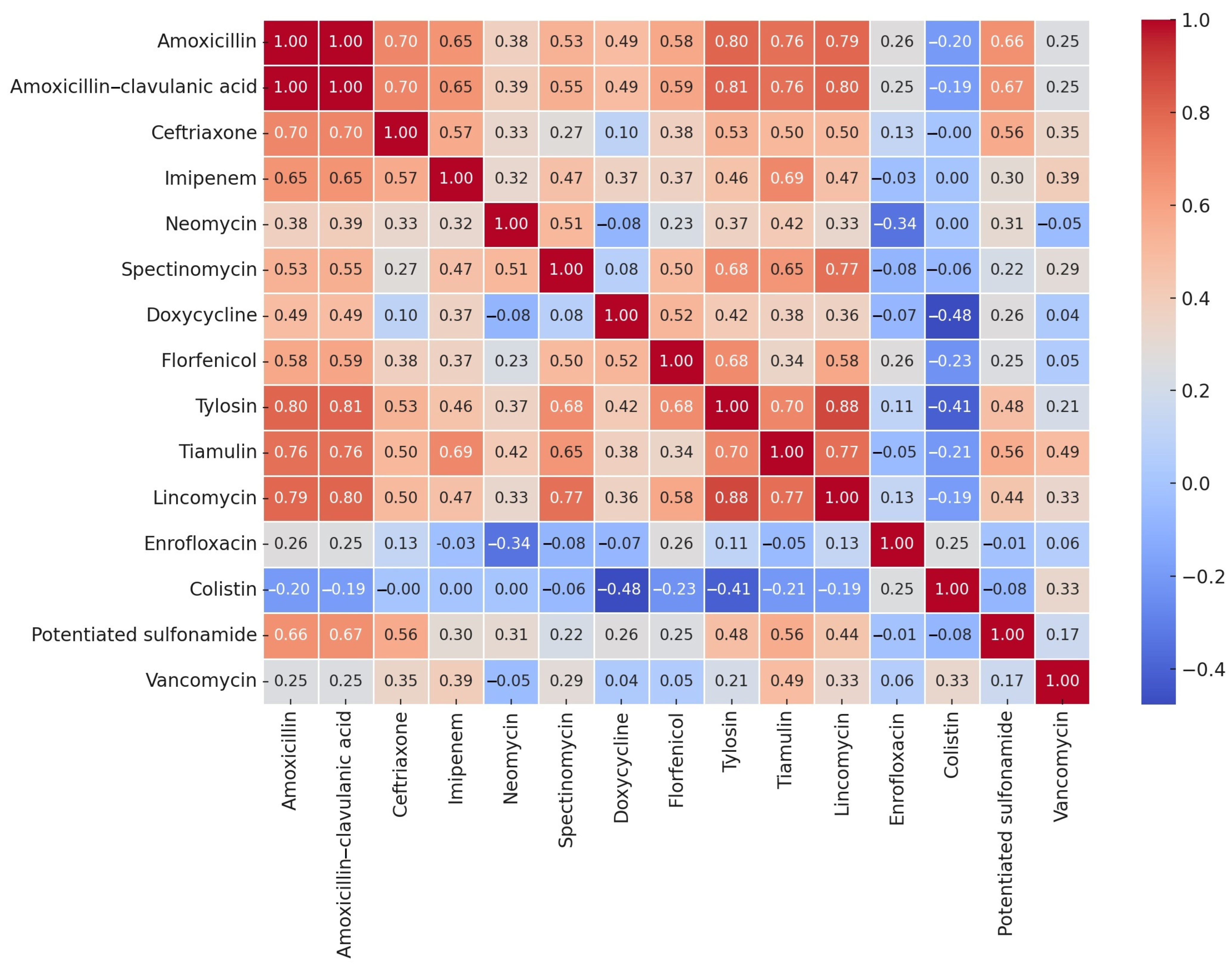
2.2. Minimum Inhibitory Concentration Values

The S. aureus isolates (n = 8) exhibited variable susceptibility to β-lactams. Although 50% of the population displayed high sensitivity to amoxicillin (MIC50 = 0.5 µg/mL), the MIC value for 90% of the population was notably high (MIC90 = 64 µg/mL). A considerably lower MIC was observed for amoxicillin–clavulanic acid (MIC50 = 0.25 µg/mL, MIC90 = 4 µg/mL), suggesting enhanced efficacy. Ceftriaxone also demonstrated high variability, but 50% of the population showed high sensitivity (MIC50 = 1 µg/mL). In contrast, imipenem retained its efficacy, with an MIC50 value of 0.03 µg/mL and an MIC90 of 0.125 µg/mL.
Among protein synthesis inhibitors, the isolates exhibited high minimum inhibitory concentration (MIC) values for neomycin and spectinomycin. Amoxicillin resistance was observed in 62.5% (n = 5/8) of the isolates, whereas 37.5% (n = 3/8) remained susceptible. A similar resistance trend was noted for doxycycline. The efficacy of tylosin was 62.5% (n = 5/8), that of tiamulin was 25.0% (n = 2/8), and imipenem exhibited 100.0% (n = 8/8) efficacy.
For enrofloxacin, 25.0% (n = 2/8) of the isolates were resistant, with MIC50 = 1 µg/mL and MIC90 = 4 µg/mL. As expected, colistin displayed high MIC values, given its specificity against Gram-negative bacteria. Moderate resistance was observed for trimethoprim–sulfamethoxazole (25% resistance, n = 2/8), whereas 100% (n = 8/8) of isolates remained susceptible to vancomycin.
The susceptibility profile of the isolates, based on established clinical breakpoints, is summarized in Figure 1. The distribution of MIC values is presented in Table 2, and is presented in box plots for each antibiotic in Supplementary Figure S1. All of the MIC values are in a Supplementary Excel File.
Epidemiological cut-off values (ECOFFs) provide valuable insight into distinguishing wild-type from acquired resistance. Imipenem was the only case where the observed MIC90 value (0.125 µg/mL) coincided with the ECOFF, suggesting that the majority of isolates belong to the wild-type population. For amoxicillin–clavulanic acid (0.25 µg/mL), ceftriaxone (1 µg/mL), florfenicol (4 µg/mL), spectinomycin (64 µg/mL), and vancomycin (0.5 µg/mL), the MIC50 values were lower than the ECOFF, indicating that at least half of the isolates were part of the wild-type population. These findings highlight the importance of integrating ECOFF values with clinical breakpoints for comprehensive resistance assessments.
Among β-lactam antibiotics, 50% of the Streptococcus isolates exhibited high susceptibility to amoxicillin and amoxicillin–clavulanic acid (MIC50 = 0.5 µg/mL). However, 42.1% (n = 8/19) of isolates were resistant to amoxicillin–clavulanic acid, with 31.6% (n = 6/19) remaining susceptible and 26.3% showing intermediate susceptibility. Similar trends were observed for ceftriaxone (MIC50 = 0.06 µg/mL), whereas imipenem demonstrated strong efficacy (MIC90 = 1 µg/mL).
Regarding protein synthesis inhibitors, high MIC values were recorded for neomycin and spectinomycin. Doxycycline resistance was observed in 89.5% (n = 17/19) of isolates, whereas 63.2% (n = 12/19) of isolates were resistant to florfenicol. Elevated MIC values were also detected for tylosin, tiamulin, and lincomycin.
Enrofloxacin showed variable susceptibility patterns, with 42.1% (n = 8/19) of isolates susceptible, 31.6% (n = 6/19) exhibiting intermediate susceptibility, and 26.3% (n = 5/19) classified as resistant. As expected, colistin exhibited high MIC values, reinforcing its inefficacy against Gram-positive bacteria. Trimethoprim–sulfamethoxazole displayed limited efficacy, with only 15.8% (n = 3/19) of isolates susceptible, 31.6% (n = 6/19) exhibiting intermediate resistance, and 52.6% (n = 10/19) classified as resistant. Vancomycin susceptibility was maintained in 94.7% (n = 18/19) of isolates, whereas 5.3% (n = 1/19) displayed resistance.
The susceptibility profile of Streptococcus isolates, based on clinical breakpoints, is depicted in Figure 2, and the corresponding MIC distributions are summarized in Table 3 and presented in box plots for each antibiotic in Supplementary Figure S2. All of the MIC values are in a Supplementary Excel File.
Colistin was included as part of the standard testing panel, despite its lack of clinical application in Gram-positive infections. The high MIC values observed are in line with the known intrinsic resistance of S. aureus and Streptococcus spp.
Vancomycin was the only case where the MIC90 value (0.5 µg/mL) was below the ECOFF, suggesting that the majority of isolates belong to the wild-type population. For enrofloxacin (1 µg/mL), the MIC50 value was lower than the ECOFF, indicating that at least half of the isolates could be classified as part of the wild-type population.
Given the sample size for Streptococcus spp. (n = 19), a Spearman correlation analysis was performed, as MIC values typically follow a non-normal distribution. This allowed for the assessment of both linear and monotonic relationships, which are crucial in evaluating cross-resistance potential. The correlation results are visualized in a heatmap (Figure 3).
A strong correlation was observed between amoxicillin and amoxicillin–clavulanic acid (1.00), as well as between tylosin and lincomycin (0.88). Lincomycin was strongly correlated with tiamulin and tylosin, as well as amoxicillin (0.79) and amoxicillin–clavulanic acid (0.80). Conversely, colistin exhibited weak negative correlations with most other antibiotics but a strong negative correlation with doxycycline (−0.48) and with tylosin (−0.41).

3. Discussion

The present study revealed high rates of AMR in S. aureus and Streptococcus spp. isolates from ducks and geese in Hungary, with particularly elevated resistance observed against β-lactams, macrolides, aminoglycosides, and doxycycline. These findings underscore the emerging threat of multidrug-resistant (MDR) Gram-positive pathogens in waterfowl production systems.
AMR is a major global challenge that requires a multidisciplinary approach integrating public health, animal health, and environmental sciences. The poultry industry, a cornerstone of global food security, faces substantial challenges due to the widespread use of antibiotics, which has driven the selection and dissemination of MDR bacteria [58,59,60]. These resistant bacteria can be transmitted to humans via multiple pathways, including through the food chain, direct contact with animals, and environmental exposure [61].
Given the intersection between public and animal health, integrated approaches, such as the One Health framework, are urgently needed. In Hungary, the veterinary antimicrobial consumption rate is 69.9 mg/PCU, positioning the country in the mid-range among European nations. Although Hungary’s total antibiotic sales (120 tons) may seem relatively low compared to Spain (1027.2 tons) or Italy (585.4 tons), the proportional use in livestock sectors remains a significant concern, particularly in poultry production [62,63].
Most poultry-related AMR research (85–90%) focuses on broiler chickens, leaving the waterfowl sector significantly underrepresented in scientific literature. This disparity reflects the economic and nutritional significance of chickens, which places them at the forefront of AMR research priorities [58]. Our study addresses this knowledge gap by characterizing AMR profiles in waterfowl pathogens, providing novel insights into S. aureus and Streptococcus spp. infections in ducks and geese.
S. aureus and Streptococcus spp. negatively impact poultry production, causing significant economic losses through arthritis, tenosynovitis, osteomyelitis, pododermatitis, and femoral head necrosis, which ultimately lead to lameness and compromised flock performance. Despite ongoing efforts, antibiotic treatment of such infections remains a major challenge, particularly in light of the high resistance rates observed in this study [64,65].
Our S. aureus isolates showed resistance rates of 62.5% (n = 5/8) to amoxicillin, 25% (n = 3/8) to enrofloxacin, and elevated MICs to neomycin and spectinomycin, indicating substantial therapeutic limitations. In some developing countries, misuse of antibiotics in poultry production has led to an increase in MDR Staphylococcus strains, defined as strains that are resistant to at least three classes of antibiotics, posing an escalating public health risk [66,67]. In a study by Eid et al. (2019), no enrofloxacin resistance was observed in S. aureus isolates, whereas neomycin resistance was reported at 20%, potentiated sulfonamide resistance at 80%, spectinomycin resistance at 90%, and universal resistance to amoxicillin [68]. In contrast, our findings demonstrated 25% (n = 2/8) enrofloxacin resistance, an MIC50 of 32 µg/mL for neomycin, 25% (n = 2/8) resistance to trimethoprim–sulfamethoxazole, an MIC50 of 64 µg/mL for spectinomycin, and 62.5% (n = 5/8) resistance to amoxicillin. Compared to these data, our findings suggest regional variation in resistance profiles, with lower sulfonamide resistance but higher enrofloxacin resistance.
Over recent years, the selective pressure exerted by antibiotics in poultry production has accelerated the spread of resistant bacteria [4,69,70]. Our results confirmed high levels of resistance to amoxicillin and doxycycline, whereas no vancomycin-resistant S. aureus strains were detected. Fortunately, vancomycin-resistant S. aureus strains remain relatively rare [66].
Similarly, Streptococcus spp. isolates exhibited significant resistance to β-lactams, macrolides, doxycycline, and lincosamides. However, some antibiotics, such as vancomycin, remained effective, whereas others, such as doxycycline, displayed high resistance rates. These findings emphasize the need for continuous AMR surveillance and the identification of alternative therapeutic options in the waterfowl sector.
The correlation analysis revealed a strong association between amoxicillin and amoxicillin–clavulanic acid (1.00), which is expected given their shared β-lactam structure. Notably, β-lactamase production is not a common resistance mechanism in Streptococcus spp. [51]. A strong correlation was also observed between tylosin and tiamulin (0.88), as well as lincomycin with tiamulin and tylosin, suggesting the presence of macrolide–lincosamide–streptogramin (MLS) cross-resistance mechanisms. This ribosomal target modification-based resistance is typically plasmid- or transposon-mediated [71]. Additionally, a significant positive correlation was detected between doxycycline and spectinomycin (0.72). Although these antibiotics inhibit protein synthesis via different mechanisms (doxycycline: ribosomal binding, spectinomycin: elongation factor inhibition), their co-occurrence suggests potential co-selection mechanisms, possibly linked to plasmid- or transposon-associated resistance determinants, such as Tn1545 and Tn917, which are known to carry tet and erm genes [72]. In contrast, colistin displayed weak correlations with other antibiotics, reinforcing its inefficacy against Gram-positive bacteria [73].
The zoonotic potential of S. aureus and Streptococcus spp. further elevates the public health relevance of our findings. Potential transmission routes from waterfowl to humans include direct contact, foodborne exposure, and environmental contamination, emphasizing the need for stringent biosecurity protocols and antimicrobial stewardship.
This study has several limitations. First, the number of isolates was relatively limited, which may restrict the generalizability of the findings. Second, molecular analyses of resistance determinants (e.g., mecA, erm, tet, or integron elements) were not performed, which limits mechanistic interpretation. Third, the isolates were obtained from clinical submissions, not from a systematic epidemiological sampling scheme. Despite these constraints, this study provides important baseline data for AMR monitoring in waterfowl and highlights key areas for future research.
The findings of this antimicrobial susceptibility study highlight significant antibiotic resistance patterns among Streptococcus and S. aureus isolates from waterfowl. The correlation analysis of MIC values identified strong associations between certain antibiotics, suggesting the potential presence of cross-resistance or co-selection mechanisms.
These results underscore the critical need for ongoing antimicrobial resistance monitoring in the waterfowl industry, particularly for the detection of multidrug-resistant strains. Based on the observed resistance patterns, further genetic investigations, such as the molecular detection of resistance genes, are recommended to enhance our understanding of resistance mechanisms and transmission pathways.
The increasing trend of resistant pathogenic bacteria in waterfowl raises significant public health and veterinary concerns, emphasizing the need for responsible antibiotic use strategies and the development of alternative therapeutic approaches in veterinary practice.

4. Materials and Methods

4.1. Origin of Strains

Sample collection was conducted between 2022 and 2023. The isolates were obtained from clinical cases, with isolation and pure culture preparation performed by the National Food Chain Safety Office, Directorate of Veterinary Diagnostics. Bacterial isolation was conducted using standard aerobic culture techniques on blood agar and selective media from tissue samples collected during necropsy (brain ventricle, joint, liver, pericardium, and lung). Preliminary identification was based on colony morphology and hemolysis patterns, followed by confirmation using standard biochemical tests. The isolates originated from 27 clinical cases submitted to the National Reference Laboratory between 2022 and 2023, including 21 geese and 6 ducks. Sampling was targeted, as all samples were obtained from animals with diagnosed bacterial infections. All animals were from commercial waterfowl farms. The geographical origins of the samples were classified into Hungary’s seven administrative regions: Közép-Magyarország, Közép-Dunántúl, Nyugat-Dunántúl, Dél-Dunántúl, Észak-Magyarország, Észak-Alföld, and Dél-Alföld. The pure cultures were then provided to our research group based on a mutual agreement. The isolates received on Petri dishes were stored in a Microbank™ system (Pro-Lab Diagnostics, Richmond Hill, ON, Canada) at −80°C until further analysis.
Each sample was assigned a unique identifier, and relevant metadata, including the host species (duck or goose), the affected organ (brain ventricle, joint, liver, pericardium, and lung), and geographical origin, were recorded. The geographical locations were categorized into Hungary’s seven administrative regions for systematic data management.

4.2. Preparation of Antibiotic Stock Solutions

The preparation of antibiotic stock solutions was conducted according to Clinical and Laboratory Standards Institute (CLSI) guidelines [74,75]
Amoxicillin and amoxicillin–clavulanic acid (2:1 ratio) were dissolved in phosphate-buffered solution (pH 7.2, 0.01 mol/L). Imipenem was prepared in phosphate-buffered solution (pH 6, 0.1 mol/L). Ceftriaxone, doxycycline, spectinomycin, neomycin, colistin, tiamulin, tylosin, lincomycin, and vancomycin were dissolved in distilled water. Trimethoprim–sulfamethoxazole (in a 19:1 ratio) was prepared by dissolving sulfamethoxazole in hot water with a few drops of 2.5 mol/L NaOH, whereas trimethoprim was dissolved in 0.05 mol/L HCl in distilled water. Enrofloxacin was dissolved in distilled water with a few drops of 1 mol/L NaOH. Florfenicol was prepared using a few drops of 95% ethanol and distilled water.
For each antibiotic, a 1024 µg/mL stock solution was prepared and was adjusted for purity according to the manufacturer’s specifications (Merck KGaA, Darmstadt, Germany).

4.3. Determination of Minimum Inhibitory Concentration

The phenotypic expression of AMR was assessed by determining the MIC of each isolate. This study was performed following CLSI methodology [75] and breakpoints were determined based on CLSI guidelines [74].
Bacterial isolates stored at −80 °C were cultured overnight in 3 mL Mueller–Hinton broth (MHB) at 37 °C for 18–24 h prior to testing. MIC determinations were carried out in 96-well microtiter plates (VWR International, LLC, Debrecen, Hungary). All wells, except those in the first column, were pre-filled with 90 µL of MHB. Two-fold serial dilutions of antibiotic stock solutions were prepared in MHB, starting at 512 µg/mL, covering a concentration range of 512 to 0.0009 µg/mL.
A 180 µL aliquot of the prepared antibiotic dilutions was transferred into the first column of the test plate, followed by two-fold serial dilutions across subsequent columns. After preparing the dilution series, 10 µL of a 0.5 McFarland standard bacterial suspension (105 CFU/mL) was inoculated into wells from column 11 backward using a nephelometer (ThermoFisher Scientific, Budapest, Hungary) [75]. Column 11 was the positive control (containing only bacterial suspension and broth). Column 12 was the negative control (containing only broth).
The plates were incubated at 41 °C for 18–24 h, and the MIC values were determined using the Sensititre™ SWIN™ automatic MIC reader (ThermoFisher Scientific, Budapest, Hungary) and VIZION™ system software (v3.4, ThermoFisher Scientific, Budapest, Hungary, 2024). The quality control strains used were S. aureus (ATCC 12600) and Streptococcus suis (ATCC 43765).

4.4. Statistical Analysis

Statistical analyses were performed using R (v4.2.2) in the RStudio environment [76]. Descriptive statistics and correlation analyses were applied to evaluate antimicrobial susceptibility data. The MIC values were log-transformed for visualization, and Spearman’s rank correlation test was conducted to assess potential relationships between the MIC values [77].
Spearman’s correlation coefficient was calculated for each pair of antibiotics. This non-parametric test is appropriate for detecting monotonic relationships, regardless of normality assumptions. Statistical significance was determined using p-values (<0.05), indicating significant correlations.
Results were visualized using heatmaps, allowing the identification of potential cross-resistance and co-selection patterns among antimicrobial agents.

Supplementary Materials

The following supporting information can be downloaded at: https://www.mdpi.com/article/10.3390/antibiotics14050496/s1, Figure S1: The distribution of minimum inhibitory concentration (MIC) values of Staphylococcus aureus isolates (n = 8) from waterfowl, visualized using a box plot for each antibiotic. Figure S2: The distribution of minimum inhibitory concentration (MIC) values of Streptococcus isolates (n = 8) from waterfowl, visualized using a box plot for each antibiotic. Supplementary Excel.

Author Contributions

Conceptualization, Á.K. and Á.J.; methodology, Á.K.; software, Á.K.; validation, Á.J.; formal analysis, Á.S.; investigation, Á.J.; resources, Á.K.; data curation, Á.S.; writing—original draft preparation, Á.S.; writing—review and editing, Á.K.; visualization, Á.S.; supervision, Á.J.; project administration, Á.S.; funding acquisition, Á.J. All authors have read and agreed to the published version of the manuscript.

Funding

This research was funded by Project no. RRF-2.3.1-21-2022-00001 and was implemented with the support provided by the Recovery and Resilience Facility (RRF), financed under the National Recovery Fund budget estimate, RRF-2.3.1-21 funding scheme.

Data Availability Statement

The data presented in this study are available from the corresponding author upon reasonable request.

Acknowledgments

We would like to thank Katalin Balogh and Tamásné Pénzes Imre for preparing the laboratory work. We also extend our thanks to Ákos Thuma for isolating the strains.

Conflicts of Interest

The authors declare no conflicts of interest.

Abbreviations

The following abbreviations are used in this manuscript:
AMRAntimicrobial resistance
CLSIClinical and Laboratory Standards Institute
ECOFFEpidemiological cut-off values
MHBMueller–Hinton broth
MICMinimum inhibitory concentration

References

  1. Jamali, H.; Radmehr, B.; Ismail, S. Prevalence and Antimicrobial Resistance of Listeria, Salmonella, and Yersinia Species Isolates in Ducks and Geese. Poult. Sci. 2014, 93, 1023–1030. [Google Scholar] [CrossRef] [PubMed]
  2. Anand, U.; Reddy, B.; Singh, V.K.; Singh, A.K.; Kesari, K.K.; Tripathi, P.; Kumar, P.; Tripathi, V.; Simal-Gandara, J. Potential Environmental and Human Health Risks Caused by Antibiotic-Resistant Bacteria (ARB), Antibiotic Resistance Genes (ARGs) and Emerging Contaminants (ECs) from Municipal Solid Waste (MSW) Landfill. Antibiotics 2021, 10, 374. [Google Scholar] [CrossRef] [PubMed]
  3. Laxminarayan, R.; Chaudhury, R.R. Antibiotic Resistance in India: Drivers and Opportunities for Action. PLoS Med. 2016, 13, e1001974. [Google Scholar] [CrossRef]
  4. Witte, W. Selective Pressure by Antibiotic Use in Livestock. Int. J. Antimicrob. Agents 2000, 16 (Suppl. 1), S19–S24. [Google Scholar] [CrossRef]
  5. Hetényi, N.; Bersényi, A.; Hullár, I. Physiological Effects of Medium-Chain Fatty Acids and Triglycerides, and Their Potential Use in Poultry and Swine Nutrition: A Literature Review. Magy. Állatorvosok Lapja 2024, 146, 651–659. [Google Scholar] [CrossRef]
  6. Jerzsele, Á.; Somogyi, Z.; Szalai, M.; Kovács, D. Effects of Fermented Wheat Germ Extract on Artificial Salmonella Typhimurium Infection in Broiler Chickens. Magy. Állatorvosok Lapja 2020, 142, 77–85. [Google Scholar]
  7. Kerek, Á.; Csanády, P.; Jerzsele, Á. Antibacterial Efficiency of Propolis—Part 1. Magy. Állatorvosok Lapja 2022, 144, 285–298. [Google Scholar]
  8. Kerek, Á.; Csanády, P.; Jerzsele, Á. Antiprotozoal and Antifungal Efficiency of Propolis—Part 2. Magy. Állatorvosok Lapja 2022, 144, 691–704. [Google Scholar]
  9. Kovács, L.; Nagy, D.; Könyves, L.; Jerzsele, Á.; Kerek, Á. Antimicrobial Properties of Essential Oils—Animal Health Aspects. Magy. Állatorvosok Lapja 2023, 145, 497–510. [Google Scholar] [CrossRef]
  10. Olasz, Á.; Jerzsele, Á.; Balta, L.; Dobra, P.F.; Kerek, Á. In Vivo Efficacy of Different Extracts of Propolis in Broiler Salmonellosis. Magy. Állatorvosok Lapja 2023, 145, 461–475. [Google Scholar] [CrossRef]
  11. Petrilla, J.; Mátis, G.; Molnár, A.; Jerzsele, Á.; Pál, L.; Gálfi, P.; Neogrády, Z.; Dublecz, K. In Vitro Investigation of the Antibacterial Efficacy of Butyrate on Various Campylobacter jejuni Strains. MÁL 2021, 143, 57–64. [Google Scholar]
  12. Acikgoz, Z.; Yucel, B.; Altan, O. The Effects of Propolis Supplementation on Broiler Performance and Feed Digestibility. Arch. Geflugelkd. 2005, 69, 117–122. [Google Scholar]
  13. Sebők, C.; Márton, R.A.; Meckei, M.; Neogrády, Z.; Mátis, G. Antimicrobial Peptides as New Tools to Combat Infectious Diseases. Magy. Állatorvosok Lapja 2024, 146, 181–191. [Google Scholar] [CrossRef]
  14. Kovács, L.; Hejel, P.; Farkas, M.; László, L.; Könyves, L. Study Report on the Effect of a Litter Treatment Product Containing Bacillus licheniformis and Zeolite in Male Fattening Turkey Flock. Magy. Állatorvosok Lapja 2024, 146, 291–305. [Google Scholar] [CrossRef]
  15. Such, N.; Molnár, A.; Pál, L.; Farkas, V.; Menyhárt, L.; Husvéth, F.; Dublecz, K. The Effect of Pre- and Probiotic Treatment on the Gumboro-Titer Values of Broilers. Magy. Állatorvosok Lapja 2021, 143, 119–127. [Google Scholar]
  16. Abd El-Ghany, W.A. Paraprobiotics and Postbiotics: Contemporary and Promising Natural Antibiotics Alternatives and Their Applications in the Poultry Field. Open Vet. J. 2020, 10, 323–330. [Google Scholar] [CrossRef]
  17. Essősy, M.; Fodor, I.; Ihnáth, Z.; Karancsi, Z.; Kovács, D.; Szalai, K.V.; Szentmiklósi, D.; Jerzsele, Á. The Possibilities of Antibiotic-Free Broiler-Hen Fattening, with Special Reference to the Use of Pre- and Probiotics. Magy. Állatorvosok Lapja 2020, 142, 397–407. [Google Scholar]
  18. Kovács, D.; Palkovicsné Pézsa, N.; Farkas, O.; Jerzsele, Á. Usage of Antibiotic Alternatives in Pig Farming: Literature Review. Magy. Állatorvosok Lapja 2021, 143, 281–282. [Google Scholar]
  19. Bager, F.; Madsen, M.; Christensen, J.; Aarestrup, F.M. Avoparcin Used as a Growth Promoter Is Associated with the Occurrence of Vancomycin-Resistant Enterococcus faecium on Danish Poultry and Pig Farms. Prev. Vet. Med. 1997, 31, 95–112. [Google Scholar] [CrossRef]
  20. Zhang, S.; Chen, S.; Rehman, M.U.; Yang, H.; Yang, Z.; Wang, M.; Jia, R.; Chen, S.; Liu, M.; Zhu, D.; et al. Distribution and Association of Antimicrobial Resistance and Virulence Traits in Escherichia coli Isolates from Healthy Waterfowls in Hainan, China. Ecotoxicol. Environ. Saf. 2021, 220, 112317. [Google Scholar] [CrossRef]
  21. Albarella, U. Alternate Fortunes? The Role of Domestic Ducks and Geese from Roman to Medieval Times in Britain. Doc. Archaeobiologiae 2005, III, 249–258. [Google Scholar]
  22. Farkas, M.; Könyves, L.; Csorba, S.; Farkas, Z.; Józwiák, Á.; Süth, M.; Kovács, L. Biosecurity Situation of Large-Scale Poultry Farms in Hungary According to the Databases of National Food Chain Safety Office Centre for Disease Control and Biosecurity Audit System of Poultry Product Board of Hungary in the Period of 2021–2022. Magy. Állatorvosok Lapja 2024, 146, 723–742. [Google Scholar] [CrossRef]
  23. Adebowale, O.; Makanjuola, M.; Bankole, N.; Olasoju, M.; Alamu, A.; Kperegbeyi, E.; Oladejo, O.; Fasanmi, O.; Adeyemo, O.; Fasina, F.O. Multi-Drug Resistant Escherichia coli, Biosecurity and Anti-Microbial Use in Live Bird Markets, Abeokuta, Nigeria. Antibiotics 2022, 11, 253. [Google Scholar] [CrossRef] [PubMed]
  24. Mag, P.; Németh, K.; Somogyi, Z.; Jerzsele, Á. Antibacterial therapy based on pharmacokinetic/ pharmacodynamic models in small animal medicine-1. Literature review. Magy. Állatorvosok Lapja 2023, 145, 419–438. [Google Scholar] [CrossRef]
  25. Adams, H.R. Veterinary Pharmacology and Therapeutics; Iowa State University Press: Ames, Iowa, 2001; ISBN 978-0-8138-1743-9. [Google Scholar]
  26. KSH Baromfiállomány (19.1.1.29.). Available online: https://www.ksh.hu/stadat_files/mez/hu/mez0029.html (accessed on 18 May 2023).
  27. Szoke, Z.; Fauszt, P.; Mikolas, M.; David, P.; Szilagyi-Tolnai, E.; Pesti-Asboth, G.; Homoki, J.R.; Kovacs-Forgacs, I.; Gal, F.; Stundl, L.; et al. Comprehensive Analysis of Antimicrobial Resistance Dynamics among Broiler and Duck Intensive Production Systems. Sci. Rep. 2025, 15, 4673. [Google Scholar] [CrossRef]
  28. Heaton, C.J.; Gerbig, G.R.; Sensius, L.D.; Patel, V.; Smith, T.C. Staphylococcus aureus Epidemiology in Wildlife: A Systematic Review. Antibiotics 2020, 9, 89. [Google Scholar] [CrossRef] [PubMed]
  29. Yuko, S. Staphylococcosis in Poultry–Poultry. Available online: https://www.merckvetmanual.com/poultry/staphylococcosis/staphylococcosis-in-poultry (accessed on 3 May 2025).
  30. Teresa, Y.M. Streptococcosis in Poultry–Poultry. Available online: https://www.msdvetmanual.com/poultry/streptococcosis/streptococcosis-in-poultry (accessed on 3 May 2025).
  31. Fulde, M.; Valentin-Weigand, P. Epidemiology and Pathogenicity of Zoonotic Streptococci. Curr. Top. Microbiol. Immunol. 2013, 368, 49–81. [Google Scholar] [CrossRef] [PubMed]
  32. Chen, H.; Zhang, J.; He, Y.; Lv, Z.; Liang, Z.; Chen, J.; Li, P.; Liu, J.; Yang, H.; Tao, A.; et al. Exploring the Role of Staphylococcus aureus in Inflammatory Diseases. Toxins 2022, 14, 464. [Google Scholar] [CrossRef]
  33. Abbasi, K.; Tajbakhsh, E.; Momtaz, H. Antimicrobial Resistance, Virulence Genes, and Biofilm Formation in Staphylococcus aureus Strains Isolated from Meat and Meat Products. J. Food Saf. 2021, 41, e12933. [Google Scholar] [CrossRef]
  34. Bortolaia, V.; Espinosa-Gongora, C.; Guardabassi, L. Human Health Risks Associated with Antimicrobial-Resistant Enterococci and Staphylococcus aureus on Poultry Meat. Clin. Microbiol. Infect. 2016, 22, 130–140. [Google Scholar] [CrossRef]
  35. Benrabia, I.; Hamdi, T.M.; Shehata, A.A.; Neubauer, H.; Wareth, G. Methicillin-Resistant Staphylococcus aureus (MRSA) in Poultry Species in Algeria: Long-Term Study on Prevalence and Antimicrobial Resistance. Vet. Sci. 2020, 7, 54. [Google Scholar] [CrossRef] [PubMed]
  36. European Food Safety Authority (EFSA); European Centre for Disease Prevention and Control (ECDC). The European Union Summary Report on Antimicrobial Resistance in Zoonotic and Indicator Bacteria from Humans, Animals and Food in 2020/2021. EFSA J. 2023, 21, e07867. [Google Scholar] [CrossRef]
  37. Abdullahi, I.N.; Zarazaga, M.; Campaña-Burguet, A.; Eguizábal, P.; Lozano, C.; Torres, C. Nasal Staphylococcus aureus and S. pseudintermedius Carriage in Healthy Dogs and Cats: A Systematic Review of Their Antibiotic Resistance, Virulence and Genetic Lineages of Zoonotic Relevance. J. Appl. Microbiol. 2022, 133, 3368–3390. [Google Scholar] [CrossRef]
  38. Zehra, A.; Gulzar, M.; Singh, R.; Kaur, S.; Gill, J.P.S. Prevalence, Multidrug Resistance and Molecular Typing of Methicillin-Resistant Staphylococcus aureus (MRSA) in Retail Meat from Punjab, India. J. Glob. Antimicrob. Resist. 2019, 16, 152–158. [Google Scholar] [CrossRef]
  39. de Boer, E.; Zwartkruis-Nahuis, J.T.M.; Wit, B.; Huijsdens, X.W.; de Neeling, A.J.; Bosch, T.; van Oosterom, R.A.A.; Vila, A.; Heuvelink, A.E. Prevalence of Methicillin-Resistant Staphylococcus aureus in Meat. Int. J. Food Microbiol. 2009, 134, 52–56. [Google Scholar] [CrossRef]
  40. AVEC. Annual Report 2022; Association of Poultry Processors and Poultry Trade in the EU Countries: Brussels, Belgium, 2022; Available online: http://avec-poultry.eu/wp-content/uploads/2022/09/AVEC-annual-report-2022_FINAL-WEB.pdf (accessed on 3 May 2025).
  41. Bourély, C.; Cazeau, G.; Jarrige, N.; Jouy, E.; Haenni, M.; Lupo, A.; Madec, J.-Y.; Leblond, A.; Gay, E. Co-Resistance to Amoxicillin and Tetracycline as an Indicator of Multidrug Resistance in Escherichia coli Isolates From Animals. Front. Microbiol. 2019, 10, 2288. [Google Scholar] [CrossRef] [PubMed]
  42. Fessler, A.T.; Kadlec, K.; Hassel, M.; Hauschild, T.; Eidam, C.; Ehricht, R.; Monecke, S.; Schwarz, S. Characterization of Methicillin-Resistant Staphylococcus aureus Isolates from Food and Food Products of Poultry Origin in Germany. Appl. Environ. Microbiol. 2011, 77, 7151–7157. [Google Scholar] [CrossRef] [PubMed]
  43. Dahms, C.; Huebner, N.-O.; Kossow, A.; Mellmann, A.; Dittmann, K.; Kramer, A. Occurrence of ESBL-Producing Escherichia coli in Livestock and Farm Workers in Mecklenburg-Western Pomerania, Germany. PLoS ONE 2015, 10, e0143326. [Google Scholar] [CrossRef] [PubMed]
  44. Krupa, P.; Bystroń, J.; Bania, J.; Podkowik, M.; Empel, J.; Mroczkowska, A. Genotypes and Oxacillin Resistance of Staphylococcus aureus from Chicken and Chicken Meat in Poland. Poult. Sci. 2014, 93, 3179–3186. [Google Scholar] [CrossRef]
  45. Pyzik, E.; Marek, A.; Stępień-Pyśniak, D.; Urban-Chmiel, R.; Jarosz, Ł.S.; Jagiełło-Podębska, I. Detection of Antibiotic Resistance and Classical Enterotoxin Genes in Coagulase -Negative Staphylococci Isolated from Poultry in Poland. J. Vet. Res. 2019, 63, 183–190. [Google Scholar] [CrossRef]
  46. Boost, M.V.; Wong, A.; Ho, J.; O’Donoghue, M. Isolation of Methicillin-Resistant Staphylococcus aureus (MRSA) from Retail Meats in Hong Kong. Foodborne Pathog. Dis. 2013, 10, 705–710. [Google Scholar] [CrossRef] [PubMed]
  47. Lim, S.-K.; Nam, H.-M.; Park, H.-J.; Lee, H.-S.; Choi, M.-J.; Jung, S.-C.; Lee, J.-Y.; Kim, Y.-C.; Song, S.-W.; Wee, S.-H. Prevalence and Characterization of Methicillin-Resistant Staphylococcus aureus in Raw Meat in Korea. J. Microbiol. Biotechnol. 2010, 20, 775–778. [Google Scholar] [CrossRef]
  48. Cha, S.-Y.; Kang, M.; Yoon, R.-H.; Park, C.-K.; Moon, O.-K.; Jang, H.-K. Prevalence and Antimicrobial Susceptibility of Salmonella Isolates in Pekin Ducks from South Korea. Comp. Immunol. Microbiol. Infect. Dis. 2013, 36, 473–479. [Google Scholar] [CrossRef] [PubMed]
  49. Zhao, F.; He, S.; Tan, A.; Guo, X.; Jiang, L.; Liu-Fu, C.; Deng, Y.; Zhang, R. Isolation, Identification and Character Analysis of Streptococcus dysgalactiae from Megalobrama Terminalis. J. Fish Dis. 2020, 43, 239–252. [Google Scholar] [CrossRef] [PubMed]
  50. Andam, C.P.; Hanage, W.P. Mechanisms of Genome Evolution of Streptococcus. Infect. Genet. Evol. 2015, 33, 334–342. [Google Scholar] [CrossRef]
  51. Hakenbeck, R.; Grebe, T.; Zähner, D.; Stock, J.B. Beta-Lactam Resistance in Streptococcus pneumoniae: Penicillin-Binding Proteins and Non-Penicillin-Binding Proteins. Mol. Microbiol. 1999, 33, 673–678. [Google Scholar] [CrossRef]
  52. Dewachter, L.; Dénéréaz, J.; Liu, X.; de Bakker, V.; Costa, C.; Baldry, M.; Sirard, J.-C.; Veening, J.-W. Amoxicillin-Resistant Streptococcus pneumoniae Can Be Resensitized by Targeting the Mevalonate Pathway as Indicated by sCRilecs-Seq. eLife 2022, 11, e75607. [Google Scholar] [CrossRef]
  53. McMullin, P. A Pocket Guide to Poultry Health and Disease; 5M Books Ltd.: Sheffield, UK, 2004; ISBN 978-0-9530150-5-4. [Google Scholar]
  54. Hogg, R.; Pearson, A. Streptococcus gallolyticus subspecies gallolyticus Infection in Ducklings. Vet. Rec. 2009, 165, 297–298. [Google Scholar] [CrossRef]
  55. Logue, C.M.; Andreasen, C.B.; Borst, L.B.; Eriksson, H.; Hampson, D.J.; Sanchez, S.; Fulton, R.M. Other Bacterial Diseases. In Diseases of Poultry; John Wiley & Sons, Ltd.: Hoboken, NJ, USA, 2020; pp. 995–1085. ISBN 978-1-119-37119-9. [Google Scholar]
  56. Mark, P.; Paul, M.; Janet, M.B.; Dennis, A. Poultry Diseases; Elsevier Health Sciences: Edingurg, UK, 2007; ISBN 978-0-7020-2862-5. [Google Scholar]
  57. Rodrigo, M.K.D.; Saiganesh, A.; Hayes, A.J.; Wilson, A.M.; Anstey, J.; Pickering, J.L.; Iwasaki, J.; Hillas, J.; Winslow, S.; Woodman, T.; et al. Host-Dependent Resistance of Group A Streptococcus to Sulfamethoxazole Mediated by a Horizontally-Acquired Reduced Folate Transporter. Nat. Commun. 2022, 13, 6557. [Google Scholar] [CrossRef]
  58. Abreu, R.; Semedo-Lemsaddek, T.; Cunha, E.; Tavares, L.; Oliveira, M. Antimicrobial Drug Resistance in Poultry Production: Current Status and Innovative Strategies for Bacterial Control. Microorganisms 2023, 11, 953. [Google Scholar] [CrossRef] [PubMed]
  59. Al-Hadidi, S.H.; Al Mana, H.; Almoghrabi, S.Z.; El-Obeid, T.; AlAli, W.Q.; Eltai, N.O. Retail Chicken Carcasses as a Reservoir of Multidrug-Resistant Salmonella. Microb. Drug Resist. 2022, 28, 824–831. [Google Scholar] [CrossRef] [PubMed]
  60. da Costa, P.M.; Loureiro, L.; Matos, A.J.F. Transfer of Multidrug-Resistant Bacteria between Intermingled Ecological Niches: The Interface between Humans, Animals and the Environment. Int. J. Environ. Res. Public Health 2013, 10, 278–294. [Google Scholar] [CrossRef] [PubMed]
  61. Hafez, H.M.; Attia, Y.A. Challenges to the Poultry Industry: Current Perspectives and Strategic Future After the COVID-19 Outbreak. Front. Vet. Sci. 2020, 7, 516. [Google Scholar] [CrossRef] [PubMed]
  62. European Medicines Agency (EU Body or Agency). Sales of Veterinary Antimicrobial Agents in 31 European Countries in 2022: Trends from 2010 to 2022: Thirteenth ESVAC Report; Publications Office of the European Union: Luxembourg, 2023; ISBN 978-92-9155-072-2. [Google Scholar]
  63. Bank, W. People, Pathogens and Our Planet: The Economics of One Health; World Bank: Washington, DC, USA, 2012. [Google Scholar]
  64. Awad, N.F.S.; Abd El-Hamid, M.I.; Nabil, N.M.; Tawakol, M.M.; Eid, S.; Al-Zaban, M.I.; Farouk, H.; Zakai, S.A.; Elkelish, A.; Ibrahim, M.S.; et al. Multidrug Resistant and Multivirulent Avian Bacterial Pathogens: Tackling Experimental Leg Disorders Using Phytobiotics and Antibiotics Alone or in Combination. Poult. Sci. 2023, 102, 102889. [Google Scholar] [CrossRef] [PubMed]
  65. Garcia-Llorens, J.; Monroy, I.; Torres-Boncompte, J.; Soriano, J.M.; Catalá-Gregori, P.; Sevilla-Navarro, S. Tracking the Prevalence of Antibiotic Resistance in Enterococcus Within the Spanish Poultry Industry: Insights from a One Health Approach. Antibiotics 2024, 14, 16. [Google Scholar] [CrossRef]
  66. Wang, J.; Meng, Y.; Zhang, R.; Yan, H.; Xu, G.; Zhu, Y.; Xie, Z.; Jiang, S. Coagulase-Negative Staphylococci Are the Main Causes of Bacterial Meningitis in Duck. Poult. Sci. 2024, 103, 103592. [Google Scholar] [CrossRef] [PubMed]
  67. Alves, R.R.N.; Rosa, I.M.L. Biodiversity, Traditional Medicine and Public Health: Where Do They Meet? J. Ethnobiol. Ethnomed. 2007, 3, 14. [Google Scholar] [CrossRef]
  68. Eid, H.M.; Algammal, A.M.; Elfeil, W.K.; Youssef, F.M.; Harb, S.M.; Abd-Allah, E.M. Prevalence, Molecular Typing, and Antimicrobial Resistance of Bacterial Pathogens Isolated from Ducks. Vet. World 2019, 12, 677–683. [Google Scholar] [CrossRef]
  69. Xu, Z.; Shah, H.N.; Misra, R.; Chen, J.; Zhang, W.; Liu, Y.; Cutler, R.R.; Mkrtchyan, H.V. The Prevalence, Antibiotic Resistance and mecA Characterization of Coagulase Negative Staphylococci Recovered from Non-Healthcare Settings in London, UK. Antimicrob. Resist. Infect. Control 2018, 7, 73. [Google Scholar] [CrossRef]
  70. Kolár, M.; Urbánek, K.; Látal, T. Antibiotic Selective Pressure and Development of Bacterial Resistance. Int. J. Antimicrob. Agents 2001, 17, 357–363. [Google Scholar] [CrossRef]
  71. Leclercq, R.; Courvalin, P. Bacterial Resistance to Macrolide, Lincosamide, and Streptogramin Antibiotics by Target Modification. Antimicrob. Agents Chemother. 1991, 35, 1267–1272. [Google Scholar] [CrossRef] [PubMed]
  72. Okitsu, N.; Kaieda, S.; Yano, H.; Nakano, R.; Hosaka, Y.; Okamoto, R.; Kobayashi, T.; Inoue, M. Characterization of ermB Gene Transposition by Tn1545 and Tn917 in Macrolide-Resistant Streptococcus pneumoniae Isolates. J. Clin. Microbiol. 2005, 43, 168–173. [Google Scholar] [CrossRef] [PubMed]
  73. Gurjar, M. Colistin for Lung Infection: An Update. J. Intensive Care 2015, 3, 3. [Google Scholar] [CrossRef] [PubMed]
  74. Clinical and Laboratory Standards Institute CLSI. Methods for Antimicrobial Susceptibility Testing of Infrequently Isolated or Fastidious Bacteria Isolated From Animals, 1st ed.; CLSI Standards VET06; Clinical and Laboratory Standards Institute: Wayne, PA, USA, 2017. [Google Scholar]
  75. Clinical and Laboratory Standards Institute CLSI. Methods for Dilution Antimicrobial Susceptibility Tests for Bacteria That Grow Aerobically, 12th ed.; CLSI standards M07; Clinical and Laboratory Standards Institute: Wayne, PA, USA, 2024. [Google Scholar]
  76. R Core Team. R: A Language and Environment for Statistical Computing; R Foundation for Statistical Computing: Vienna, Austria, 2020. [Google Scholar]
  77. Heck, D.W.; Moshagen, M. RRreg: Correlation and Regression Analyses for Randomized Response Data; R package Version 1.1.0, 2022. Available online: https://CRAN.R-project.org/package=RRreg (accessed on 3 May 2025).
Figure 1. Susceptibility of Staphylococcus aureus (n = 8) strains to drugs with clinical breakpoints. The chart shows the percentage of isolates classified as susceptible (green), intermediate (yellow), or resistant (red) for each antimicrobial agent. The values inside bars represent the percentage of isolates per category. All isolates were recovered from clinical waterfowl cases between 2022 and 2023. The antibiotics for which no CLSI/EUCAST breakpoint was available are not displayed in this figure. The MIC values for these agents are provided in a Supplementary Excel File.
Antibiotics 14 00496 g001
Figure 2. Susceptibility of Streptococcus strains (n = 19) to drugs with clinical breakpoints. The chart illustrates the percentage of isolates classified as susceptible (green), intermediate (yellow), or resistant (red) for each antimicrobial agent, based on minimum inhibitory concentration (MIC) values. The values inside each bar represent the proportion of isolates (%) in each susceptibility category. All isolates were derived from clinical samples obtained from waterfowl between 2022 and 2023. The antibiotics for which no CLSI/EUCAST breakpoint was available are not displayed in this figure. The MIC values for these agents are provided in a Supplementary Excel File.
Antibiotics 14 00496 g002
Figure 3. Correlation analysis of minimum inhibitory concentration (MIC) values for Streptococcus strains (n = 19) by active substance. The heatmap shows pairwise Pearson correlation coefficients between the MIC values of 15 antibiotics tested across all isolates. The warmer colors (red) indicate stronger positive correlations (r > 0), suggesting potential co-resistance patterns or shared resistance mechanisms. The cooler colors (blue) indicate negative correlations (r < 0), and white indicates weak or no correlation. The values in each cell represent the exact correlation coefficient (r) between the respective antibiotic pairs.
Antibiotics 14 00496 g003
Table 1. Distribution of organ localization of isolated bacterial strains.
Bacterial SpeciesPericardiumLungsLiverArticulation
n (%)
Staphylococcus aureus (n = 8)0 (0.0)0 (0.0)6 (75.0)2 (25.0)
Streptococcus spp. (n = 19)2 (10.5)1 (5.3)15 (78.9)1 (5.3)
Total (n = 27)2 (7.4)1 (3.7)21 (77.8)3 (11.1)
Table 2. Distribution of the minimum inhibitory concentrations (MICs) of Staphylococcus aureus strains isolated from waterfowl (n = 8) and the calculated values for 50% (MIC50) and 90% (MIC90) of the population. The vertical red lines indicate clinical breakpoints. The vertical green lines indicate the threshold distinguishing wild-type from non-wild-type strains.
AntibioticsBreakpoint 1
(μg/mL)
Distribution of Strains by MIC (μg/mL)MIC50MIC90ECOFF 4
0.0150.030.060.1250.250.512481632641282565121024µg/mL
Amoxicillin0.5 20120010001001 0.5640.5
Amoxicillin–clavulanic acid 21 2120020001 0.2540.5
Ceftriaxone- 3001011100001 1168
Colistin- 1400012321024-
Doxycycline0.5 300020201 280.5
Enrofloxacin4 10221100001 14-
Florfenicol- 2112100001 4168
Imipenem813210001 0.030.1250.125
Lincomycin- 2110001101145122
Neomycin- 132011 322561
Trimethoprim–sulfamethoxazole 34 4200011 1320.25
Spectinomycin- 14011164512128
Tiamulin4 11020101100141282
Tilozin64 300011000110012128-
Vancomycin32 4112 0.542
1 Clinical Laboratory Standard Institute (CLSI); 2 2:1 ratio; 3 1:19 ratio; 4 European Committee on Antimicrobial Susceptibility Testing—Epidemiological cut-off value; MIC50—Minimum Inhibitory Concentration at which 50% of isolates are inhibited; MIC90—Minimum Inhibitory Concentration at which 90% of isolates are inhibited; ECOFF—Epidemiological Cut-Off Value separating wild-type (non-resistant) populations from those with acquired resistance.
Table 3. Distribution of the minimum inhibitory concentrations (MICs) of Streptococcus strains isolated from waterfowl (n = 19) and the calculated values for 50% (MIC50) and 90% (MIC90) of the population. The vertical red lines indicate clinical breakpoints. The vertical green lines indicate the threshold distinguishing wild-type from non-wild-type strains.
AntibioticsBreakpoint 1
(μg/mL)
Distribution of Strains by MIC (μg/mL)MIC50MIC90ECOFF 4
0.0150.030.060.1250.250.512481632641282565121024µg/mL
Amoxicillin- 123053203 0.580.06 5
Amoxicillin–clavulanic acid 21 123052006 0.580.06 5
Ceftriaxone- 46201113001 0.0640.06 6
Colistin- 1100860200116128-
Doxycycline0.5 1010000306701 16320.5 7
Enrofloxacin4 26422111 1162 8
Florfenicol8 10425011212 85124 7
Imipenem- 8340031 0.061-
Lincomycin- 1001201111303512810241 9
Neomycin- 14113 64512-
Trimethoprim–sulfamethoxazole 34 2011523005 4640.25 7
Spectinomycin- 102313641024-
Tiamulin- 10000013281031281024-
Tilozin- 401001111121336410241 8
Vancomycin2 2313001 0.50.51 6
1 Clinical Laboratory Standard Institute (CLSI); 2 2:1 ratio; 3 1:19 ratio; 4 European Committee on Antimicrobial Susceptibility Testing—Epidemiological cut-off value; 5 Streptococcus equi; 6 Streptococcus pneumonia; 7 Streptococcus suis; 8 Streptococcus dysgalactiae; 9 Streptococcus canis; MIC50—Minimum Inhibitory Concentration at which 50% of isolates are inhibited; MIC90—Minimum Inhibitory Concentration at which 90% of isolates are inhibited; ECOFF—Epidemiological Cut-Off Value separating wild-type (non-resistant) populations from those with acquired resistance.
Disclaimer/Publisher’s Note: The statements, opinions and data contained in all publications are solely those of the individual author(s) and contributor(s) and not of MDPI and/or the editor(s). MDPI and/or the editor(s) disclaim responsibility for any injury to people or property resulting from any ideas, methods, instructions or products referred to in the content.

