Article

A Snapshot of Antimicrobial Resistance in Semi-Wild Oryx: Baseline Data from Qatar

1
Biomedical Research Centre, Microbiology Department, Qu Health, Qatar University, Doha P.O. Box 2713, Qatar
2
International School for Medical Science and Engineering, Doha P.O. Box 7582, Qatar
3
Centre for Food Safety, University of Georgia, Griffin, GA 30609, USA
4
Faculty of Agricultural and Food Sciences, American University of Beirut, Riad El Solh, Beirut 1107, Lebanon
5
Department of Natural Reserves, Ministry of Environment and Climate Change, Doha P.O. Box 7634, Qatar
*
Author to whom correspondence should be addressed.
Antibiotics 2025, 14(3), 248; https://doi.org/10.3390/antibiotics14030248
Submission received: 28 January 2025 / Revised: 17 February 2025 / Accepted: 19 February 2025 / Published: 1 March 2025
(This article belongs to the Special Issue Antimicrobial Resistance in Veterinary Science, 2nd Edition)

Abstract

:
Background/Objectives: The spread of antimicrobial resistance (AMR) is a growing global health concern. Wild animals can play an important role in the amplification and dissemination of AMR and in conservation efforts aiming at controlling diseases in vulnerable wild animal populations. These animals can serve as reservoirs for antibiotic resistance genes and are key in the spread of AMR across ecosystems and hosts. Therefore, monitoring AMR in wild animals is crucial in tackling the spread of resistance in the environment and human population. This study investigated the phenotypic and genotypic resistance of Escherichia coli (E. coli) isolated from semi-wild oryx (Oryx leucoryx) in Qatar. Methods: One hundred fecal samples were collected from oryx in diverse natural reserves across Qatar. A selective agar medium was used to isolate E. coli, and the identity of the isolates was further confirmed using the VITEK® 2 Compact system. The Kirby–Bauer disk diffusion method was used to test antibiotic susceptibility. Genetic resistance determinants were identified through polymerase chain reaction (PCR) analyses and sequencing using the Oxford Nanopore Technology (ONT). Results: The results revealed that 18% (n = 18) of the samples harbored E. coli with resistance to a single antibiotic, 28% (n = 28) were resistant to at least one antibiotic, and 2% (n = 2) were multidrug-resistant (MDR). No resistance was observed against colistin. tetA and tetB encode tetracycline resistance were the most frequently detected genes (57.7%). Whole genome sequencing (WGS) was used to expand on AMR gene-PCR analyses and analyze the resistome of 12E. coli isolates. WGS identified several important antibiotic resistance determinates, including blaCTX-M-encoding Extended Spectrum Beta-Lactamase (ESBL) resistance, soxR associated with tetracycline target alteration, and mdtE, emrB, AcrE, mdtF, and marA related to ciprofloxacin efflux pump resistance. Conclusions: This study provides essential information regarding AMR in Qatari semi-wild animals, which will guide conservation strategies and wildlife health management in a world experiencing increasing antibiotic-resistant infections. Furthermore, these findings can inform policies to mitigate AMR spread, improve ecosystems, and enhance public and environmental health while paving the way for future research on AMR dynamics in wildlife.

1. Introduction

The misuse and abuse of antibiotics have fueled the rise of resistant bacteria among animals and humans, complicating infection treatments and threatening public health [1,2]. Clinically significant antibiotic-resistant bacteria (ARB) are being isolated from various animals, including food animals and wildlife species [3] and synanthropic birds [4,5]. The factors influencing the presence of ARB in wildlife are diverse and remain incompletely understood, with anthropogenic sources, directly or indirectly (e.g., via the environment), playing key roles in the spread of ARB in wildlife [4,6]. Examples of anthropogenic sources include ineffectively treated wastewater draining into natural waters like rivers and lakes and waste from intensive livestock farming [7,8,9,10]. The spread of ARB in the natural environment has harmful economic and health consequences [11]. Therefore, investigations have increasingly focused on wild animals to recognize the existence of antibiotic resistance in diverse environmental sources [12]. If wild animals become colonized with ARB via exposure to contaminated environments, these animals can then amplify ARB and transmit it to pristine environments or agricultural areas [13,14,15]. This will result in ARB amplification and/or transmission cycle across the human, animal, and environment continuum. Therefore, monitoring ARB in wildlife is an integral component of One Health strategies that aim to control the spread of resistance.
Escherichia coli (E. coli) is a Gram-negative bacterium that resides in the intestinal tract of mammals and birds and has been associated with fecally-contaminated environments [16,17]. Importantly, E. coli serves as a paradigm organism for understanding the spread of ARB within specific populations, because monitoring antimicrobial resistance in E. coli can serve as an indicator for the emergence and spread of resistance in bacterial communities across hosts and niches [7,18]. Notably, E. coli is considered a main element of antimicrobial resistance surveillance programs in food-producing animals. It can act as a reservoir or carrier of antibiotic resistance, further complicating AMR combating efforts. In recent years, antibiotic-resistant (AR) strains of E. coli have been detected in livestock farming and wild animals, indicating transmission of resistance outside the clinical and agricultural settings [19]. The detection of AR E. coli in wildlife has been associated with inadequately treated human and livestock waste, highlighting the spread of resistance into the broader environment [20]. Therefore, monitoring E. coli isolated from wild animals is an effective approach for assessing the spread of AMR bacteria into the environment.
Many studies have demonstrated that the bacteria found in wildlife, including important sequence types and strains carrying clinically relevant antibiotic resistance determinants, were associated with those found in humans and other animals [21]. Subsequently, AMR research in wild animals also helps in deciphering the complex dynamics of resistance dissemination across ecosystems and hosts and via zoonosis and reverse zoonotic pathways. Despite the importance of wildlife and the environment in the various One Health approaches targeting AMR, there is a significant gap in research on AMR in wildlife species, particularly endangered ones. This gap limits our understanding of how health threats (e.g., AMR or infectious agents) in wildlife species affect ecosystems (e.g., transmission of disease across wildlife and other environmental reservoirs), biodiversity (e.g., decline of wildlife due to anthropozoonotic disease), and human health (e.g., zoonosis). Taken together, studying AMR and infectious diseases in wild animals aids biodiversity conservation and expands the One Health approach to include species that can impact the ecosystem and human health.
In this study, we targeted wild animals, because they serve as valuable indicators of environmental and public health [22]. Specifically, we focused on Oryx leucoryx (Arabian Oryx), an endangered species native to the desert and steppe regions of the Arabian Peninsula [23]. The Arabian oryx holds significant cultural and national importance in Qatar, where it is recognized as the national animal, symbolizing the country’s heritage and connection to its desert environment. It represents national pride and the ongoing efforts to preserve Qatar’s natural heritage [24]. Classified as endangered and extinct in the wild by the International Union for Conservation of Nature (IUCN) Red List [25] the Arabian oryx has benefitted from several captive breeding and reintroduction programs across the Middle East, in collaboration with international organizations. As a result, the oryx’s status was downgraded to vulnerable in 2011. Studying AMR in endangered or vulnerable species addresses a gap in understanding how AMR might contribute to biodiversity loss, ecosystem degradation and the spread of zoonotic disease. These species inhabit unique ecosystems with under-researched health dynamics, and their conservation helps in preventing broader public health by bridging wildlife conservation and public health under a One Health strategy.
In Qatar, the Arabian oryx is among the four most iconic national animals, alongside the Arabian horse, the dromedary or Arabian camel, and falcons. For this reason, the State of Qatar, particularly the Ministry of Environment and Climate Change (MECC), has been implementing a captive breeding and conservation project for over 16 years to prevent the oryx from becoming extinct. This study fills an important knowledge gap, as there is no data on ARB in Qatar’s wild and semi-wild animals. Oryx leucoryx was chosen because of its importance to Qatar’s semi-wild ecosystems, where this species may be exposed to environmental pollutants and AMR pathogens. This research goes beyond conservation, offering new insights into the role of wildlife, particularly endangered or vulnerable species, in AMR transmission, a largely overlooked aspect in the Middle Eastern region. We aim to benchmark the prevalence of antibiotic-resistant E. coli strains in Qatar’s semi-wild animals and establish a nationwide monitoring system to track ARB in wildlife. Understanding the extent of AMR in these animal populations is essential for effective surveillance and preventive measures to mitigate the insidious health impacts of AMR on humans, animals, and the environment.

2. Results

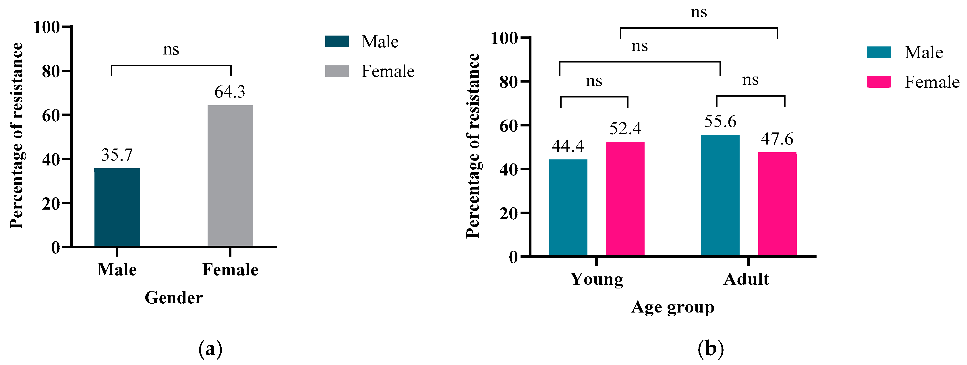
2.1. Demographic Data

In this study, 100 E. coli were isolated from otherwise healthy semi-wild oryx, 33% of the isolates (n = 33) were from males and 67% (n = 67) from females, and all the isolates were screened for AMR. Demographic data included the age and gender of the oryx and the collection locations. Notably, 49% (n = 49) of the isolates were collected from young animals (under three years old), while 51% (n = 51) were from adults (more than 8 years old). Additionally, 24% (n = 24) of the isolates were from the northwestern region, 50% (n = 50) from the central zone, and 26% (n = 26) from the southwestern part of Qatar. Since this is the first-ever study on AMR in Oryx, it was important to collect demographic data, which would serve two main purposes: (1) identify if age and/or gender influenced AMR carriage in the Oryx, which will help in conservation efforts, and (2) determine if the location of the animal might have contributed to acquisition of AMR. The latter might reveal anthropogenic influencing factors such as pollution of the location.

2.2. Detection of Phenotypically Antimicrobial Resistant E. coli

Antimicrobial resistance profiles of the E. coli isolates were assessed using 15 antibiotics as detailed in Table 1. Twenty-eight E. coli isolates were at least resistant to one of the tested antibiotics (Table A1). Resistance was observed only against five antibiotics: ampicillin, trimethoprim-sulfamethoxazole, ciprofloxacin, cefotaxime, and tetracycline. Among the 100 E. coli isolates, 8% exhibited resistance to ampicillin, while 26% of the isolates were resistant to tetracycline. Additionally, 3% of the isolates were resistant to trimethoprim-sulfamethoxazole and ciprofloxacin, while 1% were resistant to cefotaxime and 2% were multidrug- resistant (MDR) as shown in Figure 1. The profiles of the MDR isolates were AMP-SXT-CTX-TE and AMP-SXT-TE, respectively.
Analysis using SPSS (Version 29.0.0.0) indicated a higher percentage of antibiotic-resistant E. coli among female oryx (64.3%, n = 18/28) compared to males (35.7%, n = 10/28), although this difference was not statistically significant (p > 0.05) (Figure 2a). Age-related carriage of resistant isolates was equally distributed, with both young and adult groups yielding resistance in 50% (n = 14/28) of the samples (Figure 2b). In the young group, females exhibited higher carriage of resistant E. coli (52.4%, n = 11/21) compared to males (44.4%, n = 8/18) (Figure 2b). Conversely, the adult group showed higher carriage of resistant E. coli in males (55.6%, n = 10/18) compared to females (47.6%, n = 10/21) (Figure 2b). Regarding sample location, the carriage of resistant E. coli was highest in the central region (39.3%, n = 11), followed by the northwestern (35.7%, n = 10) and southwestern (25%, n = 7) locations (Figure 3). However, the binary logistic regression analysis (Table A1) yielded a p-value > 0.05 for resistance categorized by age, gender, and location, indicating no statistically significant relationship between these factors and the observed resistance percentages.

2.3. Screening Genetic Determinants of Resistance Using AMR Gene-Specific PCR Analyses

PCR analysis revealed that 57.7% of the tetracycline-resistant E. coli isolates carried both the tetA and tetB genes. Additionally, 11.55% of the isolates contained were positive only for tetA, while 15.4% were positive only for tetB. (Figure 4). tetC and tetE were not detected by PCR. For the eight ampicillin-resistant isolates, 12.5% (n = 1/8) harbored blaCTX-M, while 25% (n = 2/8) of the isolates were positive for blaTEM-1. The remaining 62.5% (n = 5/8) of the ampicillin-resistant isolates did not have any of the four PCR-screened genes associated with ampicillin resistance. None of the ampicillin-resistant E. coli tested positive for blaSHV.

2.4. Whole Genome Sequencing Using the Oxford Nanopore Technology (ONT)

Whole Genome Sequencing was performed on 12 E. coli isolates that revealed phenotypic resistance against ampicillin, ciprofloxacin, tetracycline, or cefotaxime but their resistance genetic determinants were not detected by PCR. The sequencing results revealed the resistance mechanisms, including relevant mutations and genes, which are listed in Table 2.

3. Discussion

The One Health approach has been implemented in Qatar as a multidisciplinary framework to address the increasing public health concern posed by AMR. The National Antimicrobial Resistance Action Plan (2024–2030) of Qatar illustrates the interconnectedness of people, animals, and the environment [26]. By studying AMR in wild animals, this study addresses a knowledge gap that will enhance this action plan and provide insights on antibiotic resistance acquisition and spread in these nationally important hosts. Notably, research on antibiotic resistance is limited in Qatari wild and semi-wild species. Consequently, this study evaluated antibiotic resistance in E. coli isolated from the semi-wild Qatari Oryx (Oryx leucoryx).
In this study, 28% (n = 28) of the bacterial isolates exhibited resistance to at least one antibiotic. The most common resistance was observed to tetracycline in 26% (n = 26) of the isolates, followed by ampicillin (8%, n = 8), trimethoprim-sulfamethoxazole (3%, n = 3), ciprofloxacin (3%, n = 3), and cefotaxime (1%, n = 1). Additionally, 2% of the isolates were identified as multidrug-resistant (MDR). These findings indicate a relatively low resistance level compared to similar studies in other regions. For instance, in a study conducted in Costa Rica, 93% (n = 63) of the bacterial isolates showed resistance to cephalexin (58%, n = 39), ampicillin (43%, n = 29), and oxytetracycline (22%, n = 15). The bacterial isolates in that study were sourced from Bradypus variegatus (Three-Toed Sloth), Choloepus hoffmanni (Two-Toed Sloth), and Alouatta palliata (Howler Monkey). Notably, 48% (n = 32) of the isolates in the Costa Rican study were classified as MDR, highlighting a significantly higher prevalence of resistance compared to our findings [27]. The differences in antibiotic resistance observed between the two studies are likely attributable to variations in the targeted animal populations, geographic locations, antibiotic practices and sampling techniques among others. In the Costa Rican study, the samples were collected from wild animal rehabilitation facilities, where the isolates were divided into two groups: 30% from restored animals and 70% from recently admitted animals. Remarkably, 21% of the rehabilitated animals had received prior antibiotic treatment, which may have contributed to the higher resistance rates observed in that study [27]. Antibiotics are infrequently used in healthy, semi-wild Oryx leucoryx, and are primarily reserved for treating diseased animals in Qatar. The main antibiotics employed in these cases include tetracyclines (e.g., oxytetracycline), fluoroquinolones (e.g., marbofloxacin, enrofloxacin), and beta-lactams (e.g., amoxicillin) (personal communication). Therefore, the relatively low levels of resistance observed in our study, particularly to ampicillin, ciprofloxacin, and tetracycline, may be attributed to the limited and targeted use of antibiotics. In contrast, a study by Alhababi et al. (2020) [28] investigating AMR profiles in food animals in Qatar, specifically cattle, camels, and pigeons, found a 42.8% resistance rate (n = 114/266). Furthermore, resistance rates were higher at 36.5% in cattle, 20.6% in camels, 70% in pigeons, and 90% in broiler chickens. In comparison, the restricted and focused use of antibiotics in Oryx probably explains the reduced resistance rates seen in this study [28]. This highlights the significant differences in resistance patterns between wild and domesticated animal populations, potentially due to variations in antibiotic use and exposure [28]. The oryx in this study inhabited a semi-conserved area, which is separated from livestock and wastewater sources. As a result, the oryx should have no direct contact with these potential environmental contributors to AMR. While separation likely plays a role in the relatively low levels of AMR observed within this population, other contributing factors such as dust storms can potentially transport AMR from distant sources to the regions occupied by the oryx [29].
Alhababi et al. [28] also reported that the highest resistance in E. coli isolated from food animals in Qatar was observed against tetracycline (64% in pigeons, 27.9% in cattle, and 15% in camels), followed by ampicillin (55.1%, 14%, and 7%, respectively) [28]. In our study, comparatively modest resistance rates to ampicillin (8%) and tetracycline (26%) were also identified in the oryx. The widespread use of tetracycline and ampicillin globally likely contributes to these trends, as ampicillin and tetracycline are commonly used as first-line treatments for animal infections worldwide [30]. This suggests that frequent and widespread antibiotic exposure in food animals might potentially be a significant factor driving the higher resistance rates observed in domesticated species compared to wild animals [31]. Perhaps, it can also be highlighted that the AMR observed in food animals might be difficult to spread to the oryx or their environment in Qatar, likely due to the separation between these animals and proper handling of food animal waste. However, in our study, one multi-drug-resistant E. coli isolated from Oryx had an MARI > 0.2, suggesting that it might have originated in high-risk contaminated sources that have experienced frequent use of antibiotics. This latter was concerning and highlighted the need for further investigations into oryx and other wild animals and other factors such as AMR transported by dust to confirm our observations and detect potential sources of AMR that might affect these animals.
This study found a unique profile in one E. coli isolate which was resistant to cefotaxime but not ceftazidime, despite both being third-generation cephalosporins. This differential resistance, although not common, can be attributed to specific bacterial mechanisms that selectively affect resistance to these antibiotics. WGS analysis revealed that cefotaxime resistance might be primarily due to efflux pump action and/or antibiotic target changes, not beta-lactamase production (Table 2). The active expulsion by efflux pumps reduces cefotaxime’s intracellular concentration, rendering it ineffective. At the same time, the altered target site may prevent the binding of the antibiotic. Ceftazidime, in our isolate, appears less affected by these resistance mechanisms, possibly due to differences in pumping ability or modified target affinity. The absence of beta-lactamase production in this isolate suggests other mechanisms, possibly involving chromosomal mutations that enhance the bacterium’s resistance to cefotaxime without affecting ceftazidime susceptibility. These findings highlight the importance of comprehensive resistance profiling, including WGS, to identify the mechanisms behind resistance in wildlife samples. Further investigation into the genetic basis of the efflux pumps and target alterations may provide valuable insights into how bacteria adapt to third-generation cephalosporins and how resistance can be differentially expressed between closely related antibiotics.
Taken together, our findings provide valuable insights into AMR in the Oryx, an ecologically important semi-wild species. Although the observed AMR levels were relatively low, this study empahsized the need for continuous monitoring and highlighted the potential impact of AMR on wildlife conservation practices. The data can inform future AMR management strategies by emphasizing the need for targeted interventions in areas with higher AMR risks, including in wildlife populations. Additionally, the study underscores the significance of maintaining isolated environments, like the semi-conserved areas studied here, to minimize wildlife exposure to potential AMR anthropogenic sources. This research could aid in developing AMR management practices within wildlife conservation programs, promoting sustainable and healthy ecosystems.

4. Materials and Methods

4.1. Sample Collection

A total of 100 rectal faecal samples were collected from 100 individual Oryx leucoryx. Given that there are about 1000 Arabian Oryx in semi-wild conditions currently in Qatar, sampling 100 individual oryx will result in a statistical power of 0.8 for this study. Therefore, the sample number was selected based on statistical guidelines that will provide an adequate representation of the population, capturing key insights while balancing the available resources for AMR analyses from healthy semi-wild Arabian Oryx (Oryx leucoryx) between 18 February and 20 May 2024. Along with the samples, information about the age, sex, and antibiotic exposure was also collected. The samples were collected by a qualified veterinarian using sterile techniques (Figure 5) in collaboration with the Ministry of Environment and Climate Change (MECC), Department of Wildlife Protection, from Qatar’s central, northwestern, and southwestern regions. During sample collection, rigorous welfare protocols were implemented to ensure the animals’ well-being throughout the study. Sampling was performed by a trained and experienced veterinarian and technologist following established guidelines designed to minimize stress and discomfort. The animals were approached gently and calmly to reduce anxiety. Additionally, the sampling process was completed swiftly, within approximately 3 min, and was conducted exclusively during the early morning hours, when temperatures are cooler, to avoid any potential thermal shock.
The samples were labelled and transported in cooled boxes (4–8 °C) to the Microbiology Laboratory at the Biomedical Research Center (BRC), Qatar University (QU). Upon arrival, the samples were stored at 4 °C and processed within 24 h. Ethical approval for this study was obtained from the Institutional Biosafety Committee (IBC) at Qatar University, with approval number QU-IBC-005/2024.

4.2. Escherichia coli Isolation and Identification

One gram of the faecal sample was suspended in 3 mL of Phosphate-Buffered Saline (PBS, Aton Scientific, Hyde, UK) and vortexed vigorously. Subsequently, 10 μL of each suspension was streaked onto selective CHROMagarTM E. coli plates (Hi-Media, Mumbai, India) and incubated at 37 °C for 18 to 24 h [32]. Typical E. coli colonies, characterized by a green colour with a smooth surface, were selected and streaked onto nutrient agar plates (Hi-Media, Mumbai, India). The identity of the colonies was confirmed using VITEK® 2 Compact system (bioMerieux, Marcy l’Etoile, France). The confirmed isolates were transferred to Cryovial tubes (Technical Service Consultant, Lancashire, UK) and stored at −80 °C until further analysis.

4.3. Phenotypic Antibiotic Susceptibility Testing (AST)

E. coli isolates were screened for resistance to 15 important antibiotics (Table 3) (Liofilchem, Roseto degli Abruzzi, Italy) using the standard Kirby–Bauer disk diffusion method as outlined by CLSI (2020) [33]. The microdilution method was used to assess colistin resistance with the SensiTest colistin kit (Liofilchem, Roseto degli Abruzzi, Italy) following the manufacturer’s guidelines.
For the disk diffusion assay, overnight cultures of E. coli were suspended in PBS (Aton Scientific, Hyde, UK) to achieve an inoculum equivalent to 0.5 McFarland standard as measured by DensiCHEK™ Plus instrument (bioMéerieux, Craponne, France). Suspensions were spread using swabs on Cation-adjusted Mueller–Hinton agar (MHA) plates (Himedia-India, Maharashtra, India). Then, the antibiotic disks (Liofilchem, Roseto degli Abruzzi, Italy) were applied on the surface of the plate, which was incubated at 37 °C for 18 to 24 h. The zone of inhibition was measured and interpreted according to the CLSI (2020) guidelines [33]. E. coli ATCC 25922 and ATCC 35218, a beta-lactamase-producing strain, were used as quality controls (QCs). If any isolate exhibited resistance to at least one agent in three or more antimicrobial categories, it was classified as MDR.
The multiple antibiotic resistance index (MARI) for each resistant isolate was calculated as described in Woh et al. (2023) [34]. MARI > 0.2 suggests that an isolate is from high-risk contaminated sources (that experience frequent use of antibiotics), while MARI ≤ 0.2 suggest that the isolates belong to sources with relatively low antibiotic use (Table A1).
Molecular Detection of Antibiotic Resistance Determinants The genomic DNA of E. coli isolates exhibiting phenotypic resistance was extracted using the QIAamp UCP Pathogen Mini Kit (Qiagen, Hilden, Germany) following the manufacturer’s instructions. The concentration and purity of the extracted DNA were assessed using a Nanodrop Lite Spectrophotometer (Thermo Scientific, Waltham, MA, USA). The extracted DNA was then stored at −20 °C until it was needed for further experiments.
The extracted DNA was used to run PCR for the following genes: tetA, tetB, tetC, tetE, blaTEM-1, blaCTX-M, blaSHV using previously published primers (Table A2). The PCR mixture was made in a volume of 25 μL, using HotStar Taq® Plus Master mix (Qiagen, Hilden, Germany) containing 10 μL of HotStar, 0.5 μL of forward and reverse primer, 9 μL of nuclease-free H2O and 2 μL of Red Color reagent as a loading dye, and 3 μL of DNA. The reaction mixture was amplified using a Biometra TAdvanced PCR Thermal Cycler (Analytik Jena, Jena, Germany). Each PCR reaction had specific conditions used for the amplification of specific genes, which are provided in Table A3. E. coli NCTC® 13,461™, E. coli NCTC ® 13,351™, and Klebsiella pneumoniae NCTC ®13,368™ were used as positive controls for blaCTX-M G1, bla TEM and bla SHV, respectively. For tet genes, E. coli strains isolated from hospital patients and identified as tetracycline-resistant by sequencing were used as controls. The amplified PCR products were subjected to electrophoresis in 1.5% agarose (Agarose-LE, Ambion®, Thermo Scientific, Waltham, MA, USA), stained with ethidium bromide (Promega, Madison, WI, USA) for 45 min, and visualized using iBrightTM CL1000 Imaging System (Thermo Fisher, Waltham, MA, USA).

4.4. WGS Using Oxford Nanopore Technology (ONT)

WGS analyses targeted only 12 isolates that exhibited phenotypic antibiotic resistance (including one cefotaxime-resistant isolate) but were negative for specific AMR genes by the PCR-based screening. The WGS was performed using the Oxford Nanopore Technology (ONT) protocols (https:// nanoporetech.com/document/ligation-sequencing-amplicons-native-barcoding-v14-sqk-nbd114-24 (accessed on 18 February 2025)).
Briefly, high molecular weight genomic DNA (gDNA) from the isolates was first quantified using a Qubit fluorometer (Thermo Fisher Scientific, Waltham, MA, USA). The DNA was then repaired and prepared for adapter ligation using the NEBNext FFPE Repair Mix (NEB, M6630) and Ultra II End Repair/dA-tailing Module (NEB, E7546) to address nicks, gaps, or deaminated bases, which are critical for preserving DNA. Native barcoding was performed by ligating barcodes (NB01-24) from the Native Barcoding Kit 24 V14 (SQK-NBD114.24) to the DNA ends using NEB Blunt/TA Ligase Master Mix (NEB, M0367), which was followed by addition of EDTA to terminate ligation. Barcoded samples were pooled and excess adapters were removed using 0.4X AMPure XP bead. Sequencing adapters were then ligated to the pooled barcoded DNA using the Quick Ligation Module (NEB, E6056) with a final clean-up using Long Fragment Buffer (LFB) to retain fragments > 3 kb. The R10.4.1 flow cell (FLO-MIN114) was primed to stabilize the nanopore array. A priming mix containing Flow Cell Flush (FCF), Flow Cell Tether (FCT) and Bovine Serum Albumin (BSA; 0.2 mg/mL final concentration) was prepared to minimize pore blockage and enhance DNA capture. After removing the storage buffer and ensuring that there were no air bubbles, the priming mix was loaded into the priming port, followed by a 5 min incubation. The E. coli library was diluted in Sequencing Buffer (SB) and Library Beads (LIB) and a total of 75 µL of the library-bead mix was added dropwise to the SpotON sample port to ensure uniform distribution. The flow cell was shielded with a light protector to prevent signal interference. Sequencing was initiated using the MinKNOW software (version 23.11.7) on the GridION 5X GXB01496 platform (Oxford Nanopore Technologies, Oxford Science Park, Oxford, UK). MinKNOW controlled data acquisition, real-time base-calling, and sequencing monitoring performance. The base-called data were demultiplexed to separate individual barcoded samples, and further downstream analysis was performed in a Linux environment. Post-base-calling, quality control was performed using FastQC (version 0.12.1), a bioinformatics tool that generates per-base sequence quality scores, GC content distribution, sequence length distribution, and the presence of overrepresented sequences such as adapters or contaminants. A specialized tool, Porechop, was used to trim adapter sequences and handle chimeric reads by splitting them at the junctions. Furthermore, de novo assembly of the sequence was performed using Flye. The assembly process involved multiple stages, including error correction, repeat graph construction, contig generation, and final polishing. The final assemblies (n = 12) are available on the NCBI website under BioProject (the genome accessions are listed in Table 2). The assembled sequences were analyzed using CARD: RGI analyzer software (https://card.mcmaster.ca/analyze/rgi, accessed on 18 February 2025) to identify the AMR genes and determinants responsible for resistance [35].

4.5. Data Analysis

Data were imported into Microsoft Excel 2016 to generate figures and run analyses to compare antibiotic resistance percentages. All the graphs were then generated using GraphPad Prism (Version 10.4.0). The demographic data, including the age and gender and the location of the oryx, were statistically analyzed using the Statistical Package for Social Sciences (SPSS) (IBM SPSS Statistics, Version 29.0.0.0). Oryx under 3 years of age were considered young, and 3 to 8 years old were categorized as adults. A binary logistic regression test was conducted to examine the correlation and statistical significance between age, gender, and location for the observed antibiotic resistance. The analysis used a confidence interval of 95%. p-value < 0.05 was considered statistically significant, while a p-value > 0.05 was deemed statistically insignificant.

5. Conclusions

This study establishes a crucial preliminary baseline for AMR in oryx, highlighting the current low resistance levels. Early AMR trend identification allows for proactive measures to prevent future increases in resistance. The findings will inform policy decisions and guide Oryx antibiotic stewardship programs, helping the country maintain low AMR levels in these national animals. Overall, the study supports sustainable practices to protect wildlife and informs future management strategies to disrupt the AMR cycle that affects humans, animals, and the environment. Future research should build on the findings of this study by tracking AMR trends in oryx over time and expanding the scope to include additional bacterial species and antimicrobial agents. It is also essential to examine environmental factors that may contribute to resistance (such as dust storms) and to conduct comparative studies with other wildlife species to identify common risk factors. Furthermore, investigating the potential for zoonotic and anthropozoonosis transmission of AMR (e.g., between oryx and human caretakers, and conservationists) will provide valuable insights.
The study has some limitations, including focusing mainly on E. coli, and potentially overlooking other relevant resistance patterns. Furthermore, selecting E. coli on agar without using specific antibiotics might have favored susceptible strains. Other relevant environmental factors were not extensively evaluated, which could provide important insights linking the oryx habitats to human-affected sources. Additionally, the study offers cross-sectional data without tracking long-term trends. Lastly, the oryx ecology should have been analyzed further to identify further exposure to AMR sources.

Author Contributions

Conceptualization: N.O.E.; methodology, N.O.E., H.A., R.K., A.M.R., S.E.A., S.A.O., R.A.A.-H., A.D., A.A. and H.A.; software, R.K. and A.M.R.; validation, N.O.E. and I.I.K.; formal analysis, R.K. and A.M.R. resources, N.O.E.; data curation, N.O.E., I.I.K. and A.D.; writing original draft preparation, A.M.R., S.E.A. and R.K. writing—review and editing, N.O.E. and I.I.K.; supervision, N.O.E.; project administration, N.O.E.; funding acquisition, N.O.E. All authors have read and agreed to the published version of the manuscript.

Funding

This work was financially supported by the Qatar National Research Fund grant no (HSREP05-1014-230038) and by the BRC, Qatar University.

Institutional Review Board Statement

Not applicable.

Informed Consent Statement

Not applicable.

Data Availability Statement

The genome assemblies included in this study are available at the NCBI website (https://www.ncbi.nlm.nih.gov/bioproject, accessed on 1 January 2025) The BioProject number and assembly accessions are listed in Table 2.

Acknowledgments

We thank the Ministry of Environment and Climate Change and Department of Wildlife Protection staff members for cooperating in providing the samples.

Conflicts of Interest

The authors declare no conflicts of interest. The funders had no role in the study’s design, in the collection, analyses, or interpretation of data, in the writing of the manuscript or in the decision to publish the results.

Appendix A

Table A1. Phenotypic resistant profiles of E. coli isolates from Oryx leucoryx rectal swabs (n = 100).
Resistance PhenotypeFrequency Percentage (%)Multiple Antimicrobial Resistance Indices (MARI)
CIP; TE330.14
AMP; TE440.14
* AMP; SXT; CTX; TE110.26
AMP; SXT110.14
* AMP; SXT; TE110.2
No resistance 7272Not applicable
Resistant to only one antibiotic1818Not applicable
* MDR multidrug-resistant; AMP: Ampicillin; CIP: Ciprofloxacin; TE: Tetracycline; SXT: Trimethoprim/Sulfamethoxazole; CTX: Cefotaxime. MARI was calculated as described in Woh et al. (2023) [34]. MARI > 0.2 suggests that an isolate is from high-risk contaminated sources (that experience frequent use of antibiotics), while MARI ≤ 0.2 suggests that the isolates belong to sources with relatively low antibiotic use.
Table A2. Primer sequences for tetA, tetB, tetC, tetE, blaTEM-1, blaCTX-M, blaSHV targeted by PCR and the amplified fragment size.
AntibioticTarget GenePrimer Sequence (5′–3′)PCR Fragment
Size (bp)
References
Tetracycline (TE)tetAF: GCT ACA TCC TGC TTG CCT TC210[36]
R: CAT AGA TCG CCG TGA AGA GG
tetBF: TTG GTT AGG GGC AAG TTT TG659[36]
R: GTA ATG GGC CAA TAA CAC CG
tetCF: CTT GAG AGC CTT CAA CCC AG418[37]
R: ATG GTC GTC ATC TAC CTG CC
tetEF: AAA CCA CAT CCT CCA TAC GC278[37]
R: AAA TAG GCC ACA ACC GTC AG
Ampicillin (AMP)blaTEM-1F: TCG CCG CAT ACA CTA TTC TCA GAA TGA431[38]
R: CTG ACT CCC CGT CGT GTA GAT A
blaCTX-MF: ACG CTC ACC GGC TCC AGA TTT AT688[39]
R: CGATATCGTTGGTGGTGCCATA
blaSHVF: GGG TTA TTC TTA TTT GTC GCT567[38]
R: TTAGCGTTGCCAAGTGCTC
Table A3. PCR conditions for tetA, tetB, tetC, tetE, blaTEM-1, blaCTX-M, and blaSHV targeted genes.
Target GenePCR Conditions (Temperature/Duration)
Initial DenaturationDenaturationAnnealing ExtensionFinal Extension
tetA95 °C for 5 min94 °C for 1 min55 °C for 1 min72 °C for 1 min
(35 cycles)
72 °C for 5 min
tetB95 °C for 5 min94 °C for 1 min55 °C for 1 min72 °C for 1 min
(35 cycles)
72 °C for 5 min
tetC95 °C for 5 min94 °C for 1 min55 °C for 1 min72 °C for 1 min
(35 cycles)
72 °C for 5 min
tetE95 °C for 5 min94 °C for 1 min55 °C for 1 min72 °C for 1 min
(35 cycles)
72 °C for 5 min
blaTEM-196 °C for 5 min96 °C for 30 s44 °C for 45 s72 °C for 60 s
(35 cycles)
72 °C for 10 min
blaCTX-M95 °C for 5 min94 °C for 30 s60 °C for 40 s72 °C for 1 min
(30 cycles)
72 °C for 7 min
blaSHV95 °C for 5 min94 °C for 1 min58 °C for 1 min72 °C for 1 min
(35 cycles)
72 °C for 5 min

References

  1. Marshall, B.M.; Levy, S.B. Food Animals and Antimicrobials: Impacts on Human Health. Clin. Microbiol. Rev. 2011, 24, 718–733. [Google Scholar] [CrossRef]
  2. Salam, M.d.A.; Al-Amin, M.d.Y.; Salam, M.T.; Pawar, J.S.; Akhter, N.; Rabaan, A.A.; Alqumber, M.A.A. Antimicrobial Resistance: A Growing Serious Threat for Global Public Health. Healthcare 2023, 11, 1946. [Google Scholar] [CrossRef] [PubMed]
  3. Skarżyńska, M.; Leekitcharoenphon, P.; Hendriksen, R.S.; Aarestrup, F.M.; Wasyl, D. A Metagenomic Glimpse into the Gut of Wild and Domestic Animals: Quantification of Antimicrobial Resistance and More. PLoS ONE 2020, 15, e0242987. [Google Scholar] [CrossRef]
  4. Dolejska, M.; Literak, I. Wildlife Is Overlooked in the Epidemiology of Medically Important Antibiotic-Resistant Bacteria. Antimicrob. Agents Chemother. 2019, 63, e01167-19. [Google Scholar] [CrossRef] [PubMed]
  5. Al-Hadidi, S.H.; Al mana, H.; Almoghrabi, S.Z.; El-Obeid, T.; AlAli, W.Q.; Eltai, N.O. Retail Chicken Carcasses as a Reservoir of Multidrug-Resistant Salmonella. Microb. Drug Resist. 2022, 28, 824–831. [Google Scholar] [CrossRef]
  6. Allen, S.E.; Boerlin, P.; Janecko, N.; Lumsden, J.S.; Barker, I.K.; Pearl, D.L.; Reid-Smith, R.J.; Jardine, C. Antimicrobial Resistance in Generic Escherichia coli Isolates from Wild Small Mammals Living in Swine Farm, Residential, Landfill, and Natural Environments in Southern Ontario, Canada. Appl. Env. Microbiol. 2011, 77, 882–888. [Google Scholar] [CrossRef] [PubMed]
  7. Dagher, L.A.; Hassan, J.; Kharroubi, S.; Jaafar, H.; Kassem, I.I. Nationwide Assessment of Water Quality in Rivers across Lebanon by Quantifying Fecal Indicators Densities and Profiling Antibiotic Resistance of Escherichia coli. Antibiotics 2021, 10, 883. [Google Scholar] [CrossRef]
  8. Hassan, J.; Osman, M.; Xu, T.; Naas, T.; Schiff, S.J.; Mann, D.; Esseili, M.A.; Deng, X.; Kassem, I.I. Monitoring Sewage and Effluent Water Is an Effective Approach for the Detection of the Mobile Colistin Resistance Genes (Mcr) and Associated Bacterial Hosts in the Human Population and Environment in the USA. Environ. Pollut. 2025, 366, 125515. [Google Scholar] [CrossRef]
  9. Kassem, I.I. Chinks in the Armor: The Role of the Nonclinical Environment in the Transmission of Staphylococcus Bacteria. Am. J. Infect. Control 2011, 39, 539–541. [Google Scholar] [CrossRef] [PubMed]
  10. Guenther, S.; Ewers, C.; Wieler, L.H. Extended-Spectrum Beta-Lactamases Producing E. coli in Wildlife, yet Another Form of Environmental Pollution? Front. Microbiol. 2011, 2, 246. [Google Scholar] [CrossRef] [PubMed]
  11. Ventola, C.L. The Antibiotic Resistance Crisis: Part 2: Management Strategies and New Agents. Pharm. Ther. 2015, 40, 344–352. [Google Scholar]
  12. Arnold, K.E.; Williams, N.J.; Bennett, M. ‘Disperse Abroad in the Land’: The Role of Wildlife in the Dissemination of Antimicrobial Resistance. Biol. Lett. 2016, 12, 20160137. [Google Scholar] [CrossRef]
  13. Esposito, E.; Scarpellini, R.; Celli, G.; Marliani, G.; Zaghini, A.; Mondo, E.; Rossi, G.; Piva, S. Wild Birds as Potential Bioindicators of Environmental Antimicrobial Resistance: A Preliminary Investigation. Res. Vet. Sci. 2024, 180, 105424. [Google Scholar] [CrossRef]
  14. Bourdonnais, E.; Le Bris, C.; Brauge, T.; Midelet, G. Tracking Antimicrobial Resistance Indicator Genes in Wild Flatfish from the English Channel and the North Sea Area: A One Health Concern. Environ. Pollut. 2024, 343, 123274. [Google Scholar] [CrossRef]
  15. Tallon, A.K.; Smith, R.K.; Rush, S.; Naveda-Rodriguez, A.; Brooks, J.P. The Role of New World Vultures as Carriers of Environmental Antimicrobial Resistance. BMC Microbiol. 2024, 24, 487. [Google Scholar] [CrossRef] [PubMed]
  16. Cao, J.; Hu, Y.; Liu, F.; Wang, Y.; Bi, Y.; Lv, N.; Li, J.; Zhu, B.; Gao, G.F. Metagenomic Analysis Reveals the Microbiome and Resistome in Migratory Birds. Microbiome 2020, 8, 26. [Google Scholar] [CrossRef]
  17. Osińska, M.; Nowakiewicz, A.; Zięba, P.; Gnat, S.; Łagowski, D.; Trościańczyk, A. A Rich Mosaic of Resistance in Extended-Spectrum β-Lactamase-Producing Escherichia coli Isolated from Red Foxes (Vulpes Vulpes) in Poland as a Potential Effect of Increasing Synanthropization. Sci. Total Environ. 2022, 818, 151834. [Google Scholar] [CrossRef] [PubMed]
  18. Kassem, I.I.; Nasser, N.A.; Salibi, J. Prevalence and Loads of Fecal Pollution Indicators and the Antibiotic Resistance Phenotypes of Escherichia coli in Raw Minced Beef in Lebanon. Foods 2020, 9, 1543. [Google Scholar] [CrossRef] [PubMed]
  19. Rousham, E.K.; Unicomb, L.; Islam, M.A. Human, Animal and Environmental Contributors to Antibiotic Resistance in Low-Resource Settings: Integrating Behavioural, Epidemiological and One Health Approaches. Proc. R. Soc. B Biol. Sci. 2018, 285, 20180332. [Google Scholar] [CrossRef]
  20. Kümmerer, K.; Henninger, A. Promoting Resistance by the Emission of Antibiotics from Hospitals and Households into Effluent. Clin. Microbiol. Infect. 2003, 9, 1203–1214. [Google Scholar] [CrossRef] [PubMed]
  21. Atterby, C.; Börjesson, S.; Ny, S.; Järhult, J.D.; Byfors, S.; Bonnedahl, J. ESBL-Producing Escherichia coli in Swedish Gulls—A Case of Environmental Pollution from Humans? PLoS ONE 2017, 12, e0190380. [Google Scholar] [CrossRef]
  22. Radhouani, H.; Silva, N.; Poeta, P.; Torres, C.; Correia, S.; Igrejas, G. Potential Impact of Antimicrobial Resistance in Wildlife, Environment and Human Health. Front. Microbiol. 2014, 5, 23. [Google Scholar] [CrossRef]
  23. Qatar e-Nature Qatar E-Nature—Arabian Oryx, White Oryx. Available online: https://www.enature.qa/specie/arabian-oryx-white-oryx/ (accessed on 25 December 2024).
  24. Julien Meet The National Animal of Qatar: The Arabian Oryx. Available online: https://www.explorationjunkie.com/qatar-national-animal/ (accessed on 14 December 2024).
  25. Leo Arabian Oryx—Antelope IUCN. Antelope IUCN. Available online: https://antelopesg.org/arabian-oryx/ (accessed on 25 December 2024).
  26. WHO Qatar: National Antimicrobial Resistance Action Plan 2024–2030. Available online: https://www.who.int/publications/m/item/qatar--national-antimicrobial-resistance-action-plan-2024-2030 (accessed on 14 December 2024).
  27. Fernandes, R.; Abreu, R.; Serrano, I.; Such, R.; Garcia-Vila, E.; Quirós, S.; Cunha, E.; Tavares, L.; Oliveira, M. Resistant Escherichia coli Isolated from Wild Mammals from Two Rescue and Rehabilitation Centers in Costa Rica: Characterization and Public Health Relevance. Sci. Rep. 2024, 14, 8039. [Google Scholar] [CrossRef] [PubMed]
  28. Alhababi, D.A.; Eltai, N.O.; Nasrallah, G.K.; Farg, E.A.; Al Thani, A.A.; Yassine, H.M. Antimicrobial Resistance of Commensal Escherichia coli Isolated from Food Animals in Qatar. Microb. Drug Resist. 2020, 26, 420–427. [Google Scholar] [CrossRef]
  29. Erkorkmaz, B.A.; Zeevi, D.; Rudich, Y. Dust Storm-Driven Dispersal of Potential Pathogens and Antibiotic Resistance Genes in the Eastern Mediterranean. Sci. Total Environ. 2025, 958, 178021. [Google Scholar] [CrossRef]
  30. Gens, K.D.; Singer, R.S.; Dilworth, T.J.; Heil, E.L.; Beaudoin, A.L. Antimicrobials in Animal Agriculture in the United States: A Multidisciplinary Overview of Regulation and Utilization to Foster Collaboration: On Behalf Of the Society of Infectious Diseases Pharmacists. Open Forum Infect. Dis. 2022, 9, ofac542. [Google Scholar] [CrossRef] [PubMed]
  31. Van Boeckel, T.P.; Brower, C.; Gilbert, M.; Grenfell, B.T.; Levin, S.A.; Robinson, T.P.; Teillant, A.; Laxminarayan, R. Global Trends in Antimicrobial Use in Food Animals. Proc. Natl. Acad. Sci. USA 2015, 112, 5649–5654. [Google Scholar] [CrossRef] [PubMed]
  32. Eltai, N.; Al Thani, A.A.; Al-Hadidi, S.H.; Abdfarag, E.A.; Al-Romaihi, H.; Mahmoud, M.H.; Alawad, O.K.; Yassine, H.M. Antibiotic Resistance Profile of Commensal Escherichia coli Isolated from Healthy Sheep in Qatar. J. Infect. Dev. Ctries. 2020, 14, 138–145. [Google Scholar] [CrossRef] [PubMed]
  33. Weinstein, M.P. Performance Standards for Antimicrobial Susceptibility Testing; Clinical and Laboratory Standards Institute: Wayne, PA, USA, 2020; ISBN 9781684400324. [Google Scholar]
  34. Woh, P.Y.; Yeung, M.P.S.; Goggins, W.B. Multiple Antibiotic Resistance Index (MARI) of Human-Isolated Salmonella Species: A Practical Bacterial Antibiotic Surveillance Tool. J. Antimicrob. Chemother. 2023, 78, 1295–1299. [Google Scholar] [CrossRef] [PubMed]
  35. Alcock, B.P.; Huynh, W.; Chalil, R.; Smith, K.W.; Raphenya, A.R.; Wlodarski, M.A.; Edalatmand, A.; Petkau, A.; Syed, S.A.; Tsang, K.K.; et al. CARD 2023: Expanded Curation, Support for Machine Learning, and Resistome Prediction at the Comprehensive Antibiotic Resistance Database. Nucleic Acids. Res. 2023, 51, D690–D699. [Google Scholar] [CrossRef]
  36. Ramírez-Bayard, I.E.; Mejía, F.; Medina-Sánchez, J.R.; Cornejo-Reyes, H.; Castillo, M.; Querol-Audi, J.; Martínez-Torres, A.O. Prevalence of Plasmid-Associated Tetracycline Resistance Genes in Multidrug-Resistant Escherichia coli Strains Isolated from Environmental, Animal and Human Samples in Panama. Antibiotics 2023, 12, 280. [Google Scholar] [CrossRef] [PubMed]
  37. Jia, S.; He, X.; Bu, Y.; Shi, P.; Miao, Y.; Zhou, H.; Shan, Z.; Zhang, X.-X. Environmental Fate of Tetracycline Resistance Genes Originating from Swine Feedlots in River Water. J. Environ. Sci. Health Part B 2014, 49, 624–631. [Google Scholar] [CrossRef]
  38. Eltai, N.O.; Al Thani, A.A.; Al-Ansari, K.; Deshmukh, A.S.; Wehedy, E.; Al-Hadidi, S.H.; Yassine, H.M. Molecular Characterization of Extended Spectrum β-Lactamases Enterobacteriaceae Causing Lower Urinary Tract Infection among Pediatric Population. Antimicrob. Resist. Infect. Control 2018, 7, 90. [Google Scholar] [CrossRef] [PubMed]
  39. Ugwu, M.C.; Shariff, M.; Nnajide, C.M.; Beri, K.; Okezie, U.M.; Iroha, I.R.; Esimone, C.O. Phenotypic and Molecular Characterization of β-Lactamases among Enterobacterial Uropathogens in Southeastern Nigeria. Can. J. Infect. Dis. Med. Microbiol. 2020, 2020, 5843904. [Google Scholar] [CrossRef]
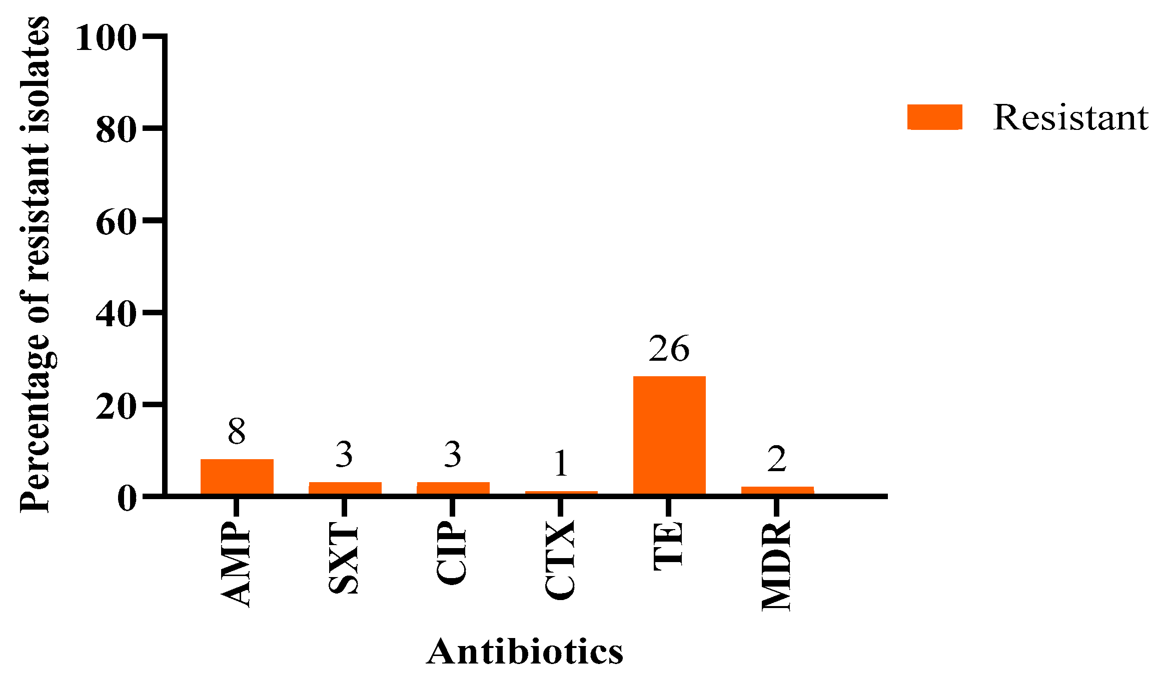
Figure 1. The percentages of resistance to each antibiotic and multidrug resistance (MDR) in E. coli isolated from Oryx leucoryx. AMP: Ampicillin; CIP: Ciprofloxacin; TE: Tetracycline; SXT: Trimethoprim/Sulfamethoxazole; CTX: Cefotaxime. MDR: multidrug-resistant.
Antibiotics 14 00248 g001
Figure 2. (a) The percentages of resistant E. coli isolates (out of 28 resistant isolates from a total of 100) among males and females from Qatari semi-wild Oryx leucoryx. Using SPSS statistical software, a higher percentage of resistance was detected among females than males, with 64.3% (n = 18/28) and 35.7% (n = 10/28), respectively. This difference was not statistically significant (p > 0.05). (b) Comparing the number of resistant E. coli isolates among different age groups and genders. The adult males showed a higher resistance percentage than adult females. ns: not significant.
Antibiotics 14 00248 g002
Figure 3. The percentages of resistant E. coli isolates across northwestern, central, and southwestern locations. The results did not show statistically significant differences. The highest percentage of resistance was detected in the central region. ns: not significant.
Antibiotics 14 00248 g003
Figure 4. The percentage of genes associated with the phenotypic resistance to 2 antibiotics, tetracycline and ampicillin, was determined using gene-specific PCR. The “Other Genes” were determined using WGS and are listed in Table 2.
Antibiotics 14 00248 g004
Figure 5. A full view of the studied Arabian Oryx along with two images depicting the sample collection process in this study.
Antibiotics 14 00248 g005
Table 1. Resistance of Escherichia coli isolated from Oryx to individual antibiotics.
No.AntibioticConcentrationSusceptible (n, %)Resistant (n, %)
1Ampicillin (AMP)10 μg92 (92%)8 (8%)
2Amoxicillin-clavulanic acid (AUG)30 μg100 (100%)0 (0%)
3Piperacillin-tazobactam (TZP)25 μg100 (100%)0 (0%)
4Ertapenem (ETP)10 μg100 (100%)0 (0%)
5Meropenem (MRP)10 μg100 (100%)0 (0%)
6Amikacin (AK)30 μg100 (100%)0 (0%)
7Gentamicin (CN)10 μg100 (100%)0 (0%)
8Fosfomycin (FOS)200 μg100 (100%)0 (0%)
9Trimethoprim-sulfamethoxazole (SXT)25 μg97 (3%)3 (3%)
10Ciprofloxacin (CIP)5 μg97 (3%)3 (3%)
11Cefotaxime (CTX)30 μg99 (99%)1 (1%)
12Ceftazidime (CAZ)30 μg100 (100%)0 (0%)
13Nitrofurantoin (F)300 μg100 (100%)0 (0%)
14Tetracycline (TE)30 μg74 (74%)26 (26%)
15Colistin (Broth microdilution)0.25–16 mg/mL100 (100%)0 (0%)
Table 2. Phenotypic resistant profiles of E. coli isolates identifying AMR genes using ONT genome sequencing technique.
Isolate No.Phenotypic ResistanceAMR GenesDrug ClassResistance MechanismAccession NumberBio Project Number
91TetracyclineacrS, kpnETetracyclineAntibiotic effluxJBKFEU000000000PRJNA1203416
11TetracyclinesoxR with mutationTetracyclineAntibiotic target alteration
Antibiotic efflux
JBKFDZ000000000PRJNA1203247
34CiprofloxacinemrAFluoroquinolone
antibiotic
Antibiotic effluxJBKFEA000000000PRJNA1203252
32AmpicillinacrAB-toIC with
acrR mutation
PenicillinsAntibiotic target alteration
Antibiotic efflux
JBKFEO000000000PRJNA1203389
29Tetracycline, ampicillinkpnETetracyclineAntibiotic efflux
fabl mutationsMulti-drugAntibiotic target alterationJBKFEP000000000PRJNA1203404
35TetracyclinekpnETetracyclineAntibiotic effluxJBKFEQ000000000PRJNA1203405
47AmpicillinqacGSmall MDR antibiotic
efflux pump
Antibiotic effluxJBKFES000000000PRJNA1203414
55AmpicillinacrAB-toIC with
marR mutations
Multi-drugAntibiotic target alteration
Antibiotic efflux
JBKFET000000000PRJNA1203415
92AmpicillinacrAB-toIC with
acrR mutation
penicillinAntibiotic target alteration
Antibiotic efflux
JBKFEV000000000PRJNA1203417
39CefotaximesoxR with
mutation
Cephalosporin drugantibiotic target alteration,
antibiotic efflux
JBKFER000000000PRJNA1203408
78CiprofloxacinmdtE, emrB, AcrE,mdtF
and marA
FluoroquinoloneAntibiotic effluxCP182564 PRJNA1224957
06CiprofloxacinmdtE, emrB, AcrE,
mdtF and marA
FluoroquinoloneAntibiotic effluxCP180189PRJNA1218299
Table 3. List of antibiotics used in the experiments with corresponding categories, concentrations, and CLSI 2020 susceptibility range (mm).
No.AntibioticAntibiotic
Class
ConcentrationCLSI Susceptibility
Range (mm)
1Ampicillin (AMP)Penicillin 10 μg≥17 S/R 13≤
2Amoxicillin-clavulanic acid (AUG)Penicillin30 μg≥18 S/R 13≤
3Piperacillin-tazobactam (TZP)Penicillin-beta-lactamase inhibitor25 μg≥21 S/R 17≤
4Ertapenem (ETP)Carbapenem10 μg≥22 S/R 18≤
5Meropenem (MRP)Carbapenem10 μg≥23 S/R 19≤
6Amikacin (AK)Aminoglycoside30 μg≥17 S/R 16≤
7Gentamicin (CN)Aminoglycoside10 μg≥15 S/R 12≤
8Fosfomycin (FOS)phosphonic acid derivative200 μg≥16 S/R 12≤
9Trimethoprim-sulfamethoxazole (SXT)-Sulfonamide,25 μg≥ 16 S/R 10 ≤
10Ciprofloxacin (CIP)Fluoroquinolone5 μg≥21 S/R 15≤
11Cefotaxime (CTX)Cephalosporin 30 μg≥26 S/R 22≤
12Ceftazidime (CAZ)Cephalosporin 30 μg≥21 S/R 17≤
13Nitrofurantoin (F)Nitrofuran 300 μg≥17 S/R 14≤
14Tetracycline (TE)Tetracycline30 μg≥15 S/R 11≤
15Colistin (Broth microdilution)Polymyxin0.25–16 mg/mL≤1 S/R 4≥
Disclaimer/Publisher’s Note: The statements, opinions and data contained in all publications are solely those of the individual author(s) and contributor(s) and not of MDPI and/or the editor(s). MDPI and/or the editor(s) disclaim responsibility for any injury to people or property resulting from any ideas, methods, instructions or products referred to in the content.

