Article

A National Surveillance of the Antibiotic Susceptibility of Acinetobacter baumannii in Saudi Arabia

by
Abrar K. Thabit
1,*,
Feras S. Alharbi
2,
Anas F. Jawah
2,
Ammar M. Alghamdi
2,
Musaab Y. Miaji
2,
Fatimah Alturki
3,
Nehal Hosin
4,
Mohammed Bazuqamah
5,
Masaad Saeed Almutairi
6,
Hamad Alhamed
7,
Alaa Elhendawy
8,
Dalya Atallah
9,
Abdulaziz A. Humadi
10,
Khalid A. Alfifi
11,
Khadija Alfadel
12 and
Khalid Eljaaly
1 on behalf of the Saudi AntiMicrobial Surveillance (SAMS) Study Group
1
Department of Pharmacy Practice, Faculty of Pharmacy, King Abdulaziz University, Jeddah 22254, Saudi Arabia
2
Faculty of Pharmacy, King Abdulaziz University, Jeddah 22254, Saudi Arabia
3
Microbiology Department, King Fahad Hospital of the University, Khobar 34445, Saudi Arabia
4
Department of Microbiology, College of Medicine, Imam Abdulrahman bin Faisal University, Dammam 34212, Saudi Arabia
5
Microbiology Department, King Khaled Hospital, Najran 66262, Saudi Arabia
6
Department of Pharmacy Practice, College of Pharmacy, Qassim University, Qassim 51452, Saudi Arabia
7
Laboratory and Blood Bank Department, King Fahad Specialist Hospital, Qassim 52366, Saudi Arabia
8
Microbiology Department, King Fahad Hospital, Albaha 65732, Saudi Arabia
9
Department of Clinical Microbiology, King Abdulaziz University Hospital, Jeddah 22254, Saudi Arabia
10
Laboratory and Blood Bank Department, King Fahad Specialist Hospital, Tabuk 47717, Saudi Arabia
11
Microbiology Department, King Fahad Specialist Hospital, Tabuk 47717, Saudi Arabia
12
Microbiology Department, Maternity and Children Hospital, King Salman Medical City, Madinah 42319, Saudi Arabia
*
Author to whom correspondence should be addressed.
Antibiotics 2025, 14(2), 209; https://doi.org/10.3390/antibiotics14020209
Submission received: 14 January 2025 / Revised: 11 February 2025 / Accepted: 17 February 2025 / Published: 19 February 2025
(This article belongs to the Special Issue Multidrug-Resistance Patterns in Infectious Pathogens)

Abstract

:
Most surveillance studies in Saudi Arabia have been single-centered or did not use the gold standard broth microdilution (BMD) antimicrobial susceptibility test. This is the first study from Saudi Arabia to evaluate the resistance profiles of Acinetobacter baumannii by using BMD on a national level. Between November 2022 and April 2023, isolates from several infection sites were collected from seven hospitals in seven regions of Saudi Arabia. On testing days, BMD was done following Clinical Laboratory Standards Institute standards. Antibiotic susceptibility percentages and MIC50 and MIC90 were calculated. One hundred A. baumannii isolates were included. The highest susceptibility was to tigecycline (39%) and aminoglycosides (22–25%). The MIC90 of all antibiotics were higher than the resistance breakpoint. All isolates (100%) were multidrug-resistant, of which 52% were classified as extensive-drug-resistant, and 42% were identified as pandrug-resistant. The isolates collected from the ear, peritoneal fluid, and the cerebrospinal fluid were all XDR, while 2/3 of the urine isolates (10/15; 66.7%), more than 1/2 of the skin/soft tissue and respiratory isolates (9/16; 56.3% and 22/43; 51.7%, respectively), and 3/8 (37.5%) of the blood isolates met this definition. Conversely, PDR isolates made up 5/8 of blood isolates (62.5%), 8/15 of body fluid isolates (57.14%), and 19/43 (44.2%) of respiratory isolates. A. baumannii showed a surprisingly high resistance to multiple commonly used antibiotics. Infection control policies and antimicrobial stewardship should be implemented by hospitals throughout the country to improve treatment, track resistance trends with local antibiograms, and prevent the development of resistant strains.

1. Introduction

Acinetobacter baumannii, a non-fermentative Gram-negative coccobacillus from the Moraxellaceae family, has resulted in increasing global attention as a nosocomial pathogen in recent years. Over the previous decade, A. baumannii strains, often displaying multidrug resistance, have emerged as a prominent global health challenge as they have been implicated in various infections, primarily of nosocomial origin [1]. The prevalence of A. baumannii is particularly pronounced in intensive care units, where controlling numerous outbreaks has proven exceptionally challenging [2]. A. baumannii is intrinsically resistant to numerous antibiotics, including ampicillin, ertapenem, aztreonam, and fosfomycin [3].
Bacterial resistance to antimicrobial agents is a growing global concern that significantly impacts both patient clinical outcomes and the economic stability of hospitals and governments [1]. A list of antibiotic-resistant priority pathogens was published by the World Health Organization (WHO) in 2024, classifying them as either critical, high, or medium priority [4]. In this list, carbapenem-resistant A. baumannii was categorized under “critical priority”, which indicates that this organism poses a threat to patients due to their association with deadly infections, such as bacteremia and pneumonia. In a recent review article of antimicrobial resistance (AMR) in Saudi Arabia between 2013 and 2023 that included nine studies evaluating antibiotic resistance by A. baumannii, the reported high resistance rates have increased over the last decade [5]. In fact, all the reported isolates were multidrug-resistant (MDR). The antibiotic to which the isolates (n = 1082) exhibited the highest susceptibility rates was colistin (97.5%), though it should be noted that such susceptibilities were tested using automated susceptibility testing (AST) methods, such as Vitek2, Phoenix, and Microscan, rather than the recommended reference method of broth microdilution (BMD) [6,7,8], except in one study [5,9]. Conversely, the susceptibility to ciprofloxacin was the lowest (1.25%). Furthermore, the review revealed elevated resistance to carbapenems, including to imipenem and meropenem, with average percentages of susceptibility below 20%.
Numerous methods for testing antimicrobial susceptibility have been documented in the literature. However, only BMD stands out as the ‘gold standard’ according to international laboratory standards [6,7,8]. Since no study in the literature from Saudi Arabia has evaluated the susceptibility of A. baumannii using the gold standard method of BMD, we aimed to test the susceptibility of A. baumannii on a national level against a panel of antibiotics. Our aim was to be a leading project to report AMR rates of A. baumannii on a large scale in Saudi Arabia by including isolates from various hospitals across the country. Another objective of the study was to ultimately contribute a scientifically grounded foundation to advocate for the formulation and implementation of antimicrobial stewardship programs within Saudi hospitals, as well as foster the adoption of infection control and prevention strategies to further enhance healthcare practices in the region.

2. Results

2.1. Isolates Distribution by Participating Hospitals

A total of 100 A. baumannii isolates were collected, mostly from Dammam (23%), followed by Qassim (20%) and Tabuk (20%), as shown in Table 1.

2.2. Sources of Isolates

The sources of the isolates are presented in Table 2, where most isolates came from a respiratory source (43%), skin and soft tissue (16%), urine (15%), or body fluids (14%). All the isolates (100%) were MDR, whereas 52% and 42% of isolates were XDR and PDR, respectively.

2.3. Susceptibility Results

Table 3 shows the susceptibility percentages and MIC data of the isolates. Susceptibility was the highest to tigecycline (39%) with MIC50 and MIC90 of 4 and >8 μg/mL, respectively. Aminoglycosides ranked second to tigecycline in terms of activity, with the highest percentage susceptibility being observed with tobramycin (25%), followed by gentamicin (23%) and amikacin (22%). In contrast, susceptibility to the remaining tested antibiotics was extremely low, reflecting a high resistance rate to β-lactams (including ampicillin/sulbactam and carbapenems), fluoroquinolones, and polymyxins.
Figure 1 shows the number of isolates reported for each MIC value, with color shades reflecting CLSI breakpoints for each antibiotic.

2.4. Distribution of Drug-Resistant Isolates

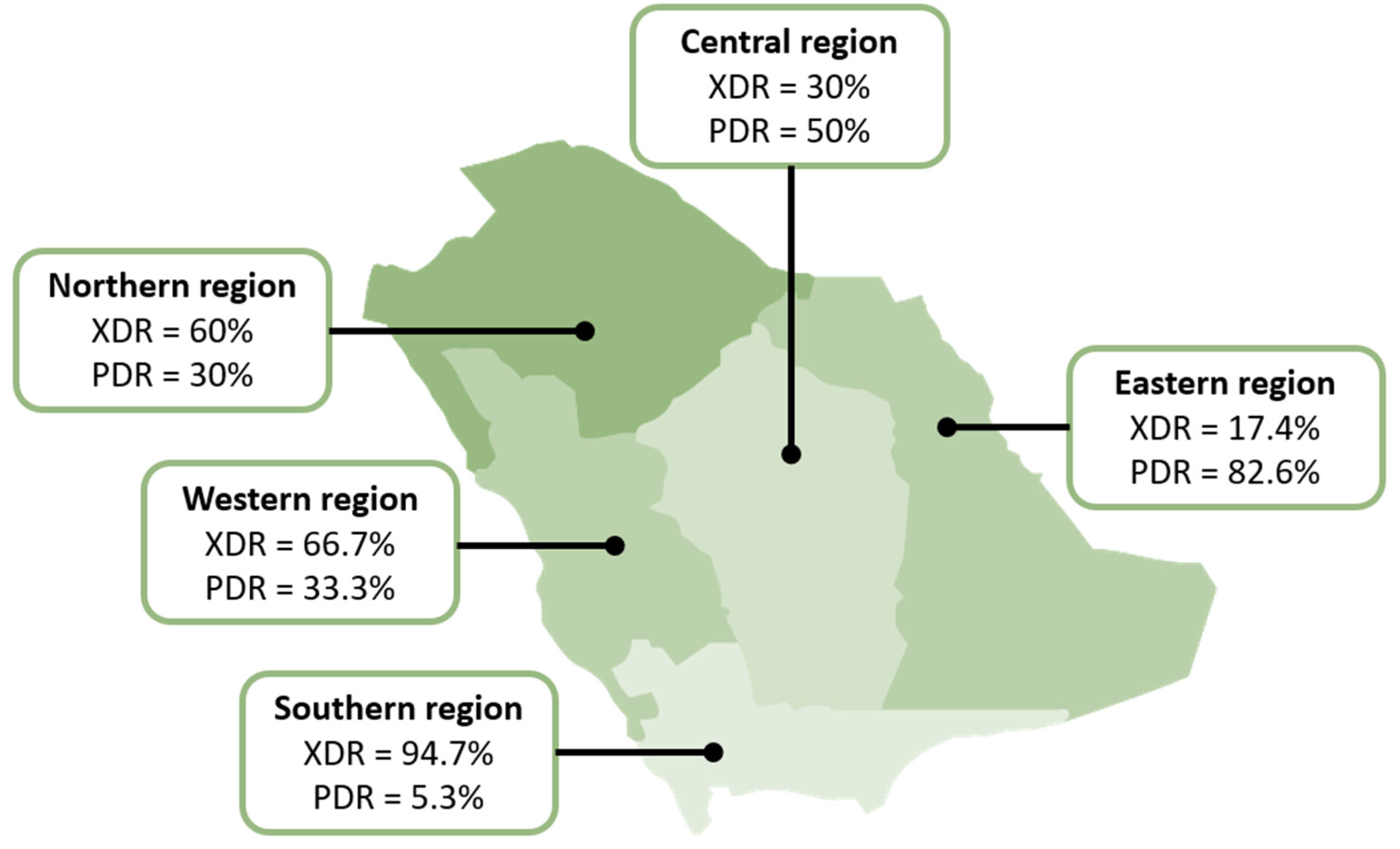
Of the 100 isolates collected, all met the definition of MDR, 52% were XDR, and 42% were PDR. The distribution of these resistance patterns based on the region is shown in Figure 2, where a significant difference was observed between the regions in the distribution of XDR and PDR isolates (p < 0.0001 for both resistance patterns). The isolates collected from the ear, peritoneal fluid, and the cerebrospinal fluid were all XDR, while 2/3 of the urine isolates (10/15; 66.7%), more than 1/2 of the skin/soft tissue and respiratory isolates (9/16; 56.3% and 22/43; 51.7%, respectively), and 3/8 (37.5%) of the blood isolates met this definition. PDR isolates made up 5 of the 8 blood isolates (62.5%) and 8 of the 15 body fluid isolates (57.14%). Of the 43 respiratory isolates, 19 (44.2%) were PDR. No statistical difference was observed in the distribution of XDR isolates (p = 0.512) and PDR isolates (p = 0.425) between infection sites (Table 2). The exact MICs and susceptibility results are provided in the Supplementary Table (Supplementary Table S1: MIC results of all tested antibiotics against A. baumannii).

3. Materials and Methods

3.1. Study Period and Collection Process

Seven large hospitals from seven administrative regions of Saudi Arabia participated in this study, where unique A. baumannii clinical isolates from various infection sites were collected between November 2022 and April 2023. All isolates were transported on blood agar plates (BAPs) (Saudi Prepared Media Laboratory, Co., Ltd., Riyadh, Saudi Arabia) to a central microbiology laboratory (at the Faculty of Pharmacy, King Abdulaziz University, Jeddah). Ethical approval was obtained from the Saudi Ministry of Health (reference no. 607/44/5113) and the Biomedical Research Ethics Committee of the Faculty of Medicine of King Abdulaziz University (ref No. 559–22).

3.2. Bacterial Isolates

Consecutive clinical isolates of A. baumannii labeled with the infection site were prospectively collected by the participating hospitals. Isolate identification was done at each hospital using automated antimicrobial susceptibility testing methods, such as BD Phoenix 100 (Becton, Dickinson and Company, Franklin Lakes, NJ, USA) or VITEK 2 (bioMérieux, Craponne, France). Upon reception, the isolates were subcultured on BAPs, then stored at −80 °C in glycerol until the testing day. Isolates that exhibited poor growth patterns were subcultured again from the newly grown colonies to ensure the storage of sufficient bacterial counts. Before antibiotic susceptibility testing, the isolates to be tested were thawed and then subcultured on BAPs and incubated for 24 h at 36 °C.

3.3. Antimicrobial Susceptibility Testing

Sensititre GNX3F plates (Thermo Fisher Scientific, Waltham, MA, USA) were used to conduct BMD according to the Clinical Laboratory Standards Institute (CLSI) standard (M100–33) [3]. The list of antibiotics included in these plates is shown in Table 3. These plates comprise a panel of various antibiotics against Gram-negative bacteria. To ensure the accuracy of the plates, the first microtiter plate of the batch was assessed using ATC27853 (Pseudomonas aeruginosa quality control isolate). On testing days, 50 μL of prepared Mueller–Hinton Broth (Biolab, Inc., Budapest, Hungary) were instilled in the microtiter plates. In the meantime, a bacterial inoculum was prepared with a density of 0.5 McFarland (equivalent to 1.5 × 10⁸ CFU/mL). The density of the prepared inocula was confirmed using a daily calibrated densitometer (Grant Instruments, Ltd., Royston, UK). A volume of 10 μL of the inoculum was then instilled in the microtiter plates, followed by incubation for 16–24 h at 36 °C prior to reading.

3.4. Definitions

An organism is labeled as MDR if it exhibits resistance to three or more antibiotics from different classes, whereas isolates that are resistant to at least one agent in all but 2 or fewer antimicrobial classes are labeled extensive drug-resistant (XDR) [10]. An isolate that is resistant to all antibiotics from all antibiotic classes is known as pandrug-resistant (PDR) [10]. Of note, these terms apply to acquired resistance against antibiotics, to which wild-type A. baumannii is susceptible (i.e., not intrinsically resistant).

3.5. Data Analysis

The percentages of susceptible organisms (% susceptibility) to each antibiotic, minimum inhibitory concentrations required to inhibit 50% of the isolates (MIC50), MIC required to inhibit 90% of the isolates (MIC90), and percentages of different drug-resistant patterns were calculated from the data that were logged in Microsoft Excel (Microsoft, Corp., Seattle, WA, USA). The susceptibility of all antibiotics, except tigecycline, was determined based on CLSI breakpoints (document M100–33) [3]. For tigecycline, the susceptibility breakpoints approved by the US Food and Drug Administration for Enterobacterales that are listed in the drug’s package insert were used for MIC interpretation [11]. The chi-square test was done using SPSS version 24.0 (IBM Corp., Armonk, NY, USA) to compare the distribution of XDR and PDR isolates among regions and infection sites. Since all the isolates were MDR, the test could not be carried out on them.

4. Discussion

In this study, 100 isolates of A. baumannii were collected from seven hospitals throughout Saudi Arabia as part of the country’s first national multicentric antimicrobial surveillance study. These isolates were obtained from various infection sites, mostly from the respiratory tract, urinary tract, skin/soft tissue, and body fluids. A recent review that evaluated the epidemiology of A. baumannii in Saudi Arabia noticed that this pathogen is largely responsible for respiratory tract infections, mostly in intensive care units (ICUs) [12]. This is probably because A. baumannii is a major contaminant of respiratory support equipment and suction devices commonly used in ICUs [13].
While infection types were not collected, the knowledge of infection sites could provide an insight into the type of infections the patients had due to A. baumannii. The most common site was the respiratory tract, which implies that pneumonia was prevalent among the patients from whom the isolates were collected. This aligns with the previous knowledge that nosocomial pneumonia is the top infection caused by A. baumannii, especially in critically ill patients [14]. Interestingly, only eight isolates were from the blood (a common infection site for A. baumannii) compared with 16 from skin and soft tissue and 15 from the urinary tract. Most skin infections due to A. baumannii are usually surgical site infections [14,15].
Results from our surveillance study revealed high susceptibility to aminoglycosides compared to carbapenems, which are considered the broadest-spectrum β-lactams. Moreover, tigecycline exhibited the highest in vitro activity, whereas ciprofloxacin and trimethoprim/sulfamethoxazole demonstrated the lowest activity. In our study, all A. baumannii isolates (100%) were MDR, whereas 52% and 42% of isolates were XDR and PDR, respectively. A recent review that evaluated AMR among various bacterial pathogens, including A. baumannii, over the last decade showed an increasing prevalence of AMR [5]. The review included nine studies that were mostly single-centered and used automated systems for susceptibility testing. In that review, the highest average susceptibility from all the studies was to colistin (97.5%), followed by amikacin (36.3%). It is worth noting that the discrepancy in the susceptibility rates to colistin between our study and these previous studies could be attributed to the difference in the testing method used (automated susceptibility tests vs. BMD), where BMD is the method recommended by the latest international colistin guidelines [16]. Furthermore, the extremely low susceptibility to colistin demonstrated in our study could be attributed to a phenomenon of the attachment of polymyxins to the plastic bottom of the microtiter plates used in BMD. Previous studies on BMD observed that polymyxins, including colistin and polymyxin B, have an affinity for plastic—the material from which BMD microtiter plates are made [17,18]. This interaction with plastic can affect MIC measurements. To mitigate this effect, Sutherland et al. recommended using polysorbate 80 by adding it to the microtiter plates wells when assessing colistin MIC using BMD. Polysorbate 80 serves as a surfactant, preventing colistin from adhering to the plastic plate bottom. Our study reported a low susceptibility rate to colistin, which may be partly attributed to this phenomenon. However, since our study used pre-filled plates with lyophilized antibiotics, adding polysorbate 80 was not feasible.
A large study from a hospital in Jeddah (in the western region of Saudi Arabia) between September 2013 and May 2014 included 527 A. baumannii isolates, of which 244 were respiratory, 93 were urinary, and 190 were from miscellaneous infection sites [19]. In that study, 35.7% of the respiratory isolates and 34.4% of the urinary isolates were susceptible to all tested antimicrobials, while 64.3% and 65.6% were resistant to one or more antimicrobials from these sites, respectively. Of the isolates collected from miscellaneous sites, 28.4% were susceptible to all tested antimicrobials, while 71.6% were resistant to one or more antimicrobials. In another study from the same region by Shah et al. between 2015 and 2016, 58.5% of the 135 A. baumannii isolates were MDR, but none were PDR, which is inconsistent with our findings [9]. Similar to our study, the highest susceptibility was to tigecycline, though it was much higher than the value reported in our study (96% vs. 39%). While all isolates were susceptible to colistin in that study, our study showed a very low susceptibility rate (5%) [6]. Nonetheless, it should be noted that these results were generated about a decade ago. Hence, the low susceptibilities shown in our study may indicate the rapid increase in AMR rates. In addition, newer studies from various regions of Saudi Arabia showed higher resistance rates for A. baumannii [20,21,22,23]. For instance, a study from the Eastern region (2017–2018) showed that all the collected 103 isolates were MDR exhibiting a substantial resistance to carbapenems (only 1.9% were susceptible to imipenem and 42.7% were susceptible meropenem) and aminoglycosides, with a susceptibility rate reaching around 52% [20]. The highest susceptibility was to tigecycline (95.1%). Another recent study in 2020 from Hail (in the northern part of Saudi Arabia) showed that all the included 82 A. baumannii isolates were XDR, with the highest susceptibility being reported to amikacin (61.3%), followed by tigecycline (12.8%), whereas the susceptibility to colistin was only 5%, which matches the rate reported in our study [21]. Using disk diffusion methods, a study from Riyadh (in the central region of Saudi Arabia) showed that all the 54 A. baumannii isolates that were collected between 2013–2014 were MDR, though all were susceptible to colistin [24].
The β-lactamase inhibitor sulbactam was used for decades against A. baumannii when it was only available as a combination with ampicillin. However, due to the fixed concentration of sulbactam in this combination and to limit the unnecessary exposure to ampicillin, two new combinations of β-lactamase inhibitors involving sulbactam have been developed: sulbactam/durlobactam and sulbactam/avibactam. Sulbactam/durlobactam received US FDA approval in May 2023 for hospital-acquired and ventilator-associated pneumonia caused by A. baumannii [25]. In an in vitro study against 5032 A. baumannii isolates from 33 hospitals across the globe, 98.3% of the isolates exhibited susceptibility to this combination (based on a presumed susceptibility breakpoint of 4 μg/mL), including MDR and XDR isolates with an MIC50 and MIC90 of 1 and 2 μg/mL, respectively [26]. Similarly, sulbactam/avibactam (as ampicillin/sulbactam plus ceftazidime/vibactam), has shown potent activity against drug-resistant A. baumannii, where 124 of 127 (97.6%) XDR isolates had MICs ≤ 4 μg/mL [27]. This study, as well as another study by Pasteran et al., demonstrated this activity mostly against OXA-producers but less against NDM-producers [28]. In the study by Pasteran et al., 14% of 187 isolates were inhibited by sulbactam alone at 4 μg/mL, but this percentage increased to 69% when sulbactam was combined with avibactam (91% inhibition of OXA-producers) [28]. In our study, only 2% of the isolates were susceptible to sulbactam (in ampicillin/sulbactam). However, this percentage is expected to rise if avibactam or durlobactam were added.
Our study involved a nationwide investigation across various regions of Saudi Arabia. These regions may exhibit distinct patterns of antibiotic usage, leading to differences in susceptibility rates. Although we intended to include more hospitals to expand the sample size, logistical challenges with certain hospitals posed a barrier. Moreover, the prefilled microtiter plates lacked ceftazidime/avibactam, sulbactam/avibactam, sulbactam/durlobactam, and cefiderocol, which are important antibiotics clinically used against MDR A. baumannii. Lastly, genotyping could have provided a deeper understanding of the underlying resistance mechanisms, which we recommend for future studies.

5. Conclusions

By incorporating samples from seven different hospitals situated across various regions of Saudi Arabia, our study aimed to identify a wide range of A. baumannii strains that are prevalent throughout the country, which showed high rates of AMR to antibiotics commonly used in therapy. Data generated from our study are crucial for policymakers and healthcare providers as they offer valuable insights into the current status of AMR in Saudi Arabia. Equipped with this knowledge, policymakers and hospital antimicrobial stewardship committees can make well-informed decisions to develop effective strategies for combating AMR, while clinicians may adapt their treatment plans in accordance with the prevalent susceptibility patterns. The findings from this research will provide a crucial resource for informing public health initiatives, developing antimicrobial stewardship programs, and ultimately improving patient outcomes in the face of rising antibiotic resistance challenges.

Supplementary Materials

The following supporting information can be downloaded at: https://www.mdpi.com/article/10.3390/antibiotics14020209/s1. Table S1: MIC values of all isolates; Table S2: MIC50 and MIC90 of all antibiotics.

Author Contributions

Conceptualization, methodology, data curation, investigation, formal analysis, funding, resources, supervision, writing—original draft, and writing—review and editing, A.K.T.; Data curation, investigation, writing—original draft, F.S.A., A.F.J., A.M.A. and M.Y.M.; Data acquisition. resources, writing—review and editing, F.A., N.H., M.B., M.S.A., H.A., A.E., D.A., A.A.H., K.A.A. and K.A.; Supervision, data interpretation, writing—review and editing, K.E. All authors have read and agreed to the published version of the manuscript.

Funding

Funding was provided by the Ministry of Education and King Abdulaziz University, DSR, Jeddah, Saudi Arabia.

Institutional Review Board Statement

Ethical approval was obtained from the Research Ethics Committee of King Abdulaziz University (reference No. 559–22) and the Saudi Ministry of Health (reference No. 607/44/5113).

Informed Consent Statement

Not applicable.

Data Availability Statement

The original contributions presented in this study are included in the article/Supplementary Material. Further inquiries can be directed to the corresponding author.

Acknowledgments

This research work was funded by Institutional Fund Projects under grant no. IFPIP: 721–249–1443. The authors gratefully acknowledge technical and financial support provided by the Ministry of Education and King Abdulaziz University, DSR, Jeddah, Saudi Arabia. We would also like to thank the graphic designer, Raghad Thabit, for creating the regional map of Saudi Arabia.

Conflicts of Interest

The authors declare no conflicts of interest. The funders had no role in the design of the study; in the collection, analyses, or interpretation of data; in the writing of the manuscript; or in the decision to publish the results.

Abbreviations

The following abbreviations are used in this manuscript:
AMRAntimicrobial resistance
BMDBroth microdilution
CLSIClinical Laboratory Standards Institute
ICUIntensive care unit
MDRMultidrug-resistant
MICMinimum inhibitory concentration
XDRExtensive drug-resistant
PDRPandrug-resistant
WHOWorld Health Organization

References

  1. Thabit, A.K.; Crandon, J.L.; Nicolau, D.P. Antimicrobial resistance: Impact on clinical and economic outcomes and the need for new antimicrobials. Expert Opin. Pharmacother. 2015, 16, 159–177. [Google Scholar] [CrossRef] [PubMed]
  2. Asif, M.; Alvi, I.A.; Rehman, S.U. Insight into Acinetobacter baumannii: Pathogenesis, global resistance, mechanisms of resistance, treatment options, and alternative modalities. Infect. Drug Resist. 2018, 11, 1249–1260. [Google Scholar] [CrossRef] [PubMed]
  3. Clinical and Laboratory Standards Institute. Performance Standards for Antimicrobial Susceptibility Testing: Thertieth Informational Supplement [Document M100-S33]; National Committee for Clinical Laboratory Standards: Wayne, PA, USA, 2023. [Google Scholar]
  4. World Health Organization. WHO Bacterial Priority Pathogens List, 2024: Bacterial Pathogens of Public Health Importance to Guide Research, Development and Strategies to Prevent and Control Antimicrobial Resistance. Available online: https://www.who.int/publications/i/item/9789240093461 (accessed on 11 February 2025).
  5. Thabit, A.K.; Alabbasi, A.Y.; Alnezary, F.S.; Almasoudi, I.A. An Overview of Antimicrobial Resistance in Saudi Arabia (2013–2023) and the Need for National Surveillance. Microorganisms 2023, 11, 2086. [Google Scholar] [CrossRef]
  6. Humphries, R.M.; Ambler, J.; Mitchell, S.L.; Castanheira, M.; Dingle, T.; Hindler, J.A.; Koeth, L.; Sei, K.; Development, C.M.; CLSI Methods Development and Standardization Working Group of the Subcommittee on Antimicrobial Susceptibility Testing. CLSI Methods Development and Standardization Working Group Best Practices for Evaluation of Antimicrobial Susceptibility Tests. J. Clin. Microbiol. 2018, 56, e01934-17. [Google Scholar] [CrossRef]
  7. Clinical and Laboratory Standards Institute. Performance Standards for Antimicrobial Susceptibility Testing: Thertieth Informational Supplement [Document M100-S35]; National Committee for Clinical Laboratory Standards: Wayne, PA, USA, 2025. [Google Scholar]
  8. European Committee on Antimicrobial Susceptibility Testing. Breakpoint Tables for Interpretation of MICs and Zone Diameters. Version 13.0. Available online: http://www.eucast.org (accessed on 11 February 2025).
  9. Shah, M.W.; Yasir, M.; Farman, M.; Jiman-Fatani, A.A.; Almasaudi, S.B.; Alawi, M.; El-Hossary, D.; Azhar, E.I. Antimicrobial susceptibility and molecular characterization of clinical strains of Acinetobacter baumannii in Western Saudi Arabia. Microb. Drug Resist. 2019, 25, 1297–1305. [Google Scholar] [CrossRef]
  10. Magiorakos, A.P.; Srinivasan, A.; Carey, R.B.; Carmeli, Y.; Falagas, M.E.; Giske, C.G.; Harbarth, S.; Hindler, J.F.; Kahlmeter, G.; Olsson-Liljequist, B.; et al. Multidrug-resistant, extensively drug-resistant and pandrug-resistant bacteria: An international expert proposal for interim standard definitions for acquired resistance. Clin. Microbiol. Infect. 2012, 18, 268–281. [Google Scholar] [CrossRef]
  11. Product Information: TYGACIL®. Tigecycline Intravenous Injection. Pfizer, Inc.: Philadelphia, PA, USA, 2020. [Google Scholar]
  12. Kharaba, A.; Abdelaziz Hussein, M.A.; Al-Hameed, F.M.; Mandourah, Y.; Almekhlafi, G.A.; Algethamy, H.; Hamdan, A.; Azem, M.A.; Fatani, J.; al Beshabshe, A.; et al. Acinetobacter baumannii in Saudi Arabia: The New Growing Threat. Saudi Crit. Care J. 2019, 3, 54–57. [Google Scholar] [CrossRef]
  13. Jung, J.; Park, W. Acinetobacter species as model microorganisms in environmental microbiology: Current state and perspectives. Appl. Microbiol. Biotechnol. 2015, 99, 2533–2548. [Google Scholar] [CrossRef]
  14. Cavallo, I.; Oliva, A.; Pages, R.; Sivori, F.; Truglio, M.; Fabrizio, G.; Pasqua, M.; Pimpinelli, F.; Di Domenico, E.G. Acinetobacter baumannii in the critically ill: Complex infections get complicated. Front. Microbiol. 2023, 14, 1196774. [Google Scholar] [CrossRef]
  15. Howard, A.; O’Donoghue, M.; Feeney, A.; Sleator, R.D. Acinetobacter baumannii: An emerging opportunistic pathogen. Virulence 2012, 3, 243–250. [Google Scholar] [CrossRef]
  16. Tsuji, B.T.; Pogue, J.M.; Zavascki, A.P.; Paul, M.; Daikos, G.L.; Forrest, A.; Giacobbe, D.R.; Viscoli, C.; Giamarellou, H.; Karaiskos, I.; et al. International Consensus Guidelines for the Optimal Use of the Polymyxins: Endorsed by the American College of Clinical Pharmacy (ACCP), European Society of Clinical Microbiology and Infectious Diseases (ESCMID), Infectious Diseases Society of America (IDSA), International Society for Anti-infective Pharmacology (ISAP), Society of Critical Care Medicine (SCCM), and Society of Infectious Diseases Pharmacists (SIDP). Pharmacotherapy 2019, 39, 10–39. [Google Scholar] [CrossRef] [PubMed]
  17. Sharafi, T.; Ardebili, A. Plastic binding feature of polymyxins: The effect on MIC susceptibility measurements. Infect. Drug Resist. 2019, 12, 2649–2653. [Google Scholar] [CrossRef] [PubMed]
  18. Sutherland, C.A.; Nicolau, D.P. To add or not to add polysorbate 80: Impact on colistin MICs for clinical strains of Enterobacteriaceae and Pseudomonas aeruginosa and quality controls. J. Clin. Microbiol. 2014, 52, 3810. [Google Scholar] [CrossRef]
  19. Alam, M.Z.; Alam, Q.; Jiman-Fatani, A.A.; Shukri, H.A.; Haque, A. A surveillance study on the prevalence and antimicrobial resistance pattern among different groups of bacteria isolated from Western province of Saudi Arabia. Biomed. Res. 2017, 28, 898–906. [Google Scholar]
  20. AlAmri, A.M.; AlQurayan, A.M.; Sebastian, T.; AlNimr, A.M. Molecular surveillance of multidrug-resistant Acinetobacter baumannii. Curr. Microbiol. 2020, 77, 335–342. [Google Scholar] [CrossRef] [PubMed]
  21. Said, K.B.; Alsolami, A.; Khalifa, A.M.; Khalil, N.A.; Moursi, S.; Osman, A.; Fahad, D.; Rakha, E.; Rashidi, M.; Moussa, S.; et al. A multi-point surveillance for antimicrobial resistance profiles among clinical isolates of Gram-negative bacteria recovered from major Ha’il hospitals, Saudi Arabia. Microorganisms 2021, 9, 2024. [Google Scholar] [CrossRef]
  22. Kabrah, A. Extended-spectrum beta-lactamase and carbapenem-resistant Gram-negative pathogens in Makkah, Saudi Arabia. Ethiop. J. Health Sci. 2022, 32, 1221–1230. [Google Scholar] [CrossRef]
  23. Saleem, M.; Syed Khaja, A.S.; Hossain, A.; Alenazi, F.; Said, K.B.; Moursi, S.A.; Almalaq, H.A.; Mohamed, H.; Rakha, E. Molecular characterization and antibiogram of Acinetobacter baumannii clinical isolates recovered from the patients with ventilator-associated pneumonia. Healthcare 2022, 10, 2210. [Google Scholar] [CrossRef]
  24. Marie, M.A.; Krishnappa, L.G.; Alzahrani, A.J.; Mubaraki, M.A.; Alyousef, A.A. A prospective evaluation of synergistic effect of sulbactam and tazobactam combination with meropenem or colistin against multidrug resistant Acinetobacter baumannii. Bosn. J. Basic Med. Sci. 2015, 15, 24–29. [Google Scholar] [CrossRef]
  25. Anand, A.; Verma, A.; Kaur, S.; Kathayat, P.; Manoj, R.M.; Aakanksha, A.; Turzin, J.K.; Satapathy, P.; Khatib, M.N.; Gaidhane, S.; et al. An overview of sulbactam-durlobactam approval and implications in advancing therapeutics for hospital-acquired and ventilator-associated pneumonia by Acinetobacter baumannii-calcoaceticus complex: A narrative review. Health Sci. Rep. 2024, 7, e70066. [Google Scholar] [CrossRef]
  26. Karlowsky, J.A.; Hackel, M.A.; McLeod, S.M.; Miller, A.A. In Vitro Activity of Sulbactam-Durlobactam against Global Isolates of Acinetobacter baumannii-calcoaceticus Complex Collected from 2016 to 2021. Antimicrob. Agents Chemother. 2022, 66, e0078122. [Google Scholar] [CrossRef] [PubMed]
  27. Rodriguez, C.H.; Brune, A.; Nastro, M.; Vay, C.; Famiglietti, A. In vitro synergistic activity of the sulbactam/avibactam combination against extensively drug-resistant Acinetobacter baumannii. J. Med. Microbiol. 2020, 69, 928–931. [Google Scholar] [CrossRef] [PubMed]
  28. Pasteran, F.; Cedano, J.; Baez, M.; Albornoz, E.; Rapoport, M.; Osteria, J.; Montana, S.; Le, C.; Ra, G.; Bonomo, R.A.; et al. A New Twist: The Combination of Sulbactam/Avibactam Enhances Sulbactam Activity against Carbapenem-Resistant Acinetobacter baumannii (CRAB) Isolates. Antibiotics 2021, 10, 577. [Google Scholar] [CrossRef] [PubMed]
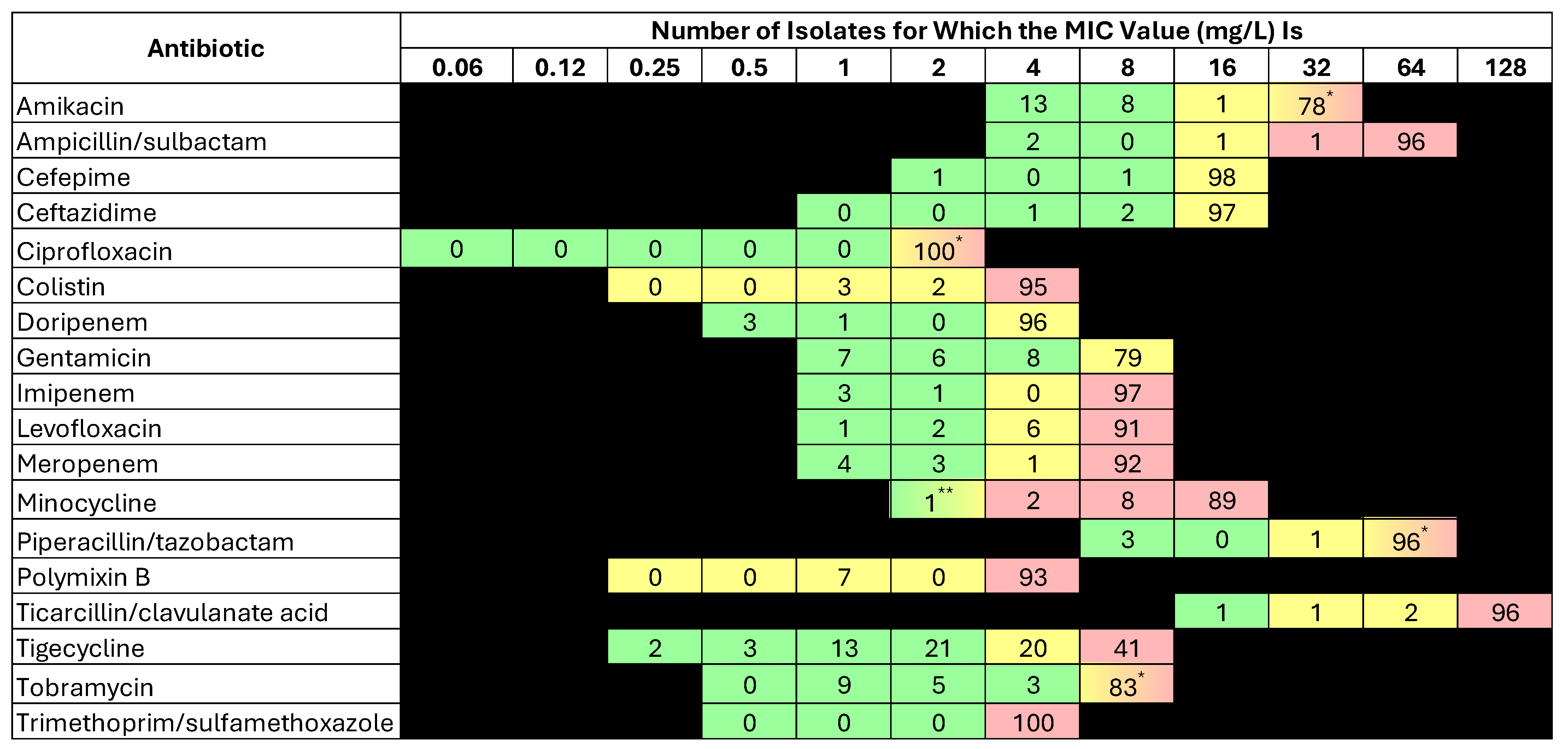
Figure 1. Number of isolates reported for each MIC value. The color shades reflect the susceptibility breakpoints according to CLSI standards [7], except tigecycline, which’s breakpoints is based on US FDA breakpoint. The green shade indicates susceptibility, the yellow shade indicates intermediate susceptibility, and the red shade indicates resistance. * The intermediate susceptibility breakpoint begins at this value, followed by resistance at the next dilution. ** The susceptibility breakpoint begins at this value, followed by intermediate susceptibility at the next dilution.
Antibiotics 14 00209 g001
Figure 2. Distribution of XDR and PDR Acinetobacter baumannii isolates in Saudi Arabia.
Antibiotics 14 00209 g002
Table 1. Characteristics of the included hospitals and the number of isolates collected.
CharacteristicHospital
King Abdulaziz University HospitalKing Fahad Hospital of the UniversityKing Khaled HospitalKing Fahad Specialist HospitalKing Fahad HospitalKing Fahad Specialist HospitalMaternity and Children Hospital of King Salman Medical City
CityJeddahDammamNajranQassimAlbahaTabukMadinah
RegionWesternEasternSouthernCentralSouthernNorthernWestern
Bed capacity879633330600320205500
Number of ICUs5 (99 beds)5 (77 beds)4 (56 beds)4 (49 beds)4 (64 beds)2 (43 beds)3 (117 beds)
N (%) of isolates16 (16)23 (23)17 (17)20 (20)2 (2)20 (20)2 (2)
Table 2. Sources of Acinetobacter baumannii isolates (n = 100).
Sourcen (%)XDR *PDR *
Respiratory43 (43)22 (51.2)19 (44.2)
Skin or soft tissue16 (16)9 (56.3)4 (25)
Urine15 (15)10 (66.7)5 (33.3)
Body fluid14 (14)4 (30.8)8 (61.5)
Blood8 (8)3 (37.5)5 (62.5)
Eye2 (2)1 (50)1 (50)
Cerebrospinal fluid1 (1)1 (100)0 (0)
Ear1 (1)1 (100)0 (0)
p-value-0.514/0.400 **0.425
PDR, pandrug-resistant; XDR, extensive drug-resistant. Note: All collected isolates were reported multidrug-resistant (MDR); therefore, the distribution of MDR isolates is the same as the distribution of the source. * The percentages were calculated based on the total number of isolates from that source. ** After combining isolates of eye, cerebrospinal fluid, and ear into one group due to the very small sample size of each one.
Table 3. Antibiotic susceptibility data of Acinetobacter baumannii.
Antibiotic% SusceptibilityMIC50MIC90MIC Range
Amikacin22>32>32≤4–>32
Ampicillin/sulbactam2>64/32>64/32≤4/2–>64/32
Cefepime2>16>16≤2–>16
Ceftazidime3>16>164–>16
Ciprofloxacin0>2>2-
Colistin *5>4>41–>4
Doripenem4>4>4≤0.5–>4
Gentamicin23>8>8≤1–>8
Imipenem4>8>8≤1–>8
Levofloxacin3>8>8≤1–>8
Meropenem7>8>8≤1–>8
Minocycline3>16>16≤2–>16
Piperacillin/tazobactam3>64/4>64/4≤8/4–>64/4
Polymixin B *1>4>41–>4
Ticarcillin/clavulanate acid1>128/2>128/2≤16/2–>128/2
Tigecycline394>8≤0.25–>8
Tobramycin25>8>8≤1–>8
Trimethoprim/sulfamethoxazole0>4/76>4/76>4/76–>4–76
MIC, minimum inhibitory concentration. * The percentages presented are the percentage of isolates with intermediate resistance since the Clinical and Laboratory Standards Institute M100 document includes intermediate and resistant breakpoints only, which are ≤2 and ≥4 μg/mL, respectively.
Disclaimer/Publisher’s Note: The statements, opinions and data contained in all publications are solely those of the individual author(s) and contributor(s) and not of MDPI and/or the editor(s). MDPI and/or the editor(s) disclaim responsibility for any injury to people or property resulting from any ideas, methods, instructions or products referred to in the content.