Article

The Long-Term Survivorship and Cause of Failure of Metal-on-Metal Total Hip Arthroplasty

Department of Orthopaedic Surgery, Mie University Graduate School of Medicine, 2-174 Edobashi, Tsu 514-8507, Japan
*
Author to whom correspondence should be addressed.
Antibiotics 2025, 14(2), 161; https://doi.org/10.3390/antibiotics14020161
Submission received: 16 December 2024 / Revised: 30 January 2025 / Accepted: 5 February 2025 / Published: 6 February 2025

Abstract

:
Background: Complications associated with metal-on-metal (MOM) prostheses, such as adverse reactions to metal debris (ARMDs), include pseudotumor (PT) formation, metallosis, and soft tissue necrosis. High short-term failure rates have been reported for various MOM total hip arthroplasties (THAs) due to ARMDs. ARMDs in MOM THAs can potentially lead to secondary failure modes, such as dislocation or infection. Objectives: This study aims to examine the cumulative incidence of revisions due to ARMDs and periprosthetic joint infection (PJI) in primary MOM total hip arthroplasty and to compare the outcomes of ARMD and PJI cases. Methods: Between 2006 and 2011, 247 primary MOM THAs were performed on 230 patients (39 men, 191 women) with a mean age of 64.1 years. The average follow-up duration was 10.5 years. Results: Thirty-eight hips were converted to metal-on-polyethylene articulation between 1.2 and 14.7 years postoperatively (mean: 7.2 years) due to pain, swelling, infection, and/or implant failure. Eight hips (3.2%) were complicated by infection, while 30 hips (12.1%) were diagnosed with ARMDs. Rheumatoid arthritis (RA) was significantly more prevalent in patients with PJI. Preoperative C-reactive protein (CRP) levels were significantly elevated in THAs diagnosed with PJI compared to ARMD cases. Additionally, the preoperative white blood cell (WBC) counts, neutrophil counts, and neutrophil-to-WBC ratios were significantly higher in THAs with PJI, while the lymphocyte-to-WBC ratios were significantly lower. Conclusion: The incidence of postoperative infection in MOM THA cases was 3.2%, with a notable occurrence of late-onset infections. Differentiating ARMDs from PJI in MOM THA cases remains crucial.

1. Introduction

Total hip arthroplasty (THA) is an established treatment for managing pain and dysfunction in the hip due to progressive osteoarthritis. Over the years, numerous innovations have been developed to enhance implant stability, longevity, and clinical performance. Large-diameter heads (LDHs) and dual-mobility implants were introduced to improve stability and reduce dislocation risks. Metal-on-metal (MOM) bearings and implants with LDHs have been widely utilized for their stabilizing benefits [1]. The popularity of MOM bearing surfaces in THAs peaked in 2007, when over 30% of THA procedures in the United States employed these surfaces [2].
MOM prostheses offer several advantages, including an enhanced range of motion, reduced dislocation risks, decreased volumetric wear, and durable bearing surfaces. For younger, active individuals, concerns about wear have encouraged the exploration of alternatives like MOM, theoretically believed to exhibit lower wear rates than conventional THAs [3].
However, MOM bearings have been linked to biological complications, such as adverse reactions to metal debris (ARMDs) and pseudotumor formation [4,5]. ARMDs encompass a spectrum of disorders caused by implant wear and longevity [6]. Long-term studies on MOM THAs, specifically regarding ARMDs and revision rates, remain limited. Although MOM surfaces are now rarely used, analyzing long-term data may provide insights into their management [7].
Failed MOM THAs have been associated with higher infection rates [8]. Browne et al. [4] reported that 19% of various MOM implant revisions were due to infection. Similarly, data from the Australian Orthopaedic Association’s National Joint Replacement Registry indicated that 20% of ASR XL implant revisions were infection related [9].
Clinical testing, imaging, and aspirated cell counts may yield similar findings in aseptic reactions (ARMDs) and septic conditions, making it challenging to differentiate the two. Failures in MOM THAs may mimic hip infections, and both conditions can coexist, necessitating careful investigation [3,10,11,12]. This study investigates the cumulative incidence of ARMD- and periprosthetic joint infection (PJI)-related revisions in MOM THAs and compares these outcomes.

2. Results

A total of 230 patients (247 hips) were evaluated, with a mean follow-up period of 10.5 years (range, 1–16 years) in the MOM THA group. Postoperative MRIs were performed on all 232 hips. Pseudotumors (PTs) attributable to MOM articulation were observed in 80 hips (34.5%) by MRI.
In the MOM THA group, thirty-eight hips (36 patients) were converted to metal-on-polyethylene articulation between 1.2 and 14.7 years postoperatively (mean: 7.2 years) due to pain, swelling, infection, and/or implant failure. An additional hip underwent cup revision and conversion to metal-on-polyethylene articulation due to aseptic cup loosening, occurring 4 months after a fall-related trauma.
Eight hips (3.2%) in seven patients were complicated by infection at an average of 7.4 years (median: 6.6 years; range: 1.9–14.7 years) following primary THA. These cases were diagnosed as PJIs using the Musculoskeletal Infection Society (MSIS) International Consensus Meeting (ICM) 2013 criteria [13]. PTs were identified in six of these hips. Seven hips had late-onset infections occurring more than two years after primary THA, with four cases arising after nine years. The affected hips included three in two males and five in five females. Pathogenic bacteria were identified in six hips from five patients: Staphylococcus aureus (four hips), Streptococcus anginosus (one hip), and Listeria (one hip). No causative bacteria were identified in two hips (Figure 1, Table 1).
Clinically, pain was reported in seven hips, with two presenting with cup loosening as a preoperative symptom of infection. One hip exhibited only swelling, and another demonstrated swelling and fistula formation. All patients underwent revision surgery. The revision approaches included the following: irrigation and debridement followed by modular component replacement (head, liner, S-ROM stem, and Profemur TL modular neck) in seven hips from six patients. A one-stage revision was performed for implants exhibiting osteolysis, loosening, or both. A two-stage revision was performed in one scheduled case.
PJI was treated with antibiotic-impregnated calcium hydroxyapatite in seven hips. During the follow-ups (9 months to 12 years; average: 8 years and 4 months), no recurrence of infection was observed.
Thirty hips (29 patients) were diagnosed with ARMDs, with PTs identified in 21 hips. The ARMD diagnosis was based on the presence of PTs, progressive osteolytic lesions, or both in patients with clinical symptoms. The final diagnosis of ARMDs and PJI was confirmed through intraoperative and histopathological findings.
In THAs diagnosed with ARMDs, the head, liner, modular components, and implants exhibiting osteolysis or loosening (e.g., cup and stem) were removed at an average of 7.1 years (median: 6.2 years; range: 1.2–14.5 years) after the primary THA. The revision THAs involved modular component exchange and implant replacement, converting the articulation to metal-on-polyethylene.
The timing of revision surgeries exhibited distinct patterns: ARMD-related revisions showed a progressive increase over time, whereas PJI-related revisions followed a biphasic pattern, occurring either within four years or after nine years postoperatively (Figure 1).
The patient demographics for PJI and ARMD cases are summarized in Table 2. Among the PJI cases, primary hip osteoarthritis (OA) was the indication for primary THA in 62.5% (5/8 hips), while rheumatoid arthritis (RA) accounted for 37.5% (3/8 hips). In ARMD cases, primary hip OA was the indication in 90% (27/30 hips), RA in 6.7% (2/30 hips), and the avascular necrosis of the femoral head following a post-traumatic fracture occurred in one case (3.3%).
Patient demographics were not significantly prevalent between THA patients diagnosed with PJI and those with ARMDs. The rate of PT positivity was similar between PJI cases (75%, 6/8 hips) and ARMD cases (70%, 21/30 hips). Although the preoperative CRP positivity rate was higher in PJI cases (85.7%, 6/7 patients) than in ARMD cases (44.8%, 13/29 patients), the difference was not statistically significant. However, the mean preoperative CRP level was significantly higher in PJI cases (3.89 mg/dL) compared to ARMD cases (0.41 mg/dL). Furthermore, the preoperative white blood cell (WBC) counts and neutrophil counts were significantly higher in PJI cases (Table 3).
In the non-MOM THA group, only one hip required revision surgery on the cup side due to dislocation in a case of metal-on-polyethylene THA, which occurred 11 years postoperatively. No revisions were observed in cases of ceramic-on-polyethylene THAs.

3. Discussion

Metal-on-metal (MOM) bearings release metallic particles ranging from nanometer to submicrometer dimensions, significantly increasing the corrosion-prone surface area when particle numbers are high [14,15]. The release of ions and particles from MOM implants is associated with severe localized biological responses, including adverse reactions to metal debris (ARMDs), metallosis, and pseudotumor formation. These complications can lead to functional impairment and extensive tissue destruction [3,16].
Pseudotumors and ARMDs are significant concerns after MOM hip arthroplasty. Some patients with pseudotumors experience severe soft tissue damage, resulting in early implant failure [17]. Pseudotumors may be asymptomatic or present with various symptoms, such as groin pain, discomfort, cup loosening, or nerve palsy [18,19]. Local inflammation mediated by wear debris and ions released from corrosion may contribute to implant loosening and bone resorption around the prosthesis [20]. Furnes O et al. reported a comprehensive analysis of the MOM implant outcomes in younger patients using uncemented fixation and found that large-head-size MOM implants were associated with an increased risk of revision after two years compared with metal-on-highly cross-linked polyethylene implants, with the effect becoming more pronounced over time [21]. In our study of the non-MOM THA group, one hip required revision surgery on the cup side due to dislocation in a case of metal-on-polyethylene THA. And no revision surgeries were observed in cases of ceramic-on-polyethylene THAs.
The deposition of these particles in periprosthetic tissue can lead to extensive necrosis, severe foreign body macrophage reactions, and pronounced cell-mediated immune responses [22,23]. The relationship between the inflammatory and necrotic changes observed in MOM periprosthetic tissues and the development of PJI remains poorly understood. While there is little evidence in the literature to support the theory that the infection risk is inherently higher in MOM THAs, some reports suggest a higher-than-expected prevalence of infection. For instance, prior studies have described a case series involving nine hips in eight patients who underwent revision surgery for local soft tissue reactions to MOM THA; three of these cases were found to have concurrent infections [3]. Similarly, Prieto HA et al. followed 124 patients who received MOM hip arthroplasty between 2006 and 2010 with a minimum follow-up of three years, identifying eight cases of acute delayed or late PJI [2].
The Australian Orthopaedic Association National Joint Replacement Registry recorded 3115 revisions of MOM THA. Among these, the most common reasons were metal-associated pathology (41.2%), implant loosening or osteolysis (27.3%), and infections (10.9%). The 10-year cumulative revision rate for infections in MOM THAs was 2.5%, compared to 0.8% for other bearing surfaces nationwide [24]. Although it remains unresolved whether MOM THAs carry a greater infection risk than other THA implants, prior studies suggest a potential elevation in risk [25]. Cobalt and chromium wear and corrosion products from MOM THAs have been reported to suppress immune responses, either hinder or enhance bacterial growth, and promote antibiotic resistance. Additionally, some metals can interfere with the efficacy of certain antibiotics against bacteria [26].
The presence of pseudotumors, along with the associated necrosis and fluid accumulation, may provide an ideal environment for acute hematogenous late PJI. A higher-than-expected incidence of PJI has been observed in cases of revision for failed aseptic MOM hip arthroplasty.
Patients with ARMDs, pseudotumors, or both typically present with generalized complaints of pain and hip dysfunction. Radiological findings often reveal cystic masses in these patients, which can be difficult to distinguish from infections [11,27]. Diagnosing local reactions in MOM total hip replacements is particularly challenging because the clinical presentation of ARMDs often mimics infection. Since treatment strategies and outcomes differ substantially, a thorough evaluation is essential for patients with painful MOM hip implants suspected of infection or ARMDs [28].
Differentiating ARMDs from infection remains challenging due to the absence of reliable and standardized diagnostic tests in most medical facilities [29]. However, this differentiation is critical for determining appropriate management strategies. Commonly used diagnostic tests, including ESR, CRP, and histopathology, are employed to distinguish septic from aseptic implant failure [30,31]. The MSIS diagnostic criteria for PJI, based on clinical, microbiological, and histological parameters [13,30], were used in this study to diagnose PJI. The final diagnoses of ARMDs and PJI were determined through intraoperative findings and histopathological confirmation.
The standard evaluation for diagnosing infected joint prostheses includes serum ESR and CRP levels. When these values are elevated or suspicion is high, a hip aspiration is performed to assess the WBC count (total nucleated cell count), white cell differential (especially the percentage of polymorphonuclear cells [PMNs]), and bacterial cultures with sensitivity testing. While nonspecific, elevated ESR and CRP levels are reliable indicators of systemic inflammation. In this study, the CRP levels, WBC counts, neutrophil counts, and neutrophil-to-WBC ratios were significantly higher in PJI cases compared to ARMD cases. In contrast, the lymphocyte-to-WBC ratios were significantly lower in PJI cases. A positive culture from hip aspiration fluid remains as the gold standard for the preoperative diagnosis of infected THAs. Even if fluid cultures are negative, elevated inflammatory markers strongly suggest revision surgery for PJI.
This study has limitations, including those inherent to any retrospective design. The sample size was relatively small, there was no control group, and the dropout rate was high. Furthermore, the data on blood cobalt and chromium levels were unavailable for analysis.

4. Materials and Methods

This retrospective study followed patients who underwent cementless MOM THAs at a single institution between 2006 and 2011. A total of 247 primary MOM THAs was performed using the following acetabular prostheses: 89 Pinnacle (DePuy, Warsaw, IN, USA), 108 Cormet (Corin, Cirencester, UK), and 50 Conserve Plus (Wright Medical Technology, Inc., Arlington, TN, USA). The cohort included 39 men and 191 women, with a mean age of 64.1 years and an average follow-up duration of 10.5 years (Table 4). During the observation period (2006–2011), 158 patients (159 hips) underwent cementless non-MOM THA. All primary THA patients during the observation period were included. However, twelve patients who underwent bilateral THAs during this period and received both MOM THAs and non-MOM THAs were excluded from the non-MOM THA group. After excluding these 12 patients, the patient demographics for 146 patients (147 hips) treated with non-MOM THAs are summarized in Table 5. Of these, 69 hips underwent metal-on-polyethylene procedures, while 78 hips underwent ceramic-on-polyethylene procedures, with the respective average follow-up durations of 11.7 and 10.7 years.
Ethical approval was obtained from the institutional review board, and all participants provided informed consent. The procedures adhered to the ethical standards of the institutional committee and the 1964 Helsinki Declaration and its later amendments.

4.1. Clinical Evaluation

The cases of revision during follow-up were reviewed retrospectively to assess the frequency and timing of revisions due to ARMDs and PJI. A comparative analysis of pre-revision test results and medical histories was performed between ARMD and PJI cases.
The diagnosis of ARMDs was based on the presence of pseudotumors (PTs), progressive OL lesions, or both in patients with clinical symptoms. The diagnosis of infection was based on clinical criteria, including the presence of a discharging sinus, purulent fluid, or pus found during preoperative hip aspiration, or positive laboratory and histopathological findings. PJI was defined as the presence of a sinus tract communicating with the prosthesis, at least two identical positive cultures, or both. PJIs were diagnosed using the Musculoskeletal Infection Society (MSIS) International Consensus Meeting (ICM) 2013 criteria. We evaluated joint-related factors, such as the presence of PTs and revision surgeries, based on the number of hip joints, while systemic factors, including patient background and blood test data, were assessed per patient.

4.2. Radiological Evaluation

Radiological assessments were conducted at the final follow-up or before revision surgery. Osteolysis (OL) was evaluated using the Gruen and DeLee classifications [32,33]. MRI for PT screening was performed two years postoperatively and, subsequently, every 2–3 years until the 10-year mark.

4.3. Statistical Analysis

The statistical analyses employed the Wilcoxon signed-rank test, analysis of variance, and Spearman’s rank correlation coefficient. A p-value < 0.05 was deemed to be statistically significant. The Kaplan–Meier method estimated THA survivorship, considering the time to revision, last follow-up, or death. The analyses were performed using IBM SPSS Statistics 26 (IBM Japan, Tokyo, Japan).
In conclusion, the incidence of postoperative infection in MOM THA cases was 3.2%. Differentiating MOM THA cases from ARMDs is essential to guiding appropriate management. Positive cultures from hip aspiration fluid remain the gold standard for diagnosing infected THAs preoperatively. Even when fluid culture results are negative, elevated inflammatory markers, such as CRP, WBC count, and neutrophil count, strongly suggest PJI and justify recommending revision surgery.

Author Contributions

H.W., Y.N. and S.T. contributed to the data collection and analysis. All patients underwent surgery by A.S. and M.H.; H.W., M.H. and A.S. have contributed majorly to writing the manuscript. All authors have read and agreed to the published version of the manuscript.

Funding

This research received no external funding.

Institutional Review Board Statement

The study was conducted in accordance with the Declaration of Helsinki and approved by the Institutional Review Board of our institution (approval number: H2018-083).

Informed Consent Statement

Informed consent was obtained from all subjects involved in the study.

Data Availability Statement

Data are contained within the article.

Conflicts of Interest

The authors declare no conflicts of interest.

References

  1. Cannella, A.; Greco, T.; Polichetti, C.; De Martino, I.; Mascio, A.; Maccauro, G.; Perisano, C. A Rare Case of Adverse Reaction to Metal Debris in a Ceramic-on-Ceramic Total Hip Replacement. J. Funct. Biomater. 2022, 13, 145. [Google Scholar] [CrossRef] [PubMed]
  2. Prieto, H.A.; Berbari, E.F.; Sierra, R.J. Acute delayed infection: Increased risk in failed metal on metal total hip arthroplasty. J. Arthroplast. 2014, 29, 1808–1812. [Google Scholar] [CrossRef] [PubMed]
  3. Judd, K.T.; Noiseux, N. Concomitant infection and local metal reaction in patients undergoing revision of metal on metal total hip arthroplasty. Iowa Orthop. J. 2011, 31, 59–63. [Google Scholar] [PubMed]
  4. Browne, J.A.; Bechtold, C.D.; Berry, D.J.; Hanssen, A.D.; Lewallen, D.G. Failed metal-on-metal hip arthroplasties: A spectrum of clinical presentations and operative findings. Clin. Orthop. Relat. Res. 2010, 468, 2313–2320. [Google Scholar] [CrossRef] [PubMed]
  5. Gill, H.S.; Grammatopoulos, G.; Adshead, S.; Tsialogiannis, E.; Tsiridis, E. Molecular and immune toxicity of CoCr nanoparticles in MoM hip arthroplasty. Trends Mol. Med. 2012, 18, 145–155. [Google Scholar] [CrossRef]
  6. Langton, D.J.; Jameson, S.S.; Joyce, T.J.; Hallab, N.J.; Natu, S.; Nargol, A.V.F. Early failure of metal-on-metal bearings in hip resurfacing and large-diameter total hip replacement: A consequence of excess wear. J. Bone Jt. Surg. Ser. B 2010, 92, 38–46. [Google Scholar] [CrossRef]
  7. Bozza, N.; Guindani, N.; Pezzotta, G.; Alberto, F.; Castelli, C.C. 15-year follow-up of MoM 36-mm THA: Clinical, laboratory, and radiological (CT and MRI) prospective assessment. HIP Int. 2020, 30, 42–51. [Google Scholar] [CrossRef]
  8. Wyles, C.C.; Van Demark, R.E., 3rd; Sierra, R.J.; Trousdale, R.T. High rate of infection after aseptic revision of failed metal-on-metal total hip arthroplasty. Clin. Orthop. Relat. Res. 2014, 472, 509–516. [Google Scholar] [CrossRef]
  9. de Steiger, R.N.; Hang, J.R.; Miller, L.N.; Graves, S.E.; Davidson, D.C. Five-year results of the ASR XL Acetabular System and the ASR Hip Resurfacing System: An analysis from the Australian Orthopaedic Association National Joint Replacement Registry. J. Bone Jt. Surg. Am. 2011, 93, 2287–2293. [Google Scholar] [CrossRef]
  10. Galbraith, J.G.; Butler, J.S.; Browne, T.J.; Mulcahy, D.; Harty, J.A. Infection or metal hypersensitivity? The diagnostic challenge of failure in metal-on-metal bearings. Acta Orthop. Belg. 2011, 77, 145–151. [Google Scholar]
  11. Mikhael, M.M.; Hanssen, A.D.; Sierra, R.J. Failure of metal-on-metal total hip arthroplasty mimicking hip infection. A report of two cases. J. Bone Jt. Surg. Am. 2009, 91, 443–446. [Google Scholar] [CrossRef] [PubMed]
  12. Wyles, C.C.; Larson, D.R.; Houdek, M.T.; Sierra, R.J.; Trousdale, R.T. Utility of synovial fluid aspirations in failed metal-on-metal total hip arthroplasty. J. Arthroplast. 2013, 28, 818–823. [Google Scholar] [CrossRef] [PubMed]
  13. Alijanipour, P.; Bakhshi, H.; Parvizi, J. Diagnosis of periprosthetic joint infection: The threshold for serological markers. Clin. Orthop. Relat. Res. 2013, 471, 3186–3195. [Google Scholar] [CrossRef] [PubMed]
  14. Campbell, P.; Doorn, P.; Dorey, F.; Amstutz, H.C. Wear and morphology of ultra-high molecular weight polyethylene wear particles from total hip replacements. Proc. Inst. Mech. Eng. H 1996, 210, 167–174. [Google Scholar] [CrossRef] [PubMed]
  15. Doorn, P.F.; Campbell, P.A.; Worrall, J.; Benya, P.D.; McKellop, H.A.; Amstutz, H.C. Metal wear particle characterization from metal on metal total hip replacements: Transmission electron microscopy study of periprosthetic tissues and isolated particles. J. Biomed. Mater. Res. 1998, 42, 103–111. [Google Scholar] [CrossRef]
  16. Aroukatos, P.; Repanti, M.; Repantis, T.; Bravou, V.; Korovessis, P. Immunologic adverse reaction associated with low-carbide metal-on-metal bearings in total hip arthroplasty. Clin. Orthop. Relat. Res. 2010, 468, 2135–2142. [Google Scholar] [CrossRef]
  17. Hasegawa, M.; Naito, Y.; Yamaguchi, T.; Miyazaki, S.; Wakabayashi, H.; Sudo, A. Factors associated with symptomatic pseudotumors following metal-on-metal total hip arthroplasty. BMC Musculoskelet. Disord. 2016, 17, 456. [Google Scholar] [CrossRef]
  18. Hasegawa, M.; Yoshida, K.; Wakabayashi, H.; Sudo, A. Prevalence of adverse reactions to metal debris following metal-on-metal THA. Orthopedics 2013, 36, e606–e612. [Google Scholar] [CrossRef] [PubMed]
  19. Sutphen, S.A.; MacLaughlin, L.H.; Madsen, A.A.; Russell, J.H.; McShane, M.A. Prevalence of Pseudotumor in Patients After Metal-On-Metal Hip Arthroplasty Evaluated with Metal Ion Analysis and MARS-MRI. Radiology 2012, 265, 848–857. [Google Scholar] [CrossRef]
  20. Korovessis, P.; Petsinis, G.; Repanti, M.; Repantis, T. Metallosis after contemporary metal-on-metal total hip arthroplasty. Five to nine year follow-up. J. Bone Jt. Surg. Am. 2006, 88, 1183–1191. [Google Scholar] [CrossRef]
  21. Furnes, O.; Paxton, E.; Cafri, G.; Graves, S.; Bordini, B.; Comfort, T.; Rivas, M.C.; Banerjee, S.; Sedrakyan, A. Distributed analysis of hip implants using six national and regional registries: Comparing metal-on-metal with metal-on-highly cross-linked polyethylene bearings in cementless total hip arthroplasty in young patients. J. Bone Jt. Surg. Am. 2014, 96 (Suppl. S1), 25–33. [Google Scholar] [CrossRef]
  22. Grammatopoulos, G.; Pandit, H.; Kamali, A.; Maggiani, F.; Glyn-Jones, S.; Gill, H.S.; Murray, D.W.; Athanasou, N. The correlation of wear with histological features after failed hip resurfacing arthroplasty. J. Bone Jt. Surg. Am. 2013, 95, e81. [Google Scholar] [CrossRef] [PubMed]
  23. Willert, H.G.; Buchhorn, G.H.; Fayyazi, A.; Flury, R.; Windler, M.; Köster, G.; Lohmann, C.H. Metal-on-metal bearings and hypersensitivity in patients with artificial hip joints. A clinical and histomorphological study. J. Bone Jt. Surg. Am. 2005, 87, 28–36. [Google Scholar] [CrossRef]
  24. Huang, P.; Lyons, M.; O’Sullivan, M. The Infection Rate of Metal-on-Metal Total Hip Replacement Is Higher When Compared to Other Bearing Surfaces as Documented by the Australian Orthopaedic Association National Joint Replacement Registry. HSS J. 2018, 14, 99–105. [Google Scholar] [CrossRef]
  25. Merritt, K.; Brown, S.A. Tissue reaction and metal sensitivity. Acta Orthop. Scand. 1980, 51, 403–411. [Google Scholar] [CrossRef] [PubMed]
  26. Rushton, N.; Coakley, A.J.; Tudor, J.; Wraight, E.P. The value of technetium and gallium scanning in assessing pain after total hip replacement. J. Bone Jt. Surg. Br. 1982, 64, 313–318. [Google Scholar] [CrossRef] [PubMed]
  27. Pandit, H.; Glyn-Jones, S.; McLardy-Smith, P.; Gundle, R.; Whitwell, D.; Gibbons, C.L.; Ostlere, S.; Athanasou, N.; Gill, H.S.; Murray, D.W. Pseudotumours Associated with Metal-on-Metal Hip Resurfacing. J. Bone Jt. Surg. Br. 2008, 90, 847–851. [Google Scholar] [CrossRef] [PubMed]
  28. Cooper, H.J.; Della Valle, C.J.; Berger, R.A.; Tetreault, M.; Paprosky, W.G.; Sporer, S.M.; Jacobs, J.J. Corrosion at the head-neck taper as a cause for adverse local tissue reactions in total hip arthroplasty. J. Bone Jt. Surg. Am. 2012, 94, 1655–1661. [Google Scholar] [CrossRef]
  29. MacDonald, S.J.; Brodner, W.; Jacobs, J.J. A consensus paper on metal ions in metal-on-metal hip arthroplasties. J. Arthroplast. 2004, 19 (Suppl. S3), 12–16. [Google Scholar] [CrossRef]
  30. Parvizi, J.; Zmistowski, B.; Berbari, E.F.; Bauer, T.W.; Springer, B.D.; Della Valle, C.J.; Garvin, K.L.; Mont, M.A.; Wongworawat, M.D.; Zalavras, C.G. New definition for periprosthetic joint infection: From the Workgroup of the Musculoskeletal Infection Society. Clin. Orthop. Relat. Res. 2011, 469, 2992–2994. [Google Scholar] [CrossRef]
  31. Nilsdotter-Augustinsson, A.; Briheim, G.; Herder, A.; Ljunghusen, O.; Wahlström, O.; Ohman, L. Inflammatory response in 85 patients with loosened hip prostheses: A prospective study comparing inflammatory markers in patients with aseptic and septic prosthetic loosening. Acta Orthop. 2007, 78, 629–639. [Google Scholar] [CrossRef]
  32. DeLee, J.G.; Charnley, J. Radiological Demarcation of Cemented Sockets in Total Hip Replacement. Clin. Orthop. Relat. Res. 1976, 121, 20–32. [Google Scholar] [CrossRef]
  33. Gruen, T.A.; McNeice, G.M.; Amstutz, H.C. “Modes of failure” of cemented stem-type femoral components: A radiographic analysis of loosening. Clin. Orthop. Relat. Res. 1979, 141, 17–27. [Google Scholar] [CrossRef]
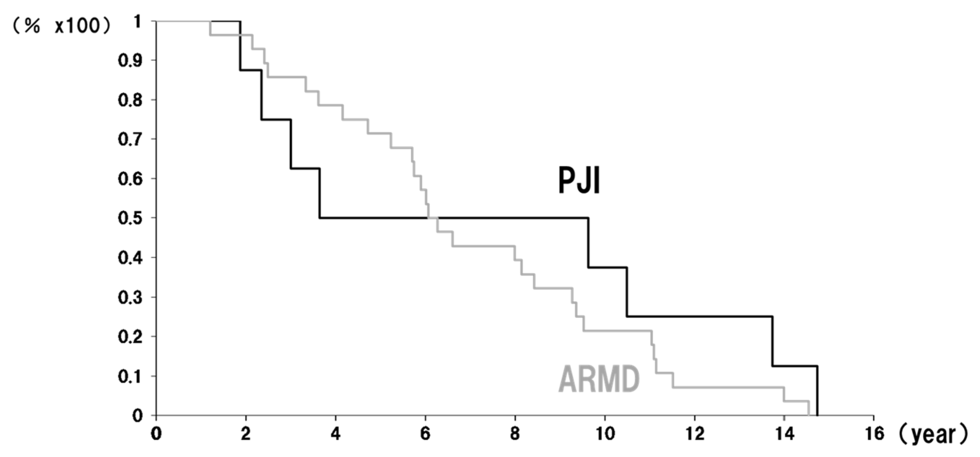
Figure 1. Implant survival curves at the revision endpoints for PJI and ARMDs: Black line—PJI (periprosthetic joint infection). Gray line—ARMDs (adverse reactions to metal debris).
Antibiotics 14 00161 g001
Table 1. Revision cases for PJI.
CaseAge, Sex, and DiagnosisBMI
(kg/m2)
Observed PT After Primary ArthroplastyTime to Revision Causative BacteriaCup Inclination Cup Anteversion SymptomCup RevisedStem Revised
Case153 F RA16.56.5 years10.5 yearsListeria55.724.2Swelling Revised (S-ROM)
Case268 M RA27.0Not observed3.5 yearsMSSA39.622.2Pain Revised (S-ROM)
Case268 M RA27.0Not observed3 yearsMSSA36.714.1Pain
Case369 F OA21.85.1 years9.7 yearsStreptococcus anginosus57.73.1Pain, Cup and stem osteolysis2stage2stage
Case466 F OA18.913.6 years13.7 yearsMSSA32.72.7Swelling, Fistula,
Cup and stem osteolysis
Revised
Case560 F OA22.52.3 years2.3 yearsno causative42.811.9PainRevised
Case658 M OA31.71.7 years1.9 yearsno causative53.26.0PainRevised
Case763 F OA21.410.1 years14.7 yearsMSSA58.827.8Pain, Cup osteolysisRevised
Table 2. Patient demographics for revisions related to PJI and ARMD.
Revision for PJI (3.2%)Revision for ARMD (12.1%)p
Male/Female2 patients (3 hips)/5 patients4 patients/25 patients (26 hips)ns
Age at revisionMean 62.4 (58–69) Mean 64.5 (52–69) ns
Preoperative diagnosisOA 5 patients
RA 2 patients (3 hips)
OA 26 patients (27 hips)
RA 2 patients
Fx 1 patient
ns
CRP
(≦0.14 mg/dL)
CRP-positive 7 patients
CRP-negative 1 patient
CRP-positive 13 patients (14 hips)
CRP-negative 16 patients
ns
OA—osteoarthritis; RA—rheumatoid arthritis; Fx—secondary hip OA in post-traumatic fracture.
Table 3. Evaluation metrics among revision surgeries for ARMDs and PJI.
Revision forPJI (8 Hips)ARMDs (30 Hips)p
Frequency (%)3.2%12.1% ns
PT-positive (%)75%70%ns
Revision forPJI (7 Patients)ARMDs (29 Patients)p
CRP-positive (%)85.7%44.8%ns
CRP level (mean ± SD)3.89 ± 3.060.41 ± 0.67<0.005
WBC counts (mean ± SD)8060 ± 31815651 ± 1357<0.05
Neutrophil counts (mean ± SD)5927 ± 31813664 ± 1280<0.05
Lymphocyte counts (mean ± SD)1390 ± 3191410 ± 484ns
Neutrophil-to-WBC ratios (mean ± SD)71.0 ± 8.564.2 ± 9.4ns
Lymphocyte-to-WBC ratios (mean ± SD)19.2 ± 6.925.5 ± 8.0ns
ARMDs—adverse reactions to metal debris; PJI—periprosthetic joint infection; SD—standard deviation; ns—not significant.
Table 4. Demographics of patients undergoing MOM THA.
Male/Female39 Patients/191 Patients
Age (years)Mean 64.1 (34–85)
Preoperative diagnosisOsteoarthritis (OA)223 hips
Rheumatoid arthritis (RA)15 hips
Osteonecrosis (ON)7 hips
Secondary OA in post hip fracture 2 hips
ImplantPinnacle82 patients89 hips
Cormet98 patients108 hips
Conserve plus50 patients50 hips
Follow-up periodsMean 10.5 years
Table 5. Demographics of patients undergoing non-MOM THA.
Metal on PolyethyleneCeramic on Polyethylene
68 patients/69 hips78 patients/78 hips
Age (years)Mean 60.8 Mean 64.9
Preoperative diagnosis
Osteoarthritis (OA)57 hips72 hips
Rheumatoid arthritis (RA)5 hips2 hips
Osteonecrosis (ON)3 hips (2 patients)
Rapidly destructive coxarthrosis2 hips3 hips
Secondary OA in infected hip2 hips
Pigmented villonodular synovitis 1 hip
Follow-up periodsMean 11.7 yearsMean 10.7 years
Disclaimer/Publisher’s Note: The statements, opinions and data contained in all publications are solely those of the individual author(s) and contributor(s) and not of MDPI and/or the editor(s). MDPI and/or the editor(s) disclaim responsibility for any injury to people or property resulting from any ideas, methods, instructions or products referred to in the content.