

The Behavior of Some Bacterial Strains Isolated from Fallow Deer Compared to Antimicrobial Substances in Western Romania


 ,
, 
Abstract
1. Introduction
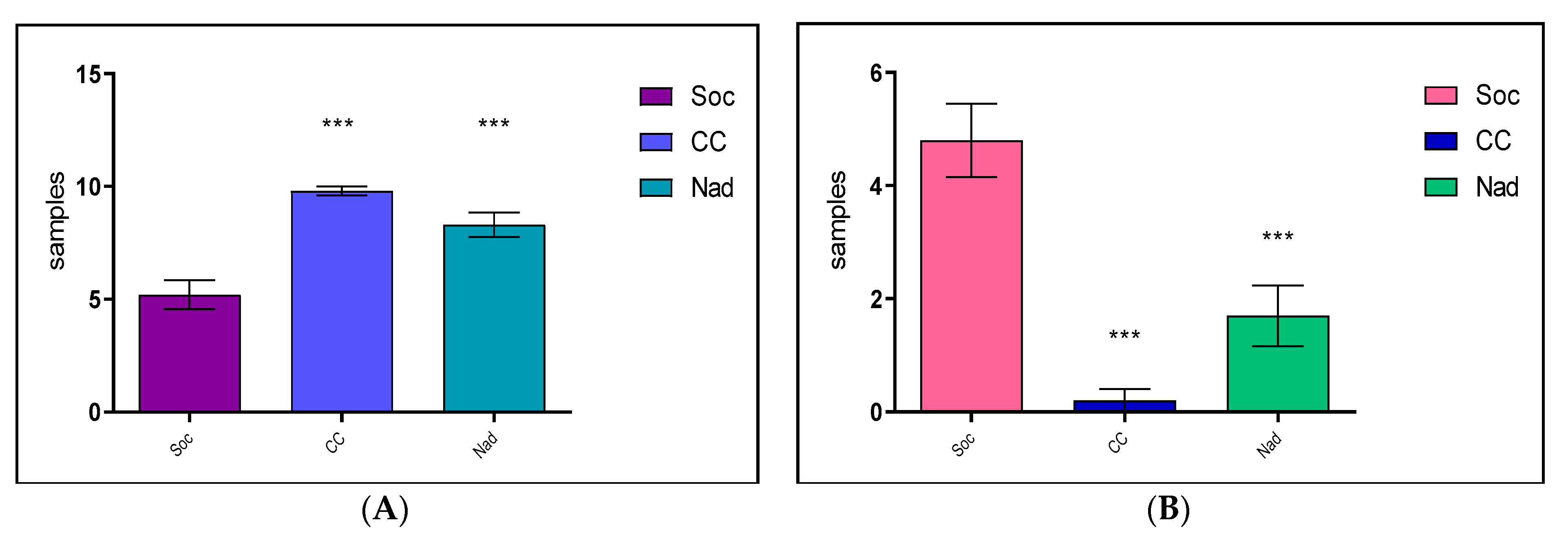
2. Results
2.1. The Antimicrobial Susceptibility after the Diffusimetric Method
2.1.1. Socodor Hunting Ground
2.1.2. Chișineu Criș–Sălișteanca Hunting Ground
2.1.3. Nadăș Hunting Ground
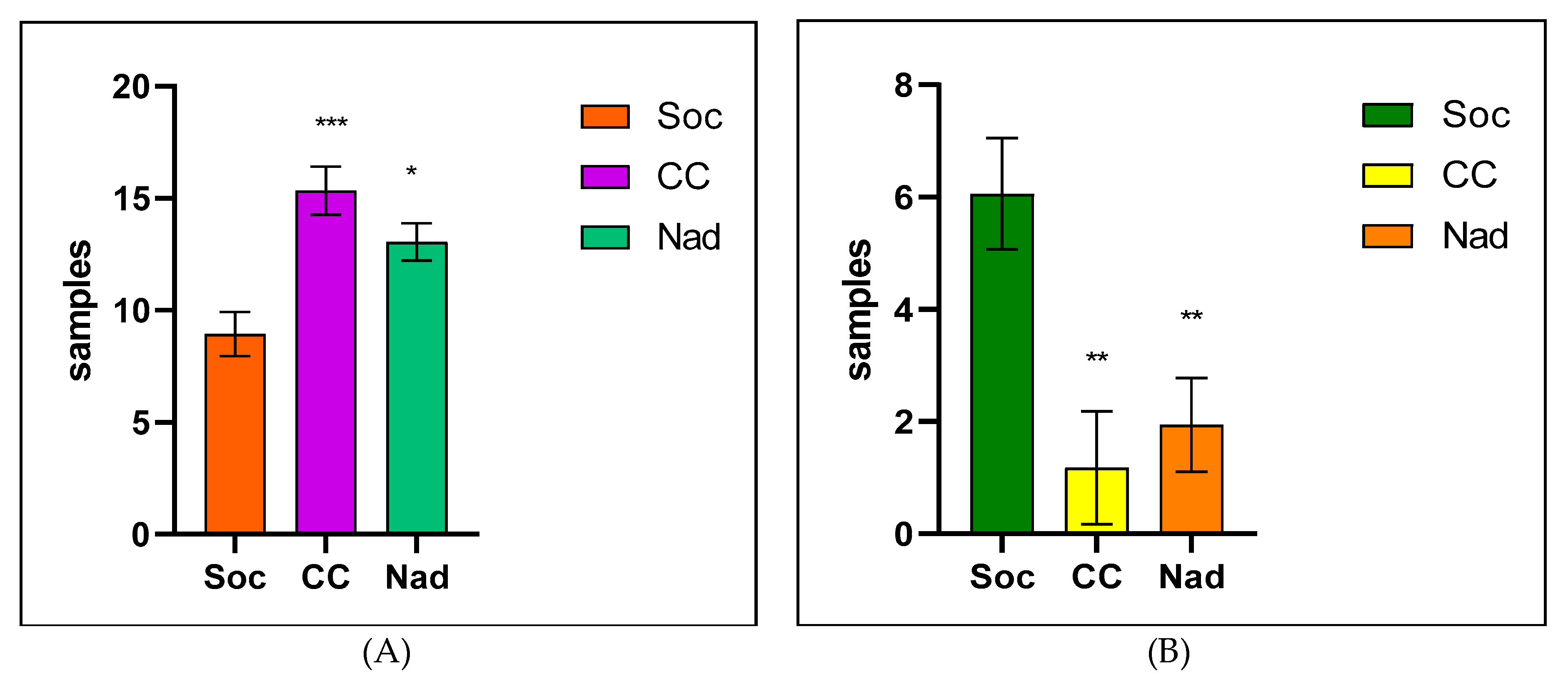
2.2. The Antimicrobial Susceptibility of Some Gram-Negative Bacterial Species with the Vitek-2 Compact
2.2.1. Socodor Hunting Ground
2.2.2. Chișinău Criș–Sălișteanca Hunting Ground
2.2.3. Nadăș Hunting Ground
2.3. Antimicrobial Susceptibility Results of Some Gram-Positive Bacterial Species with the Vitek 2 Compact
2.3.1. Socodor Hunting Ground
2.3.2. Chișinău Criș–Sălișteanca Hunting Ground
2.3.3. Nadăș Hunting Ground
3. Discussion
4. Materials and Method
4.1. Samples Source
4.2. The Antimicrobial Susceptibility of Bacterial Strains, Isolated by the Diffusimetric Method
4.3. The Antimicrobial Susceptibility of the Isolated Bacterial Strains by Vitek-2 Compact (BioMérieux, Craponne, France)
4.4. The Statistical Analysis
5. Conclusions
Supplementary Materials
Author Contributions
Funding
Institutional Review Board Statement
Informed Consent Statement
Data Availability Statement
Conflicts of Interest
References
- Prestinaci, F.; Pezzotti, P.; Pantosti, A. Antimicrobial Resistance: A Global Multifaceted Phenomenon. Pathog. Glob. Health 2015, 109, 309–318. [Google Scholar] [CrossRef] [PubMed]
- Poirel, L.; Madec, J.-Y.; Lupo, A.; Schink, A.-K.; Kieffer, N.; Nordmann, P.; Schwarz, S. Antimicrobial Resistance in Escherichia coli. In Antimicrobial Resistance in Bacteria from Livestock and Companion Animals; Schwarz, S., Cavaco, L.M., Shen, J., Eds.; ASM Press: Washington, DC, USA, 2018; pp. 289–316. [Google Scholar] [CrossRef]
- Schwarz, S.; Kehrenberg, C.; Doublet, B.; Cloeckaert, A. Molecular Basis of Bacterial Resistance to Chloramphenicol and Florfenicol. FEMS Microbiol. Rev. 2004, 28, 519–542. [Google Scholar] [CrossRef] [PubMed]
- Aarestrup, F.M. The Origin, Evolution, and Local and Global Dissemination of Antimicrobial Resistance. In Antimicrobial Resistance in Bacteria of Animal Origin; Aarestrup, F.M., Ed.; ASM Press: Washington, DC, USA, 2019; pp. 339–359. [Google Scholar] [CrossRef]
- Tiseo, K.; Huber, L.; Gilbert, M.; Robinson, T.P.; Van Boeckel, T.P. Global Trends in Antimicrobial Use in Food Animals from 2017 to 2030. Antibiotics 2020, 9, 918. [Google Scholar] [CrossRef]
- Van Boeckel, T.P.; Glennon, E.E.; Chen, D.; Gilbert, M.; Robinson, T.P.; Grenfell, B.T.; Levin, S.A.; Bonhoeffer, S.; Laxminarayan, R. Reducing Antimicrobial Use in Food Animals. Science 2017, 357, 1350–1352. [Google Scholar] [CrossRef]
- Arnold, K.E.; Williams, N.J.; Bennett, M. “Disperse Abroad in the Land”: The Role of Wildlife in the Dissemination of Antimicrobial Resistance. Biol. Lett. 2016, 12, 20160137. [Google Scholar] [CrossRef] [PubMed]
- Guardabassi, L.; Courvalin, P. Modes of Antimicrobial Action and Mechanisms of Bacterial Resistance. In Antimicrobial Resistance in Bacteria of Animal Origin; Aarestrup, F.M., Ed.; ASM Press: Washington, DC, USA, 2019; pp. 1–18. [Google Scholar] [CrossRef]
- EUCAST. EUCAST Guidelines for Detection of Resistance Mechanisms and Specific Resistances of Clinical and/or Epidemiological Importance. Version 2.01. July 2017. Available online: https://aurosan.de/images/mediathek/servicematerial/EUCAST_detection_of_resistance_mechanisms.pdf (accessed on 30 June 2022).
- EMA. Antibiotic Resistance in European Union Associated with the Therapeutic Use of Veterinary Medicines, Doc. EMA/CVMP/342/99-Final. 14 July 1999. Available online: https://www.ema.europa.eu/en/documents/report/antibiotic-resistance-european-union-associated-therapeutic-use-veterinary-medicines-report_en-0.pdf (accessed on 30 June 2022).
- Khoshbakht, R.; Tabatabaei, M.; Shirzad Aski, H.; Shayegh, H. Distribution of Salmonella, arcobacter, and Thermophilic Campylobacter spp. among Persian Fallow deer (Dama mesopotamica) Population in Dasht-e-Arzhan Wildlife Refuge, Southern Iran. Comp. Clin. Pathol. 2015, 24, 777–781. [Google Scholar] [CrossRef]
- Köck, R.; Daniels-Haardt, I.; Becker, K.; Mellmann, A.; Friedrich, A.W.; Mevius, D.; Schwarz, S.; Jurke, A. Carbapenem-Resistant Enterobacteriaceae in Wildlife, Food-Producing, and Companion Animals: A Systematic Review. Clin. Microbiol. Infect. 2018, 24, 1241–1250. [Google Scholar] [CrossRef]
- Ballash, G.A.; Munoz-Vargas, L.; Albers, A.; Dennis, P.M.; LeJeune, J.T.; Mollenkopf, D.F.; Wittum, T.E. Temporal Trends in Antimicrobial Resistance of Fecal Escherichia coli from Deer. EcoHealth 2021, 18, 288–296. [Google Scholar] [CrossRef]
- Dias, D.; Torres, R.T.; Kronvall, G.; Fonseca, C.; Mendo, S.; Caetano, T. Assessment of Antibiotic Resistance of Escherichia coli Isolates and Screening of Salmonella spp. in Wild Ungulates from Portugal. Res. Microbiol. 2015, 166, 584–593. [Google Scholar] [CrossRef]
- Botti, V.; Navillod, F.V.; Domenis, L.; Orusa, R.; Pepe, E.; Robetto, S.; Guidetti, C. Salmonella ssp. and Antibiotic-Resistant Strains in Wild Mammals and Birds in North-Western Italy from 2002 to 2010. Vet. Ital. 2013, 49, 195–202. [Google Scholar]
- European Food Safety Authority; European Centre for Disease Prevention and Control. The European Union Summary Report on Antimicrobial Resistance in Zoonotic and Indicator Bacteria from Humans, Animals and Food in 2010. EFSA J. 2012, 10, 2598. [Google Scholar] [CrossRef]
- Dionisi, A.M.; Filetici, E.; Ocwzarek, S.; Arena, S.; Benedetti, I.; Lucarelli, C.; Luzzi, I.; Scavia, G.; Minelli, F.; Ciaravino, G.; et al. ENTER-NET: Sorveglianza delle infezioni trasmesse da alimenti e acqua. Rapporto dell’attività 2007-2009. Not. Ist. Super. Sanità 2011, 24, 3–10. [Google Scholar]
- Alonso, C.A.; González-Barrio, D.; Tenorio, C.; Ruiz-Fons, F.; Torres, C. Antimicrobial Resistance in Faecal Escherichia coli Isolates from Farmed Red Deer and Wild Small Mammals. Detection of a Multiresistant E. coli Producing Extended-Spectrum Beta-Lactamase. Comp. Immunol. Microbiol. Infect. Dis. 2016, 45, 34–39. [Google Scholar] [CrossRef] [PubMed]
- Livermore, D.M.; Struelens, M.; Amorim, J.; Baquero, F.; Bille, J.; Canton, R.; Henning, S.; Gatermann, S.; Marchese, A.; Mittermayer, H.; et al. Multicentre Evaluation of the VITEK 2 Advanced Expert System for Interpretive Reading of Antimicrobial Resistance Tests. J. Antimicrob. Chemother. 2002, 49, 289–300. [Google Scholar] [CrossRef] [PubMed]
- Radhouani, H.; Silva, N.; Poeta, P.; Torres, C.; Correia, S.; Igrejas, G. Potential Impact of Antimicrobial Resistance in Wildlife, Environment and Human Health. Front. Microbiol. 2014, 5, 23. [Google Scholar] [CrossRef]
- Costa, D.; Poeta, P.; Sáenz, Y.; Vinué, L.; Coelho, A.C.; Matos, M.; Rojo-Bezares, B.; Rodrigues, J.; Torres, C. Mechanisms of Antibiotic Resistance in Escherichia coli Isolates Recovered from Wild Animals. Microb. Drug Resist. 2008, 14, 71–77. [Google Scholar] [CrossRef]
- van den Honert, M.S.; Gouws, P.A.; Hoffman, L.C. Escherichia coli Antibiotic Resistance Patterns from Co-Grazing and Non-Co-Grazing Livestock and Wildlife Species from Two Farms in the Western Cape, South Africa. Antibiotics 2021, 10, 618. [Google Scholar] [CrossRef]
- Katakweba, A.A.S.; Møller, K.S.; Muumba, J.; Muhairwa, A.P.; Damborg, P.; Rosenkrantz, J.T.; Minga, U.M.; Mtambo, M.M.A.; Olsen, J.E. Antimicrobial Resistance in Faecal Samples from Buffalo, Wildebeest and Zebra Grazing Together with and without Cattle in Tanzania. J. Appl. Microbiol. 2015, 118, 966–975. [Google Scholar] [CrossRef]
- Woolhouse, M.; Ward, M.; van Bunnik, B.; Farrar, J. Antimicrobial Resistance in Humans, Livestock and the Wider Environment. Philos. Trans. R. Soc. Lond B Biol. Sci. 2015, 370, 20140083. [Google Scholar] [CrossRef]
- Bouza, E.; Cercenado, E. Klebsiella and Enterobacter: Antibiotic Resistance and Treatment Implications. Semin. Respir. Infect. 2002, 17, 215–230. [Google Scholar] [CrossRef]
- Szczerba-Turek, A.; Kordas, B. Fallow deer (Dama Dama) as a Reservoir of Shiga Toxin-Producing Escherichia coli (STEC). Animals 2020, 10, 881. [Google Scholar] [CrossRef] [PubMed]
- Szczerba-Turek, A.; Siemionek, J.; Socha, P.; Bancerz-Kisiel, A.; Platt-Samoraj, A.; Lipczynska-Ilczuk, K.; Szweda, W. Shiga Toxin-Producing Escherichia coli Isolates from Red deer (Cervus elaphus), Roe deer (Capreolus capreolus) and Fallow deer (Dama Dama) in Poland. Food Microbiol. 2020, 86, 103352. [Google Scholar] [CrossRef] [PubMed]
- Kozak, G.K.; Boerlin, P.; Janecko, N.; Reid-Smith, R.J.; Jardine, C. Antimicrobial Resistance in Escherichia coli Isolates from Swine and Wild Small Mammals in the Proximity of Swine Farms and in Natural Environments in Ontario, Canada. Appl. Environ. Microbiol. 2009, 75, 559–566. [Google Scholar] [CrossRef] [PubMed]
- Lachmayr, K.L.; Kerkhof, L.J.; Dirienzo, A.G.; Cavanaugh, C.M.; Ford, T.E. Quantifying Nonspecific TEM Beta-Lactamase (BlaTEM) Genes in a Wastewater Stream. Appl. Environ. Microbiol. 2009, 75, 203–211. [Google Scholar] [CrossRef] [PubMed]
- Torres, R.T.; Fernandes, J.; Carvalho, J.; Cunha, M.V.; Caetano, T.; Mendo, S.; Serrano, E.; Fonseca, C. Wild Boar as a Reservoir of Antimicrobial Resistance. Sci. Total Environ. 2020, 717, 135001. [Google Scholar] [CrossRef]
- Euden, P.R. Salmonella Isolates from Wild Animals in Cornwall. Br. Vet. J. 1990, 146, 228–232. [Google Scholar] [CrossRef]
- Doi, Y.; Arakawa, Y. 16S Ribosomal RNA Methylation: Emerging Resistance Mechanism against Aminoglycosides. Clin. Infect. Dis. 2007, 45, 88–94. [Google Scholar] [CrossRef]
- Gnat, S.; Trościańczyk, A.; Nowakiewicz, A.; Majer-Dziedzic, B.; Ziółkowska, G.; Dziedzic, R.; Zięba, P.; Teodorowski, O. Experimental Studies of Microbial Populations and Incidence of Zoonotic Pathogens in the Faeces of Red Deer (Cervus elaphus). Lett. Appl. Microbiol. 2015, 61, 446–452. [Google Scholar] [CrossRef]
- Lasko, M.J.; Nicolau, D.P. Carbapenem-Resistant Enterobacterales: Considerations for Treatment in the Era of New Antimicrobials and Evolving Enzymology. Curr. Infect. Dis. Rep. 2020, 22, 6. [Google Scholar] [CrossRef]
- Zhou, Y.; Yu, H.; Guo, Q.; Xu, X.; Ye, X.; Wu, S.; Guo, Y.; Wang, M. Distribution of 16S RRNA Methylases among Different Species of Gram-Negative Bacilli with High-Level Resistance to Aminoglycosides. Eur. J. Clin. Microbiol. Infect. Dis. 2010, 29, 1349–1353. [Google Scholar] [CrossRef]
- Doma, A.O.; Cristina, R.T.; Muselin, F.; Dumitrescu, E.; Dégi, J.; Imre, K.; Boldea, M.; Vlad, D.C.; Popescu, R.; Cimporescu, A.; et al. The Role of Methyl-(Z)-11-Tetradecenoate Acid from the Bacterial Membrane Lipid Composition in Escherichia coli Antibiotic Resistance. BioMed Res. Int. 2022, 2022, 6028045. [Google Scholar] [CrossRef] [PubMed]
- Jiang, F.; Wu, Z.; Zheng, Y.; Frana, T.S.; Sahin, O.; Zhang, Q.; Li, G. Genotypes and Antimicrobial Susceptibility Profiles of Hemolytic Escherichia coli from Diarrheic Piglets. Foodborne Pathog. Dis. 2019, 16, 94–103. [Google Scholar] [CrossRef] [PubMed]
- Doma, A.O.; Popescu, R.; Mitulețu, M.; Muntean, D.; Dégi, J.; Boldea, M.V.; Radulov, I.; Dumitrescu, E.; Muselin, F.; Puvača, N.; et al. Comparative Evaluation of QnrA, QnrB, and QnrS Genes in Enterobacteriaceae Ciprofloxacin-Resistant Cases, in Swine Units and a Hospital from Western Romania. Antibiotics 2020, 9, 698. [Google Scholar] [CrossRef]
- Lillehaug, A.; Bergsjø, B.; Schau, J.; Bruheim, T.; Vikøren, T.; Handeland, K. Campylobacter Spp., Salmonella Spp., Verocytotoxic Escherichia coli, and Antibiotic Resistance in Indicator Organisms in Wild Cervids. Acta Vet. Scand. 2005, 46, 23–32. [Google Scholar] [CrossRef] [PubMed]
- Liu, G.; Bogaj, K.; Bortolaia, V.; Olsen, J.E.; Thomsen, L.E. Antibiotic-Induced, Increased Conjugative Transfer Is Common to Diverse Naturally Occurring ESBL Plasmids in Escherichia coli. Front. Microbiol. 2019, 10, 2119. [Google Scholar] [CrossRef]
- Ramey, A.M.; Ahlstrom, C.A. Antibiotic resistant bacteria in wildlife: Perspectives on trends, acquisition and dissemination, data gaps, and future directions. J. Wildl. Dis. 2020, 56, 1–15. [Google Scholar] [CrossRef]
- Dec, M.; Stępień-Pyśniak, D.; Gnat, S.; Fratini, F.; Urban-Chmiel, R.; Cerri, D.; Winiarczyk, S.; Turchi, B. Antibiotic susceptibility and virulence genes in Enterococcus isolates from wild mammals living in Tuscany, Italy. Microb. Drug Res. 2020, 26, 505–519. [Google Scholar] [CrossRef]
- Bertelloni, F.; Cilia, G.; Bogi, S.; Ebani, V.V.; Turini, L.; Nuvoloni, R.; Cerri, D.; Fratini, F.; Turchi, B. Pathotypes and antimicrobial susceptibility of Escherichia coli isolated from wild boar (Sus scrofa) in Tuscany. Animals 2022, 10, 744. [Google Scholar] [CrossRef]
- Elsby, D.T.; Zadoks, R.N.; Boyd, K.; Silva, N.; Chase-Topping, M.; Mitchel, M.C.; Currie, C.; Taggart, M.A. Antimicrobial resistant Escherichia coli in Scottish wild deer: Prevalence and risk factors. Environ. Pollut. 2022, 314, 120129. [Google Scholar] [CrossRef]
- Clinical and Laboratory Standards Institute (CLSI). Performance Standards for Antimicrobial Susceptibility Testing, 28th ed.; CLSI Document M100-28; CLSI: Wayne, PA, USA, 2018. [Google Scholar]
- Clinical and Laboratory Standards Institute (CLSI). Performance Standards for Antimicrobial Susceptibility Testing, 28th ed.; CLSI Supplement M100; CLSI: Wayne, PA, USA, 2019. [Google Scholar]
- CA-SFM/EUCAST; Comité de l’antibiogramme. French Society of Microbiology/European Committee on Antimicrobial Susceptibility Testing: Växjö, Sweden, 2019.


| Antimicrobial Susceptibility of Species Isolated from the Socodor Hunting Ground | ||||||||||
|---|---|---|---|---|---|---|---|---|---|---|
| Antimicrobial | Bacterial Species/Susceptibility/Drug | |||||||||
| E. coli | P. oleovorans | P. rettgeri | Enterob. spp. | Enterob. aerogenes | Enterob. spp. | |||||
| 1 | 2 | 3 | 4 | 5 | ||||||
| Ampicillin | R | R | R | R | R | S | S | R | R | S |
| Amoxi/Clavulanic ac | R | R | R | R | R | S | S | R | R | S |
| Cefuroxime | R | R | S | R | R | S | R | S | S | S |
| Ceftazidim | S | S | S | S | S | S | S | R | S | S |
| Ciprofloxacin | R | R | R | R | R | S | S | S | S | R |
| Gentamicin | R | R | S | R | R | S | S | S | S | S |
| Nitrofurantoin | S | S | R | S | R | S | S | R | S | S |
| Trimetho/Sulfametho | S | R | S | R | R | S | S | S | S | S |
| Chloramphenicol | S | R | S | R | R | S | R | S | R | S |
| Azithromycin | R | R | R | R | R | S | S | R | R | S |
| Resistant (%) | 60 | 80 | 50 | 80 | 90 | 0 | 20 | 50 | 40 | 10 |
| Sensitive (%) | 40 | 20 | 50 | 20 | 10 | 100 | 80 | 50 | 60 | 90 |
| Antimicrobial Susceptibility of Species Isolated from the Chișinău Criș–Sălișteanca Hunting Ground | ||||||||||
| Antimicrobial | E. coli | Salmonella spp. | Enterobacter spp. | |||||||
| 1 | 2 | 3 | 4 | 5 | 1 | 2 | 3 | 1 | 2 | |
| Ampicillin | S | R | R | S | S | S | S | S | S | S |
| Amoxi/Clavulanic ac | S | S | S | S | S | S | S | S | S | S |
| Cefuroxime | S | S | S | S | S | S | S | S | S | S |
| Ceftazidim | S | S | S | S | S | S | S | S | S | S |
| Ciprofloxacin | S | S | S | S | S | S | S | S | S | S |
| Gentamicin | S | S | S | S | S | S | S | S | S | S |
| Nitrofurantoin | S | S | S | S | S | S | S | S | S | S |
| Trimetho/Sulfametho | S | S | S | S | S | S | S | S | S | S |
| Chloramphenicol | S | S | S | S | S | S | S | S | S | S |
| Azithromycin | S | S | S | S | S | S | S | S | S | S |
| Resistant (%) | 0 | 10 | 10 | 0 | 0 | 0 | 0 | 0 | 0 | 0 |
| Sensitive (%) | 100 | 90 | 90 | 100 | 100 | 100 | 100 | 100 | 100 | 100 |
| Antimicrobial Susceptibility of Species Isolated from the Nadăș Hunting Ground | ||||||||||
| Antimicrobial | E. coli | Salmonella spp. | Enterobacter spp. | |||||||
| 1 | 2 | 3 | 4 | 5 | 1 | 2 | 3 | 1 | 2 | |
| Ampicillin | S | R | R | S | S | S | S | R | S | S |
| Amoxi/Clavulanic ac | S | S | S | S | S | S | S | S | S | S |
| Cefuroxime | S | S | R | R | S | S | S | S | R | R |
| Ceftazidim | S | S | R | R | S | S | S | S | S | S |
| Ciprofloxacin | S | S | S | S | S | S | S | S | S | S |
| Gentamicin | S | R | S | S | S | S | R | R | S | S |
| Nitrofurantoin | S | S | S | S | S | S | S | S | S | S |
| Trimetho/Sulfametho | S | S | S | R | S | S | S | S | S | S |
| Chloramphenicol | S | S | S | S | S | S | S | S | S | S |
| Azithromycin | S | S | S | S | R | S | R | S | R | R |
| Resistant (%) | 0 | 20 | 30 | 30 | 10 | 0 | 20 | 20 | 20 | 20 |
| Sensitive (%) | 100 | 80 | 70 | 70 | 90 | 100 | 80 | 80 | 80 | 80 |
| Antimicrobial Susceptibility of Species Isolated from the Socodor Hunting Ground | |||||||||||||||||
|---|---|---|---|---|---|---|---|---|---|---|---|---|---|---|---|---|---|
| Species | Antimicrobial Class | ||||||||||||||||
| Beta-Lactamins | Aminoglicosides | Quinolones | Tcy. | Fourans | |||||||||||||
| AMP | AMC | CN | CF | CEC | CFP | FUR | IPM | GM | AK | N | UMB | ENR | MAR | TE | FT | SXT * | |
| Ps. oleovorans | R | R | R | R | S | S | S | S | S | S | S | S | S | S | S | S | S |
| Pr. rettgeri | R | R | R | R | S | S | S | S | S | S | S | R | S | S | R | S | S |
| Enterobacter sp. | R | R | R | R | S | S | S | S | S | S | S | S | S | S | S | S | S |
| Enterobacter sp. | S | R | R | R | S | S | S | S | S | S | S | S | S | S | S | S | S |
| Ent. aerogenes | R | R | R | R | S | S | S | S | S | S | S | S | S | S | S | S | S |
| Escherichia coli | S | S | R | I | R | S | S | S | S | S | S | S | S | S | S | S | S |
| Escherichia coli | R | S | R | I | R | R | S | S | S | S | S | S | S | S | R | S | S |
| Escherichia coli | R | R | R | R | R | R | R | S | R | S | R | R | R | R | R | S | R |
| Escherichia coli | R | R | R | R | R | R | R | S | R | S | R | R | R | R | R | R | R |
| Escherichia coli | R | R | R | R | R | R | R | S | R | S | S | R | R | R | R | R | R |
| Escherichia coli | R | R | R | R | R | R | R | S | R | S | R | R | R | R | R | S | R |
| Escherichia coli | R | R | R | R | S | S | S | S | R | S | S | R | R | R | R | S | R |
| Escherichia coli | S | S | R | R | S | S | S | S | S | S | S | S | S | S | S | S | S |
| Escherichia coli | R | S | R | R | S | R | S | S | S | S | S | S | S | S | R | S | S |
| Escherichia coli | S | S | R | I | S | S | S | S | S | S | S | S | S | S | S | S | S |
| Antimicrobial Susceptibility of Species Isolated from the Chișinău Criș–Sălișteanca hunt Ground | |||||||||||||||||
| Species | Beta-Lactamins | Aminoglicosides | Quinolones | Tcy. | Fourans | ||||||||||||
| AMP | AMC | CN | CF | CEC | CFP | FUR | IPM | GM | AK | N | UMB | ENR | MAR | TE | FT | SXT * | |
| Escherichia coli | S | S | R | R | S | S | S | S | S | S | S | S | S | S | S | S | S |
| Escherichia coli | S | S | R | I | S | S | S | S | S | S | S | S | S | S | S | S | S |
| Escherichia coli | S | S | R | R | S | S | S | S | S | S | S | S | S | S | S | S | S |
| Escherichia coli | S | S | R | I | S | S | S | S | S | S | S | S | S | S | S | S | S |
| Escherichia coli | S | S | R | I | S | S | S | S | S | S | S | S | S | S | S | S | S |
| Escherichia coli | S | S | R | R | S | S | S | S | S | S | S | S | S | S | S | S | S |
| Escherichia coli | S | S | R | I | S | S | S | S | S | S | S | S | S | S | S | S | S |
| Escherichia coli | S | S | R | R | S | S | S | S | S | S | S | S | S | S | S | S | S |
| Escherichia coli | S | S | R | R | S | S | S | S | S | S | S | S | S | S | S | S | S |
| Escherichia coli | S | S | R | I | S | S | S | S | S | S | S | S | S | S | S | S | S |
| Escherichia coli | S | S | R | R | S | S | S | S | S | S | S | R | S | S | S | S | S |
| Escherichia coli | S | S | R | R | S | S | S | S | S | S | S | R | S | S | S | S | S |
| Salmonella sp. | S | S | R | I | S | S | S | S | S | S | S | S | S | S | S | S | S |
| Salmonella sp. | S | S | R | R | S | S | S | S | S | S | S | R | S | S | S | S | S |
| Salmonella sp. | S | S | R | I | S | S | S | S | S | S | S | S | S | S | S | S | S |
| Enterobacter sp. | S | S | R | I | S | S | S | S | S | S | S | S | S | S | S | S | S |
| Enterobacter sp. | S | S | R | I | S | S | S | S | S | S | S | S | S | S | S | S | S |
| Antimicrobial Susceptibility of Species Isolated from the Nadăș Hunting Stock | |||||||||||||||||
| Species | Beta-lactamins | Aminoglicosides | Quinolones | Tcy. | Fourans | ||||||||||||
| AMP | AMC | CN | CF | CEC | CFP | FUR | IPM | GM | AK | N | UMB | ENR | MAR | TE | FT | SXT * | |
| Escherichia coli | R | R | S | S | S | S | S | S | S | S | S | S | S | S | S | S | S |
| Escherichia coli | S | S | S | S | S | S | S | S | S | S | S | S | S | S | S | S | S |
| Escherichia coli | S | S | R | S | S | S | S | S | S | S | S | S | S | S | S | S | S |
| Escherichia coli | S | S | R | R | R | S | S | S | S | S | S | S | S | S | S | S | S |
| Escherichia coli | S | S | R | R | R | R | S | S | S | S | S | S | S | S | S | S | S |
| Escherichia coli | S | S | R | I | R | R | S | S | S | S | S | S | S | S | S | S | S |
| Escherichia coli | S | S | R | R | S | S | S | S | S | S | S | R | R | S | I | S | S |
| Escherichia coli | S | S | R | R | S | S | S | S | S | S | S | S | S | S | S | S | S |
| Escherichia coli | R | S | R | R | S | S | S | S | S | S | S | R | S | S | S | S | S |
| Escherichia coli | S | S | R | R | R | R | S | S | S | S | S | S | S | S | S | S | S |
| Salmonella sp. | S | S | R | I | S | S | S | S | S | S | S | S | S | S | S | S | S |
| Salmonella sp. | S | S | R | I | S | S | S | S | S | S | S | S | S | S | S | S | S |
| Salmonella sp. | S | S | R | R | S | S | S | S | S | S | S | S | S | S | S | S | S |
| Enterobacter sp. | S | S | R | I | S | S | S | S | S | S | S | S | S | S | S | S | S |
| Enterobacter sp. | S | S | R | I | S | S | S | S | S | S | S | S | S | S | S | S | S |
| Antimicrobial Susceptibility of Species Isolated from the Socodor Hunting Ground | ||||||||||||
|---|---|---|---|---|---|---|---|---|---|---|---|---|
| Species/Antimicrobial | AN | GM | K | N | ENR | E | TIL | TYL | CM | TE | FLO | SXT |
| A. viridans | S | S | S | S | S | R | R | R | R | R | S | S |
| A. viridans | S | S | S | S | I | R | R | R | R | R | S | S |
| Kocuria kristinae | S | S | R | S | R | R | R | R | R | R | R | S |
| Kocuria kristinae | S | S | R | S | I | I | R | R | R | R | R | S |
| E. faecium | S | S | S | S | I | R | S | S | R | R | S | S |
| S. lentus | S | R | S | S | R | R | R | S | R | R | S | R |
| S. lentus | S | S | S | S | R | I | R | R | R | R | S | S |
| S. lentus | S | S | S | S | S | R | R | R | R | R | S | S |
| S. lentus | S | I | S | S | I | R | S | R | R | R | S | S |
| S. sciuri | S | S | S | S | S | S | S | S | S | S | S | S |
| S. vitulinus | S | S | S | S | I | S | S | R | S | R | S | S |
| S. vitulinus | S | S | S | S | I | S | R | R | R | S | S | S |
| S. vitulinus | S | S | S | S | S | S | S | S | R | S | S | S |
| S. xylosus | S | S | S | S | S | R | R | R | R | S | S | S |
| S. xylosus | S | S | S | S | S | R | R | R | R | S | S | S |
| Antimicrobial Susceptibility of Species Isolated from the Chișinău Criș–Sălișteanca Hunting Ground | ||||||||||||
| Species/Antimicrobial | AN | GM | K | N | ENR | E | TIL | TYL | CM | TE | FLO | SXT |
| A. viridans | S | S | S | S | S | R | S | S | S | S | S | S |
| A. viridans | S | S | S | S | S | S | S | S | S | R | S | S |
| Kocuria/Dermacoccus spp. | S | S | S | S | R | I | S | S | S | R | S | S |
| S. aureus | S | S | S | S | S | S | R | R | R | S | S | S |
| S. lentus | S | S | S | S | S | S | S | S | S | S | S | S |
| S. lentus | S | S | S | S | I | S | S | S | S | S | S | S |
| S. lentus | S | S | S | S | R | S | S | R | R | S | S | S |
| S. sciuri | S | S | S | S | S | S | R | S | R | S | S | S |
| S. sciuri | S | S | S | S | S | S | S | R | I | S | S | S |
| S. sciuri | I | S | I | S | S | I | R | S | S | |||
| S. vitulinus | S | S | S | S | S | S | S | S | S | S | S | S |
| S. vitulinus | S | S | S | S | S | S | S | R | R | S | S | S |
| S. vitulinus | S | S | S | S | S | S | S | S | R | S | S | S |
| S. xylosus | S | S | S | S | S | R | S | R | S | S | S | S |
| S. xylosus | S | S | S | S | S | I | R | R | R | R | R | S |
| Antimicrobial Susceptibility of Gram-Positives Isolated from the Nadăș Hunting Ground | ||||||||||||
| Species/antimicrobial | AN | GM | K | N | ENR | E | TIL | TYL | CM | TE | FLO | SXT |
| A. viridans | S | S | S | S | I | S | R | S | S | S | S | S |
| A. viridans | S | S | S | S | S | S | R | S | S | S | S | S |
| Kocuria kristinae | S | S | S | S | S | I | R | R | S | R | R | S |
| Kocuria kristinae | S | S | S | S | S | S | S | R | S | S | S | S |
| E. faecium | S | S | S | S | S | S | R | R | S | S | S | S |
| S. lentus | S | S | S | S | S | S | S | R | S | S | S | S |
| S. lentus | S | S | S | S | S | S | S | S | S | S | S | S |
| S. lentus | S | S | S | S | S | S | S | S | S | S | S | S |
| S. lentus | S | S | S | S | S | S | S | S | S | S | S | S |
| S. sciuri | S | S | S | S | S | S | S | S | S | S | S | S |
| S. vitulinus | S | S | S | S | S | S | S | S | S | S | S | S |
| S. vitulinus | S | S | S | S | S | S | S | R | S | S | S | S |
| S. vitulinus | S | S | S | S | S | S | S | S | S | S | S | S |
| S. xylosus | S | S | S | S | S | S | S | S | S | S | S | S |
| S. xylosus | S | S | S | S | S | S | S | S | S | S | S | S |
| Crt. No. | Hunting Ground | Bacterial Species | Strains No. | Sampling Place |
|---|---|---|---|---|
| 1. | Socodor | Escherichia coli | 5 | Anal |
| Pseudomonas oleovorans | 1 | Nasal | ||
| Providencia rettgeri | 1 | Nasal | ||
| Enterobacter aerogenes | 1 | Nasal | ||
| Enterobacter spp. | 2 | Anal | ||
| 2. | Chișineu Criș–Sălișteanca | Escherichia coli | 5 | Nasal |
| Salmonella spp. | 3 | Nasal | ||
| Enterobacter spp. | 2 | Anal | ||
| 3. | Nadăș | Escherichia coli | 5 | Nasal |
| Salmonella spp. | 3 | Anal | ||
| Enterobacter spp. | 2 | Anal |
| Crt. No. | Bacterial Species | Strains No. | Sampling Place |
|---|---|---|---|
| Gram-negative | |||
| 1 | Escherichia coli | 32 | anal |
| 2 | Enterobacter spp. | 6 | anal |
| 3 | Enterobacter aerogenes | 1 | nasal |
| 4 | Salmonella spp. | 6 | nasal |
| 5 | Providencia rettgeri | 1 | nasal |
| 6 | Pseudomonas oleovorans | 1 | nasal |
| Total samples | 47 | ||
| Gram-positives | |||
| 1 | Aerococcus viridans | 4 | anal |
| 2 | Enterococcus faecium | 1 | anal |
| 3 | Kocuria kristinae | 3 | nasal |
| 4 | Staphylococcus sciuri | 11 | nasal |
| 5 | Staphylococcus lentus | 10 | nasal |
| 6 | Staphylococcus vitulinus | 7 | nasal |
| 7 | Staphylococcus xylosus | 8 | nasal |
| 8 | Staphylococcus aureus | 1 | nasal |
| Total samples | 45 | ||
Disclaimer/Publisher’s Note: The statements, opinions and data contained in all publications are solely those of the individual author(s) and contributor(s) and not of MDPI and/or the editor(s). MDPI and/or the editor(s) disclaim responsibility for any injury to people or property resulting from any ideas, methods, instructions or products referred to in the content. |
© 2023 by the authors. Licensee MDPI, Basel, Switzerland. This article is an open access article distributed under the terms and conditions of the Creative Commons Attribution (CC BY) license (https://creativecommons.org/licenses/by/4.0/).
Share and Cite
Tîrziu, E.; Bulucea, A.V.; Imre, K.; Nichita, I.; Muselin, F.; Dumitrescu, E.; Tîrziu, A.; Mederle, N.G.; Moza, A.; Bucur, I.M.; et al. The Behavior of Some Bacterial Strains Isolated from Fallow Deer Compared to Antimicrobial Substances in Western Romania. Antibiotics 2023, 12, 743. https://doi.org/10.3390/antibiotics12040743
Tîrziu E, Bulucea AV, Imre K, Nichita I, Muselin F, Dumitrescu E, Tîrziu A, Mederle NG, Moza A, Bucur IM, et al. The Behavior of Some Bacterial Strains Isolated from Fallow Deer Compared to Antimicrobial Substances in Western Romania. Antibiotics. 2023; 12(4):743. https://doi.org/10.3390/antibiotics12040743
Chicago/Turabian StyleTîrziu, Emil, Alexandrina V. Bulucea, Kalman Imre, Ileana Nichita, Florin Muselin, Eugenia Dumitrescu, Andreea Tîrziu, Narcisa G. Mederle, Alexandru Moza, Iulia M. Bucur, and et al. 2023. "The Behavior of Some Bacterial Strains Isolated from Fallow Deer Compared to Antimicrobial Substances in Western Romania" Antibiotics 12, no. 4: 743. https://doi.org/10.3390/antibiotics12040743
APA StyleTîrziu, E., Bulucea, A. V., Imre, K., Nichita, I., Muselin, F., Dumitrescu, E., Tîrziu, A., Mederle, N. G., Moza, A., Bucur, I. M., & Cristina, R. T. (2023). The Behavior of Some Bacterial Strains Isolated from Fallow Deer Compared to Antimicrobial Substances in Western Romania. Antibiotics, 12(4), 743. https://doi.org/10.3390/antibiotics12040743