Article

Salmonella Prophages, Their Propagation, Host Specificity and Antimicrobial Resistance Gene Transduction

Institute of Food Safety, University of Veterinary Medicine, 1210 Vienna, Austria
*
Author to whom correspondence should be addressed.
Antibiotics 2023, 12(3), 595; https://doi.org/10.3390/antibiotics12030595
Submission received: 20 February 2023 / Revised: 10 March 2023 / Accepted: 15 March 2023 / Published: 16 March 2023
(This article belongs to the Special Issue Antimicrobial Resistance in Food-borne Pathogens)

Abstract

:
Salmonella enterica subsp. enterica is a zoonotic bacterial pathogen that causes foodborne outbreaks in humans. Lytic bacteriophages to control Salmonella in food production are already being used in scientific studies and some are commercially available. However, phage application is still controversial. In addition to virulent phages, which are used in phage therapy and lyse the bacterial host, lysogenic phages coexist in the environment and can reside as prophages in the bacterial host. Therefore, information about Salmonella prophages is essential to understand successful phage therapy. In 100 Salmonella food isolates of the serovars Enteritidis and Typhimurium, we propagated prophages by oxidative stress. In isolates of the serovars Typhimurium and Enteritidis, 80% and 8% prophages could be activated, respectively. In the phage lysates from the serovar Typhimurium, the following antibiotic resistance genes or gene fragments were detected by PCR: sul1, sul2, blaTEM, strA and cmlA; however, no tetA,B,C, blaOXA, blaCMY, aadA1, dfr1,2 or cat were detected. In contrast, no resistance genes were amplified in the phage lysates of the serovar Enteritidis. None of the phage lysates was able to transduce phenotypic resistance to WT 14028s. Most of the prophage lysates isolated were able to infect the various Salmonella serovars tested. The high abundance of prophages in the genome of the serovar Typhimurium may counteract phage therapy through phage resistance and the development of hybrid phages.

1. Introduction

Salmonella (S.) enterica is an important zoonotic bacterial pathogen transmitted to humans through food, mainly through contaminated poultry products and eggs [1]. S. enterica subsp. enterica, with its main serovars, Enteritidis and Typhimurium, is the second most important bacterial zoonotic pathogen causing foodborne outbreaks in humans in Europe [1]. Poultry meat, eggs and food thereof are the main sources, but other animal-based foods and plant products have also been implicated in foodborne outbreaks of Salmonella [1]. A good proportion of these Salmonella isolates are multi-resistant to various antibiotics [2,3,4,5,6,7,8,9]. When nontyphoid Salmonella causes invasive clinical signs of disease in humans, antibiotic treatment is required. Invasive disease occurs primarily in the YOPI group (Young, Old, Pregnant, Immune Deficient) of humans [10,11,12]. As a facultative intracellular pathogen, Salmonella can multiply in macrophages and produce endotoxins during infection. Certain antibiotics may enhance intracellular location and endotoxin release, which limits the range of treatment options [13,14].
Lytic bacteriophage to control Salmonella in food production and to combat animal colonization and disease are used intensively in scientific studies and some are already commercially available [15,16,17]. Phage therapy in humans still requires individual approval in most countries and results of phage application are still controversial. Reports of phage therapy for human salmonellosis are scarce [18].
Virulent phages are mainly used in therapy, but lysogenic phages, which can enter the bacterial genome and survive as a prophage for a period of time, appear to be most relevant to bacterial evolution. Horizontal gene transfer by phages is called transduction and refers to lysogenic phages, but even virulent phages are capable of advertently transducing genetic material from one bacterial cell to another [19]. Therefore, the fitness of bacteria can be enhanced by prophages harbouring phage morons (adding “more on” to the bacterial host), which are genes carried by the prophage and benefit the bacterial host [20]. These accessory genes can supply virulence factors, toxins and resistance genes. Furthermore, the infection of a bacterial cell by virulent and lysogenic phages can lead to so-called hybrid phages, which can change in host specificity or live style (virulent/lysogenic). In this way, initial information about the phageome of a specific bacterial species or genus can be used to secure phage therapy.
Here we analyzed the S. serovars Typhimurium and Enteritidis for inducible prophages. In addition, these phage lysates were screened for antimicrobial resistance genes (morons) and the transduction of phenotypic resistance to a standard strain. Finally, we evaluated the host specificity of these phage lysates.

2. Results

Food isolates of the S. serovars Typhimurium and Enteritidis, for the propagation of prophages, were obtained from different types of meat purchased in Austria, and human isolates were provided by the Austrian National Reference Center for Salmonellosis. As host strains to culture the activated prophages from the food and human isolates, we selected two strains from the type and culture collections (ATCC and DSM) previously described in the literature as well-defined hosts. These culture collection strains have already been used for phage transduction experiments, and do not harbor inducible prophages in their genome to interfere with phage lysates from human and food isolates. The strain ATCC 14028 WT was selected as a known strain for use as a host strain for the provision of transduction libraries, and has also been used in phage infection and transduction studies [21]. The strain DSM 17420 has also been used for transduction studies [22]. Various stressors can activate prophage induction. We tested H2O2, nitrate and mitomycin C in a study on Sty1, a serovar Typhimurium isolate, and WS27D, a serovar Enteritidis isolate, respectively. Mitomycin C, a cytostatic agent, was most effective at inducing prophage, followed by H2O2 and nitrite. Therefore, for prophage induction, H2O2 was used as a potential oxygen stressor for Salmonella (a typical activated prophage infecting host strains is shown in Figure 1). In Figure 1, a typical propagated prophage lysate was used to infect the host strains ATCC 14028 (left) and DSM 17420 (right). In the overlay method, an exponentially growing host strain at a McF concentration of 0.5 is used with a phage extract (in this case a propagated prophage lysate from a S. serovar Typhimurium isolate, left, and a S. serovar Enteritidis isolate, right, with H2O2), which is sterile filtered to avoid bacterial contamination and treated with DNAse, avoiding interaction with free DNA.
Additionally, we tested four isolates each of the serovar Typhimurium and the serovar Enteritidis, treated in parallel with H2O2 and mitomycin C, respectively. H2O2 was able to propagate prophages in two isolates of the serovar Typhimurium, whereas mitomycin C was only able to induce it in one of the isolates. No difference was seen for activating prophage with either H2O2 or mitomycin C in the four isolates of the serovar Enteritidis.
In 40 out of 50 S. serovar Typhimurium food and human isolates, prophages were inducible by H2O2 treatment. There was no difference between the human and food isolates in prophage activation potential under H2O2 treatment. In a total of 20% of the serovar Typhimurium isolates, no phage transduction was possible under H2O2 treatment and host strain ATCC 14028 WT. The activated prophage lysates of these isolates were also unable to infect DSM 17420. For the serovar Enteritidis we used DSM 17420 and ATCC14028 WT as host strains for the transduction experiments. Prophages could be activated in only four isolates. All four isolates were isolated from chicken meat. No prophages from any human S. serovar Enteritidis isolates could be activated by H2O2 treatment.
Phages are important evolutionary drivers in bacteria. Antimicrobial resistance genes can be transduced by a variety of lysogenic phages. The potential of phages for horizontal gene transfer, including antibiotic resistance genes, has only recently been discussed [23]. Therefore, we isolated DNA from the total lysates of activated prophages and analyzed it by PCR for 14 common antibiotic resistance genes. We made sure only phage DNA was extracted by filtering lysates and DNAse treatment to destroy external DNA. Of these genes, sul1 was detected in 5 prophage lysates, sul2 in 8 lysates, blaTEM in one lysate, strA in four lysates and cmlA in 10 isolates (as shown in Table 1). No lysate tested positive for tetA,B,C, blaOXA, blaCMY, aadA1, dfr1,2 or cat. All the lysates that tested positive were from the S. serovar Typhimurium isolates and none was from the serovar Enteritidis. Most of the isolates tested positive for one antibiotic resistance gene fragment, but four lysates tested positive for two and two for three genes. A combination of more than one resistance gene, namely sul1 + sul2 + strA in two isolates, sul2 + strA in two isolates, cmlA + sul1 in one, and blaTEM + sul2 in one isolate, was detected.
Transduction experiments were performed in ATCC 14028 WT from each of the lysate described in Table 1, and screening for antibiotic resistant transductans was conducted. Neither lysate was able to transduce phenotypic resistance into ATCC 14028 WT. ATCC 14028 does not harbor any of the resistance genes analyzed and has no phenotypic resistance against the tested antibiotic substances. No growth on plates with adequate antibiotic concentration (ampicillin at 15 µg/mL, sulfonamids at 25 µg/mL, streptomycin at 15 µg/mL and chloramphenicol at 30 µg/mL) was detected.
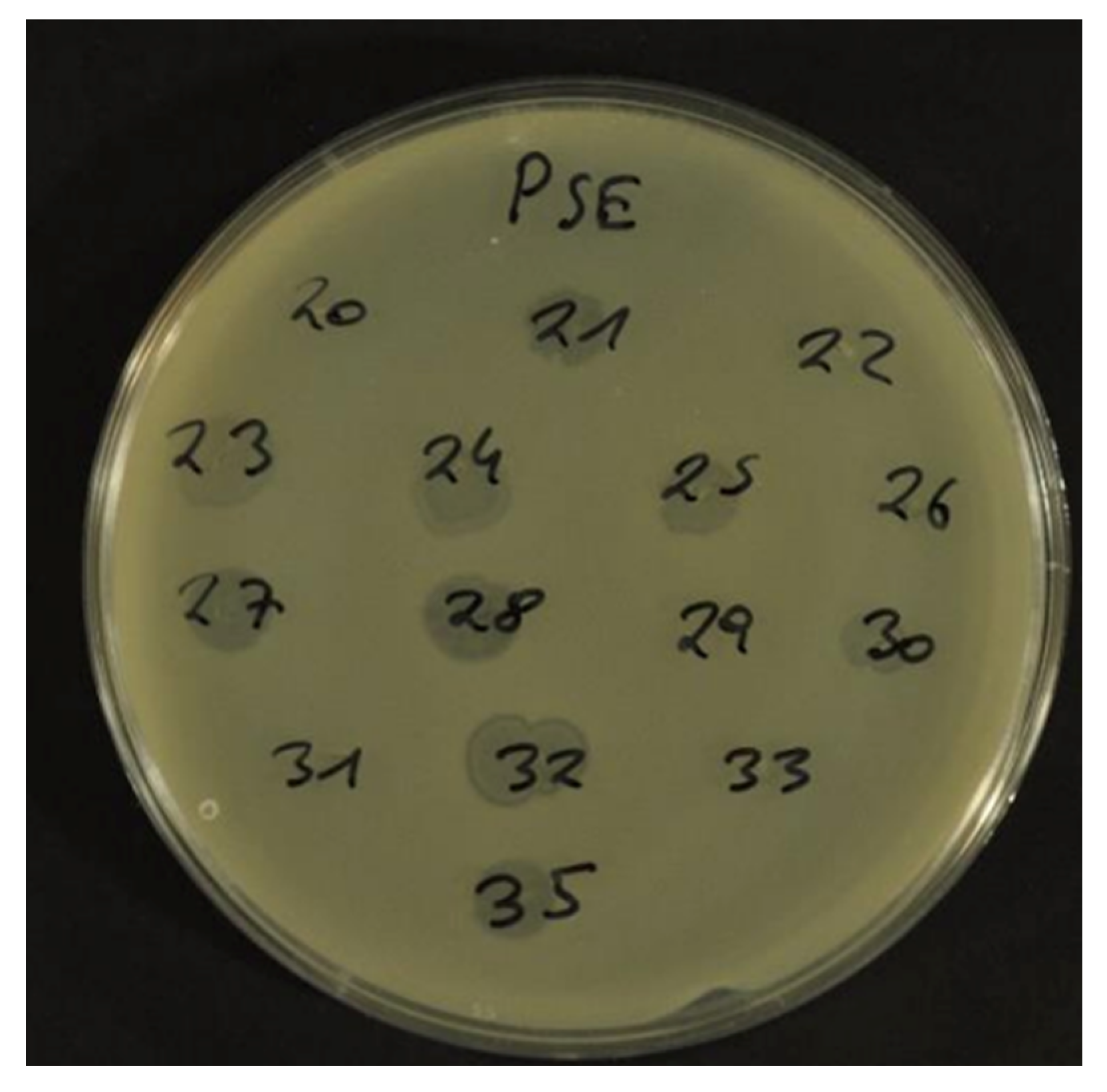
In order to test the lysing ability of all the different propagated prophages, a number of different Salmonella serovars were tested by spot lyses (as shown in Figure 2).
Most prophage lysates of the serovar Typhimurium were able to infect food isolates of the serovar Typhimurium, serovar Enteritidis, serovar Heidelberg, serovar Blockley, serovar Hadar, serovar Rissen, serovar Kentucky and serovar Parathyphi. Only two lysates could infect the serovars Blockley and Paratyphi, whereas the serovars Virchow and Heidelberg were most susceptible and could be infected by 39 and 36 of the total of 44 lysates, respectively (see Table 2). All the data on lysing ability can be found in Supplemental Data Table S1 from Supplementary.
In addition, 50 E. coli food isolates and 50 Yersinia enterocolitica food isolates were tested for susceptibility to the activated prophage lysates. None of these isolates could be infected by any of the 44 lysates extracted from S. serovar Typhimurium and serovar Enteritidis (see Supplemental Data Table S1).

3. Discussion

The S. serovars Typhimurium and Enteritidis are the major foodborne pathogens causing foodborne outbreaks worldwide. Depending on the serovar, this pathogen can be highly resistant to several antimicrobial agents. Therefore, alternative treatment options such as phage therapy are being discussed. Salmonella phage applications are already used in many countries in the food sector to avoid food contamination. Phage therapy in animal production is also reaching the stage of large-scale industrial trials [24,25,26,27]. However, these applications can be controversial, as phages drive bacterial evolution in Salmonella in the fastest and most effective way [28]. Some S. serovars such as Typhimurium are known to harbor functional and truncated prophages in their genome. Some of these prophages can confer phage resistance, while others can interact with phages infecting the cell and fuse into so-called hybrid phages with a different host specificity or lifestyle (virulent or lysogenic), and the transfer of antibiotic resistance genes is also possible [23].
To keep phage application and therapy effective, information about naturally occurring prophages in different serovars is as important as infectivity and host specificity. Here, we propagated prophage from the two most important clinical S. serovars, Typhimurium and Enteritidis, and tested their host specificity and their potential to transduce antimicrobial resistance genes.
The most potent activation of prophages in vitro is activation by mitomycin C, a cytostatic agent with potent mutagenic activity. While Salmonella isolates are unlikely to come into contact with mitomycin C in their lifecycle, we used oxidative stress from H2O2 to activate prophage induction, a stressor that Salmonella is exposed to in the animal and human host, e.g., against innate immune response, and externally by reactive oxygen species used in food production, such as nitrate, nitrite or in the environment by UV light [29]. Oxidative stress with H2O2 was almost as effective as mitomycin C in propagating prophages of the S. serovars Typhimurium and Enteritidis [30]. In our study, we also tested four isolates of the serovar Typhimurium and the serovar Enteritidis, each treated in parallel with H2O2 and mitomycin C, respectively. In two of the four serovar Typhimurium isolates, H2O2 was able to propagate prophages, whereas mitomycin C was only able to propagate prophages in one isolate. No difference was seen in the serovar Enteritidis isolates. Thus, there is a difference in the ability to propagate prophages with different methods, and genome analysis alone is not able to predict the propagation of complete prophages by various stressors.
Interestingly, a large difference was found between the serovars Typhimurium and Enteritidis in prophage propagation. In case of the serovar Typhimurium, 80% of the isolates could be propagated and only 8% of the isolates of the serovar Enteritidis. All four of the isolates from the serovar Enteritidis that could be propagated were food isolates, while the same proportion of human and food isolates of the serovar Typhimurium could be propagated. This is consistent with other studies looking at prophages in Salmonella [31,32]. These results confirm the high plasticity of the S. serovar Typhimurium genome.
We did not analyze the lysates for reinfection properties, as we did not know if the propagated lysates harbored one or more phages. It can be seen from published genome sequences that many sequenced Salmonella genomes hold more than one intact prophage and it is not possible to isolate different phages based on plaque morphology. Additionally, isolation of a single phage from a plaque plate is not always possible. Thus, reinfection studies would have been less meaningful for the definition of phage resistance or superinfection.
Antibiotic resistance genes detected in the activated prophage lysates of the S. serovar Typhimurium are also a sign of this plasticity. Gene coding for sulfonamide resistance or a gene fragment was detected most frequently in our study. The two resistance genes sul1 and sul2 were identified in two lysates. A combination of sulfonamide resistance genes with the streptomycin resistance gene strA-B was found in four lysates. The detection of streptomycin resistance genes in the lysates was always found in combination with another resistance gene or resistance gene fragment. The chloramphenicol resistance gene cmlA was detected in 10 lysates, while another chloramphenicol gene, cat, was not found in our lysates. A ß-lactam antibiotic resistance gene, the blaTEM, was identified in only one isolate. No resistance gene to tetracycline could be detected, which is a common resistance phenotype in various S. serovars [33]. This could indicate that horizontal transmission of the most common resistance traits, such as ß-lactam resistance and tetracycline resistance, occurs not through phages and transduction events, but through other mobile elements such as plasmids and transposons.
Most phages are very host specific, and broad host range phages are the exception. Regarding other genera, no propagated prophage lysate in our study was able to infect food isolates of E. coli or Y. enterocolitica. In contrast, most propagated prophage lysates were able to lyse a wide range of different S. serovars. No S. isolate of other serovars was tested in our study that could not be lysed by at least one extracted lysate, with the serovars Virchow and Heidelberg being the most susceptible to phage infection and the serovars Blockley and Paratyphi being most resistant. Therefore, phage exchange and their transduction events are very common in and between different serovars, and horizontal gene transfer by phage-induced transduction could be an important evolutionary tool for most serovars, with some being more resistant than others to phage transduction. Of the 44 propagated prophage lysates, none could infect E. coli or Y. enterocolitica two species of the Enterobacteriacae, even though E. coli shares many features with Salmonella enterica, and Yersinia phages have been reported as sharing homologies with Salmonella phage [34].

4. Materials and Methods

4.1. Bacterial Isolates

A total of 50 isolates from the S. enterica serovar Typhimurium and 50 from the serovar Enteritidis were randomly selected from the strain collection of the Institute of Food Safety, Division of Hygiene and Technology of the University of Veterinary Medicine, Vienna, Austria. Reference strains of the serovar Enteritidis DSM 17420 (German collection of Microorganisms and Cell Cultures, Braunschweig, Germany) and the serovar Typhimurium ATCC 14028 WT (American Type and Culture Collection, Manassas, VA, USA) were used. All the strains and isolates were streaked from stocks at −80 C. The isolates were all serotyped at the Austrian Reference Centre for Salmonellosis (Austrian Agency for Health and Food Safety, Graz, Austria) (see Supplementary Table S2 from Supplementary for all isolates used).

4.2. Prophage Induction

For phage induction, a protocol of Pacifico et al. [35] was used with the following modifications. Isolates were streaked on MH broth (CM0405, Oxoid, Wesel, Germany) and incubated for 24 h at 37 °C in aerobic conditions. One colony was used for overnight culture in liquid broth (LB-broth OXOICM0996B, VWR International, Radnor, TN, USA) under shaking (250 rpm/min) for 24 h at 37 °C. Next, 1 ml of stationary bacterial cells were pelleted at 10,000× g and diluted in 2 mL prophage induction buffer (MH-broth 10 mM CaCl2; 10 mM MgCl2; 2 mM H2O2), and then incubated for 1 h at 30 °C for phage activation. Lysozyme at a concentration of 200 µg/mL was added and incubated for 1 h at 30 C followed by a centrifugation step at 10,000× g for 5 min at room temperature. The supernatant was collected, filtered sterile (0.2 µm-pore filter), treated with DNAase (2U/µL) (95159-01K QuantaBio, Beverly, MA, USA) and used for the infection experiments.

4.3. Phage Infection

Phage infection was carried out according to Groisman [21]. The collected lysate for phage infection was used undiluted and in a 1:100 dilution. Overlay plates were incubated at 30 C for 24 h. All the overlay plates were screened for plaque forming by eye and with reflected light microscopy (100–1000 × ). Plaques were counted on plates containing phage forming units. A total lysate was prepared from these plates, as described in the reference above, using an additional sterile filtering step, DNAse (2U/µL) and RNase (20 mg/mL) (732-3152, VWR International, Radnor, TN, USA) treatment to digest free DNA and RNA.

4.4. Analyzing Phage DNA for Antibiotic Resistance Genes

These total lysates were used for DNA preparation using a QIAamp DNA mini kit (Qiagen, Venlo, The Netherlands) and this DNA was used for PCR of resistance genes sul1, sul2, tetA, tetB, tetC, blaOXA, blaTEM, blaCMY, strA, aadA1, dfr1, dfr2, cmlA and cat, using the primers and methods described in Sacher-Pirklbauer et al. [36]. All 44 lysates from the propagated prophage out of a total of 100 Salmonella isolates were analyzed.

4.5. Transduction of Antibiotic Resistance

Transduction assays were performed with total lysates positive for any resistance gene, using the method of Shousha et al. [37]. In brief, titration of the bacteriophage was carried out for each phage lysate in tenfold serial dilutions (10−1 to 10−6) by using plaque counting on overlay plates and expressing phage particles in phage forming units (PFU) per ml. For transduction, a 100 µL overnight culture of either the S. serovar Typhimurium ATCC 14028 WT or the S. serovar Enteritidis DSM 17420 was used, mixed with 10 µL appropriate phage dilution (102–105) and incubated at 37 °C for 30 min. After adding 2 mL Scholtens’s broth, the mixture was incubated at 37 °C for 2 h shaking. After centrifugation the pellet was diluted in 150 µL modified Scholtens’ broth and plated on MH-plates containing either ampicillin at 15 µg/mL, sulfonamids at 25 µg/mL, streptomycin at 15 µg/mL and chloramphenicol at 30 µg/mL. The plates were incubated at 37 C for 48 h and screened for colonies.

4.6. Defining Host Range of Induced Prophage Lysates

The host range of total lysates was screened using a spot lysis assay. In brief, bacterial strains to be tested for lyses were plated on MH-agar and a drop of 5 µL of the undiluted lysate was pipetted onto the agar plate (up to 15 drops per plate). Lysis was determined after appropriate incubation time and temperature (Table 1). In total, 48 S. enterica food isolates of different serovars, 50 E. coli food isolates and 50 Yersinia enterocolitica food isolates were screened for phage sensitivity.

5. Conclusions

Certain Salmonella serovars are highly resistant to antibiotics with so-called multidrug resistance phenotypes. Therefore, phage therapy could be an effective alternative to antibiotic therapy when invasive clinical signs of disease are prevalent. However, there are few reports of human phage therapy for salmonellosis, but commercialization is close in animal therapy and in the food sector. Nonetheless, reports of the possibility of virulence, toxin and antibiotic resistance gene transduction, and phage evolution (hybrid phage) must be considered before the use of phage application is widely adopted. Here we show that 80% of food and human isolates of the Salmonella enterica serovar Typhimurium contain intact prophage that could be released by oxidative stress, and that these lysates were able to infect a range of different Salmonella serovars. Although no broad host range lysate has been isolated that infects other Enterobacteriaceae, some of these lysates contain antibiotic resistance genes or gene fragments detected by PCR. The difference between the serovars Typhimurium and Enteritidis in the propagation of prophages and the detection of resistance genes is remarkable.

Supplementary Materials

The following are available online at https://www.mdpi.com/article/10.3390/antibiotics12030595/s1: Table S1 Lysing property of propagates prophages; Table S2 listing of isolates and their propagation.

Author Contributions

Conceptualization, F.H.; methodology, F.H., L.T., E.S., and J.K.; formal analysis, F.H.; investigation, F.H., L.T., E.S., and J.K.; resources, F.H.; data curation, F.H., L.T., E.S., and J.K.; writing—original draft preparation, F.H., L.T., E.S., and J.K.; writing—review and editing, F.H., L.T., E.S., and J.K.; project administration, F.H.; All authors have read and agreed to the published version of the manuscript.

Funding

This research received no external funding.

Institutional Review Board Statement

Not applicable.

Informed Consent Statement

Not applicable.

Data Availability Statement

All data have been described and made available by tables, figures and supplementary material.

Acknowledgments

Open Access Funding by the University of Veterinary Medicine Vienna. We thank Sofka D. for technical support.

Conflicts of Interest

The authors declare no conflict of interest.

References

  1. EFSA and ECDC (European Food Safety Authority and European Centre for Disease Prevention and Control). The European Union One Health 2021 Zoonoses Report. EFSA J. 2022, 20, 7666. [Google Scholar] [CrossRef]
  2. Chiu, C.-H.; Su, L.-H.; Chu, C.-H.; Wang, M.-H.; Yeh, C.-M.; Weill, F.-X.; Chu, C. Detection of Multidrug-Resistant Salmonella enterica Serovar Typhimurium Phage Types DT102, DT104, and U302 by Multiplex PCR. J. Clin. Microbiol. 2006, 44, 2354–2358. [Google Scholar] [CrossRef] [PubMed] [Green Version]
  3. El-Sharkawy, H.; Tahoun, A.; El-Gohary, A.E.-G.A.; El-Abasy, M.; El-Khayat, F.; Gillespie, T.; Kitade, Y.; Hafez, H.M.; Neubauer, H.; El-Adawy, H. Epidemiological, molecular characterization and antibiotic resistance of Salmonella enterica serovars isolated from chicken farms in Egypt. Gut Pathog. 2017, 9, 8. [Google Scholar] [CrossRef] [PubMed] [Green Version]
  4. Gebreyes, W.A.; Altier, C. Molecular Characterization of Multidrug-Resistant Salmonella enterica subsp. enterica Serovar Typhimurium Isolates from Swine. J. Clin. Microbiol. 2002, 40, 2813–2822. [Google Scholar] [CrossRef] [PubMed] [Green Version]
  5. Ghoddusi, A.; Nayeri Fasaei, B.; Zahraei Salehi, T.; Akbarein, H. Prevalence and characterization of multidrug resistance and variant Salmonella genomic island 1 in Salmonella isolates from cattle, poultry and humans in Iran. Zoonoses Public Health 2019, 66, 587–596. [Google Scholar] [CrossRef] [PubMed]
  6. Bacci, C.; Boni, E.; Alpigiani, I.; Lanzoni, E.; Bonardi, S.; Brindani, F. Phenotypic and genotypic features of antibiotic resistance in Salmonella enterica isolated from chicken meat and chicken and quail carcasses. Int. J. Food Microbiol. 2012, 160, 16–23. [Google Scholar] [CrossRef]
  7. Papadopoulou, C.; Davies, R.H.; Carrique-Mas, J.J.; Evans, S.J. Salmonella serovars and their antimicrobial resistance in British turkey flocks in 1995 to 2006. Avian Pathol. 2009, 38, 349–357. [Google Scholar] [CrossRef] [Green Version]
  8. Threlfall, E.J.; Ward, L.R.; Skinner, J.A.; Graham, A. Antimicrobial drug resistance in non-typhoidal salmonellas from humans in England and Wales in 1999: Decrease in multiple resistance in Salmonella enterica serotypes Typhimurium, Virchow, and Hadar. Microb. Drug Resist. 2000, 6, 319–325. [Google Scholar] [CrossRef]
  9. EFSA (European Food Safety Authority) and ECDC (European Centre for Disease Prevention and Control). The European Union Summary Report on Antimicrobial Resistance in zoonotic and indicator bacteria from humans, animals and food in 2019–2020. EFSA J. 2022, 20, 7209. [Google Scholar] [CrossRef]
  10. LaRock, D.; Chaudhary, A.; Miller, S. Salmonellae interactions with host process. Nat. Rev. Microbiol. 2015, 13, 191–205. [Google Scholar] [CrossRef]
  11. Aliaga, L.; Mediavilla, J.D.; López de la Osa, A.; López-Gómez, M.; de Cueto, M.; Miranda, C. Nontyphoidal salmonella intracranial infections in HIV-infected patients. Clin. Infect Dis. 1997, 25, 1118–1120. [Google Scholar] [CrossRef] [PubMed]
  12. Lee, W.S.; Puthucheary, S.D.; Parasakthi, N. Extra-intestinal non-typhoidal Salmonella infections in children. Ann. Trop. Paediatr. 2000, 20, 125–129. [Google Scholar] [CrossRef]
  13. Frost, J.A.; Kelleher, A.; Rowe, B. Increasing ciprofloxacin resistance in salmonellas in England and Wales 1991–1994. J. Antimicrob. Chemother. 1996, 37, 85–91. [Google Scholar] [CrossRef] [PubMed] [Green Version]
  14. Mon, K.K.Z.; Si, Z.; Chan-Park, M.B.; Kenney, L.J. Polyimidazolium Protects against an Invasive Clinical Isolate of Salmonella Typhimurium. Antimicrob. Agents Chemother. 2022, 66, e0059722. [Google Scholar] [CrossRef] [PubMed]
  15. Cao, Y.; Ma, R.; Li, Z.; Mao, X.; Li, Y.; Wu, Y.; Wang, L.; Han, K.; Li, L.; Ma, D.; et al. Broad-Spectrum Salmonella Phages PSE-D1 and PST-H1 Controls Salmonella in Foods. Viruses 2022, 14, 2647. [Google Scholar] [CrossRef]
  16. Lavilla, M.; Domingo-Calap, P.; Sevilla-Navarro, S.; Lasagabaster, A. Natural Killers: Opportunities and Challenges for the Use of Bacteriophages in Microbial Food Safety from the One Health Perspective. Foods 2023, 12, 552. [Google Scholar] [CrossRef]
  17. Lorenzo-Rebenaque, L.; Malik, D.J.; Catalá-Gregori, P.; Marin, C.; Sevilla-Navarro, S. Gastrointestinal Dynamics of Non-Encapsulated and Microencapsulated Salmonella Bacteriophages in Broiler Production. Animals 2022, 12, 144. [Google Scholar] [CrossRef]
  18. Kutateladze, M.; Adamia, R. Phage therapy experience at the Eliava Institute. Med. Mal. Infect. 2008, 38, 426–430. [Google Scholar] [CrossRef]
  19. Humphrey, S.; Fillol-Salom, A.; Quiles-Puchalt, N.; Ibarra-Chávez, R.; Haag, A.F.; Chen, J.; Penadés, J.R. Bacterial chromosomal mobility via lateral transduction exceeds that of classical mobile genetic elements. Nat. Commun. 2021, 12, 6509. [Google Scholar] [CrossRef]
  20. Tsao, Y.F.; Taylor, V.L.; Kala, S.; Bondy-Denomy, J.; Khan, A.N.; Bona, D.; Cattoir, V.; Lory, S.; Davidson, A.R.; Maxwell, K.L. Phage Morons Play an Important Role in Pseudomonas aeruginosa Phenotypes. J. Bacteriol. 2018, 200, e00189-18. [Google Scholar] [CrossRef] [Green Version]
  21. Groisman, E.A. In vivo Genetic Engineering with Bacteriophage Mu. Methods Enzymol. 1991, 204, 180–212. [Google Scholar] [CrossRef] [PubMed]
  22. Li, P.; Zhang, X.; Xie, X.; Tu, Z.; Gu, J.; Zhang, A. Characterization and whole-genome sequencing of broad-host-range Salmonella-specific bacteriophages for bio-control. Microb. Pathog. 2020, 143, 104119. [Google Scholar] [CrossRef] [PubMed]
  23. Blanco-Picazo, P.; Morales-Cortes, S.; Ramos-Barbero, M.D.; García-Aljaro, C.; Rodríguez-Rubio, L.; Muniesa, M. Dominance of phage particles carrying antibiotic resistance genes in the viromes of retail food sources. ISME J. 2023, 17, 195–203. [Google Scholar] [CrossRef] [PubMed]
  24. Akhtar, M.; Viazis, S.; Diez-Gonzalez, F. Isolation, identification and characterization of lytic, wide host range bacteriophages from waste effluents against Salmonella enterica serovars. Food Control 2014, 38, 67–74. [Google Scholar] [CrossRef]
  25. Pelyuntha, W.; Sanguankiat, A.; Kovitvadhi, A.; Vongkamjan, K. Broad lytic spectrum of novel Salmonella phages on ciprofloxacin-resistant Salmonella contaminated in the broiler production chain. Vet. World 2022, 15, 2039–2045. [Google Scholar] [CrossRef]
  26. Thanki, A.M.; Clavijo, V.; Healy, K.; Wilkinson, R.C.; Sicheritz-Pontén, T.; Millard, A.D.; Clokie, M.R.J. Development of a Phage Cocktail to Target Salmonella Strains Associated with Swine. Pharmaceuticals 2022, 15, 58. [Google Scholar] [CrossRef]
  27. Azari, R.; Yousefi, M.H.; Taghipour, Z.; Wagemans, J.; Lavigne, R.; Hosseinzadeh, S.; Mazloomi, S.M.; Vallino, M.; Khalatbari-Limaki, S.; Berizi, E. Application of the lytic bacteriophage Rostam to control Salmonella enteritidis in eggs. Int. J. Food Microbiol. 2023, 389, 110097. [Google Scholar] [CrossRef]
  28. Fillol-Salom, A.; Bacigalupe, R.; Humphrey, S.; Chiang, Y.N.; Chen, J.; Penadés, J.R. Lateral transduction is inherent to the life cycle of the archetypical Salmonella phage P22. Nat. Commun. 2021, 12, 6510. [Google Scholar] [CrossRef]
  29. Barbieri, G.; Bergamaschi, M.; Barbieri, G.; Franceschini, M. Kinetics of nitrite evaluated in a meat product. Meat Sci. 2013, 93, 282–286. [Google Scholar] [CrossRef]
  30. Filipiak, M.; Łoś, J.M.; Łoś, M. Efficiency of induction of Shiga-toxin lambdoid prophages in Escherichia coli due to oxidative and antibiotic stress depends on the combination of prophage and the bacterial strain. J. Appl. Genet. 2020, 61, 131–140. [Google Scholar] [CrossRef] [Green Version]
  31. Fong, K.; Lu, Y.T.; Brenner, T.; Falardeau, J.; Wang, S. Prophage Diversity Across Salmonella and Verotoxin-Producing Escherichia coli in Agricultural Niches of British Columbia, Canada. Front. Microbiol. 2022, 13, 853703. [Google Scholar] [CrossRef] [PubMed]
  32. Hiley, L.; Fang, N.X.; Micalizzi, G.R.; Bates, J. Distribution of Gifsy-3 and of variants of ST64B and Gifsy-1 prophages amongst Salmonella enterica Serovar Typhimurium isolates: Evidence that combinations of prophages promote clonality. PLoS ONE 2014, 9, e86203. [Google Scholar] [CrossRef] [PubMed]
  33. Mayrhofer, S.; Paulsen, P.; Smulders, F.J.; Hilbert, F. Antimicrobial resistance profile of five major food-borne pathogens isolated from beef, pork and poultry. Int. J. Food Microbiol. 2004, 97, 23–29. [Google Scholar] [CrossRef] [PubMed]
  34. Salem, M.; Pajunen, M.I.; Jun, J.W.; Skurnik, M. T4-like Bacteriophages Isolated from Pig Stools Infect Yersinia pseudotuberculosis and Yersinia pestis Using LPS and OmpF as Receptors. Viruses 2021, 13, 296. [Google Scholar] [CrossRef]
  35. Pacífico, C.; Hilbert, M.; Sofka, D.; Dinhopl, N.; Pap, I.J.; Aspöck, C.; Hilbert, F. Characterization of Bacteria and Inducible Phages in an Intensive Care Unit. J. Clin. Med. 2019, 8, 1433. [Google Scholar] [CrossRef] [PubMed] [Green Version]
  36. Sacher-Pirklbauer, A.; Klein-Jöbstl, D.; Sofka, D.; Blanc-Potard, A.B.; Hilbert, F. Phylogenetic Groups and Antimicrobial Resistance Genes in Escherichia coli from Different Meat Species. Antibiotics 2021, 10, 1543. [Google Scholar] [CrossRef] [PubMed]
  37. Shousha, A.; Awaiwanont, N.; Sofka, D.; Smulders, F.J.; Paulsen, P.; Szostak, M.P.; Humphrey, T.; Hilbert, F. Bacteriophages Isolated from Chicken Meat and the Horizontal Transfer of Antimicrobial Resistance Genes. Appl. Environ. Microbiol. 2015, 81, 4600–4606. [Google Scholar] [CrossRef] [Green Version]
Figure 1. Lysate of propagated prophage of (a) PSTy27 a S. enterica serovar Typhimurium isolate on host strain ATCC 14028 WT and (b) PSEn7 a S. enterica serovar Enteritidis on host strain DSM 17420 (pictures were taken by the authors).
Antibiotics 12 00595 g001
Figure 2. Spot lysis assay on DSM 17420 strain (picture was taken by the authors).
Antibiotics 12 00595 g002
Table 1. PCR detection of resistance genes in phage DNA isolated from propagated lysates of S. serovar Typhimurium.
LysateblaTEMcmlAstrAsul1sul2
PS1 +
PS2 +
PS3 +
PS4 +
PS9 +
PS14 +
PS17 +
PS19 +
PS21 +++
PS25 +
PS26 +
PS27 +++
PS28 + +
PS33 + +
PS45 +
PS46 +
PS47 +
PSL18B + +
PSL36B +
PSLs157+ +
Table 2. Lysis of different S. serovars by the propagated prophage lysates.
Serovar/StrainNumber of Lysates Serovar TyphimuriumNumber of Lysates from Serovar
Enteritidis
ATCC 14028400
DSM 17420284
Blockley30
Heidelberg342
Parathyphi B51
Typhimurium00
Virchow363
Hadar20
Rissen20
Kentucky20
B-Group60
Disclaimer/Publisher’s Note: The statements, opinions and data contained in all publications are solely those of the individual author(s) and contributor(s) and not of MDPI and/or the editor(s). MDPI and/or the editor(s) disclaim responsibility for any injury to people or property resulting from any ideas, methods, instructions or products referred to in the content.