Article

Evaluation of Commercial Anti-Listerial Products for Improvement of Food Safety in Ready-to-Eat Meat and Dairy Products

Postharvest Biology and Technology Unit, Department of Food Technology, Engineering and Science, University of Lleida, AGROTECNIO-CERCA Center, Av. Rovira Roure 191, 25198 Lleida, Spain
*
Author to whom correspondence should be addressed.
Antibiotics 2023, 12(2), 414; https://doi.org/10.3390/antibiotics12020414
Submission received: 12 January 2023 / Revised: 10 February 2023 / Accepted: 17 February 2023 / Published: 20 February 2023
(This article belongs to the Special Issue Detection and Control of Pathogens in Food Production)

Abstract

:
In ready-to-eat products, such as cooked ham, fresh cheese, and fuet in which Listeria monocytogenes is a concern, the use of biopreservation techniques represents an additional hurdle to inhibit pathogen growth during storage. The objective of this study was to apply several biopreservation techniques in three different food matrices to reduce the growth of Listeria innocua, used as a surrogate of L. monocytogenes. Several lactic acid bacteria, the bacteriocin nisin, the bacteriophage PhageGuard ListexTM P100, and the enzyme lysozyme were evaluated. Cooked ham treated with the bacteriophage PhageGuard ListexTM at 0.5% or with the lactic acid bacteria SafePro® B-SF-43 (25 g/100 kg) reduced L. innocua population to below the detection limit after 7 days of storage (4 °C plus modified atmosphere packaging). In fresh cheese, the application of PhageGuard ListexTM at 0.2 and 0.5% reduced L. innocua counts by more than 3.4 logarithmic units after 6 days at 4 °C. In fuet, the 1.0% of PhageGuard ListexTM reduced L. innocua population by 0.7 ± 0.2 logarithmic units in front of control with no significant differences to other evaluated biopreservative agents. The present results confirm that the application of biopreservation techniques was able to inhibit L. innocua in fuet, cooked ham, and fresh cheese, and suggest that the type of food matrix and its physicochemical characteristics influence the biopreservative efficacy.

1. Introduction

Listeria monocytogenes is a ubiquitous microorganism that can be found widely in the environment, but also in food processing plants where it can be protected from cleaning and disinfection operations due to its ability to produce biofilms. Therefore, L. monocytogenes can contaminate food at any point in its processing, from raw material production to household food handling. This microorganism causes invasive listeriosis which affects high-risk groups in the population: elderly people, newborns, immunocompromised people, and pregnant women.
Since L. monocytogenes is a quite resistant bacterium, able to grow at refrigeration temperatures (minimal growth temperature between −1.5 and 3.0 °C), at low pH and low water activity, ready-to-eat (RTE) foods with long shelf life are considered risky food products [1]. The main categories of RTE foods implicated in human listeriosis are ‘meat and meat products’, ‘fish and fish products’, and ‘milk and milk products’ [2]. Although the presence of L. monocytogenes in RTE foods is legislated in the European Union (EU) since 2006 as a food safety criterion (Commission Regulation (EC) 2073/2005), in the period from 2009–2013 a statistically significant increasing trend was observed in the EU [2]; meanwhile, the trend in the period from 2017–2021 was stable (neither increase nor decrease in the number of cases was reported) [3]. In 2021, listeriosis was the fifth most commonly reported zoonosis in humans in the EU [3]. It caused 2183 confirmed cases of invasive listeriosis with 923 hospitalisations and 196 deaths [3].
Data regarding the incidence of invasive listeriosis in the EU and around the world suggest that efforts carried out by food business operators, such as the application of the Hazard Analysis and Critical Control Points (HACCP) programme, good agricultural and farming practices, and good hygiene practices are not enough to avoid L. monocytogenes risk in RTE foods. Therefore, alternative approaches are necessary to reduce contamination of food and to prevent the growth of L. monocytogenes in RTE products. However, in the last years, consumer demands have changed to less processed, more natural foods without chemical additives [4]. Biopreservation and the use of natural enzymes could be attractive options to control L. monocytogenes while obtaining clean-label products [5]. Biopreservation is defined as the use of microorganisms and/or their metabolic products to extend the shelf life and enhance the safety of foods [6]. Among the protective cultures that can be used as biopreservative techniques, lactic acid bacteria (LAB) are widely applied. LAB have been involved in food preservation for years due to their capacity to inhibit spoilage and pathogenic bacteria [7] since they produce a wide range of antimicrobial metabolites during growth and food fermentation, such as hydrogen peroxide, lactic acid, acetic acid, low molecular weight substances, antifungal substances, and bacteriocins [8]. In addition, most of them are classified as GRAS (Generally Recognized as Safe) by the FDA (Food and Drug Administration) and as QPS (Qualified Presumption of Safety) by EFSA (European Food Safety Authority). Thus, several LAB cultures are commercialized for food preservation. In addition, some LAB can be considered probiotic bacteria as they have beneficial properties that can improve human and animal health. In this sense, some authors have demonstrated the protective effect of the probiotic bacteria Lactobacillus rhamnosus GG against L. monocytogenes in fresh-cut fruits [9,10,11]. Among the antimicrobial substances LAB produce, bacteriocins have great potential for use in food processing. They are antimicrobial peptides with bacteriostatic or bactericidal effect against pathogen and spoilage microorganisms [8]. Bacteriocins are considered safe for humans since gastric and pancreatic enzymes present in the stomach during food digestion degrade them into non-toxic substances [12]. Currently, nisin is the only bacteriocin approved by EFSA for its use as a food additive in the EU and accepted by the World Health Organization as a food biopreservative [13,14]. Another strategy to ensure food safety within biopreservation is the use of bacteriophages, which are viruses that infect specific bacteria. They are the most abundant microorganisms on the planet, and they can be found widely in nature. They are considered safe for humans due to their high host-specificity, which means they do not affect human microbiota or microorganisms involved in food production [15]. In 2006, EFSA published a positive scientific opinion about the use of the bacteriophage PhageGuard ListexTM P100 to control L. monocytogenes in some RTE foods (meat and poultry, fish and shellfish, and dairy products). However, EFSA highlighted that this bacteriophage should only be used as an additional tool to ensure microbial food safety used in combination with the application of good hygienic practices and good manufacturing practices, and not as a substitute for those practices [16]. Enzymes can also be used as natural preservatives in the food industry. One of the main enzymes used in products such as wine, cheese, sausage, and meat is lysozyme [17], an antimicrobial compound obtained from egg whites and bovine colostrum [18]. Lysozyme hydrolyses β-1,4-glycosidic bonds in the peptidoglycan of bacterial cell walls [19]; therefore, it is more effective against Gram-positive bacteria. In addition, it is not toxic to humans and does not change the physicochemical characteristics of food [17].
Cooked ham is an RTE meat product that can be contaminated with L. monocytogenes after thermal treatment; for example, during slicing and packing [20]. In addition, several authors have described the growth of L. monocytogenes in sliced and packed cooked ham [21,22]. Fresh cheese is a non-ripened cheese produced by enzymatic coagulation of cow milk with rennet. It is characterized by high moisture content and a high pH, and several authors have demonstrated that L. monocytogenes can grow in cheese [23,24]. In addition, a study detected L. monocytogenes in 15.4% and 8.9% of environmental samples from artisanal and industrial cheese producers, respectively [25]. Fuet is a low-acid dry-cured pork sausage from the Catalonia region (Spain) [26]. During its processing, the product undergoes fermentation that reduces its pH and water activity, increasing the stability of the product [27]. However, the presence and growth of L. monocytogenes in low-acidic fermented products has been reported [27,28,29].
In experimental studies conducted at pilot plant scale, L. monocytogenes cannot be used because of the risk of cross-contamination in the production of other foods. Therefore, a non-pathogen surrogate organism such as Listeria innocua must be used [30,31,32]. In addition, several authors have detected L. monocytogenes and L. innocua in the same samples from meat and dairy facilities [33,34,35]. Presence of L. innocua in food products is also a concern since it is a reservoir of resistance gens which may transfer between bacterial species. Gómez et al. [36] studied the resistance pattern of Listeria strains isolated from RTE meat products to various antibiotics. As a conclusion, they observed a multidrug resistance in 24 up to 71 Listeria isolates, with a higher prevalence of multidrug resistance in L. innocua specie (13.9%) than in L. monocytogenes specie (2.9%). In this scenario, control of both species, L. monocytogenes and L. innocua, could improve microbial food safety. This study aimed to assess the suitability of different commercial biopreservatives, including LAB, the bacteriocin nisin, the bacteriophage ListexTM P100, and the enzyme lysozyme to reduce the population of a Listeria innocua cocktail, used as an L. monocytogenes surrogate, in three different RTE foods (sliced cooked ham, fresh cheese, and fuet).

2. Results

2.1. Effect of Antimicrobials on Cooked Ham

A first challenge test was carried out on cooked ham plugs at worst-case conditions (13 °C) to select the most effective techniques. A remarkable L. innocua population increase was observed in the control (inoculated only with L. innocua) from the initial counts (2.2 ± 0.5 log cfu/g) to 9.0 ± 0.2 log cfu/g after 6 days of storage at 13 °C. At the end of storage (6 days), in four out of seven of the evaluated treatments L. innocua population increased considerably in cooked ham plugs (between 5.8 and 6.6 logarithmic units), close to the increase observed in the control treatment (6.8 ± 0.2 logarithmic units). In contrast, L. innocua population increased only by 1.3 ± 0.3, 2.2 ± 0.7, and 3.9 ± 0.8 logarithmic units in the treatments with L. rhamnosus GG, SafePro® B-SF-43, and PhageGuard ListexTM, respectively. Under the same storage conditions (13 °C), a second challenge test was carried out to elucidate if a reduced bacteriophage dose was also effective against L. innocua. The results showed that the three studied doses of PhageGuard ListexTM (1, 0.5, and 0.2%) had the same efficacy against L. innocua when applied to the surface of cooked ham plugs. Therefore, only the lower doses of bacteriophage were evaluated (0.2 and 0.5%) in the assay carried out at the recommended storage temperature (4 °C and air atmosphere).
During the evaluation of the selected biopreservative on cooked ham plugs stored under desirable refrigerated temperature (4 °C), only the application of the highest dose of PhageGuard ListexTM (0.5%) produced a significant reduction in L. innocua population after 3 days (Figure 1). In contrast, after 10 days of storage, all the treatments reduced L. innocua population and reached counts below 2.0 log cfu/g; meanwhile, in the control counts, it increased up to 3.4 ± 0.1 log cfu/g. The PhageGuard ListexTM at 0.5% reached the highest reduction (3.0 ± 0.2 logarithmic units) followed by the lowest bacteriophage dose (2.6 ± 0.6 logarithmic units), SafePro® B-SF-43 (1.7 ± 0.0 logarithmic reductions), and L. rhamnosus GG (1.4 ± 0.0 logarithmic units).
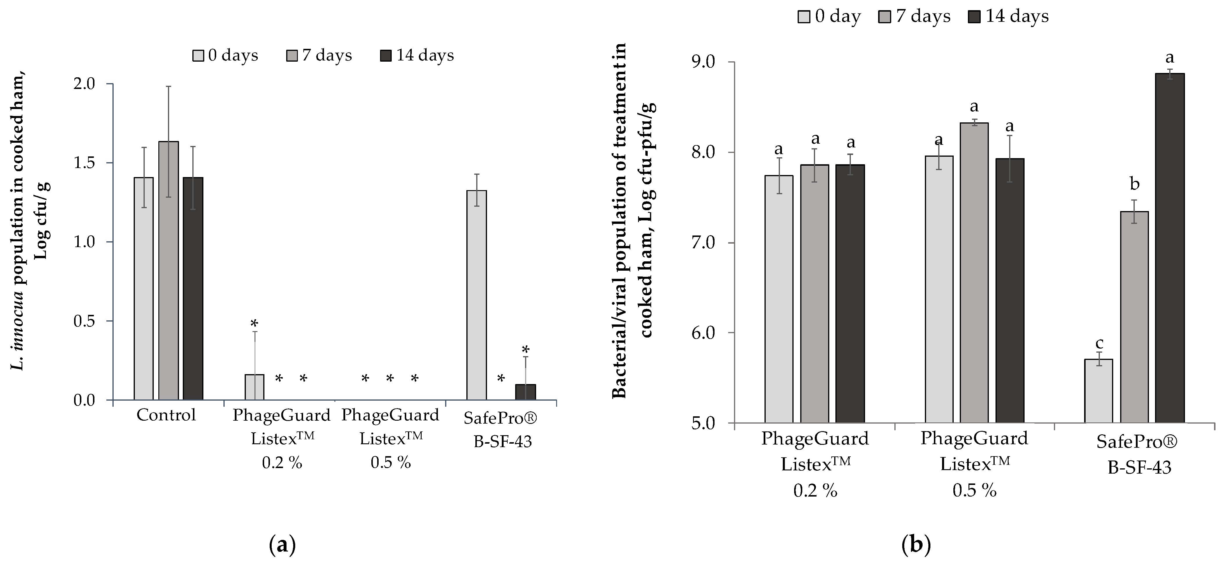
When a semi-commercial evaluation was carried out on cooked ham slices under modified atmosphere packaging (MAP) at 4 °C, an immediate reduction was observed on L. innocua counts in the PhageGuard ListexTM treatments after inoculation (Figure 2a). The decrease was higher for cooked ham treated with the highest bacteriophage concentration (0.5%, L. innocua population not detected after enrichment) and in the PhageGuard ListexTM 0.2% treatment, in which L. innocua decreased below the detection limit (<4 cfu/g). In contrast, L. innocua counts in SafePro® B-SF-43 (1.3 ± 0.1 log cfu/g) were not significantly different from the control. Nevertheless, seven days after treatment, SafePro® B-SF-43 reduced L. innocua population to no detected bacteria after enrichment as both PhageGuard ListexTM treatments. After 14 days, in the SafePro® B-SF-43 treatment only one of the three trays contained viable cell counts of L. innocua, which were below the detection limit (4 cfu/g); meanwhile, the PhageGuard ListexTM treatments (0.2 and 0.5%) reduced L. innocua to no detected bacteria after enrichment.
Regarding the evolution of the biopreservation techniques in treated cooked ham stored at 4 °C under MAP, the counts of Leuconostoc carnosus (LAB formulated in SafePro® B-SF-43) increased from 5.7 ± 0.1 log cfu/g to 7.3 ± 0.1 log cfu/g during the first 7 days of storage (Figure 2b) and up to 8.9 ± 0.1 log cfu/g at the end of storage, while bacteriophage levels did not increase during storage compared with initial bacteriophage counts (7.7 ± 0.2 and 8.0 ± 0.1 log pfu/g for 0.2% and 0.5% of PhageGuard ListexTM, respectively). Table 1 shows the O2 and CO2 gas concentration composition of trays measured before sampling. The CO2 concentration in trays treated with SafePro® B-SF-43 increased from 25.8 ± 3.4 to 36.6 ± 1.5% after 14 days of storage at 4 °C. Conversely, in the control (L. innocua only) and trays treated with PhageGuard ListexTM, the CO2 concentration decreased with the storage time up to 13.3 ± 0.4% with no significant difference between these treatments.

2.2. Effect of Antimicrobials in Fresh Cheese

When the biopreservative agents’ efficacy was evaluated in fresh cheese, antimicrobial agents were introduced directly into the milk used to elaborate the cheese. The effect of the biopreservative treatments applied during the elaboration of fresh cheese is shown in Figure 3. A remarkable L. innocua population increase was observed in the control treatment, from the initial population (3.0 ± 0.1 log cfu/g) to 5.3 ± 0.1 log cfu/g after 6 days of storage at 4 °C. Meanwhile, most of the evaluated lactic acid bacteria did not reduce L. innocua population, while L. rhamnosus GG obtained a slight reduction after 3 days (0.6 ± 0.2 logarithmic units) and 6 days (0.8 ± 0.1 logarithmic units) of storage in front of control. Conversely, all the evaluated doses of PhageGuard ListexTM reduced L. innocua population below the detection limit (<10 cfu/g) after inoculation. However, only 0.5 and 1% of PhageGuard ListexTM treatments prevented L. innocua regrowth after 6 days of storage.
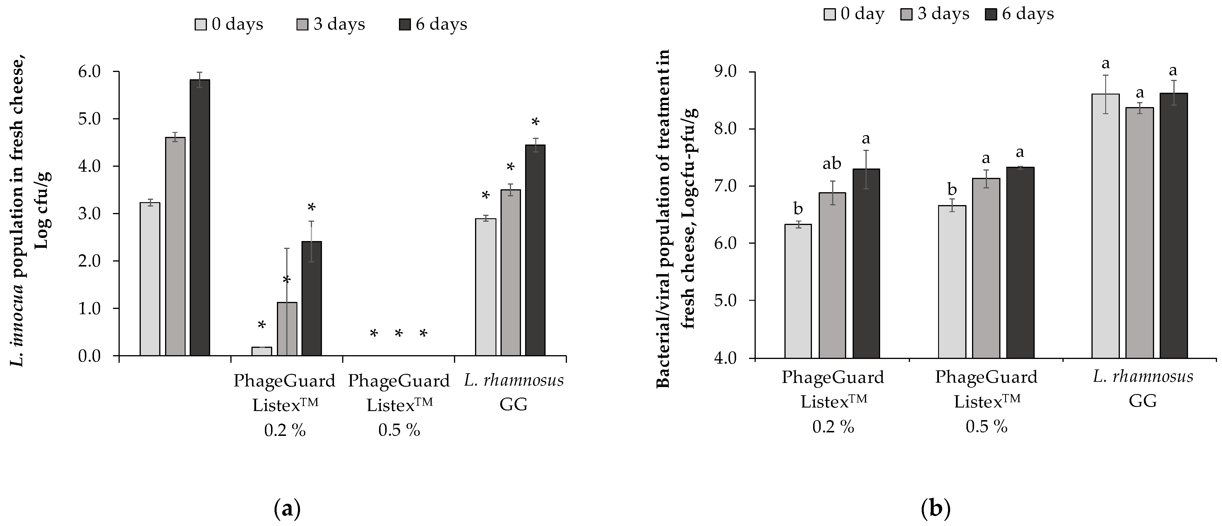
Under semi-commercial conditions, L. innocua population increased in the control from 3.2 ± 0.1 log cfu/g to 5.8 ± 0.2 log cfu/g after 6 days in cheese covered with liquid and stored at 4 °C (Figure 4a). After the obtention of the manufactured cheese, a biopreservative effect against L. innocua was observed in all the evaluated treatments with reductions higher than 3 logarithmic units in both PhageGuard ListexTM treatments (0.2 and 0.5%) and 0.3 ± 0.1 logarithmic unit reductions in L. rhamnosus GG treatment. The decrease of L. innocua population in the 0.5% PhageGuard ListexTM treatment was remarkable because it reduced the pathogen population below the detection limit (<10 cfu/g) and no L. innocua was detected after enrichment throughout storage. After 6 days of storage, L. rhamnosus GG treatment reduced L. innocua population up to 4.5 ± 0.1 log cfu/g (1.4 ± 0.1 logarithmic units in front of control) and 0.2% PhageGuard ListexTM obtained reductions of 3.4 ± 0.4 logarithmic units.
In this assay, the bacteriophage P100 concentrations in the obtained fresh cheeses were 6.3 ± 0.1 log pfu/g and 6.7 ± 0.1 log pfu/g in 0.2 and 0.5% bacteriophage treatment, respectively (Figure 4b). Regardless of the initial dose of bacteriophage applied in the milk (7.2 ± 0.1 log pfu/mL and 8.0 ± 0.0 log pfu/mL in treatment with PhageGuard ListexTM 0.2% and 0.5%, respectively), the bacteriophage increased up to 7.3 log pfu/g in fresh cheese. In contrast, the initial population of L. rhamnosus GG in the obtained fresh cheese was 8.6 ± 0.3 log cfu/g and levels remained stable throughout storage.
During the manufacturing of fresh cheese from the semi-commercial assay, the population of L. innocua and the biopreservative agents was also determined in the recovered whey after curd separation. The population of L. innocua in the recovered whey varied among treatments. Meanwhile, in the control and L. rhamnosus GG treatment, counts of 2.0 ± 0.0 and 2.2 ± 0.0 log cfu/mL of L. innocua were recovered in the whey; in both treatments with PhageGuard ListexTM the counts of L. innocua were below the detection limit (<10 cfu/mL). In addition, all used biopreservants were recovered in the whey with populations of 7.5 ± 0.1 log CFU/mL in L. rhamnosus GG treatment, 7.2 ± 0.0 log pfu/mL for 0.5% PhageGuard ListexTM, and 6.5 ± 0.1 log pfu/mL for 0.2% PhageGuard ListexTM.
The yield of the obtained cheeses with each biopreservative agent was determined. From 400 mL of inoculated milk, in the control treatment 94.75 g of fresh cheese was obtained, 97.97 g in the L. rhamnosus GG treatment, 91.99 g in the PhageGuard ListexTM 0.5% treatment, and 95.25 g in the PhageGuard ListexTM 0.2% treatment. The yield of the produced cheese ranged between 0.24 and 0.25 kg/L of milk.

2.3. Effect of Antimicrobials in Fuet

The efficacy of seven biopreservative agents to control L. innocua population in fuet after ripening and post-cold storage was evaluated. In the fuet control (meat inoculated only with L. innocua), the pathogen population increased from the initial concentration (4.5 ± 0.6 log cfu/g) to 5.3 ± 0.1 log cfu/g after fermentation (24 h at 22 °C), with an increase of 0.7 ± 0.1 logarithmic units. In contrast, during the ripening process (7 days at 15 °C and 75% R.H.), the population of L. innocua in the control treatment reduced by 0.9 ± 0.1 logarithmic units with a final count of 4.3 ± 0.1 log cfu/g (Figure 5). However, the population increased (0.5 ± 0.4 logarithmic units) during post-cold storage, reaching a final population of 4.8 ± 0.4 log cfu/g. In treated samples, SafePro® B-SF-43 obtained the highest population reduction (0.5 ± 0.3 logarithmic reductions) after the ripening step in front of the control. Lower L. innocua counts after cold storage were observed in the treatments SafePro® B-SF-43 (3.8 ± 0.3 log cfu/g), SafePro® B-LC-20 (3.9 ± 0.5 log cfu/g), and PhageGuard ListexTM (4.0 ± 0.3 log cfu/g). These three treatments reduced pathogen population around 1 logarithmic unit in front of control at the end of shelf life.
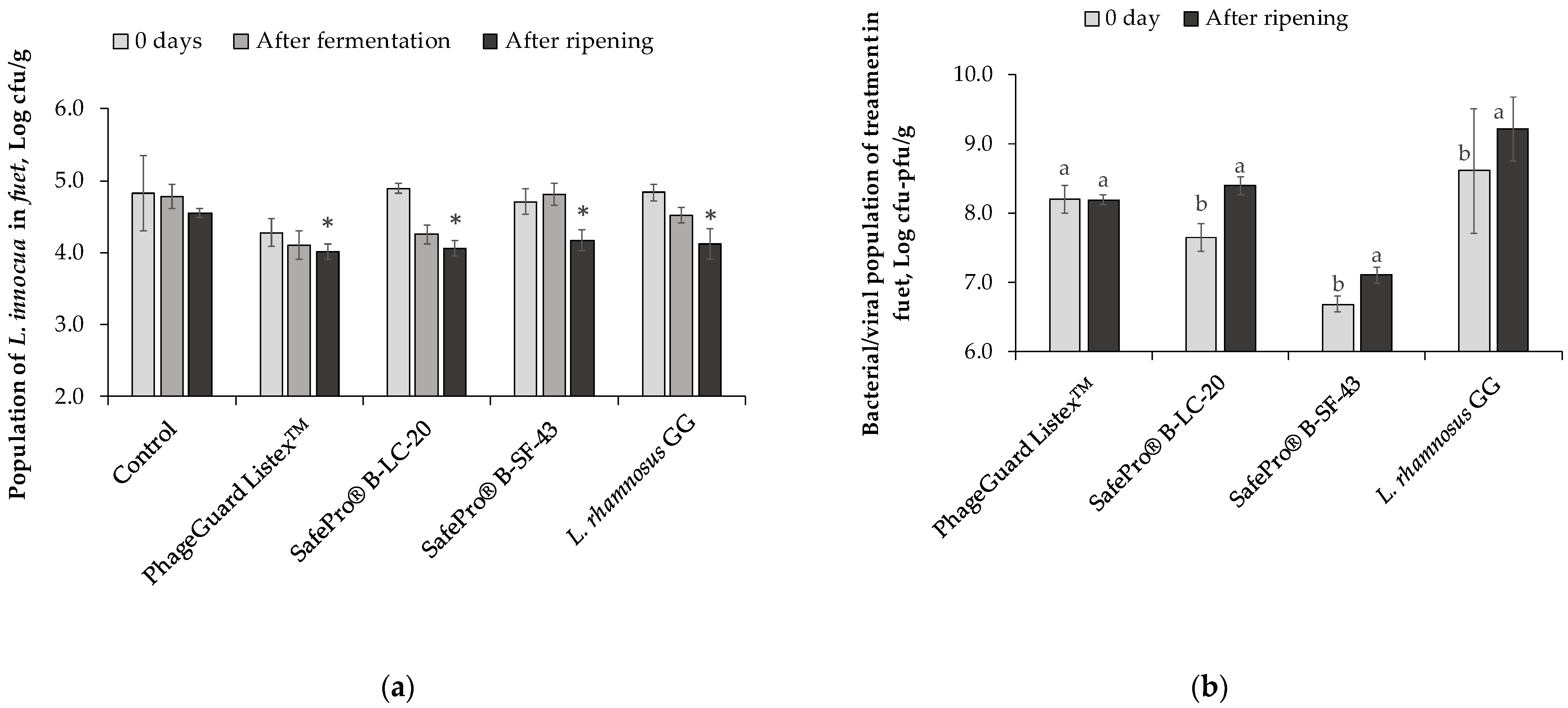
The biopreservative treatments that showed the best results were evaluated again against L. innocua in fuet (Figure 6). In this evaluation, the population of L. innocua was enumerated immediately after inoculation (before stuffing in pork casing), after 24 h of fermentation, and at the end of the ripening (Figure 6a). After inoculation, the population of L. innocua in the control was 4.8 ± 0.5 log cfu/g, and a slight reduction (0.5 ± 0.2 logarithmic unit) was reached in PhageGuard ListexTM treatment with an L. innocua population of 4.3 ± 0.2 log cfu/g. After 24 h of fermentation, no reduction was observed in treatment SafePro® B-SF-43; meanwhile, significant reductions were reached in the treatments PhageGuard ListexTM and SafePro® B-LC-20 with 0.7 ± 0.2 and 0.5 ± 0.1 logarithmic units, respectively, in front of the control. When the L. innocua population was enumerated at the end of ripening, all evaluated treatments reached reductions around 0.5 logarithmic units, with the lowest L. innocua population recovered in the treatment with 1% of PhageGuard ListexTM (4.0 ± 0.1 log cfu/g). The bacterial or viricidal population of the four evaluated techniques was determined after inoculation (before stuffing) and after ripening (Figure 6b). Whereas bacteriophage population remained stable at the initial inoculated level (8.2 ± 0.1 log pfu/g) until the end of ripening, all lactic acid bacteria populations increased during the ripening process. Pediococus acidilactici (formulated in SafePro® B-LC-20) increased 0.8 ± 0.2 logarithmic units to 8.4 ± 0.2 log cfu/g, Leuc. carnosus (formulated in SafePro® B-SF-43) increased 0.4 ± 0.2 logarithmic units to 7.1 ± 0.2 log cfu/g, and L. rhamnosus GG increased 0.6 ± 0.2 logarithmic units to 9.2 ± 0.2 log cfu/g at the end of fuet ripening.
Table 2 shows the evolution of pH during fuet production. In all treatments, pH decreased more than one unit after fermentation (from pH 6.33 ± 0.02, sausage mixture before stuffing). The pH reduction observed in the L. rhamnosus GG treatment after fermentation reached the lowest pH value of 5.06 ± 0.04.

3. Discussion

This study aimed to compare the efficacy of several biopreservative agents to control a four-strain L. innocua cocktail on sliced cooked ham (the evaluated treatments were PhageGuard ListexTM, SafePro® B-LC-20, SafePro® B-LC-48, SafePro® B-SF-43, L. rhamnosus GG, and NisinZ®), fresh cheese (the evaluated treatments were PhageGuard ListexTM, SafePro® B-LC-20, SafePro® B-LC-48, SafePro® B-SF-43, L. rhamnosus GG, NisinZ®, and Lysoch®L4), and fuet (the evaluated treatments were PhageGuard ListexTM, SafePro® B-LC-20, SafePro® B-LC-48, SafePro® B-SF-43, Fermitrat-Export®, Fermitrat-S3®, L. rhamnosus GG, and NisinZ®). The most appropriate time to apply the biopreservative agents was selected for each food matrix considering the physicochemical characteristics of the finished food, the microbiota of the raw material, and the manufacturing process steps. Therefore, cooked ham was surface treated after the slicing operation because it could be get contaminated if food manufacturing practices are not applied correctly in the operations that take place after pasteurization or thermal treatment, the last operation aimed to reduce microorganisms. In the case of fresh cheese, the biopreservative agents were added after milk treatment, since L. monocytogenes contamination in cheese could be due to survived bacteria to a weak thermal treatment or from cross-contamination after thermal treatment [25,37]. Finally, in fuet, the biopreservative agents were also added to the raw materials at the beginning of the production process because no step in the production process has the purpose of reducing microorganism concentration in the raw material. Nevertheless, pH reduction and the reduction in water activity occur in fuet during the ripening process could avoid L. monocytogenes multiplication. In addition, the introduction of biopreservative agents before a homogenization step allows the spreading of the agents uniformly in food.
In a previous study, Szczawinski et al. [38] demonstrated the ability of L. monocytogenes to grow on contaminated cooked ham at different temperatures (3, 6, 9, 12, and 15 °C), increasing the growth rate with the temperature increase. This study highlights L. innocua’s ability to grow on cooked ham plugs stored at 4 °C with an increase of 0.6 logarithmic units after 6 days of storage. However, the pathogen maintained constant population counts when the sliced cooked ham was packaged under modified atmosphere. Furthermore, the L. innocua population increased by 2.6 logarithmic units in the fresh cheese after 6 days of storage, although it was packaged in a covering liquid with 2% sodium chloride and stored at 4 °C. In queso fresco, which has similar characteristics to fresh cheese, Lourenço et al. [39] observed an L. monocytogenes population increase from 3.5 log cfu/g to 7.0 log cfu/g after 11 days of storage at 4 °C, although they inoculated L. monocytogenes in the curd instead of the milk. In contrast, the ripening step of the production process of the fuet caused a decrease of 0.3 logarithmic units of L. innocua after 7 days at 15 °C at 75% humidity. Previously, Porto-Fett et al. [40] validated that L. monocytogenes population reduced approximately by 3.6 log cfu/g during the preparation and storage of fuet.
To our knowledge, no previous research has compared the efficacy of different biopreservative agents in three food matrices of RTE products with such different characteristics. In this study, on sliced cooked ham, the most effective anti-listerial treatment was PhageGuard ListexTM followed by the SafePro® B-SF-43 agent. Furthermore, PhageGuard ListexTM showed the most effective anti-listerial activity against L. innocua in packaged fresh cheese followed by L. rhamnosus GG. In both matrices, the evaluated biopreservation techniques reduced L. innocua counts by more than 1.5 logarithmic units in front of the control. Conversely, the maximum reduction observed in the manufactured fuet was 0.5 logarithmic units with no significant difference among PhageGuard ListexTM, SafePro® B-LC-20, SafePro® B-SF-43, and L. rhamnosus GG.
The manufacturer’s recommendation for SafePro® B-SF-43 application was 25 g/100 kg of product, which corresponded to 5.7 ± 0.1 log/cfu of Leuc. carnosum per gram in our evaluated sliced cooked ham. Budde et al. [41] observed that the rate of inhibition of L. monocytogenes in a cooked ham depended on the initial concentration of Leuc. carnosum 4010 applied. They reported that when the initial concentration of the Leuc. carnosum was 6.3 × 10⁶ cfu/g, L. monocytogenes inhibition was observed after 7 days. Meanwhile, when a lower initial concentration (1.2 × 10⁵ cfu/g) was applied the inhibition effect was observed after 14 days. This may be explained by a higher initial lactic acid bacteria population that reached the exponential growth phase earlier. Nevertheless, regardless of the initial Leuc. carnosum concentration, at the end of storage (28 days at 5 °C) the cell counts of L. monocytogenes were reduced to below 10 cfu/g in both treatments. In this study, Leuc. carnosum reached the exponential growth phase after 7 days of storage at 4 °C in the sliced cooked ham. Bacteriocin production by LAB usually starts in this growth phase [42]. In our study, L. innocua inhibition was not observed immediately after the application of SafePro® B-SF-43 on the treated slices of cooked ham, and it was delayed to 7 days, when bacteriocins could have been produced, similarly to Budde et al.’s [41] results. The same authors also described the strong anti-listerial activity of Leuc. carnosum without producing undesirable flavour in the evaluated RTE meat foods. Although Leuc. carnosum established in the fuet, it multiplied slightly, around 0.4 logarithmic units after 7 days of ripening. Meanwhile, on the cooked ham, the bacteria increased fourfold compared to the fuet (1.6 logarithmic units). The adverse environment (low pH and aw, and microbial competition with indigenous fermentation strains) in the fuet could have caused the lower Leuc. carnosum multiplication and, consequently, lower bacteriocin production than in the cooked ham, in which higher anti-listerial activity was observed. The same anti-listerial rate activity was observed when the SafePro® B-LC-20 was applied in the contaminated mixed meat used to elaborate the fuet. Pediococcus acidilactici is the bacteria lyophilizate in SafePro® B-LC-20 formulation. Nieto-Lozano et al. [43] evaluated the inhibitory effect of P. acidilactici MCH14 against L. monocytogenes in Spanish dry-fermented sausage. This pediocin-producing strain reduced the L. monocytogenes counts by 2 logarithmic units compared to the control after 30 days of storage. They inoculated the minced meat at a P. acidilactici initial concentration of 5 × 10⁶ cfu/g. Although higher initial concentration was applied in our fuet (7.7 ± 0.1 log cfu/g), a lower anti-listerial activity was observed. This could be due to the temperature used in our experiment, since a previous study noticed that the activity of P. acidilactici was more effective at 4 °C than 15 °C in raw meat [44]. P. acidilactici is used as a starter culture for its capacity to reduce food pH; its efficacy as an antimicrobial against Listeria has also been confirmed. In addition, Komora et al. [45] and Barbosa et al. [46] concluded that P. acidilactici should be considered a potentially useful probiotic.
Dairy products have extensively been used as food vehicles for L. rhamnosus GG, which is the most introduced probiotic strain in food. Rubio et al. [47] confirmed its suitability as a starter culture in cured sausage, and in this study we have evaluated its suitability as an anti-listerial agent in three treated food matrices. The highest L. innocua reduction was obtained in the fresh cheese with 1.4 logarithmic units in front of control, as in the cooked ham plugs stored under air conditions a 4 °C, whilst in fuet a decrease of 0.5 logarithmic units was achieved at the end of the ripening step. Although the probiotic strain was more effective in the fresh cheese than in the fuet, L. rhamnosus GG maintained the population counts above 108 cfu/g until the end of storage in both matrices. Therefore, the obtained fresh cheese and fuet in this study could be commercialised as functional foods while L. rhamnosus GG improves food safety against L. monocytogenes.
Generally, the PhageGuard ListexTM was the most effective biopreservative agent among the evaluated techniques because it showed effective anti-listerial activity in the three evaluated food matrices. In addition, a large reduction of L. innocua population was observed in the fresh cheese and the cooked ham. Several authors have also observed anti-listerial activity when applying the bacteriophage P100 in these two food matrices [21,48,49]. Similarly to our results, Holck et al. [21] obtained a 1 logarithmic reduction of L. monocytogenes on cooked ham after application of the bacteriophage P100 (5 × 10⁷ pfu/cm2) and the surviving pathogen population remained constant until 7 days of storage at 4 °C. Furthermore, higher pathogen reduction after inoculation was observed when we increased the inoculation levels of PhageGuard ListexTM on the sliced cooked ham. The same behaviour tendency was observed in the fresh cheese. Carlton et al. [50] also noticed that the effect of bacteriophages varies with the type of product and is strongly dose dependent. This could be explained by the fact that a high concentration of bacteriophage per unit area is required to ensure interaction with the target pathogen on food surfaces [48]. The lowest initial dose of PhageGuard ListexTM allowed L. innocua regrowth in the fresh cheese throughout storage. Regarding bacteriophage multiplication ability, we observed that in the fresh cheese bacteriophage concentration increased with time. The self-replication ability of bacteriophage only occurs if there is still a host present in the food matrix, and the observed biopreservative population increase was in accordance with L. innocua survival after the treatment application of PhageGuard ListexTM (0.2%). Therefore, on cooked ham, the P100 population remained stable from inoculation to the end of the evaluation because no L. innocua survived after inoculation. Conversely to the lower reduction levels obtained in the fuet, Gutiérrez et al. [51] and Komora et al. [45] observed a reduction of 3 logarithmic units of L. monocytogenes counts respectively in a Spanish dry-cured ham and in a fermented meat sausage (Alheira). However, the addition of extra hurdles, such as high pressure, were necessary to reduce L. monocytogenes to below detection limits after 60 days of storage in the fermented sausage. Although an increase of the bacteriophage P100 population was noticed in the fresh cheese when L. innocua cells were present, in the fuet the bacteriophage population did not increase. Nevertheless, a large quantity of L. innocua was present in all evaluated sampling times in the fuet. This could be due to the food’s characteristics, such as low moisture and low pH, which did not allow bacteriophage multiplication. In addition, several authors described that in nonliquid food matrices the phage particles appear to become immobilized soon after addition due to limited interaction between the phage and the target bacteria [50,52]. For instance, when Listex® P100 was applied on fresh-cut melon and in melon juice, Oliveira et al. [53] observed the highest effectivity in the liquid matrix.
Although the results obtained in this study indicate the feasibility of applying biopreservation in the selected food matrices, it is important to highlight this study’s limitations. Firstly, due to the impossibility to work with the human pathogenic L. monocytogenes, L. innocua was used as a surrogate. However, previously, an in vitro assay was conducted to determine the sensitivity of L. innocua selected cocktail in comparison to a cocktail of five strains of L. monocytogenes against the different selected biopreservative agents. The results showed that both Listeria cocktails presented similar sensibility. In addition, when slight differences in sensibility were observed, L. monocytogenes cocktail was more sensitive than the evaluated L. innocua cocktail. For this reason, we determined that the selected L. innocua strains cocktail was adequate. Secondly, the objective of this study was to evaluate the effect of several biopreservative agents against L. innocua in three food matrices susceptible to be linked to listeriosis. Therefore, to quantify L. innocua population reduction, the initial levels of L. innocua in raw ingredients was above 100 cfu/g, which is a relatively high population. Thirdly, we used a small sample size in this study. However, the aim was to evaluate several options of commercial biopreservatives to obtain knowledge of which could be the best option to food producers. Regardless of the obtained results and the background information, before the application of a new biocontrol tool in a production system, its effectiveness must be evaluated in the specific product considering the food composition, the food physicochemical characteristic, such as pH and water availability, and the production process. Likewise, the evaluated sampling times were approximations of reality, and before biopreservative application in the food chain, longer storage times should be evaluated.

4. Material and Methods

4.1. Products

The biopreservatives’ efficacy was evaluated in three different food matrices: cooked ham, fresh cheese, and dry-cured Spanish pork sausage (fuet). A commercial cooked ham was used to evaluate the anti-listerial efficacy of the biopreservatives. The fuet and the fresh cheese were elaborated in the laboratory from the raw materials, pork minced meat and cow pasteurized milk, purchased from a supermarket. Both manufactured foods did not receive any thermal treatment.

4.2. Strains, Commercial Biopreservatives, and Stock Culture Preparation

A cocktail of four strains of L. innocua was used in this study as a surrogate of L. monocytogenes. These strains were: L. innocua CECT 910, L. innocua CECT 4030 and L. innocua CECT 8848 from Colección Española de Cultivos Tipo (CECT), and L. innocua TA-1.17 from the Department of Food Technology (UdL) collection. The stock cultures were streaked onto TSA (Tryptone Soy Agar; Biokar, Allone, France) supplemented with yeast extract (6 g/L, Biokar, France; Tryptone Yeast Extract Soy Agar, TYESA) and incubated at 37 °C for 24 h. For each strain, a single colony was picked from the plate and transferred to 10 mL of TSB (Tryptone Soy Broth; Biokar, France) supplemented with yeast extract (6 g/L, Tryptone Yeast Extract Soy Broth, TYESB) and incubated at 37 °C for 20–22 h. The four strains of L. innocua were mixed in equal amounts regarding cell numbers to obtain the same concentration of each strain (108 cfu/mL). The cocktail was centrifuged (Sorvall Legend XTR, Thermo Scientific, Waltham, MA, USA) for 10 min at 8900× g, and the pellet was resuspended in saline solution (8.5 g/L NaCl, Thermo Fisher, Markham, Canada). For food inoculation, a serial dilution of the L. innocua cocktail was prepared in saline peptone solution (SP, 8.5 g/L NaCl and 1 g/L peptone (Biokar, France)) to obtain the desired concentration.
The anti-listerial agents used were the bacteriophage PhageGuard ListexTM (Micreos, Wageningen, the Netherlands), the formulated cultures SafePro® B-LC-20, SafePro® B-LC-48, and SafePro® B-SF-43 (CHR-Hansen, Horsholm, Denmark), and Fermitrat-Export® and Fermitrat-S3® (Amerex, Colmenar Viejo, Spain), the probiotic strain L. rhamnosus GG (ATCC 53103), the bacteriocin NisinZ® (Handary, Evere, Belgium), and the enzyme Lysoch®L4 (Handary, Evere, Belgium). The evaluated biopreservative agents are currently commercialised products in the EU. According to legislation and manufacturers’ instructions, each biopreservative agent was evaluated in the food matrix in which it is recommended. To apply all biopreservation techniques using the same method, the lyophilised or dehydrated products were suspended in sterile water prior their application. When necessary, the biopreservative agent’s suspension was serially diluted to treat the samples with the same volume of each anti-listerial agent.

4.3. Food Preparation and Application of the Biopreservative Treatments

4.3.1. Cooked Ham

Before removing the packaging of the commercial cooked ham (500 g/unit), its surface was disinfected with ethanol (70%) to avoid cross-contamination. The cooked ham composition was pork (55%), water, potato starch, soy protein, sugar, corn dextrose, stabiliser (E-420 and E-407), preservatives (E-250), antioxidant (E-315), colouring (E-120), and smoked flavour.
Prior to the evaluation of the most suitable techniques on slices of cooked ham in a commercial scenario (4 °C and MAP), two experiments were performed in cooked ham portions under air conditions at different temperatures (13 °C and 4 °C). For these experiments, a slice of cooked ham was cut into plugs of 1.5 cm diameter and 0.5 cm thick (approximately 1 g/plug). Then, 10 µL of the L. innocua cocktail was spread over the plug surface to obtain a concentration of 100 cfu/g. After 15 min at room temperature, the plugs were distributed into seven treatments: (1) L. innocua control (no treatment), (2) SafePro® B-LC-20 (25 g/100 kg), (3) SafePro® B-LC-48 (12.5 g/100 kg), (4) SafePro® B-SF-43 (25 g/100 kg), (5) L. rhamnosus GG (108 cfu/g), (6) NisinZ® (2.5 g/100 kg), and (7) PhageGuard ListexTM (1%). Then, 10 µL of each treatment (previously suspended in water) was spread over the same area. In the L. innocua control treatment, 10 µL of sterile water was spread on the ham plugs. The treated inoculated ham plugs were placed in a sterile Petri dish and stored at 13 °C. L. innocua population was enumerated after 3 and 6 days of storage. After the initial results evaluation, the most effective techniques were evaluated again on cooked ham plugs stored at 4 °C for 10 days: (1) L. innocua control (no treatment), (2) PhageGuard ListexTM (0.2%), (3) PhageGuard ListexTM (0.5%), (4) SafePro® B-SF-43 (25 g/100 kg), and (5) L. rhamnosus GG (108 cfu/g). Samples were inoculated as described previously and L. innocua population was enumerated after 3, 6, and 10 days at 4 °C.
For commercial evaluation, 216 slices of cooked ham (6 cm diameter and 0.3 cm thick, average 15.0 ± 6.8 g/slice) were inoculated by spraying to obtain a population of 100 cfu/g of L. innocua. After 15 min, inoculated slices were distributed in four batches. Each batch was sprayed individually with the selected biopreservative agent (0.1 mL/slice) to obtain the treatments: (1) L. innocua control (no treatment), (2) PhageGuard ListexTM (0.2%), (3) PhageGuard ListexTM (0.5%), and (4) SafePro® B-SF-43 (25 g/100 kg). In the L. innocua control treatment, sterile water was sprayed onto inoculated slices of cooked ham. For all treatments, six slices (average 89.9 ± 6.8 g/tray) were placed in polypropylene trays (PP, Tecnofood TF013) and sealed with a non-peelable polypropylene film (P12-2050PXNP, Tecnopack, O2 permeability of 110 mL/m2·24 h·bar at 23 °C) under MAP using a mixture of 30%/70% (CO2/N2, ALIGAL13, Air Liquid) with a tray sealer machine (ILPRA, Basic, Spain). Nine trays with sliced cooked ham without L. innocua were prepared and sealed as packaging control. Sealed trays were stored at 4 °C for 14 days and L. innocua and biopreservative agents’ population were enumerated as described below (4.4.) after inoculation and 7 and 14 days of storage. The concentration of O2 and CO2 in the headspace atmosphere of the trays with MAP was analysed using a handheld gas analyser (CheckPoint, PBI Dansensor, Ringsted, Denmark) before food sampling.

4.3.2. Fresh Cheese

Commercial cow’s low pasteurized milk purchased from a supermarket was warmed at 40 °C. For the first assay, 1.0 L of warm milk was inoculated with 1.0 mL of L. innocua cocktail to obtain an initial population of 100 cfu/mL. Then, the inoculated milk was distributed into ten batches of 100 mL. Each batch was individually inoculated with one of the biopreservative agents to obtain ten treatments: (1) L. innocua control (no treatment), (2) PhageGuard ListexTM (0.2%), (3) PhageGuard ListexTM (0.5%), (4) PhageGuard ListexTM (1.0%), (5) NisinZ® (1 g/100 kg), (6) SafePro® B-LC-20 (25 g/100 kg), (7) SafePro® B-LC-48 (12.5 g/100 kg), (8) SafePro® B-SF-43 (25 g/100 kg), (9) L. rhamnosus GG (108 cfu/g), and (10) Lysoch®L4 (16 mL/100 kg). To elaborate fresh cheese, 2 mL/L of calcium chloride (E509, 33%, Laguilhoat, Madrid, Spain) and 0.7 mL/L of rennet (activity of 1 × 10.000, Nievi, Barcelona, Spain) was added to each milk batch (100 mL) and homogenized. Treated milk was incubated at 40 °C for 30 min to promote curd formation. When coagulum was formed, it was cut into 1 cm cubes, stirred, and maintained at 40 °C for 30 min. Whey was drained off using a cotton cheesecloth and curd was transferred into perforated sterile plastic circular cheese containers (5.5 cm in diameter). Samples were stored at 4 °C in a Petri dish and L. innocua population was enumerated at initial time and 3 and 6 days of storage.
For the determination of the biopreservation techniques’ efficacy under semi-commercial conditions, 2.0 L of warm milk was inoculated to obtain an initial L. innocua population of 100 cfu/mL. Milk was divided into four batches (400 mL) and individually inoculated with the appropriate biopreservative treatment (0.29 mL/100 mL of milk) to obtain: (1) L. innocua control (no treatment), (2) PhageGuard ListexTM (0.2%), (3) PhageGuard ListexTM (0.5%), and (4) L. rhamnosus GG (108 cfu/g). Then, as described above, 2 mL/L of calcium chloride and 0.7 mL/L of rennet were added to each treatment and milk was incubated at 40 °C for 30 min. Each curd was cut into 1 cm cubes, stirred, and maintained at 40 °C for 30 min. Whey was drained off using a cotton cheesecloth and small half spheres of cheese (2.6 cm diameter) were made using a food grade silicone mould. Nine cheeses (approximately 4.5 g/unit) were obtained from each treatment. Prior to packaging, cheeses were dipped in a salt slurry of 20% sodium chloride for 15 min. Three cheeses of each treatment were introduced to a plastic bag (polyethylene, 14 × 9 cm) with 15.0 mL of covering liquid (containing 2% NaCl) and thermal sealed. Packaged samples were stored at 4 °C for 6 days. L. innocua and biopreservative agents’ population were enumerated at initial time and after 3 and 6 days of storage as described below (4.4.). In the semi-commercial assay, the resulting fresh cheeses were weighed, and the yield of each treatment was determined.

4.3.3. Dry-Cured Spanish Pork Sausage (Fuet)

For fuet, the sausage was prepared with minced pork meat (70:30, lean:fat ratio) and a commercial preparation (Teifel Longaniza 807, marca Teixidor S.L., Spain) was added (52 g/kg). In the first trial, 3.0 kg of sausage mixture was inoculated with 1 mL/100 g of L. innocua cocktail to obtain a population of 105 cfu/g. After homogenization, inoculated sausage mixture was divided into nine batches of 300 g. Eight treatments and an L. innocua control were evaluated: (1) L. innocua control (no treatment), (2) SafePro® B-LC-20 (25 g/100 kg), (3) SafePro® B-LC-48 (12.5 g/100 kg), (4) SafePro® B-SF-43 (25 g/100 kg), (5) L. rhamnosus GG (108 cfu/g), (6) Fermitrat-S3® (10 g/100 kg), (7) Fermitrat-Export® (150 g/100 kg), (8) NisinZ® (1 g/100 kg), and (9) PhageGuard ListexTM (1%). In the L. innocua control treatment, 1 mL/100 g of sterile water was spread into the inoculated sausage mixture. The same volume (1 mL/100 g) of each biopreservative agent suspension was spread individually and mixed. Each of the treated mixtures were stuffed into natural pork casings (30/32 diameter, packed in salt). Previously, pork casings were conditioned in tap water during 30 min and cleaned. Manufactured fuets (approximately 30 g/fuet) were placed in a fermentation chamber at 22 °C (saturated humidity) for 24 h to promote fermentation and pH decrease. Ripening of fuets took place at 15 °C and 75% relative humidity for 7 days. Prior to biopreservation assays, a trial was realised to determine the time required to achieve a half reduction of the initial weight of the fuets. After ripening, the inoculated and treated fuets were stored at 4 °C for 7 days. L. innocua population was enumerated after ripening and cold storage (7 days at 4 °C). After no observed differences among biopreservative and control treatments in the post-refrigerated storage time of the fuet, in the second assay we decided to focus on the production steps and evaluated the action of biopreservative agents only during processing. After the initial results, the most effective techniques were evaluated again at different steps of the fuet manufacture process. Treatments were: (1) L. innocua control (no treatment), (2) PhageGuard ListexTM (1%), (3) SafePro® B-LC-20 (25 g/100 kg), (4) SafePro® B-SF-43 (25 g/100 kg), and (5) L. rhamnosus GG (108 cfu/g). As previously described, 2.0 kg of sausage mixture was inoculated to obtain an initial population of 105 cfu/g of L. innocua cocktail. Then, the inoculated sausage mixture was divided into five batches (400 g) and each batch was treated individually with 1 mL/100 g of biopreservation suspension. The control was treated with sterile water. All fuets were subjected to a fermentation step (24 h) followed by a ripening of 7 days. L. innocua counts were determined after inoculation, after fermentation, and at the end of ripening, and biopreservative agents’ populations were enumerated after their application and at the end of ripening as described below (4.4.), when weight, pH, and water activity (aw) were also determined. The pH measurement was made using a handheld pH-meter (Testo 205, Testo SE & Co. KGaA, Titisee-Neustadt, Germany) and the AquaLab (Series 3 TE, Decagon Devices, Inc., Pullman, WA, USA) was used to determine the aw.

4.4. Bacterial Enumeration

4.4.1. Food Sampling

To determine L. innocua population of the treated cylinders of cooked ham, a plug (approximately 1 g/plug) was placed in a sterile filter plastic bag (80 mL, BagPage® Internscience BagSystem, Saint Nom, France) and diluted with 5 mL of buffered peptone water (BPW, Biokar, Allone, France). Samples were homogenized in a blender for 90 s and L. innocua population was enumerated as described below (4.4.2). Each treatment was evaluated in triplicate.
To enumerate pathogen and biopreservative agents’ populations on sliced cooked ham during commercial assay, 3 trays were analysed for treatment at each sampling time. From each tray, 25 g of treated slices in a sterile filter bag (400 mL, BagPage® Internscience BagSystem, France) was homogenised with 75 mL of half Fraser broth (Biokar, France) in a paddle blender for 90 s. One millilitre of the mixture was spread onto PALCAM agar (Biokar, France) by duplicate and incubated at 37 °C for 48 h. Then, the mixture was supplemented with the half Fraser supplement (Biokar, France) and incubated at 30 °C for 24 h. After that, the enriched sample was streaked onto PALCAM, and 0.1 mL was mixed with 10 mL of complete Fraser broth to allow the detection of L. innocua. In case of a negative result, enriched tubs were incubated at 30 °C for 24 h and streaked onto PALCAM and COMPASS agar (Biokar, France).
To enumerate the L. innocua population in the fresh cheese and in the fuet, one gram of sample was placed in a sterile filter plastic bag (80 mL, BagPage® Internscience BagSystem, France) and diluted with 9 mL of BPW. Samples were homogenized in a blender for 90 s and L. innocua population was enumerated as described below (Section 4.4.2). Each treatment was evaluated in triplicate. After enumeration, the mixture bags were incubated at 30 °C for 24 h. Then, 0.1 mL of incubated mixture was added to 9 mL of complete Fraser broth when enumeration was below the detection limit. Enriched tubs were incubated, streaked onto PALCAM agar, and plates were incubated at 37 °C for 24–48 h.

4.4.2. L. innocua Enumeration

For L. innocua enumeration, regardless of the matrices evaluated, the mixture (food homogenized in BPW) was serially diluted in SP, plated onto PALCAM agar, and incubated at 37 °C for 48 h. Plates were counted and results were expressed as log cfu/g.

4.4.3. Biopreservative Agents’ Enumeration

To determine the biopreservative agent counts, the same samples obtained to enumerate L. innocua population were used. To enumerate LAB population (treatments: SafePro® B-LC-20 (in fuet), SafePro® B-SF-43 (in fuet and cooked ham), and L. rhamnosus GG (in fuet and fresh cheese)), the mixture dilution was plated onto de Man Rogosa and Sharpe agar (MRS, Biokar, France) and incubated at the LAB optimum temperature/time (SafePro® B-SF-43 (Leuc. carnosum) at 25 °C for 5 days, and SafePro® B-LC-20 (P. acidilactici) and L. rhamnosus GG at 37 °C for 24 h). Plates were counted and results were expressed as log cfu/g.
Bacteriophage (P100) counts were determined using the soft agar overlay method following instructions given by the product supplier. Aliquots of 100 µL of the diluted food mixture were mixed with 50 µL of log-phase L. innocua (strain 2627 from Micreos collection) in 4.0 mL of Luria–Bertani agar (LB, 10 g/L of Pectic Digest of Meat USP (Biokar, France), 5 g/L of yeast extract, 10 g/L of NaCl, and 4 g/L agar (Condalab, Spain)). Instantly, the soft agar mixture was poured onto solid Brain Heart Infusion agar (BHI, Biokar, France) plates and plates were incubated for 24 h at 30 °C. After incubation, the visible plaques were counted and expressed as log particle forming unit (pfu)/g.

4.5. Statistical Analysis

The recovered L. innocua counts were transformed into log10 to normalize the data. When pathogen detection was positive with an enumeration below the limit of detection (LOD), the count was considered a half of the LOD. In contrast, a negative result after enrichment was considered an absence of L. innocua. An analysis of variance (ANOVA) was performed to evaluate the significant effect of each biopreservative treatment at each evaluated time. In the biopreservative agents’ enumeration, counts were transformed into log10 to normalize the data. An analysis of variance (ANOVA) was performed to evaluate the significant effect of time at each evaluated treatment. Multiple comparisons were evaluated by Tukey’s Honest Significant Difference (HSD) test with p < 0.05 using the JMP Pro 16 software (SAS Institute Inc., Cary, NC, USA).

5. Conclusions

The obtained results have shown that some currently commercialized biopreservation techniques have great potential in reducing the microbiological risk associated with the presence of L. monocytogenes in RTE foods. However, all biopreservative agents with the potential to be applied in a food product should be evaluated for each food matrix [49] to select the most efficient method for each product (higher effectivity with lower dose) and to identify the best production step for their introduction. In this sense, it is necessary to consider that the applied LAB cultures do not allow a thermal treatment whilst bacteriophage allows low thermal treatments. Therefore, use of the appropriate biopreservative agent in each RTE product could improve food safety for consumers. However, biopreservation should be used as an extra hurdle against L. monocytogenes in combination with the application of current good hygienic practices and good manufacturing practices.

Author Contributions

Conceptualization, I.A. and P.C.-M.; methodology, I.A. and P.C.-M.; formal analysis, I.A. and P.C.-M.; investigation, I.A. and P.C.-M.; resources, I.V. and I.A.; writing—original draft preparation, P.C.-M.; writing—review and editing, I.A. and P.C.-M.; supervision, I.V. and I.A.; project administration, I.A.; funding acquisition, I.A. and I.V. All authors have read and agreed to the published version of the manuscript.

Funding

This research was funded through the 01.02.01 Technology Transfer Operation of the Rural Development Programme of Catalonia (Spain) 2014–2022 (grant number: 56 30084 2019 A2).

Institutional Review Board Statement

Not applicable.

Informed Consent Statement

Not applicable.

Data Availability Statement

The data presented in this study are available on request from the corresponding author.

Conflicts of Interest

The authors declare no conflict of interest.

References

  1. Jofré, A.; Garriga, M.; Aymerich, T.; Pérez-Rodríguez, F.; Valero, A.; Carrasco, E.; Bover-Cid, S. Closing Gaps for Performing a Risk Assessment on Listeria monocytogenes in Ready-to-eat (RTE) Foods: Activity 1, an Extensive Literature Search and Study Selection with Data Extraction on L. Monocytogenes in a Wide Range of RTE Food. EFSA Support. Publ. 2016, 13, 1–184. [Google Scholar] [CrossRef]
  2. Ricci, A.; Allende, A.; Bolton, D.; Chemaly, M.; Davies, R.; Fernández Escámez, P.S.; Girones, R.; Herman, L.; Koutsoumanis, K.; Nørrung, B.; et al. Listeria monocytogenes Contamination of Ready-to-Eat Foods and the Risk for Human Health in the EU. EFSA J. 2018, 16, 1–173. [Google Scholar] [CrossRef]
  3. European Food Safety Authority (EFSA) The European Union One Health 2021 Zoonoses Report. EFSA J. 2022, 19, 1–324. [CrossRef]
  4. Liu, G.; Nie, R.; Liu, Y.; Mehmood, A. Combined Antimicrobial Effect of Bacteriocins with Other Hurdles of Physicochemic and Microbiome to Prolong Shelf Life of Food: A Review. Sci. Total Environ. 2022, 825, 154058. [Google Scholar] [CrossRef]
  5. Engstrom, S.K.; Anderson, K.M.; Glass, K.A. Effect of Commercial Protective Cultures and Bacterial Fermentates on Listeria monocytogenes Growth in a Refrigerated High-Moisture Model Cheese. J. Food Prot. 2021, 84, 772–780. [Google Scholar] [CrossRef]
  6. Argyri, A.A.; Doulgeraki, A.I.; Tassou, C.C.; Hummerjohann, J. Editorial: Food Biopreservation Technologies: Current Trends and Approaches. Front. Microbiol. 2022, 13, 1–2. [Google Scholar] [CrossRef] [PubMed]
  7. Truchado, P.; Elsser-Gravesen, A.; Gil, M.I.; Allende, A. Post-Process Treatments Are Effective Strategies to Reduce Listeria monocytogenes on the Surface of Leafy Greens: A Pilot Study. Int. J. Food Microbiol. 2020, 313, 108390. [Google Scholar] [CrossRef] [PubMed]
  8. Castellano, P.; Ibarreche, M.P.; Massani, M.B.; Fontana, C.; Vignolo, G.M. Strategies for Pathogen Biocontrol Using Lactic Acid Bacteria and Their Metabolites: A Focus on Meat Ecosystems and Industrial Environments. Microorganisms 2017, 5, 38. [Google Scholar] [CrossRef] [PubMed] [Green Version]
  9. Alegre, I.; Viñas, I.; Usall, J.; Anguera, M.; Abadias, M. Microbiological and Physicochemical Quality of Fresh-Cut Apple Enriched with the Probiotic Strain Lactobacillus rhamnosus GG. Food Microbiol. 2011, 28, 59–66. [Google Scholar] [CrossRef] [PubMed]
  10. Iglesias, M.B.; Echeverría, G.; Viñas, I.; López, M.L.; Abadias, M. Biopreservation of Fresh-Cut Pear Using Lactobacillus rhamnosus GG and Effect on Quality and Volatile Compounds. LWT-Food Sci. Technol. 2018, 87, 581–588. [Google Scholar] [CrossRef] [Green Version]
  11. Iglesias, M.B.; Viñas, I.; Colás-Medà, P.; Collazo, C.; Serrano, J.C.E.; Abadias, M. Adhesion and Invasion of Listeria monocytogenes and Interaction with Lactobacillus rhamnosus GG after Habituation on Fresh-Cut Pear. J. Funct. Foods 2017, 34, 453–460. [Google Scholar] [CrossRef]
  12. Soltani, S.; Hammami, R.; Cotter, P.D.; Rebuffat, S.; Said, L.B.; Gaudreau, H.; Bédard, F.; Biron, E.; Drider, D.; Fliss, I. Bacteriocins as a New Generation of Antimicrobials: Toxicity Aspects and Regulations. FEMS Microbiol. Rev. 2021, 45, fuaa039. [Google Scholar] [CrossRef] [PubMed]
  13. González-González, F.; Delgado, S.; Ruiz, L.; Margolles, A.; Ruas-Madiedo, P. Functional Bacterial Cultures for Dairy Applications: Towards Improving Safety, Quality, Nutritional and Health Benefit Aspects. J. Appl. Microbiol. 2022, 212–229. [Google Scholar] [CrossRef] [PubMed]
  14. Figueiredo, A.C.L.; Almeida, R.C.C. Antibacterial Efficacy of Nisin, Bacteriophage P100 and Sodium Lactate against Listeria monocytogenes in Ready-to-Eat Sliced Pork Ham. Braz. J. Microbiol. 2017, 48, 724–729. [Google Scholar] [CrossRef] [PubMed]
  15. Rendueles, C.; Duarte, A.C.; Escobedo, S.; Fernández, L.; Rodríguez, A.; García, P.; Martínez, B. Combined Use of Bacteriocins and Bacteriophages as Food Biopreservatives. A Review. Int. J. Food Microbiol. 2022, 368, 109611. [Google Scholar] [CrossRef] [PubMed]
  16. EFSA Panel on Biological Hazards (BIOHAZ). Evaluation of the Safety and Efficacy of ListexTM P100 for Reduction of Pathogens on Different Ready-to-Eat (RTE) Food Products. EFSA J. 2016, 14, 1–94. [Google Scholar] [CrossRef]
  17. Oh, M.; Lee, J.; Jeong, Y.; Kim, M. Synergistic Antilisterial Effects of Mixtures of Lysozyme and Organic Acids. J. Food Prot. 2016, 79, 2184–2189. [Google Scholar] [CrossRef] [PubMed]
  18. Ananou, S.; Rivera, S.; Madrid, M.I.; Maqueda, M.; Martínez-Bueno, M.; Valdivia, E. Application of Enterocin AS-48 as Biopreservative in Eggs and Egg Fractions: Synergism through Lysozyme. LWT-Food Sci. Technol. 2018, 89, 409–417. [Google Scholar] [CrossRef]
  19. Wang, S.; Shao, B.; Chang, J.; Rao, P. Isolation and Identification of a Plant Lysozyme from Momordica charantia L. Eur. Food Res. Technol. 2011, 232, 613–619. [Google Scholar] [CrossRef]
  20. Lin, C.M.; Takeuchi, K.; Zhang, L.; Dohm, C.B.; Meyer, J.D.; Hall, P.A.; Doyle, M.P. Cross-Contamination between Processing Equipment and Deli Meats by Listeria monocytogenes. J. Food Prot. 2006, 69, 71–79. [Google Scholar] [CrossRef]
  21. Holck, A.; Berg, J. Inhibition of Listeria monocytogenes in Cooked Ham by Virulent Bacteriophages and Protective Cultures. Appl. Environ. Microbiol. 2009, 75, 6944–6946. [Google Scholar] [CrossRef] [PubMed] [Green Version]
  22. Hwang, C.A.; Sheen, S. Growth Characteristics of Listeria monocytogenes as Affected by a Native Microflora in Cooked Ham under Refrigerated and Temperature Abuse Conditions. Food Microbiol. 2011, 28, 350–355. [Google Scholar] [CrossRef] [PubMed]
  23. Pimentel-filho, N.D.J.; Mantovani, C.; De Carvalho, F.; Dias, R.S.; Vanetti, M.C.D. Efficacy of Bovicin HC5 and Nisin Combination against Listeria monocytogenes and Staphylococcus aureus in Fresh Cheese. Int. J. Food Sci. Technol. 2014, 49, 416–422. [Google Scholar] [CrossRef]
  24. Ibarra-Sánchez, L.A.; Van Tassell, M.L.; Miller, M.J. Antimicrobial Behavior of Phage Endolysin PlyP100 and Its Synergy with Nisin to Control Listeria monocytogenes in Queso Fresco. Food Microbiol. 2018, 72, 128–134. [Google Scholar] [CrossRef] [PubMed]
  25. Almeida, G.; Magalhães, R.; Carneiro, L.; Santos, I.; Silva, J.; Ferreira, V.; Hogg, T.; Teixeira, P. Foci of Contamination of Listeria monocytogenes in Different Cheese Processing Plants. Int. J. Food Microbiol. 2013, 167, 303–309. [Google Scholar] [CrossRef]
  26. Rubio, R.; Jofré, A.; Aymerich, T.; Guàrdia, M.D.; Garriga, M. Nutritionally Enhanced Fermented Sausages as a Vehicle for Potential Probiotic Lactobacilli Delivery. Meat Sci. 2014, 96, 937–942. [Google Scholar] [CrossRef] [PubMed]
  27. Aymerich, T.; Martín, B.; Garriga, M.; Hugas, M. Microbial Quality and Direct PCR Identification of Lactic Acid Bacteria and Nonpathogenic Staphylococci from Artisanal Low-Acid Sausages. Appl. Environ. Microbiol. 2003, 69, 4583–4594. [Google Scholar] [CrossRef] [Green Version]
  28. Martin, B.; Garriga, M.; Aymerich, T. Prevalence of Salmonella spp. and Listeria monocytogenes at Small-Scale Spanish Factories Producing Traditional Fermented Sausages. J. Food Prot. 2011, 74, 812–815. [Google Scholar] [CrossRef]
  29. Ananou, S.; Garriga, M.; Jofré, A.; Aymerich, T.; Gálvez, A.; Maqueda, M.; Martínez-Bueno, M.; Valdivia, E. Combined Effect of Enterocin AS-48 and High Hydrostatic Pressure to Control Food-Borne Pathogens Inoculated in Low Acid Fermented Sausages. Meat Sci. 2010, 84, 594–600. [Google Scholar] [CrossRef]
  30. Haykir, O.; Mohácsi-Farkas, C.; Engelhardt, T. Enhanced Heat Resistance of Listeria innocua as a Surrogate of Listeria monocytogenes after Sublethal Heat Treatment. Acta Aliment. 2022, 51, 241–248. [Google Scholar] [CrossRef]
  31. Garcia, A.; Bonilla, F.; Villasmil, E.; Reyes, V.; Sathivel, S. Antilisterial Activity of Freeze-Dried Bacteriocin-Containing Powders Produced by Lactic Acid Bacteria against Listeria innocua NRRL B-33016 on Cantaloupe (Cucumis Melo) Surface. LWT 2022, 154. [Google Scholar] [CrossRef]
  32. Lucas, J.R.; Velasco, R.; Selgas, M.D.; Cabeza, M.C. Dry-Cured Ham Thickness Is a Limiting Factor for Its Sanitization by E-Beam Treatment. J. Verbraucherschutz Leb. 2022, 1–4. [Google Scholar] [CrossRef]
  33. Chambel, L.; Sol, M.; Fernandes, I.; Barbosa, M.; Zilhão, I.; Barata, B.; Jordan, S.; Perni, S.; Shama, G.; Adrião, A.; et al. Occurrence and Persistence of Listeria spp. in the Environment of Ewe and Cow’s Milk Cheese Dairies in Portugal Unveiled by an Integrated Analysis of Identification, Typing and Spatial-Temporal Mapping along Production Cycle. Int. J. Food Microbiol. 2007, 116, 52–63. [Google Scholar] [CrossRef] [PubMed] [Green Version]
  34. Awaisheh, S.S. Incidence and Contamination Level of Listeria monocytogenes and Other Listeria spp. in Ready-to-Eat Meat Products in Jordan. J. Food Prot. 2010, 73, 535–540. [Google Scholar] [CrossRef] [PubMed]
  35. Kaszoni-Rückerl, I.; Mustedanagic, A.; Muri-Klinger, S.; Brugger, K.; Wagner, K.H.; Wagner, M.; Stessl, B. Predominance of Distinct Listeria innocua and Listeria monocytogenes in Recurrent Contamination Events at Dairy Processing Facilities. Microorganisms 2020, 8, 234. [Google Scholar] [CrossRef] [PubMed]
  36. Gómez, D.; Azón, E.; Marco, N.; Carramiñana, J.J.; Rota, C.; Ariño, A.; Yangüela, J. Antimicrobial Resistance of Listeria monocytogenes and Listeria innocua from Meat Products and Meat-Processing Environment. Food Microbiol. 2014, 42, 61–65. [Google Scholar] [CrossRef]
  37. Falardeau, J.; Trmčić, A.; Wang, S. The Occurrence, Growth, and Biocontrol of Listeria monocytogenes in Fresh and Surface-Ripened Soft and Semisoft Cheeses. Compr. Rev. Food Sci. Food Saf. 2021, 20, 4019–4048. [Google Scholar] [CrossRef]
  38. Szczawiński, J.; Szczawińska, M.E.; Łobacz, A.; Tracz, M.; Jackowska-Tracz, A. Modelling the Growth Rate of Listeria monocytogenes in Cooked Ham Stored at Different Temperatures. J. Vet. Res. 2017, 61, 45–51. [Google Scholar] [CrossRef] [Green Version]
  39. Lourenço, A.; Kamnetz, M.B.; Gadotti, C.; Diez-Gonzalez, F. Antimicrobial Treatments to Control Listeria monocytogenes in Queso Fresco. Food Microbiol. 2017, 64, 47–55. [Google Scholar] [CrossRef]
  40. Porto-Fett, A.C.S.; Espuña, E.; Shane, L.E.; Shoyer, B.A.; McGeary, L.; Vinyard, B.T.; Stahler, L.J.; Osoria, M.; Luchansky, J.B. Viability of Shiga Toxin-Producing Escherichia Coli, Salmonella spp., and Listeria monocytogenes during Preparation and Storage of Fuet, a Traditional Dry-Cured Spanish Pork Sausage. J. Food Prot. 2022, 85, 879–889. [Google Scholar] [CrossRef]
  41. Budde, B.B.; Hornbæk, T.; Jacobsen, T.; Barkholt, V.; Koch, A.G. Leuconostoc carnosum 4010 Has the Potential for Use as a Protective Culture for Vacuum-Packed Meats: Culture Isolation, Bacteriocin Identification, and Meat Application Experiments. Int. J. Food Microbiol. 2003, 83, 171–184. [Google Scholar] [CrossRef]
  42. Van Laack, R.L.J.M.; Schillinger, U.; Holzapfel, W.H. Characterization and Partial Purification of a Bacteriocin Produced by Leuconostoc carnosum LA44A. Int. J. Food Microbiol. 1992, 16, 183–195. [Google Scholar] [CrossRef] [PubMed]
  43. Nieto-Lozano, J.C.; Reguera-Useros, J.I.; Peláez-Martínez, M.d.C.; Sacristán-Pérez-Minayo, G.; Gutiérrez-Fernández, Á.J.; de la Torre, A.H. The Effect of the Pediocin PA-1 Produced by Pediococcus acidilactici against Listeria monocytogenes and Clostridium perfringens in Spanish Dry-Fermented Sausages and Frankfurters. Food Control 2010, 21, 679–685. [Google Scholar] [CrossRef]
  44. Nieto-Lozano, J.C.; Reguera-Useros, J.I.; Peláez-Martínez, M.D.C.; De La Torre, A.H. Effect of a Bacteriocin Produced by Pediococcus acidilactici against Listeria monocytogenes and Clostridium perfringens on Spanish Raw Meat. Meat Sci. 2006, 72, 57–61. [Google Scholar] [CrossRef] [Green Version]
  45. Komora, N.; Maciel, C.; Amaral, R.A.; Fernandes, R.; Castro, S.M.; Saraiva, J.A.; Teixeira, P. Innovative Hurdle System towards Listeria monocytogenes Inactivation in a Fermented Meat Sausage Model-High Pressure Processing Assisted by Bacteriophage P100 and Bacteriocinogenic Pediococcus acidilactici. Food Res. Int. 2021, 148, 110628. [Google Scholar] [CrossRef]
  46. Barbosa, J.; Borges, S.; Teixeira, P. Pediococcus acidilactici as a Potential Probiotic to Be Used in Food Industry. Int. J. Food Sci. Technol. 2015, 50, 1151–1157. [Google Scholar] [CrossRef]
  47. Rubio, R.; Aymerich, T.; Bover-Cid, S.; Guàrdia, M.D.; Arnau, J.; Garriga, M. Probiotic Strains Lactobacillus plantarum 299V and Lactobacillus rhamnosus GG as Starter Cultures for Fermented Sausages. LWT-Food Sci. Technol. 2013, 54, 51–56. [Google Scholar] [CrossRef]
  48. Silva, E.N.G.; Figueiredo, A.C.L.; Miranda, F.A.; Almeida, R.C.d.C. Control of Listeria monocytogenes Growth in Soft Cheeses by Bacteriophage P100. Braz. J. Microbiol. 2014, 45, 11–16. [Google Scholar] [CrossRef] [Green Version]
  49. Sillankorva, S.M.; Oliveira, H.; Azeredo, J. Bacteriophages and Their Role in Food Safety. Int. J. Microbiol. 2012, 2012, 863945. [Google Scholar] [CrossRef] [Green Version]
  50. Carlton, R.M.; Noordman, W.H.; Biswas, B.; De Meester, E.D.; Loessner, M.J. Bacteriophage P100 for Control of Listeria monocytogenes in Foods: Genome Sequence, Bioinformatic Analyses, Oral Toxicity Study, and Application. Regul. Toxicol. Pharmacol. 2005, 43, 301–312. [Google Scholar] [CrossRef]
  51. Gutiérrez, D.; Rodríguez-Rubio, L.; Fernández, L.; Martínez, B.; Rodríguez, A.; García, P. Applicability of Commercial Phage-Based Products against Listeria monocytogenes for Improvement of Food Safety in Spanish Dry-Cured Ham and Food Contact Surfaces. Food Control 2017, 73, 1474–1482. [Google Scholar] [CrossRef]
  52. Guenther, S.; Huwyler, D.; Richard, S.; Loessner, M.J. Virulent Bacteriophage for Efficient Biocontrol of Listeria monocytogenes in Ready-to-Eat Foods. Appl. Environ. Microbiol. 2009, 75, 93–100. [Google Scholar] [CrossRef] [Green Version]
  53. Oliveira, M.; Viñas, I.; Colàs, P.; Anguera, M.; Usall, J.; Abadias, M. Effectiveness of a Bacteriophage in Reducing Listeria monocytogenes on Fresh-Cut Fruits and Fruit Juices. Food Microbiol. 2014, 38, 137–142. [Google Scholar] [CrossRef]
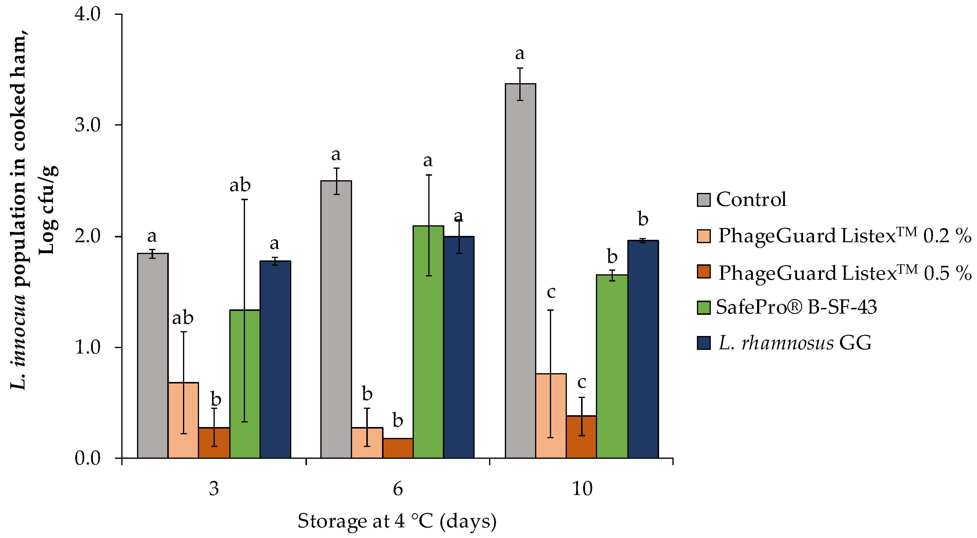
Figure 1. Effect of the biopreservative treatments in L. innocua population on cooked ham plugs throughout storage at 4 °C. Values are means ± standard deviations of three biological replicates. Bars marked with different letters (a, b and c) represent statistically significant differences among treatments (p < 0.05) at each sampling time.
Antibiotics 12 00414 g001
Figure 2. Effect of the biopreservative treatments on sliced cooked ham under semi-commercial conditions (4 °C plus modified atmosphere packaging): (a) L. innocua population on sliced cooked ham throughout storage. Values are means ± standard deviations of three biological replicates. Bars marked with an asterisk represent statistically significant differences between the treatment and the control (p < 0.05) at each sampling time. (b) Counts of Leuconostoc carnosus (formulated in SafePro® B-SF-43 treatment) and bacteriophage P100 (0.2 and 0.5% PhageGuard ListexTM treatments) on sliced cooked ham throughout storage. Values are means ± standard deviations of three biological replicates. Bars marked with different letters (a, b and c) represent statistically significant differences among sampling time (p < 0.05) at each evaluated treatment.
Antibiotics 12 00414 g002
Figure 3. Effect of the biopreservative treatments on L. innocua population in fresh cheese throughout storage at 4 °C. Values are means ± standard deviations of three biological replicates. Bars marked with different letters (a, b, c and d) represent statistically significant differences among treatments (p < 0.05) at each sampling time.
Antibiotics 12 00414 g003
Figure 4. Effect of the biopreservative treatments in fresh cheese under semi-commercial evaluation (4 °C): (a) L. innocua population in fresh cheese. Values are means ± standard deviations of three biological replicates. Bars marked with an asterisk represent statistically significant differences between the treatment and the control (p < 0.05) at each sampling time. (b) Counts of bacteriophage P100 (formulated in PhageGuard ListexTM treatment) and L. rhamnosus GG in fresh cheese at 0, 3, and 6 days of storage at 4 °C. Values are means ± standard deviations of three biological replicates. Bars marked with different letters (a and b) represent statistically significant differences among sampling time (p < 0.05) at each evaluated treatment.
Antibiotics 12 00414 g004
Figure 5. Effect of the biopreservative treatments on L. innocua population in fuet after ripening (7 days at 15 °C) and cold storage (7 days at 4 °C). Values are means ± standard deviations of three biological replicates. Bars marked with different letters (a and b) represent statistically significant differences among treatments (p < 0.05) at each sampling time.
Antibiotics 12 00414 g005
Figure 6. Effect of the biopreservative treatments in fuet: (a) L. innocua population in fuet at different time course of fuet production: inoculation day (0 day), after 24 h of fermentation, and at the end of ripening (7 days). Values are means ± standard deviations of three biological replicates. Bars marked with an asterisk represent statistically significant differences between the treatment and the control (p < 0.05) at each sampling time. (b) Counts of bacteriophage P100 (formulated in PhageGuard ListexTM treatment), P. acidilactici (formulated in SafePro® B-SF-20 treatment), Leuc. carnosus (formulated in SafePro® B-SF-43 treatment), and L. rhamnosus GG in fuet after inoculation and at the end of ripening. Values are means ± standard deviations of three biological replicates. Bars marked with different letters (a and b) represent statistically significant differences among sampling time (p < 0.05) at each evaluated treatment.
Antibiotics 12 00414 g006
Table 1. The O2 and CO2 concentration in the trays of sliced cooked ham treated with biopreservation techniques and stored at 4 °C under modified atmosphere packaging. Values are means ± standard deviations of three trays.
TreatmentO2 (%)CO2 (%)
0 Days7 Days14 Days0 Days7 Days14 Days
Control 6.1 ± 0.68.6 ± 0.19.5 ± 0.225.8 ± 3.415.6 ± 0.814.1 ± 0.1
Control L. innocua6.1 ± 0.68.2 ± 0.09.6 ± 0.025.8 ± 3.416.1 ± 0.513.5 ± 0.6
PhageGuard ListexTM 0.2%6.1 ± 0.68.4 ± 0.19.6 ± 0.125.8 ± 3.416.3 ± 0.314.2 ± 0.4
PhageGuard ListexTM 0.5%6.1 ± 0.68.3 ± 0.09.7 ± 0.425.8 ± 3.415.5 ± 0.513.3 ± 0.4
SafePro® B-SF-436.1 ± 0.67.4 ± 0.08.4 ± 0.125.8 ± 3.422.1 ± 1.1 *36.6 ± 1.5 *
*: The asterisk means statistically significant differences among treatments (p < 0.05) at each sampling time.
Table 2. Effect of the biopreservation application on pH of fuet. Values are means ± standard deviations of three biological replicates. Different letters in the same column show significant differences among treatments (p < 0.05) at each sampling time.
pH Value of Fuet
TreatmentAfter FermentationAfter Ripening
Control L. innocua5.35 ± 0.04 a5.38 ± 0.04 a
SafePro® B-LC-205.19 ± 0.02 b5.38 ± 0.04 a
SafePro® B-SF-435.39 ± 0.02 a5.44 ± 0.02 a
L. rhamnosus GG5.06 ± 0.04 c5.06 ± 0.04 b
PhageGuard ListexTM5.42 ± 0.02 a5.38 ± 0.05 a
Disclaimer/Publisher’s Note: The statements, opinions and data contained in all publications are solely those of the individual author(s) and contributor(s) and not of MDPI and/or the editor(s). MDPI and/or the editor(s) disclaim responsibility for any injury to people or property resulting from any ideas, methods, instructions or products referred to in the content.