Article

BIChromET: A Chromogenic Culture Medium for Detection of Piperacillin/Tazobactam and Cefepime Resistance in Pseudomonas aeruginosa

by
José Manuel Ortiz de la Rosa
1,2,3,
Ángel Rodríguez-Villodres
1,2,3,*,
Guillermo Martín-Gutiérrez
1,2,3,4,
Carmen Cintora Mairal
1,2,
José Luis García Escobar
1,2,
Lydia Gálvez-Benítez
1,2,3,
José Miguel Cisneros
1,2,3,5,† and
José Antonio Lepe
1,2,3,6,†
1
Clinical Unit of Infectious Diseases, Microbiology and Parasitology, University Hospital Virgen del Rocío, 41013 Seville, Spain
2
Institute of Biomedicine of Seville (IBiS), University Hospital Virgen del Rocío/CSIC/University of Seville, 41013 Seville, Spain
3
Centro de Investigación Biomédica en Red de Enfermedades Infecciosas (CIBERINFEC), Instituto de Salud Carlos III, Madrid 28029, Spain
4
Department of Health Sciences, Loyola Andalucía University, 41704 Seville, Spain
5
Department of Medicine, Faculty of Medicine, University of Seville, 41009 Seville, Spain
6
Department of Microbiology, Faculty of Medicine, University of Seville, 41009 Seville, Spain
*
Author to whom correspondence should be addressed.
These authors contributed equally to this work and participated as co-senior.
Antibiotics 2023, 12(11), 1573; https://doi.org/10.3390/antibiotics12111573
Submission received: 6 October 2023 / Revised: 24 October 2023 / Accepted: 27 October 2023 / Published: 28 October 2023

Abstract

:
Objectives: The BIChromET selective medium for detecting piperacillin-tazobactam (TZP) and cefepime (FEP) resistant Pseudomonas aeruginosa was developed. Methods: The performance of this medium was first evaluated using a collection of 100 P. aeruginosa clinical strains (70 TZP-susceptible, 30 TZP-resistant, 58 FEP-susceptible, and 42 FEP-resistant). Then, we performed clinical validation by testing 173 respiratory clinical samples. Results: The BIChromET medium showed excellent sensitivity (TZP (avg. 96.7%); FEP (avg. 92.7%)) and specificity (TZP (avg. 98.9%); FEP (avg. 98%)) in distinguishing the detection limit ranging from 104 to 108 CFU/mL. Then, testing the bronchoalveolar lavage (BAL) and tracheobronchial aspirate (TBA) clinical specimens (N = 173) revealed the excellent performance of the medium with P. aeruginosa, showing 100% and 92.6% of categorical agreements with the results obtained via the broth microdilution methods (BMD) for TZP and FEP, respectively. Conclusion: This medium allows for easy and accurate detection of TZP/FEP-resistant isolates regardless of their resistance mechanisms.

Keywords; piperacillin/tazobactam; cefepime; empiric treatment; Pseudomonas aeruginosa

1. Introduction

Pseudomonas aeruginosa is one of the most common causes of pneumonia in hospitalized and immunocompromised patients, frequently associated with high morbidity and mortality [1,2,3]. According to the National Healthcare Safety Network (NHSN) and the EU-VAP/CAP study, P. aeruginosa is the second most prevalent microorganism isolated from nosocomial pneumonia [4,5]. P. aeruginosa infections are a serious concern in hospitals. Patients in critical conditions can die from pneumonia caused by P. aeruginosa, and the elimination of P. aeruginosa is very difficult because of its wide variety of resistance mechanisms [6]. Furthermore, it is responsible for considerable additional healthcare costs and resource utilization due to the difficulty in dealing with P. aeruginosa infections [7].
The initial management of patients with P. aeruginosa infections, especially from the intensive care unit (ICU), involves obtaining culture results and administering appropriate antibiotics within the first hour [8]. Choosing an appropriate initial antibiotic is essential in managing P. aeruginosa infections, as the inappropriate initial choice of antimicrobial regimen has previously been shown to be associated with increased mortality [9,10,11,12]. A previous retrospective study found that initial inappropriate antimicrobial therapy in septic shock resulted in a 5-fold reduction in survival [13]. A large meta-analysis of patients with severe bacterial infections showed that patients who received appropriate initial antibiotic therapy had lower treatment failure rates, shorter hospital length of stay and cost, and lower mortality rates than patients who received inappropriate initial antibiotic therapy [14].
Two of the most used antibiotics for the empirical treatment of hospital-acquired P. aeruginosa infections are piperacillin-tazobactam (TZP) and cefepime (FEP) [15]. The initial empirical treatment decisions are challenged by the lack of knowledge of the bacterial resistance profile. The reference microbiological diagnostic tools available for bacteria causing respiratory tract infections may extend the time to obtain the results to 24–48 h [16] (Figure 1). During this period, patients may be receiving inappropriate treatment, increasing morbidity and mortality.
Currently, antimicrobial stewardship programs (ASPs) are being implemented. ASPs aim to promote the safe, effective, and efficient use of antibiotics to optimize patient care while mitigating individual patient risk and the impact of antimicrobial use on the greater population [17,18,19]. These kinds of programs encompass prevention, early treatment, and rapid diagnosis. Therefore, it is very important for the prognosis of the patients to have rapid diagnostic techniques to guide empirical treatment in the shortest possible time. Molecular techniques based on multiplex PCR have been developed in recent years to identify respiratory pathogens directly from the samples [20]. However, molecular techniques present a limitation due to the discrepancy between genotype and phenotype, especially in P. aeruginosa, which could lead to misinterpreting the results [21]. Thus, the main challenge in respiratory infections is rapidly detecting the antibiotic resistance profile. In this sense, we aimed to develop a selective culture medium for screening TZP/FEP-resistant P. aeruginosa regardless of the corresponding resistance mechanism in 24 h, reducing to half the time to optimize the empirical treatment (Figure 1).

2. Results

According to the minimal inhibitory concentration (MIC) results obtained via broth microdilution method (BMD), this collection included 70 TZP-susceptible isolates + 30 TZP-resistant isolates, and 59 FEP-susceptible isolates + 41 FEP-resistant isolates (Supplementary Tables S1 and S2).
On the FEP part of the bi-plates, the results showed that none of the FEP-susceptible isolates grew on the BIChromET medium, except for five isolates of 1 × 10⁸ CFU/mL of bacterial concentrations. On the other hand, all the FEP-resistant isolates were recovered within 24 h on the BIChromET medium using an inoculum between 1 × 10⁶ and 1 × 10⁸ CFU/mL. However, 11 and 3 FEP-resistant isolates did not grow onto the BIChromET selective plates when inoculated at 1 × 10⁴ and 1 × 10⁵ CFU/mL of bacterial concentration, respectively (Table 1). For the TZP part of the bi-plates, the results showed that all the TZP-susceptible isolates did not grow onto the BIChromET medium, except for four isolates at 1 × 10⁸ CFU/mL. On the other hand, all the TZP-resistant isolates were recovered within 24 h on BIChromET medium using an inoculum between 1 × 10⁵ and 1 × 10⁸ CFU/mL. However, four TZP-resistant isolates did not grow onto the BIChromET selective plates when inoculated at 1 × 10⁴ CFU/mL (Table 1). These data allowed us to set the sensitivity and specificity of the medium in each dilution tested. The sensitivity values varied between the different dilutions but remained above 90% in all the bacterial concentrations tested, most being 100%, except at 1 × 10⁴ CFU/mL. On the other hand, the BIChromET plates showed 100% in the specificity of all bacterial concentrations evaluated, except at 1 × 10⁸ CFU/mL (TZP and FEP) and 1 × 10⁷ CFU/mL (only FEP), in which a decrease in specificity [FEP (107: 98.3% and 108: 91.54%) and TZP (108: 94.3%)] was observed (Table 1).
Among the 173 clinical specimens (bronchoalveolar lavage (BAL)/tracheobronchial aspirate (TBA) analyzed, 27 P. aeruginosa were recovered via the conventional techniques used in the Microbiology Service at the University Hospital Virgen del Rocío (Seville). The BIChromET plates, compared to the reference BMD, showed a high level of categorical agreement (CA), reaching 100% and 92.6% for TZP and FEP, respectively. In particular, the 7.4% loss in categorical agreement observed in FEP corresponds to two major errors (MEs, defined as false resistance results) (Supplementary Tables S1 and S2. Despite the reduced number of P. aeruginosa (N = 27) recovered in the validation step with 173 clinical specimens (BAL and TBA), the data obtained with the BIChromET plates and BMD allowed us to calculate the sensitivity and specificity of the media. TZP showed a sensitivity and specificity of 100% (ICsensitivity: 81.6%–100%; ICspecificity: 72.2–100%), and FEP showed a sensitivity of 100% (IC: 78.5%–100%) and a specificity of 84.6% (IC: 57.8–95.7%) (Table 2).
Moreover, our media could detect some FEP/TZP-resistant Gram-negative bacteria that were also identified using conventional methods, such as MicroScan Walkaway (Beckman Coulter, Brea, Callifornia, USA) (Supplementary Table S3). Notably, no competing flora (which are frequently found on standard medium), such as Gram-positive bacteria or fungi, was identified with the BIChromET plates after 24 h of incubation, indicating that this medium has high levels of specificity and selectivity for TZP/FEP-resistant Gram-negative bacteria.

3. Discussion

In this study, we have developed the BIChromET selective medium, being the first medium that could detect TZP- and FEP-resistant P. aeruginosa isolates and reduce the time to adjust the antimicrobial therapy in 24 h in severe respiratory infections. Although the BIChromET medium showed excellent sensitivity and specificity during the evaluation, limitations were found when some isolates were inoculated at 1 × 10⁴ and 1 × 10⁸ CFU/mL, decreasing the sensitivity and specificity values, respectively (Table 1). The reduction in the sensitivity was due to the isolates with MIC values close to the breakpoint but still resistant (FEP (16 μg/mL) or TZP (32 μg/mL)) that did not grow at 1 × 10⁴ CFU/mL. In contrast, the slight reduction in the specificity was due to the isolates with MIC values close to the breakpoint but still susceptible (FEP (8 μg/mL) or TZP (16 μg/mL)) that grew at 1 × 10⁸ CFU/mL. A diagnostic algorithm was created to address the limitations presented above, especially the one significantly affecting the sensitivity, and to minimize the possible false negative obtained with the BIChromET medium (Figure 1B). This algorithm ignores plates with an absence of growth when the blood agar medium shows a bacterial concentration of 1 × 10⁴ CFU/mL. It is noteworthy that several studies and data from the University Hospital Virgen del Rocío showed that more than 95% of positive BAL and TBA contain a bacterial concentration ≥ 1 × 10⁵ CFU/mL [22,23]. This fact proves that the test will be useful in at least 95% of the samples received in the Microbiology service of the University Hospital Virgen del Rocío. Another limitation is the absence of MIC values since our media only determines susceptible or resistant bacteria. Therefore, our media will categorize as susceptible all isolates whose MIC values are ≤16 μg/mL. Notwithstanding, EUCAST guidelines recommended high doses of cefepime and piperacillin-tazobactam for all susceptible P. aeruginosa.
This medium constitutes a useful tool combo for identifying TZP/FEP-resistant strains and possibly prevents further dissemination and outbreaks by screening patients potentially infected with such resistant strains. Unlike other tests, the medium allows direct screening from clinical samples, such as BAL and TBA, usually collected from severe infection patients. The selectivity of the BIChromET medium for TZP/FEP-resistant bacteria is not impacted by the mixed flora found in many clinical samples. Moreover, the BIChromET plates showed excellent performance with respiratory samples (BAL and TBA). Using the BIChromET plates as a complement to the traditional techniques may play an important role in clinical decision-making, optimizing antibiotic usage. In 2022, P. aeruginosa encompassed 18.6% of all respiratory isolates (N = 4925) in our institution. Noteworthy, in 2022, 27% and 23% of the P. aeruginosa isolates were resistant to FEP and TZP, respectively. In this way, in around 25% of the patients with respiratory infections by P. aeruginosa, the BIChromet medium will allow to optimize the empirical treatment 24 h earlier than with the conventional methods. On the other hand, in 75% of the patients, our media will allow maintaining or simplifying the empirical treatment of the patients, preserving the utility of the last-resort antibiotics’ usefulness. Thus, this selective medium could cover a need of many patients since TZP and FEP have become two of the first options against hospital-acquired respiratory infections caused by P. aeruginosa bacteria in many places and the lack of rapid diagnostic tests in such infections [16].
In conclusion, the BIChromET medium showed excellent performance and was easy to prepare. Its cost of production would be around EUR 0.70/plate, maintaining a great cost–benefit balance. Moreover, the BIChromET medium would be added to the diagnostic toolbox dedicated to the rapid detection of antibiotic resistance in respiratory infections, which may help to optimize the empirical treatment, where the delay of the appropriate therapy is crucial for the outcome of the patients. Notwithstanding, further clinical evaluations of this medium will now be needed in daily clinical practice to further assess its usefulness.

4. Materials and Methods

Bacterial strains. A total of 100 P. aeruginosa isolates collected from different clinical samples (bronchoalveolar lavage (BAL), tracheobronchial aspirate (TBA), cerebrospinal fluid (CSF), blood cultures, sputum, wounds, and urine) from the Microbiology Service at the University Hospital Virgen del Rocío (Seville, Spain) were used in developing the BIChromET plates. This collection included 30 TZP-resistant isolates, 70 TZP-susceptible isolates, 41 FEP-resistance isolates, and 59 FEP-susceptible isolates (Supplementary Tables S1 and S2). Additionally, the BIChromET plates were clinically tested on 173 clinical specimens (BAL and TBA) recovered over 3 months. P. aeruginosa PAO1 was used as control negative, and 2 TZP/FEP-resistance clinical isolates were used as control positive. Additionally, P. aeruginosa, Stenotrophomonas maltophilia, Acinetobacter baumannii, Klebsiella pneumoniae, and Escherichia coli were used to establish the color change of the different species when growth on the BIChromET plates. The isolates were not screened at the molecular level for the presence of resistance determinants due to the medium being developed to detect resistant isolates independently of their resistance mechanism.
Susceptibility testing. MIC values for TZP and FEP were determined using the broth microdilution method following the EUCAST recommendations (https://www.eucast.org/ast_of_bacteria/mic_determination (accessed on 1 February 2023)). Clinical breakpoints were established according to the European Committee on Antimicrobial Susceptibility Testing (EUCAST; 2023) [24]. Hence, isolates with TZP MICs among >0.001 μg/mL and ≤16 μg/mL were categorized as I (susceptible, increased exposure), while those with MICs of >16 μg/mL were categorized as resistant, and isolates with FEP MICs among >0.001 μg/mL and ≤8 μg/mL were categorized as I (susceptible, increased exposure), while those with MICs of >8 μg/mL were categorized as resistant. EUCAST established the susceptible breakpoint at ≤0.001 μg/mL to categorize all susceptible P. aeruginosa as I. Thus, therapeutic success with susceptible P. aeruginosa would depend on the dosing regimen and/or the site of infection.
Selective medium for TZP and FEP resistance. CLED agar medium (reference; Biomerieux, Paris, France) was used for optimal screening following the manufacturer’s instructions. After testing several culture conditions, a selective medium was set up and supplemented with piperacillin, tazobactam, and FEP (Sigma-Aldrich, St. Louis, MO, USA) at 24, 4, and 4 µg/mL, respectively. Moreover, vancomycin (Duchefa Biochemie, Haarlem, Netherlands) and amphotericin B (Acros Organics, Morris Plains, New Jersey, USA) were added to the medium at a final concentration of 20 and 5 µg/mL to prevent the growth of Gram-positive bacteria and fungi, respectively. Additionally, a phenol red solution (0.5%) was added to the FEP section of the bi-plate to differentiate both sections visually. Thus, in the TZP section of the bi-plate (standard CLED medium), the lactose fermenter bacteria produced light beige colonies, unlike non-fermenters, which yield blue colonies. On the other hand, in the FEP part of the BIChromET medium (CLED with phenol red), the lactose fermenter bacteria produced yellow colonies, unlike non-fermenters, which yield purple colonies (Supplementary Figure S1, Table S3). The CLED powder was diluted in distilled water and autoclaved at 121 °C for 15 min. The antibiotic stock solutions were added when the medium reached 56 °C (Table 3). The prepared plates of this BIChromET medium were stored at 4 °C and were protected from direct light exposure before use for as long as 2 weeks. Candida albicans, Staphylococcus aureus, and the TZP/FEP-susceptible E. coli ATCC 25922 reference strain were sub-cultured daily on the BIChromET selective plates from a single batch of plates kept at 4 °C to test the shelf life of the medium. For at least 14 days, no growth could be observed.
Evaluation assay. The sensitivity and specificity cut-off values for detecting TZP- and FEP-resistant P. aeruginosa were established at 1 × 10³ CFU/mL, considering the positive results of only the sample, of which the isolates were recovered onto the BIChromET selective medium, which were plated at concentrations corresponding to >1 × 10³ CFU/mL. This cut-off value was fixed considering that BAL and TBA are positive when the ≥1 × 10⁴ CFU/mL bacteria are recovered from the clinical sample. Starting with a 0.5 McFarland standard (an inoculum of 1.5 × 10⁸ CFU/mL), serial 10-fold dilutions were made in 0.85% saline solution, and 100 µL aliquots of each dilution from 104 to 108 CFU/mL were plated onto the BIChromET selective medium. To quantify the viable bacteria in each dilution step, tryptic soy agar plates were inoculated concomitantly with 100 µL of each suspension and incubated overnight at 37 °C. Viable colonies were counted the following day. When no growth was observed after 18 h, incubation was extended up to 48 h to assess the negativity of the culture. The medium was designed to detect resistant P. aeruginosa from 104 to 108 CFU/mL based on the IDSA guidelines [3]. It established that a BAL culture was positive when more than 104 CFU/mL bacteria were recovered from the BAL. After evaluation with a collection of 100 P. aeruginosa, the results were analyzed for each dilution of bacterial concentration used in this study.
Validation experiments. To check the performance of the selective medium in clinical samples, a total of 173 samples obtained from patients admitted to the University Hospital Virgen del Rocío (21 BAL and 152 TBA clinical specimens) were tested using the BIChromET plates. Then, 100 μL of the BAL or TBA were inoculated onto each half of the BIChromET plates and incubated overnight at 37 °C. The colonies of different morphologies, sizes, and colors from each plate were selected for further experiments, such as identification (mass spectrometry) and resistance phenotype (gradient strips (Liofilchem, Italy) for non-P. aeruginosa isolates) (Supplementary Table S3). To confirm the resistance patterns in P. aeruginosa, the grown colonies of P. aeruginosa were tested for TZP and FEP susceptibility using BMD. The results were interpreted according to the EUCAST 2023 breakpoints [24].
Statistical analysis. The specificity (proportion of TZP- or FEP-susceptible isolates that are correctly determined) and the sensitivity (proportion of TZP- or FEP-resistant isolates that are correctly determined) were calculated for each dilution used in the evaluation step to know the limitation of the medium, its performance at the different concentration, and how to overcome these limitations using broth microdilution as the gold standard method. On the other hand, 95% confidence intervals (CI), a predictive positive value (PPV), and a predictive negative value (PNV) were also estimated. For the validation step, the sensitivity, specificity, and 95% CIs were calculated with the clinical specimens (BAL and TBA) to detect the resistance in P. aeruginosa using broth microdilution as the gold standard method. In the clinical evaluation step, true positive results were considered when a TZP or FEP-resistant P. aeruginosa (BMD MIC) growth in the BIChromET plates, while the absence of growth in the BIChromET plates when a TZP or FEP-susceptible P. aeruginosa (MD MIC) were present in the BAL/TBA samples were considered true negative. Moreover, the endpoints were considered in categorical agreement when the results were in the same susceptibility category (regardless of the MIC) for P. aeruginosa. VME is a very major error (false susceptibility), and ME is a major error (false resistance).

Supplementary Materials

The following supporting information can be downloaded at: https://www.mdpi.com/article/10.3390/antibiotics12111573/s1, Figure S1: color change of the media with different fermenter and non-fermenter species. Upper plate pictures correspond to the original color of each section of the bi-plate before bacterial growth. (A) Stenotrophomonas maltophilia, (B) Acinetobacter baumannii, (C) Pseudomonas aeruginosa, (D) Klebsiella pneumoniae, and (E) Escherichia coli.; Table S1: raw data of the BIChromET evaluation step for cefepime.; Table S2: raw data of the BIChromET evaluation step for piperacillin/tazobactam.; Table S3: raw data of the BIChromET clinical evaluation with 173 clinical specimens (TBA and BAL).

Author Contributions

Conceptualization, J.M.O.d.l.R. and Á.R.-V.; Methodology, J.M.O.d.l.R., Á.R.-V. and G.M.-G.; Validation, J.M.O.d.l.R., G.M.-G., C.C.M. and J.L.G.E.; Investigation, J.M.O.d.l.R., C.C.M., J.L.G.E. and L.G.-B.; Resources, Á.R.-V., J.M.C. and J.A.L.; Writing—original draft, J.M.O.d.l.R.; Writing—review and editing, Á.R.-V., G.M.-G., J.M.C. and J.A.L.; Supervision, J.A.L.; Funding acquisition, Á.R.-V., J.M.C. and J.A.L. All authors have read and agreed to the published version of the manuscript.

Funding

This research was funded by Instituto de Salud Carlos III (ISCIII) via the project “PI22/01464” and co-funded by the European Union. This study has also been funded by Instituto de Salud Carlos III (ISCIII) via the project “PI20/01829”. JMOR is supported by the Subprograme Sara Borrell, Instituto de Salud Carlos III, Subdirección General de Redes y Centros de Investigación Cooperativa, Ministerio de Ciencia, Innovación y Universidades, Spain (CD21/00098). ARV is supported by the Subprograme Juan Rodés, Instituto de Salud Carlos III, Subdirección General de Redes y Centros de Investigación Cooperativa, Ministerio de Ciencia, Innovación y Universidades, Spain (JR20/00023). GMG was supported by a Juan Rodés research contract from the Instituto de Salud Carlos III (JR19/00039). LGB was supported by a Rio Hortega research contract from the Instituto de Salud Carlos III (CM22/00196).

Institutional Review Board Statement

Not applicable.

Informed Consent Statement

Not applicable.

Data Availability Statement

The datasets used and/or analyzed during the present study are available from the corresponding author upon reasonable request.

Conflicts of Interest

The authors declare no conflict of interest.

References

  1. Zarb, P.; Coignard, B.; Griskeviciene, J.; Muller, A.; Vankerckhoven, V.; Weist, K.; Goossens, M.M.; Vaerenberg, S.; Hopkins, S.; Catry, B.; et al. The European Centre for Disease Prevention and Control (ECDC) pilot point prevalence survey of healthcare-associated infections and antimicrobial use. Eurosurveillance 2012, 17, 20316. [Google Scholar] [CrossRef] [PubMed]
  2. Kollef, M.H.; Chastre, J.; Fagon, J.Y.; François, B.; Niederman, M.S.; Rello, J.; Torres, A.; Vincent, J.L.; Wunderink, R.G.; Go, K.W.; et al. Global Prospective Epidemiologic and Surveillance Study of Ventilator-Associated Pneumonia due to Pseudomonas aeruginosa. Crit. Care Med. 2014, 42, 2178–2187. [Google Scholar] [CrossRef] [PubMed]
  3. Mandell, L.A.; Wunderink, R.G.; Anzueto, A.; Bartlett, J.G.; Campbell, G.D.; Dean, N.C.; Dowell, S.F.; File, T.M., Jr.; Musher, D.M.; Niederman, M.S.; et al. Infectious Diseases Society of America/American Thoracic Society Consensus Guidelines on the Management of Community-Acquired Pneumonia in Adults. Clin. Infect. Dis. 2007, 44 (Suppl. 2), S27–S72. [Google Scholar] [CrossRef]
  4. Sievert, D.M.; Ricks, P.; Edwards, J.R.; Schneider, A.; Patel, J.; Srinivasan, A.; Kallen, A.; Limbago, B.; Fridkin, S.; National Healthcare Safety Network (NHSN) Team and Participating NHSN Facilities. Antimicrobial-resistant pathogens associated with healthcare-associated infections: Summary of data reported to the National Healthcare Safety Network at the Centers for Disease Control and Prevention, 2009–2010. Infect. Control Hosp. Epidemiol. 2013, 34, 1–14. [Google Scholar] [CrossRef] [PubMed]
  5. Koulenti, D.; Tsigou, E.; Rello, J. Nosocomial pneumonia in 27 ICUs in Europe: Perspectives from the EU-VAP/CAP study. Eur. J. Clin. Microbiol. Infect. Dis. 2017, 36, 1999–2006. [Google Scholar] [CrossRef] [PubMed]
  6. Tran, C.S.; Rangel, S.M.; Almblad, H.; Kierbel, A.; Givskov, M.; Tolker-Nielsen, T.; Hauser, A.R.; Engel, J.N. The Pseudomonas aeruginosa type III translocon is required for biofilm formation at the epithelial barrier. PLoS Pathog. 2014, 10, e1004479. [Google Scholar] [CrossRef]
  7. Kaier, K.; Heister, T.; Götting, T.; Wolkewitz, M.; Mutters, N.T. Measuring the in-hospital costs of Pseudomonas aeruginosa pneumonia: Methodology and results from a German teaching hospital. BMC Infect. Dis. 2019, 19, 1028. [Google Scholar] [CrossRef]
  8. Levy, M.M.M.; Evans, L.E.M.; Rhodes, A.M. The Surviving Sepsis Campaign Bundle: 2018 Update. Crit. Care Med. 2018, 46, 997–1000. [Google Scholar] [CrossRef]
  9. Micek, S.T.; Reichley, R.M.; Kollef, M.H. Health care-associated pneumonia (HCAP): Empiric antibiotics targeting methicillin-resistant Staphylococcus aureus (MRSA) and Pseudomonas aeruginosa predict optimal outcome. Medicine 2011, 90, 390–395. [Google Scholar] [CrossRef]
  10. Micek, S.T.; Kollef, M.H.; Torres, A.; Chen, C.; Rello, J.; Chastre, J.; Antonelli, M.; Welte, T.; Clair, B.; Ostermann, H.; et al. Pseudomonas aeruginosa Nosocomial Pneumonia: Impact of Pneumonia Classification. Infect. Control Hosp. Epidemiol. 2015, 36, 1190–1197. [Google Scholar] [CrossRef]
  11. Micek, S.T.; Kollef, K.E.; Reichley, R.M.; Roubinian, N.; Kollef, M.H. Health Care-Associated Pneumonia and Community-Acquired Pneumonia: A Single-Center Experience. Antimicrob. Agents Chemother. 2007, 51, 3568–3573. [Google Scholar] [CrossRef] [PubMed]
  12. Micek, S.T.; Lloyd, A.E.; Ritchie, D.J.; Reichley, R.M.; Fraser, V.J.; Kollef, M.H. Pseudomonas aeruginosa Bloodstream Infection: Importance of Appropriate Initial Antimicrobial Treatment. Antimicrob. Agents Chemother. 2005, 49, 1306–1311. [Google Scholar] [CrossRef]
  13. Kumar, A.; Ellis, P.; Arabi, Y.; Roberts, D.; Light, B.; Parrillo, J.E.; Dodek, P.; Wood, G.; Kumar, A.; Simon, D.; et al. Initiation of Inappropriate Antimicrobial Therapy Results in a Fivefold Reduction of Survival in Human Septic Shock. Chest 2009, 136, 1237–1248. [Google Scholar] [CrossRef] [PubMed]
  14. Bassetti, M.; Rello, J.; Blasi, F.; Goossens, H.; Sotgiu, G.; Tavoschi, L.; Zasowski, E.J.; Arber, M.R.; McCool, R.; Patterson, J.V.; et al. Systematic review of the impact of appropriate versus inappropriate initial antibiotic therapy on outcomes of patients with severe bacterial infections. Int. J. Antimicrob. Agents 2020, 56, 106184. [Google Scholar] [CrossRef] [PubMed]
  15. American Thoracic Society; Infectious Diseases Society of America. Guidelines for the management of adults with hospital-acquired, ventilator-associated, and healthcare-associated pneumonia. Am. J. Respir. Crit. Care Med. 2005, 171, 388–416. [Google Scholar] [CrossRef]
  16. Cillóniz, C.; Dominedò, C.; Torres, A. Multidrug Resistant Gram-Negative Bacteria in Community-Acquired Pneumonia. Crit. Care 2019, 23, 79. [Google Scholar] [CrossRef]
  17. Teerawattanapong, N.; Kengkla, K.; Dilokthornsakul, P.; Saokaew, S.; Apisarnthanarak, A.; Chaiyakunapruk, N. Prevention and Control of Multidrug-Resistant Gram-Negative Bacteria in Adult Intensive Care Units: A Systematic Review and Network Meta-analysis. Clin. Infect. Dis. 2017, 64, S51–S60. [Google Scholar] [CrossRef]
  18. Tacconelli, E.; Cataldo, M.A.; Dancer, S.J.; De Angelis, G.; Falcone, M.; Frank, U.; Kahlmeter, G.; Pan, A.; Petrosillo, N.; Rodríguez-Baño, J.; et al. ESCMID guidelines for the management of the infection control measures to reduce transmission of multidrug-resistant Gram-negative bacteria in hospitalized patients. Clin. Microbiol. Infect. 2014, 20 (Suppl. 1), 1–55. [Google Scholar] [CrossRef]
  19. Lanckohr, C.; Bracht, H. Antimicrobial stewardship. Curr. Opin. Crit. Care. 2022, 28, 551–556. [Google Scholar] [CrossRef]
  20. Torres, A.; Lee, N.; Cilloniz, C.; Vila, J.; Van der Eerden, M. Laboratory diagnosis of pneumonia in the molecular age. Eur. Respir. J. 2016, 48, 1764–1778. [Google Scholar] [CrossRef]
  21. Weinmaier, T.; Conzemius, R.; Bergman, Y.; Lewis, S.; Jacobs, E.B.; Tamma, P.D.; Materna, A.; Weinberger, J.; Beisken, S.; Simner, P.J. Validation and Application of Long-Read Whole-Genome Sequencing for Antimicrobial Resistance Gene Detection and Antimicrobial Susceptibility Testing. Antimicrob. Agents Chemother. 2023, 67, e0107222. [Google Scholar] [CrossRef] [PubMed]
  22. Yamasaki, K.; Kawanami, T.; Yatera, K.; Fukuda, K.; Noguchi, S.; Nagata, S.; Nishida, C.; Kido, T.; Ishimoto, H.; Taniguchi, H.; et al. Significance of Anaerobes and Oral Bacteria in Community-Acquired Pneumonia. PLoS ONE 2013, 8, e63103. [Google Scholar] [CrossRef] [PubMed]
  23. Kahn, F.W.; Jones, J.M. Diagnosing Bacterial Respiratory Infection by Bronchoalveolar Lavage. J. Infect. Dis. 1987, 155, 862–869. [Google Scholar] [CrossRef] [PubMed]
  24. Breakpoint Tables for Interpretation of MICs and Zone Diameters, Version 13.0; The European Committee on Antimicrobial Susceptibility Testing: Växjö, Sweden, 2023. Available online: http://www.eucast.org(accessed on 12 February 2023).
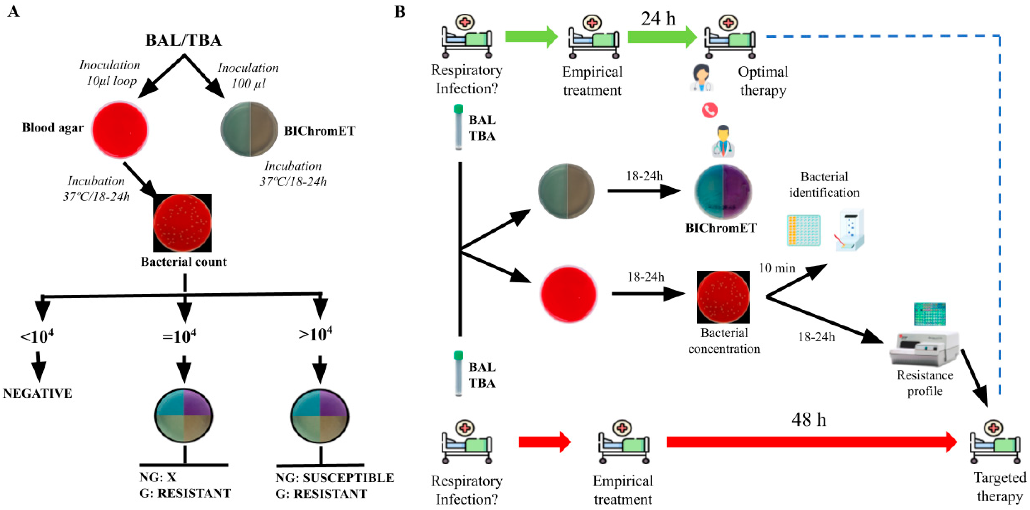
Figure 1. (A) Diagnostic algorithm designed for using the BIChromET plates in clinical practice. (B) Flowchart of the BIChromET medium methodology and its possible application in clinical practice. BAL, Bronchoalveolar lavage; TBA, Tracheobronchial aspirate; G, growth; NG, not growth.
Antibiotics 12 01573 g001
Table 1. Performance of the BIChromET medium for detecting P. aeruginosa resistance or susceptibility to TZP and/or FEP from bacterial colonies.
Bacterial Concentrations (CFU/mL)
Gold Standard1 × 1041 × 1051 × 1061 × 1071 × 108
Cefepime
Susceptible597063595854
Resistance413037414246
Sensitivity (%)-73.290.2100100100
CI (%)-58.1–84.377.5–96.191.4–10091.4–10091.4–100
Specificity (%)-10010010098.391.5
CI (%)-93.9–10093.9–10093.9–10091–99.781.6–96.3
PPV (%)-10010010097.689.1
NPV (%)-84.393.7100100100
Piperacillin-Tazobactam
Susceptible707471707066
Resistance302629303034
Sensitivity (%)-86.796.7100100100
CI (%)-70.3–94.783.3–99.488.6–10088.6–10083.8–99.4
Specificity (%)-10010010010094.3
CI (%)-94.8–10094.8–10094.8–10094.8–10086.2–97.8
PPV (%)-10010010010088.2
NPV (%)-94.698.6100100100
TZP, piperacillin/tazobactam, FEP, cefepime; CI, 95% confidence Interval, PPV, positive predictive value, NPV, negative predictive value.
Table 2. Validation of the BIChromET medium for detecting P. aeruginosa resistance or susceptibility to TZP and/or FEP from clinical samples.
BIChromETBMD
Cefepime:
Susceptible1113
Resistance1614
CA92.6%
ErrorsMEs (N = 2)
Sensitivity (a) CI100% (78.5–100%)
Specificity (a) CI84.6% (57.8–100%)
Piperacillin-Tazobactam:
Susceptible1010
Resistance1717
CA100%
Errors-
Sensitivity (a) CI100% (81.6–100%)
Specificity (a) CI100% (72.2–100%)
TZP, piperacillin/tazobactam, FEP, cefepime; CI, 95% confidence Interval; CA, Categorical agreement; BMD, broth microdilution. (a) Sensitivity and specificity were exclusively calculated using the gold standard method (BMD) as a reference.
Table 3. Preparation of the BICromET medium.
CompoundStock SolutionQuantity or Vol AddedFinal Concentration
Piperacillin-Tazobactam
   CLED agar medium 14.46 g
   Distilled water 400 mL
   Piperacillin50 mg/mL in water0.192 mL24 mg/L
   Tazobactam50 mg/mL in water0.032 mL4 mg/L
   Vancomycin50 mg/mL in water0.16 mL20 mg/L
   Amphotericin B10 mg/mL in DMSO0.2 mL5 mg/L
Cefepime
   CLED agar medium 14,46 g
   Distilled water 400 mL
   Phenol red0.5%1 mL0.00125%
   Cefepime50 mg/mL in water0.032 mL4 mg/L
   Vancomycin50 mg/mL in water0.16 mL20 mg/L
   Amphotericin B10 mg/mL in DMSO0.2 mL5 mg/L
DMSO, Dimehyl sulfoxide, TZP, piperacillin/tazobactam, FEP, cefepime; CI, 95% confidence Interval, PPV, positive predictive value, NPV, negative predictive value.
Disclaimer/Publisher’s Note: The statements, opinions and data contained in all publications are solely those of the individual author(s) and contributor(s) and not of MDPI and/or the editor(s). MDPI and/or the editor(s) disclaim responsibility for any injury to people or property resulting from any ideas, methods, instructions or products referred to in the content.