Article

Efficacy of Chlorhexidine after Oral Surgery Procedures on Wound Healing: Systematic Review and Meta-Analysis

by
María de Nuria Romero-Olid
1,2,
Elena Bucataru
1,2,
Pablo Ramos-García
1,2,* and
Miguel Ángel González-Moles
1,2
1
School of Dentistry, University of Granada, 18071 Granada, Spain
2
Instituto de Investigación Biosanitaria ibs.GRANADA, 18012 Granada, Spain
*
Author to whom correspondence should be addressed.
Antibiotics 2023, 12(10), 1552; https://doi.org/10.3390/antibiotics12101552
Submission received: 21 September 2023 / Revised: 14 October 2023 / Accepted: 18 October 2023 / Published: 20 October 2023
(This article belongs to the Special Issue Antimicrobial Activity of Dental Biomaterials)

Abstract

:
Our objective was to evaluate qualitatively and quantitatively, through a systematic review and meta-analysis, available evidence on the efficacy of chlorhexidine (CHX) when applied after oral surgery on wound healing and related clinical parameters. MEDLINE/PubMed, Embase, CENTRAL, Web of Science, and Scopus were searched for studies published before January 2023. The quality of the methodology used in primary-level studies was assessed using the RoB2 tool; meta-analyses were performed jointly with heterogeneity and small-study effect analyses. Thirty-three studies and 4766 cases were included. The results point out that the application of CHX was significantly more effective, compared to controls where CHX was not employed, providing better wound healing after oral surgery (RR = 0.66, 95% CI = 0.55–0.80, p < 0.001). Stratified meta-analyses confirmed the higher efficacy of 0.20% CHX gel vs. other vehicles and concentrations (p < 0.001, respectively). Likewise, the addition of chitosan to CHX significantly increased the efficacy of surgical wound healing (p < 0.001). The use of CHX has also been significantly beneficial in the prevention of alveolar osteitis after any type of dental extraction (RR = 0.46, 95% CI = 0.39–0.53, p < 0.001) and has also been effective when applied as a gel for a reduction in pain after the surgical extraction of third molars (MD = −0.97, 95% CI = −1.26 to −0.68, p < 0.001). In conclusion, this systematic review and meta-analysis demonstrate on the basis of evidence that the application of CHX exerts a beneficial effect on wound healing after oral surgical procedures, significantly decreasing the patient’s risk of developing surgical complications and/or poor wound healing. This benefit was greater when CHX was used at 0.20% in gel form with the addition of chitosan.

1. Introduction

Wound healing in oral surgery is a complex and dynamic process that culminates in the restitution of the tissue’s integrity [1,2]. During the healing process, a series of successive events occurred, starting with local hemostasis, followed by inflammation, tissue proliferation (mainly of fibroblasts, epithelial and endothelial cells), neoangiogenesis, repithelialization, synthesis, union and the alignment of collagen fibers, formation of granulation tissue, and definitive tissue remodeling [2,3,4]. These events can be altered by several factors, such as diseases (e.g., diabetes mellitus [5], hypothyroidism [6,7], or rheumatoid arthritis [8]), drug intake (e.g., bisphosphonates [7,9]), tobacco use [10]) or infections as a consequence of bacterial imbalance in the wound site [11,12,13]. Consequently, patients undergoing oral surgery are at an increased risk of developing complications during postoperative wound healing [14,15], such as delayed or the absence of healing, pain and, particularly, after the extraction of third molars, the development of alveolar osteitis [16,17,18,19,20]. The establishment of improvement strategies to minimize wound healing complications after oral surgery is currently a priority line of clinical research in modern dentistry [21,22].
One of the strategies that has received the most attention is the application of chlorhexidine (CHX) to wounds after oral surgical procedures [23]. This approach is justified by the broad-spectrum actions of CHX against Gram-positive and Gram-negative bacteria, with bacteriostatic properties at low doses and bactericidal properties at high concentrations [24,25], as well as an antifungal effect [23]. Its mechanism of action is exerted by increasing the permeability of the cell membrane of target microorganisms, which causes the precipitation of macromolecules in the cytoplasm and subsequent microbial death via cell lysis [23]. Therefore, the effect of CHX is mainly based on bacterial load reduction, playing an anti-infection role, which seems essential in the early stages of wound healing. Nevertheless, the problem of healing is more complicated, in which many more factors are involved.
In recent years, the use of CHX has increased exponentially in the different fields of medicine, particularly in dentistry, where it is currently the most widely used antiseptic due to its antimicrobial action and other additional advantages for clinical practice and for the patient, such as its low cost and ease of application [23]. CHX can also be presented in different vehicles (mouthwash, gel, spray, toothpaste, etc.) and in variable concentrations (mainly 0.12% and 0.20%), which allows its use to be adapted to the patient’s needs [23]. Given the proven benefits of CHX, its application has been proposed to improve wound healing after oral surgical procedures [26]. However, it is surprising that, to date, there have been no scientific publications in the form of systematic reviews and meta-analyses offering a high level of evidence on the relevance of its indication.
In light of the above, we propose the present study with the objective of evaluating qualitatively and quantitatively, based on the evidence, through a systematic review and meta-analysis, the efficacy of the application of CHX after oral surgery on wound healing for the improvement of clinical parameters in relation to this biological process and on the possible complications that may occur, among which we can include epithelialization, a reduction in erythema, suture dehiscence, the development of alveolar osteitis and pain.

2. Materials and Methods

The present study was conducted closely by rigorously following Cochrane Collaboration criteria for systematic reviews of interventions [27]. The manuscript was prepared closely in compliance with the updated PRISMA statement (Preferred Reporting Items for Systematic Reviews and Meta-Analyses) [28].

2.1. Protocol

A preliminary methodological protocol was designed a priori, which was later submitted to PROSPERO International prospective register of systematic reviews (ID465899/CRD42023465899 code was assigned), with the goal of minimizing the risk of bias by reinforcing the transparency, precision, and integrity of this research. The study protocol also adhered to specific PRISMA-P reporting guidelines [29].

2.2. Search Strategy

We searched Embase, MEDLINE/PubMed, Scopus, and Web of Sciences databases, as well as registered Cochrane Central Register of Controlled Trials (CENTRAL), for primary-level studies published before the upper limit of January 2023 without lower date filters or limits. Electronic searches were driven by combining the thesaurus with free terms, which were designed in order to maximize sensitivity (Supplementary Information [S] Table S1). Furthermore, we conducted an extra screening process by manually searching through the reference lists of the retrieved studies and utilizing Google Scholar. All references were managed using Mendeley v.1.19.8 (Elsevier, Amsterdam, The Netherlands); the duplicates’ removal process was also driven using this software.
All references were organized and managed using Mendeley v.1.19.8 (Elsevier, Amsterdam, The Netherlands), and any duplicate references were removed via this software.

2.3. Eligibility Criteria

We formulated the subsequent PICO question: “Is the application of CHX effective in patients undergoing oral surgery procedures, compared to controls not exposed to CHX or other drugs, in order to improve wound healing and related clinical parameters (i.e., epithelization, erythema, wound dehiscence, alveolar osteitis, and pain)?”. Primary-level studies were strictly included according to the following eligibility criteria.
Inclusion criteria: Randomized clinical trials (RCTs) or quasi-RCTs (q-RCTs), with parallel groups or split-mouth design, were used without restrictions for publication language or date, geographical area, age or sex; CHX either alone or in association with other antiseptics or antibiotics was applied as experimental intervention and placebo or no-treatment as the control arm; evaluation of the risk of wound healing complications after oral surgery procedures was conducted in study groups. When two research arms were investigated by applying CHX in different concentrations or vehicles, both were included and considered as separate analysis units.
Exclusion criteria were as follows: retracted articles, non-randomized clinical trials or observational studies, case reports, preclinical experiments (animal experimentation or in vitro research), articles without scientific method and/or results (letters, editorials, personal opinions, commentaries, meeting abstracts, literature narrative reviews, or book chapters), as well as secondary/tertiary-evidence level studies (scoping reviews, systematic reviews with or without meta-analysis, overviews of reviews, umbrella studies, etc.); surgical procedures from anatomic areas distinct to the oral cavity; primary-level studies without control groups, or controls exposed to an experimental intervention with known antiseptic effect (e.g., CHX, antibiotics or other drugs); no analysis of clinical outcomes of interest or a lack of essential data for the statistical estimation of effect size metrics with their corresponding confidence intervals; inter-study overlapping populations.

2.4. Study Selection Process

Two blinded authors (EB and MNRO) independently applied eligibility criteria, later resolving any discrepancies via consensus with a third supervisor author (PRG). The records were selected across two subsequent stages. In stage I, titles and abstracts were screened, looking for potential records meeting inclusion criteria. In stage II, the records were read in full text and excluded if eligibility criteria were not met. Initially, all systematic reviewers underwent training and calibration rounds by piloting 80 random papers in order to become proficient in the process of identifying and selecting studies. An optimal inter-agreement proportional score (relative frequency of agreement = 98.75%) was obtained. The inter-rater reliability was also measured by calculating Cohen’s kappa statistic and obtaining an almost perfect agreement (κ = 0.90).

2.5. Data Extraction

Two authors (EB and PRG) systematically extracted data from included primary-level studies by employing standardized data collection forms within Excel and Word software (v.16, respectively; Microsoft. Redmond, WA, USA), solving discrepancies via a consensus. Datasets were gathered on the study’s first author, language and publication date, country, sample size, study design, type of oral surgery, CHX concentration and vehicle, control group’s intervention, recruitment and follow-up period, time intervals between checkup visits, sex, age, and outcomes of interest.

2.6. Evaluation of Quality and Risk of Bias

The authors critically appraised the methodological quality and risk of bias across primary-level studies using the updated Cochrane risk-of-bias tool (aka RoB2 tool) [30]. The following five potential bias domains were explored: (1) bias arising from the de-randomization process, (2) bias due to deviations from intended intervention, (3) bias due to missing outcome data, (4) bias in the measurement of the outcome, and (5) bias in the selection of reported result. After assessing these items, we identified each included study as low with some concerns or a high potential risk of bias for each domain.
Finally, an overall score was also estimated based on the following criteria: we rated the study as “low risk of bias” if the study was critically judged as having a potentially low risk of bias for all domains for this result; “some concerns” if the study was critically judged to raise some concerns in at least one domain for this result, but not at high risk of bias for any domain; and “high risk of bias” if the study was critically judged to be at a potential high risk of bias in at least one domain for this result or if the study was judged to have some concerns for multiple domains in a way that substantially lowered confidence in the result [30].

2.7. Statistical Analysis

Pain was analyzed using primary-level studies alongside a visual analog scale and reported as the absolute difference between the mean values of the study groups. As all results were expressed as continuous outcome measurements on the same scale, the means ± SD were extracted to calculate the mean difference (MD) with their corresponding 95% confidence intervals (CI). Data were expressed as medians, interquartile ranges, and/or maximum-minimum values, which were computed and transformed, if possible, into means ± SD using the methods proposed by Luo et al. (2018) and Wan et al. (2014) [31,32]. When data were only expressed graphically, extraction was performed using Engauge-Digitizer 4.1. If it was desirable to combine two or more different datasets expressed as the means ± SD from subgroups into a single group, the Cochrane Handbook formula was applied [27]. This meta-analysis was conducted using the inverse–variance method under a random-effects model (based on the DerSimonian and Laird method). This approach was a priori planned in our study protocol since considerable sources of clinical heterogeneity were expected (e.g., differences among surgical approaches, variations due to patients’ subjective perception of pain and the challenge of scoring it, etc.). The rest of the parameters expressed dichotomous outcomes, generally presenting low event rates and/or small sample sizes. Therefore, relative risks (RR) with 95% CIs were pooled using the Mantel–Haenszel method (fixed-effect model), which showed better statistical properties when data were sparse. These results were also re-expressed in terms of RR reduction (RRR = [1-RR] × 100%). Forest plots were constructed in all meta-analyses in order to graphically represent the effect sizes and for subsequent visual inspection analysis.
Heterogeneity between the studies was assessed using the χ2-based Cochran’s Q-test. Given the low statistical power of the Q-test, p < 0.10 was considered significant. We also applied the Higgins I2 statistic to estimate what proportion of variance in the observed effects reflected variation in true effects rather than a sampling error. The percentage of inter-study heterogeneity was quantified considering values of 50–75% as showcasing a moderate-to-high degree of inconsistency [33,34]. Preplanned subgroup meta-analyses were carried out to identify potential sources of heterogeneity and the influence of the specific study subpopulations (i.e., CHX association with other antiseptics or antibiotics, variations in vehicles or concentrations, type of oral surgery, and/or study design by parallel-group/split-mouth). Furthermore, small-study effect analyses were carried out to identify potential biases [35], such as a publication bias, the construction of funnel plots, and using the Egger regression test (performing a linear regression of the effect estimates on their standard errors, weighing using one/[variance of the effect estimate], considering a pEgger-value < 0.10 as significant) [36]. Stata software was used for all statistical analyses (v.16.1, Stata Corp, College Station, TX, USA).

3. Results

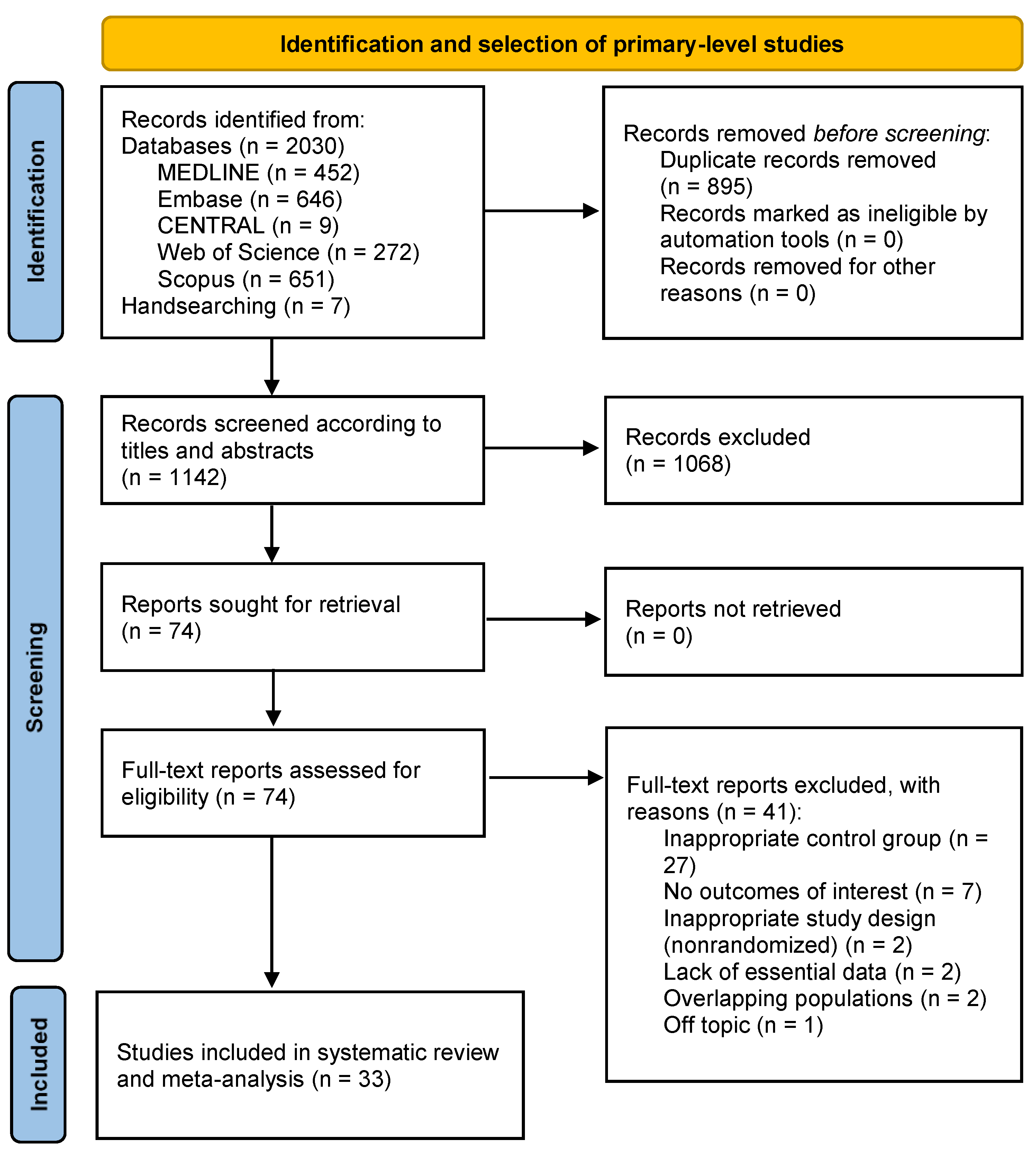
3.1. Results of the Literature Search

The flow diagram (Figure 1) graphically depicts the searching and subsequent process of identification, screening, and selection studies. Overall, 2037 records were retrieved: 646 from Embase, 452 from MEDLINE, 651 from Scopus, 272 from Web of Science, 9 from CENTRAL, and 7 from hand-searching reference lists. After duplicates were removed, 1144 records were piloted and screened according to titles and abstracts, leaving a sample of 77 papers for full-text evaluation (in the Supplementary Information, exhibits the studies excluded jointly with their corresponding exclusion criteria). Finally, 33 studies meeting all eligibility criteria were included for qualitative evaluation and meta-analysis [37,38,39,40,41,42,43,44,45,46,47,48,49,50,51,52,53,54,55,56,57,58,59,60,61,62,63,64,65,66,67,68,69].

3.2. Study Characteristics

Table 1 summarizes the main characteristics of our study sample, and Table S2 (Supplementary Information) exhibits in detail the main variables gathered. These 33 primary-level studies analyzed a total of 4766 cases (2525 from the intervention group and 2241 controls), ranging between 20 and 744. Twenty-three studies were designed as RCTs and ten as q-RCTs. In relation to the interventions under investigation, the single application of CHX was the most frequently used (n = 31), followed by its association with chitosan (n = 4) and antibiotics (n = 4). Twenty-one of them were applied in rinse form, and the rest in a gel vehicle (n = 18), while 0.2% and 0.12% CHX were the most common concentrations (n = 26 and n = 11, respectively). Finally, the oral surgery procedures investigated were third molar (n = 28) and simple tooth extractions (n = 4), periodontal surgery (n = 4), and oral biopsies (n = 4).

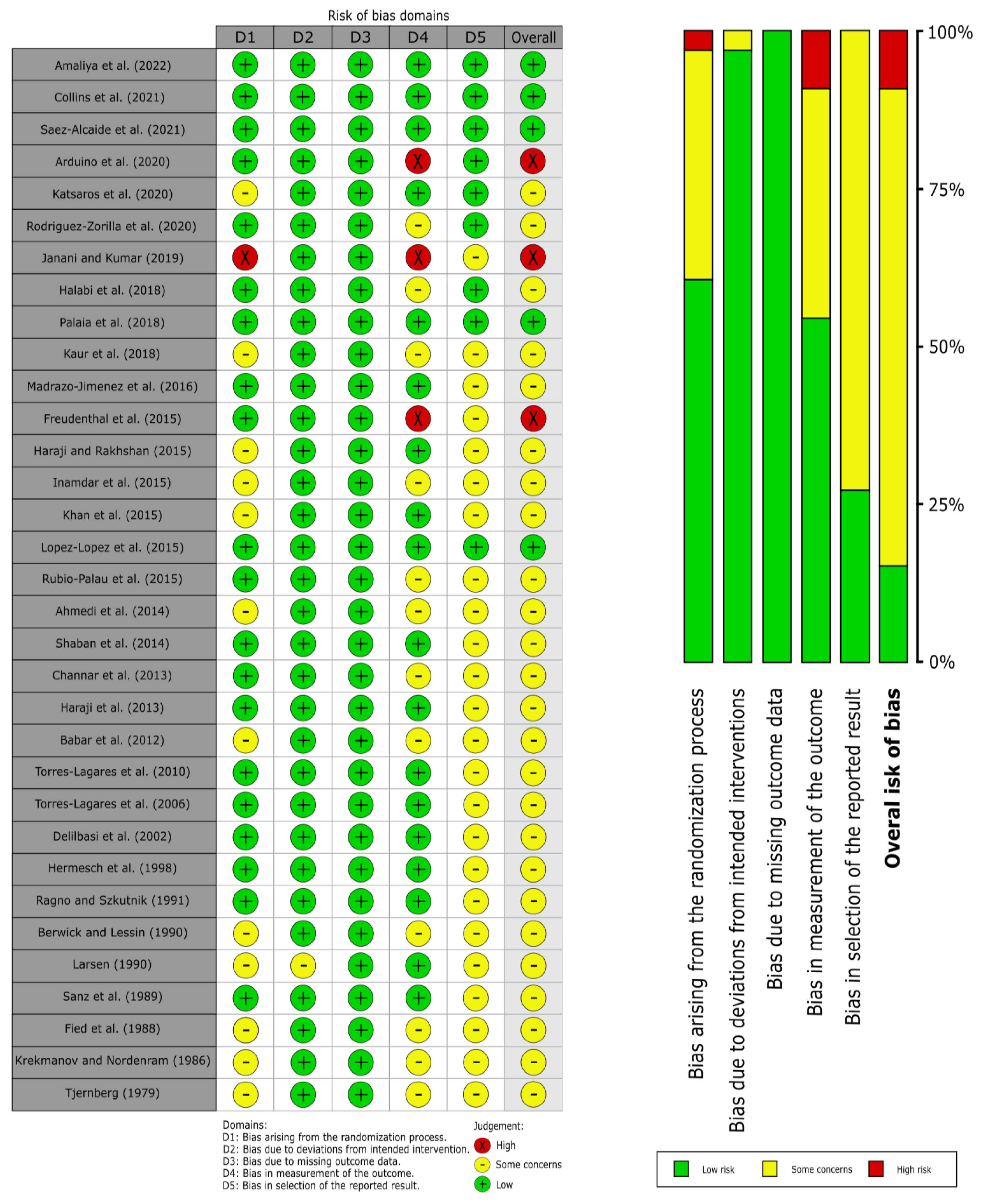
3.3. Qualitative Evaluation

The evaluation of the risk of bias for each domain was carried out by applying the RoB2 tool (Figure 2) [30], and the following results were obtained:
Domain 1 (bias arising from the randomization process) obtained a low RoB in 60.61% of the studies, some concerns in 36.36%, and a high RoB in 3.03%. The most common bias was not specifying the randomization process of the studies since many of them only mentioned the study being randomized and did not provide more information on how said randomization was carried out. The most relevant bias was a failure to blind the allocation sequence (allocation sequence concealed).
Domain 2 (bias due to deviations from intended intervention) obtained a low RoB in 96.97% of the studies and some concerns in 3.03%. The most common bias did not provide clear information on whether participants and clinics knew the allocation of each group and whether that knowledge affected the results. The most relevant bias was the lack of information on whether any deviation from the planned study intervention occurred due to knowledge of the allocation sequence for each group and whether or not such deviation affected the results.
Domain 3 (bias due to missing outcome data) obtained a low RoB in 100% of the studies since all of them provided data on almost the entire studied cases; missing data had a size small enough to not affect the results.
Domain 4 (bias in the measurement of the outcome) obtained a low RoB in 54.55% of studies, some concerns in 36.36%, and a high RoB in 9.09%. The most relevant bias was using an inappropriate measurement method. The most frequent bias was that the person in charge of carrying out the measurements knew the assignment of each group, and the study did not provide clear information about the said knowledge.
Domain 5 (bias in the selection of the reported result) obtained a low RoB in 27.27% of the studies and some concerns in 72.73%. The most common and most relevant bias did not provide information on the existence of a prior analysis plan or protocol completed before the analysis of the results in order to compare whether the subsequent analysis was carried out according to that protocol or not.

3.4. Quantitative Evaluation

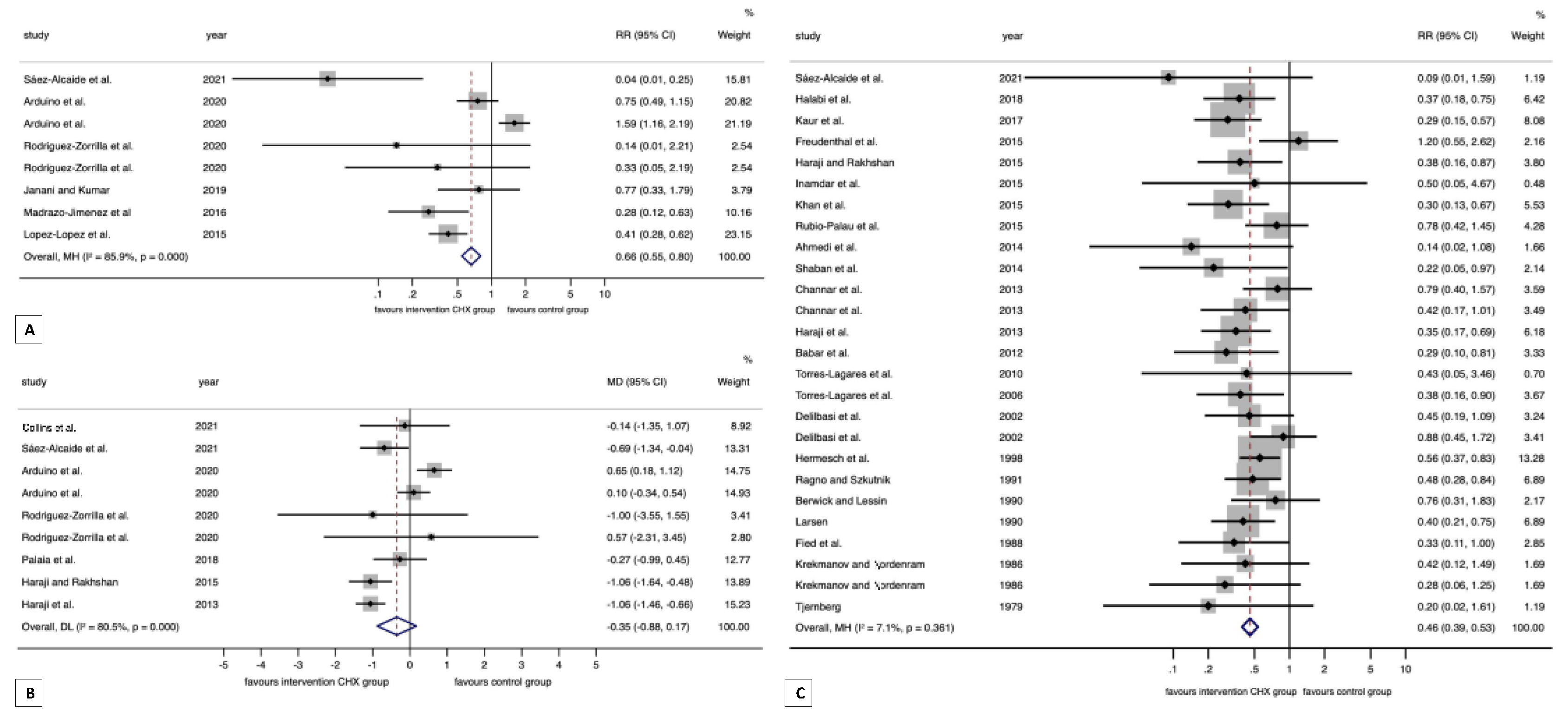
3.4.1. Meta-Analysis on Wound Healing

A significant association was found between the application of CHX and better wound healing (RR = 0.66, 95% CI = 0.55 to 0.80, p < 0.001; RRR = 34%), although a considerable degree of heterogeneity was observed (p < 0.001, I2 = 85.9%). More homogeneous subgroups were found after stratified meta-analyses and most of them preserved the statistically significant association (CHX + chitosan: RR = 0.25, 95% CI = 0.17 to 0.37, p < 0.001; CHX gel: RR = 0.26, 95% CI = 0.18 to 0.37, p < 0.001; CHX 0.12%: RR = 1.59, 95% CI = 1.16 to 2.19, p = 0.004; CHX 0.20%: RR = 0.41, 95% CI = 0.32 to 0.53, p < 0.001; third molar surgery: RR = 0.29, 95% CI = 0.21 to 0.41, p < 0.001; parallel-group design: RR = 0.70, 95% CI = 0.57 to 0.86, p = 0.001; split-mouth design: RR = 0.41, 95% CI = 0.23 to 0.74, p = 0.003; low RoB: RR = 0.26, 95% CI = 0.17 to 0.40, p < 0.001; Some concerns RoB: RR = 0.24, 95% CI = 0.11 to 0.53, p < 0.001) (Table 2, Figure 3A and Figures S1–S6).

3.4.2. Meta-Analysis on Alveolar Osteitis

A significant association after the application of CHX and a lower risk of alveolar osteitis (RR = 0.46, 95% CI = 0.39 to 0.53, p < 0.001) considerably reduced the incidence of alveolar osteitis when compared with the controls (RRR = 54%) and obtained homogeneous results across primary-level studies (heterogeneity: p = 0.36, I2 = 7.1%). Several subgroups also maintained this statically significant result (CHX single: RR = 0.49, 95% CI = 0.41 to 0.57, p < 0.001; CHX + antibiotics: RR = 0.35, 95% CI = 0.23 to 0.54, p < 0.001; CHX + chitosan: RR = 0.09, 95% CI = 0.01 to 1.59, p < 0.001; CHX gel: RR = 0.40, 95% CI = 0.31 to 0.51, p < 0.001; CHX rinse: RR = 0.50, 95% CI = 0.41 to 0.62, p < 0.001; CHX 0.12%: RR = 0.47, 95% CI = 0.37 to 0.59, p < 0.001; CHX 0.20%: RR = 0.46, 95% CI = 0.37 to 0.57, p < 0.001; third molar: RR = 0.48, 95% CI = 0.40 to 0.56, p < 0.001; simple extraction: RR = 0.34, 95% CI = 0.21 to 0.54, p < 0.001; parallel-group design: RR = 0.50, 95% CI = 0.42 to 0.60, p < 0.001; split-mouth design: RR = 0.35, 95% CI = 0.25 to 0.48, p < 0.001; Some concerns RoB: RR = 0.47, 95% CI = 0.38 to 0.52, p < 0.001) (Table 2, Figure 3C and Figures S7–S12).

3.4.3. Meta-Analysis on Erythema

A significant association was found between the application of CHX and better healing, with fewer erythematous wounds (RR = 0.60, 95% CI = 0.39 to 0.93, p = 0.02; RRR = 40%), although a considerable degree of heterogeneity was observed (p = 0.02, I2 = 76.3%) (Table 2, Figure S13). Subgroup meta-analyses were not performed for this variable, where only three primary-level studies entered into the meta-analysis.

3.4.4. Meta-Analysis on Epithelization

A significant association was not found between the application of CHX and epithelization (RR = 1.05, 95% CI = 0.77 to 1.42, p = 0.76), and moderate heterogeneity was also observed (p = 0.06, I2 = 55.6%) (Table 2, Figure S14). Subgroup meta-analyses were not performed for this parameter, where only five primary-level studies entered into the meta-analysis.

3.4.5. Meta-Analysis on Pain during Wound Healing

Pain levels were not significantly different in the CHX group compared to the controls using a visual analog scale (MD = −0.35, 95% CI = −0.88 to 0.17, p = 0.19), which also showed significant heterogeneity (p < 0.001, I2 = 80.5%) (Table 2, Figure 3B and Figures S15–S20). More homogeneous subgroups were found after the stratified meta-analyses, and some of them showed significant differences, indicating slightly less pain in the CHX group (CHX gel: MD = −0.97, 95% CI = −1.26 to −0.68, p < 0.001; Split-mouth design: MD = −1.06, 95% CI = −1.39 to −0.73, p < 0.001; Low RoB: MD = −0.45, 95% CI = −0.90 to −0.003, p = 0.05; Some concerns RoB: MD = −1.04, 95% CI = −1.36 to −0.71, p < 0.001).

3.4.6. Small-Study Effects Analysis

The visual examination of funnel plots’ asymmetry and the corresponding statistical tests were run with the same purpose affirmed for the absence of small-study effects for the variables wound healing (pEgger = 0.11) and pain (pEgger = 0.96), except for alveolar osteitis (pEgger = 0.03) for which biases, e.g., publication bias, could not be ruled out (Figures S21–S23).
Table 2. Meta-analysis of the efficacy of chlorhexidine on wound healing after oral surgery procedures.
Pooled DataHeterogeneity
Meta-AnalysesNo. of Studies *No. of
Cases *
Stat. ModelWtES (95% CI)p-ValuephetI2
(%)
Wound healing
all (poor vs. better wound healing) a8771FEMM-HRR = 0.66 (0.55 to 0.80)<0.001<0.00185.9
Subgroup analysis by type of intervention b<0.001 c
CHX single4527FEMM-HRR = 1.10 (0.87 to 1.40)0.430.0271.3
CHX + chitosan4244FEMM-HRR = 0.25 (0.17 to 0.37)<0.0010.0270.8
Subgroup analysis by type of vehicle b<0.001 c
CHX gel5272FEMM-HRR = 0.26 (0.18 to 0.37)<0.0010.0460.1
CHX rinse3499FEMM-HRR = 1.14 (0.90 to 1.45)0.280.0177
Subgroup analysis by type of concentration b<0.001 c
CHX 0.12%1239RR = 1.59 (1.16 to 2.19)0.004
CHX 0.20%7532FEMM-HRR = 0.41 (0.32 to 0.53)<0.0010.00766.0
Subgroup analysis by type of oral surgery<0.001 c
biopsy2474FEMM-HRR = 1.18 (0.92 to 1.51)0.200.00587.0
third molar6297FEMM-HRR = 0.29 (0.21 to 0.41)<0.0010.0262.2
Subgroup analysis by study design0.09 c
Parallel-group design6696FEMM-HRR = 0.70 (0.57 to 0.86)0.001<0.00188.4
Split-mouth design275FEMM-HRR = 0.41 (0.23 to 0.74)0.0030.0867.3
Subgroup analysis by overall RoB<0.001 c
Low2166FEMM-HRR = 0.26 (0.17 to 0.40)<0.0010.00289.3
Some concerns3106FEMM-HRR = 0.24 (0.11 to 0.53)<0.0010.830.0
High3166FEMM-HRR = 1.14 (0.90 to 1.45)0.280.0177.0
Alveolar osteitis
All (alveolar osteitis vs. healing) a264205FEMM-HRR = 0.46 (0.39 to 0.53)<0.0010.367.1
Subgroup analysis by type of intervention b0.17 c
CHX single213504FEMM-HRR = 0.49 (0.41 to 0.57)<0.0010.3210.7
CHX + antibiotics4629FEMM-HRR = 0.35 (0.23 to 0.54)<0.0010.840.0
CHX + chitosan172RR = 0.09 (0.01 to 1.59)<0.001
Subgroup analysis by type of vehicle b<0.14 c
CHX gel131523FEMM-HRR = 0.40 (0.31 to 0.51)<0.0010.1727.5
CHX rinse132682FEMM-HRR = 0.50 (0.41 to 0.62)<0.0010.710.0
Subgroup analysis by type of concentration b0.52 c
CHX 0.12%71936FEMM-HRR = 0.47 (0.37 to 0.59)<0.0010.780.0
CHX 0.20%182219FEMM-HRR = 0.46 (0.37 to 0.57)<0.0010.1823.6
CHX 1%150RR = 0.14 (0.02 to 1.08)0.06
Subgroup analysis by type of oral surgery b0.17 c
Third molar232992FEMM-HRR = 0.48 (0.40 to 0.56)<0.0010.339.4
Simple extraction31213FEMM-HRR = 0.34 (0.21 to 0.54)<0.0010.920.0
Subgroup analysis by study design0.051 c
Parallel-group design203363FEMM-HRR = 0.50 (0.42 to 0.60)<0.0010.395.4
Split-mouth design6842FEMM-HRR = 0.35 (0.25 to 0.48)<0.0010.750.0
Subgroup analysis by overall RoB b
Low172RR = 0.09 (0.01 to 1.59)0.99
Some concerns244038FEMM-HRR = 0.47 (0.38 to 0.52)<0.0010.640.0
High195RR = 1.20 (0.55 to 2.62)0.65
Erythema
All (erythematous vs. better healing) a388FEMM-HRR = 0.60 (0.39 to 0.93)0.020.0276.3
Epithelization
All (not-epithelized vs. epithelized) a5140FEMM-HRR = 1.05 (0.77 to 1.42)0.760.0655.6
Dehiscence
All (open vs. closed)125RR = 0.77 (0.33 to 1.79)0.55
Pain
All (absolute difference) a9919REMD-LMD = −0.35 (−0.88 to 0.17)0.19<0.00180.5
Subgroup analysis by type of intervention b0.52 c
CHX single7819REMD-LMD = −0.33 (−0.95 to 0.28)0.29<0.00184.9
CHX + chitosan2100REMD-LMD = −0.63 (−1.26 to 0.01)0.0520.400.0
Subgroup analysis by type of vehicle b<0.001 c
CHX gel5378REMD-LMD = −0.97 (−1.26 to−0.68)<0.0010.720.0
CHX rinse4541REMD-LMD = 0.18 (−0.25 to 0.60)0.410.1346.8
Subgroup analysis by type of concentration b0.71 c
CHX 0.12%3436REMD-LMD = −0.19 (−1.48 to 1.10)0.77<0.00193.3
CHX 0.20%6483REMD-LMD = −0.45 (−0.95 to 0.05)0.080.0556.0
Subgroup analysis by type of oral surgery b<0.001 c
Biopsy3504REMD-LMD = 0.21 (−0.29 to 0.70)0.410.0761.9
Periodontal137MD = −0.14 (−1.35 to 1.07)0.82
Third molar5378REMD-LMD = −0.97 (−1.26 to −0.68)<0.0010.720.0
Subgroup analysis by study design b<0.001 c
Parallel-group design7669REMD-LMD = −0.03 (−0.48 to 0.42)0.900.0553.4
Split-mouth design2250REMD-LMD = −1.06 (−1.39 to −0.73)<0.0010.990.0
Subgroup analysis by overall RoB b<0.001 c
Low3138REMD-LMD = −0.45
(−0.90 to −0.003)
0.050.600.0
Some concerns4306REMD-LMD = −1.04
(−1.36 to −0.71)
<0.0010.750.0
High2475REMD-LMD = 0.37 (−0.17 to 0.91)0.180.1064.1
Abbreviations: Stat., statistical; Wt, method of weighting; M-H, Mantel–Haenszel method; D-L, DerSimonian and Laird method; ES, effect size; FEM, fixed-effect model; REM, random-effects model; RR, relative risk; MD, mean difference; CI, confidence intervals; RoB, risk of bias. *—Note that more than one analysis unit was analyzed per study. a—Meta-analysis. b—Subgroup meta-analysis. c—Test for between-subgroup differences.

4. Discussion

The present systematic review and meta-analysis carried out on 33 studies and 4766 cases provides evidence-based results on the benefits of using CHX after oral surgical procedures. The application of CHX was 1.52 times more effective (RR = 0.66, 95% CI = 0.55 to 0.80, p < 0.001) in the healing of oral surgical wounds compared to the controls in which CHX was not used; this translates into 34% of patients who underwent oral surgery treated with CHX presenting with a significant reduction in the risk of developing surgical complications and/or having poor surgical wound healing. The benefits of applying CHX probably derive from its antimicrobial effect mediated by its control of biofilm through a reduction in the oral pathogen load [61]. The development of infections has been directly associated with delayed wound healing, which is due to local inflammation, the inhibition of angiogenesis, decreased epithelization, and delayed final tissue remodeling [12]. Therefore, the use of antiseptic agents is necessary for better microbial control of wounds after oral surgery. In this sense, our meta-analysis confirmed that CHX is an effective agent for this purpose, with broad additional benefits for clinical practice derived from its low cost, easy application, and safety, which makes its routine and use possible; an additional advantage is its availability in different vehicles [23]. In relation to the forms of application, our study has also demonstrated a significantly higher efficacy for the use of CHX in gel vs. the use of CHX in rinses (p < 0.001), at a concentration of 0.20% vs. 0.12% (p < 0.001). The use of CHX gel appears to exert a more extended pharmacological activity than rinse at the point of application [70], despite the fact that in this form of application, the contact time of the drug with the lesion cannot be effectively controlled; however, in cases of deep wounds that are difficult to access, the patient may have some difficulty in applying the drug adequately. On the other hand, the greater efficacy of CHX at a concentration of 0.20% is justified by its demonstrated dose-dependent effect, with bacteriostatic properties at low doses and bactericidal properties at high concentrations [24,25].
Interestingly, in our meta-analysis, oral wound healing was much higher in the subgroup of patients in which CHX was associated with chitosan, reaching a benefit of wound healing that was four times higher than that observed in the controls that did not use CHX plus chitosan; this translates into a reduced risk of complications and/or poor healing in 75% of the cases. In spite of the fact that there are not a large number of clinical trials that justify this result (n = 4), a large magnitude of the effect observed (RR = 0.25) and the narrow confidence interval obtained in the analysis (95% CI = 0.17 to 0.37) certify the robustness of this result. Furthermore, the use of CHX plus chitosan showed significantly higher efficacy than the use of CHX alone (p < 0.001); all this provides high-quality scientific evidence indicating that the joint use of CHX and chitosan should be recommended in clinical practice. The efficacy of chitosan has been contrasted in vitro in preclinical studies [71,72], in vivo by animal experimentation [73], and in clinical research with patients, where beneficial properties such as improved epithelialization have also been observed [74,75,76] alongside the activation of neoangiogenesis, promotion of fibroblastic colonization [77,78], and acceleration of wound healing time [79,80].
Another important result of our meta-analysis demonstrates the efficacy of CHX in preventing the development of alveolar osteitis. In cases in which CHX was applied, both in gel and rinses, the incidence of alveolar osteitis after any type of exodontia was 2.17 times lower than in the controls, implying that 54% of patients presented a decreased risk of developing this complication; the magnitude of this effect (RR = 0.46) together with the narrow confidence interval obtained (95% CI = 0.39–0.53) points to the robustness of this statistically significant result (p < 0.001). This result has been confirmed in previous meta-analytical studies [81,82,83,84,85], although our meta-analysis, by including twice as many studies and cases as those collected by any of the preceding meta-analyses, reinforces evidence of the reported results and advises the routine use of CHX after tooth extractions, since it is known that the development of alveolar osteitis is a serious complication associated with severe pain and impaired quality of life for patients [86,87]. Only one study addresses the effect of the combined use of CHX (0.20%) and chitosan applied as a gel in the prevention of alveolar osteitis after surgical third-molar extractions [41]. Although a single clinical trial cannot support solid evidence, the results show a remarkable trend effect that makes it advisable to further investigate this research line.
Our meta-analysis also indicates that the application of CHX in gel form (no data on rinses) generated a significant decrease in post-surgical pain after third molar surgery compared to the controls (MD = −0.97, 95% CI = −1.26 to −0.68, p < 0.001). Moreover, the statistical results were homogeneous (phet = 0.72, I2 = 0.0%), confirming that this effect is consistent and robust. This decrease in pain was also significantly higher after the application of CHX with third molar surgery compared to periodontal surgery and biopsy (p < 0.001). Again, in this aspect, evidence on the goodness of using CHX plus chitosan for the prevention of pain after third molar surgery derives exclusively from two clinical trials which, although by definition do not support solid evidence, yield a result very close to significance (p = 0.052), suggesting that new research studies probably allow confirmation of the benefits regarding the combination of both drugs.
Although all primary-level studies included in the present systematic review had the same study design (i.e., randomized clinical trials), not all of them were designed with the same conscientiousness. The domain that had the higher risk of bias was bias in the selection of the reported result (72.73% as some concerns) since many studies did not mention having a previous protocol with which to compare the methodology used to see if the measurements were made from different methods; instead, the most favorable ones were chosen for the study results. After stratifying the meta-analysis by overall study quality, we could also demonstrate that the subgroup of studies that had a better methodological design and quality identified significant differences for the parameters of wound healing, alveolar osteitis, and pain, as well as a larger effect size. This led us to confirm that the better the study was designed, the better it could demonstrate the efficacy of CHX in wound healing. Therefore, clinical trials focusing on this research topic should be more rigorous in the methodological design in order to reduce the risk of bias, obtain reliable results, and standardize future research. Future studies should preferably be prospective RCTs with a pre-established full trial protocol—accessible, precise, and transparent—and a larger sample size. Furthermore, they should standardize the clinical procedures for the application of CHX, the postoperative follow-up periods, as well as the criteria used to determine the efficacy of this drug on oral wound healing.
Some potential limitations of our systematic reviews and meta-analysis should also be discussed. First, considerable clinical heterogeneity sources were expected, and some degree of statistical heterogeneity was also demonstrated through meta-analytical techniques for the parameters of wound healing and pain. In order to overcome this limitation, stratified meta-analyses were conducted, showing more homogeneous subgroups of studies and confirming the potential sources of heterogeneity (e.g., the relevance of the CHX vehicle -gel vs. rinse- or variations due to different surgical approaches). Second, the presence of publication bias could not be ruled out for all parameters. Unfortunately, the trend of mostly positive results when published—rejecting findings on the basis of the direction or strength of the study results—constitutes a challenge that is hard to overcome in the current biomedical sciences era. Finally, an inherent limitation of primary-level studies included in this systematic review—highlighted after our risk of bias critical appraisal and data analysis—was the failure to report important information and datasets (e.g., within-patient correlations in split-mouth study designs). This drawback limited the number of observations gathered and challenged the performance of relevant adjusted secondary analyses through subgroup meta-analyses or meta-regressions. Given the clinical and methodological importance of these reflections, we encourage future studies to communicate their datasets in a more rigorous way, preferably reporting individual patient data. Despite the above limitations, the robust nature of the present study is remarkable, providing relevant findings with direct translational applicability to clinical practice, analyzing the effect of CHX on oral wound healing in a comprehensive approach on a large sample of primary-level studies and patients using meta-analytical techniques.

5. Conclusions

In conclusion, the results of our systematic review and meta-analysis demonstrate evidence that the application of CHX exerts a beneficial effect on oral surgical wound healing, significantly decreasing patients’ risk of developing surgical complications and/or poor wound healing. This benefit is greater when CHX is used at 0.20% in gel form. Furthermore, it has been shown that the addition of chitosan to CHX significantly increases its beneficial effect on surgical wound healing; therefore, the use of CHX plus chitosan for the preventive treatment of oral surgical wounds should be advised. The use of CHX has been especially beneficial in the prevention of alveolar osteitis after any type of dental extraction, and it has also been effective—when applied as a gel—in the reduction in pain after the surgical extraction of third molars. There is insufficient evidence, due to scarce research on the efficacy of the joint use of CHX and chitosan for the prevention of alveolar osteitis and pain after dental extractions, although the results of a few clinical trials on the subject seem to indicate a favorable effect, and it is advisable to increase research in this regard.

Supplementary Materials

The following supporting information can be downloaded at: https://www.mdpi.com/article/10.3390/antibiotics12101552/s1. Table S1. Search strategy for each database, number of results, and execution date; Table S2. Characteristics of analyzed studies (n = 33); Figure S1. Forest plot graphically representing the stratified meta-analysis by type of intervention on the association between the application of CHX and wound healing; Figure S2. Forest plot graphically representing the stratified meta-analysis by type of vehicle on the association between the application of CHX and wound healing; Figure S3. Forest plot graphically representing the stratified meta-analysis by type of concentration on the association between the application of CHX and wound healing; Figure S4. Forest plot graphically representing the stratified meta-analysis by type of oral surgery on the association between the application of CHX and wound healing; Figure S5. Forest plot graphically representing the stratified meta-analysis by study design on the association between the application of CHX and wound healing; Figure S6. Forest plot graphically representing the stratified meta-analysis by overall RoB on the association between the application of CHX and wound healing; Figure S7. Forest plot graphically representing the stratified meta-analysis by type of intervention on the association between the application of CHX and alveolar osteitis; Figure S8. Forest plot graphically representing the stratified meta-analysis by type of vehicle on the association between the application of CHX and alveolar osteitis; Figure S9. Forest plot graphically representing the stratified meta-analysis by type of concentration on the association between the application of CHX and alveolar osteitis; Figure S10. Forest plot graphically representing the stratified meta-analysis by type of oral surgery on the association between the application of CHX and alveolar osteitis; Figure S11. Forest plot graphically representing the stratified meta-analysis by type of study design on the association between the application of CHX and alveolar osteitis; Figure S12. Forest plot graphically representing the stratified meta-analysis by overall RoB on the association between the application of CHX and alveolar osteitis; Figure S13. Forest plot graphically representing the meta-analysis on the association between the application of CHX and wound erythema; Figure S14. Forest plot graphically representing the meta-analysis on the association between the application of CHX and wound epithelization; Figure S15. Forest plot graphically representing the stratified meta-analysis by type of intervention on the differences in pain between the CHX group and controls; Figure S16. Forest plot graphically representing the stratified meta-analysis by type of vehicle on the differences in pain between the CHX group and controls; Figure S17. Forest plot graphically representing the stratified meta-analysis by type of concentration on the differences in pain between the CHX group and controls; Figure S18. Forest plot graphically representing the stratified meta-analysis by type of oral surgery on the differences in pain between the CHX group and controls; Figure S19. Forest plot graphically representing the stratified meta-analysis by type of study design on the differences in pain between the CHX group and controls; Figure S20. Forest plot graphically representing the stratified meta-analysis by overall RoB on the differences in pain between the CHX group and controls; Figure S21. A funnel plot of estimated effect sizes against their standard errors, graphically representing the analysis of small-study effects on the association between the application of CHX and wound healing; Figure S22. A funnel plot of estimated effect sizes against their standard errors, graphically representing the analysis of small-study effects on the association between the application of CHX and alveolar osteitis; Figure S23. A funnel plot of the differences in pain between CHX group and controls, expressed as mean differences against their standard errors; List of full-text articles excluded with reasons.

Author Contributions

Conceptualization, M.d.N.R.-O., E.B., P.R.-G. and M.Á.G.-M.; methodology, M.d.N.R.-O., E.B., P.R.-G. and M.Á.G.-M.; software, M.d.N.R.-O., E.B., P.R.-G. and M.Á.G.-M.; validation, M.d.N.R.-O., E.B., P.R.-G. and M.Á.G.-M.; formal analysis, M.d.N.R.-O., E.B., P.R.-G. and M.Á.G.-M.; investigation, M.d.N.R.-O., E.B., P.R.-G. and M.Á.G.-M.; resources, M.d.N.R.-O., E.B., P.R.-G. and M.Á.G.-M.; data curation, M.d.N.R.-O., E.B., P.R.-G. and M.Á.G.-M.; writing—original draft preparation, M.d.N.R.-O., E.B., P.R.-G. and M.Á.G.-M.; writing—review and editing, M.d.N.R.-O., E.B., P.R.-G. and M.Á.G.-M.; visualization, M.d.N.R.-O., E.B., P.R.-G. and M.Á.G.-M.; supervision, M.d.N.R.-O., P.R.-G. and M.Á.G.-M. All authors have read and agreed to the published version of the manuscript.

Funding

This research received no external funding.

Institutional Review Board Statement

Not applicable.

Informed Consent Statement

Not applicable.

Data Availability Statement

Data are contained within the article or Supplementary Material.

Acknowledgments

We would like to thank the research group CTS-392 (Plan Andaluz de Investigación, Junta de Andalucía, Spain).

Conflicts of Interest

The authors declare no conflict of interest.

References

  1. Broughton, G.; Janis, J.E.; Attinger, C.E. Wound Healing: An Overview. Plast. Reconstr. Surg. 2006, 117, 1e-S–32e-S. [Google Scholar] [CrossRef] [PubMed]
  2. Politis, C.; Schoenaers, J.; Jacobs, R.; Agbaje, J.O. Wound Healing Problems in the Mouth. Front. Physiol. 2016, 7, 507. [Google Scholar] [CrossRef] [PubMed]
  3. Bielefeld, K.A.; Amini-Nik, S.; Alman, B.A. Cutaneous wound healing: Recruiting developmental pathways for regeneration. Cell. Mol. Life Sci. 2013, 70, 2059–2081. [Google Scholar] [CrossRef] [PubMed]
  4. Eming, S.A.; Martin, P.; Tomic-Canic, M. Wound repair and regeneration: Mechanisms, signaling, and translation. Sci. Transl. Med. 2014, 6, 265sr6. [Google Scholar] [CrossRef] [PubMed]
  5. González-Moles, M.Á.; Ramos-García, P. State of Evidence on Oral Health Problems in Diabetic Patients: A Critical Review of the Literature. J. Clin. Med. 2021, 10, 5383. [Google Scholar] [CrossRef] [PubMed]
  6. Loftus, M.J.; Peterson, L.J. Delayed healing of mandibular fracture in idiopathic myxedema. Oral Surg. Oral Med. Oral Pathol. 1979, 47, 233–237. [Google Scholar] [CrossRef] [PubMed]
  7. Aghaloo, T.; Pi-Anfruns, J.; Moshaverinia, A.; Sim, D.; Grogan, T.; Hadaya, D. The Effects of Systemic Diseases and Medications on Implant Osseointegration: A Systematic Review. Int. J. Oral Maxillofac. Implants 2019, 34, S35–S49. [Google Scholar] [CrossRef] [PubMed]
  8. Gál, P.; Brábek, J.; Holub, M.; Jakubek, M.; Šedo, A.; Lacina, L.; Strnadová, K.; Dubový, P.; Hornychová, H.; Ryška, A.; et al. Autoimmunity, cancer and COVID-19 abnormally activate wound healing pathways: Critical role of inflammation. Histochem. Cell Biol. 2022, 158, 415–434. [Google Scholar] [CrossRef]
  9. Kalyan, S.; Wang, J.; Quabius, E.S.; Huck, J.; Wiltfang, J.; Baines, J.F.; Kabelitz, D. Systemic immunity shapes the oral microbiome and susceptibility to bisphosphonate-associated osteonecrosis of the jaw. J. Transl. Med. 2015, 13, 212. [Google Scholar] [CrossRef]
  10. Vellappally, S.; Fiala, Z.; Smejkalová, J.; Jacob, V.; Somanathan, R. Smoking related systemic and oral diseases. Acta Medica-Hradec Kral. 2007, 50, 161–166. [Google Scholar] [CrossRef]
  11. Hampton, S. Bacteria and wound healing. J. Community Nurs. 2007, 21, 32–40. [Google Scholar]
  12. Jones, S.G.; Edwards, R.; Thomas, D.W. Inflammation and Wound Healing: The Role of Bacteria in the Immuno-Regulation of Wound Healing. Int. J. Low. Extrem. Wounds 2004, 3, 201–208. [Google Scholar] [CrossRef] [PubMed]
  13. Robson, M.C. Wound infection. Surg. Clin. N. Am. 1997, 77, 637–650. [Google Scholar] [CrossRef]
  14. Bolton, L.L.; van Rijswijk, L.; Shaffer, F.A. Quality wound care equals cost-effective wound care: A clinical model. Adv. Wound Care 1997, 10, 33–38. [Google Scholar] [CrossRef] [PubMed]
  15. Steed, D.L.; Attinger, C.; Colaizzi, T.; Crossland, M.; Franz, M.; Harkless, L.; Johnson, A.; Moosa, H.; Robson, M.; Serena, T.; et al. Guidelines for the treatment of diabetic ulcers. Wound Repair Regen. 2006, 14, 680–692. [Google Scholar] [CrossRef] [PubMed]
  16. Moreno-Drada, J.A.; García-Perdomo, H.A. Effectiveness of Antimicrobial Prophylaxis in Preventing the Spread of Infection as a Result of Oral Procedures: A Systematic Review and Meta-Analysis. J. Oral Maxillofac. Surg. 2016, 74, 1313–1321. [Google Scholar] [CrossRef] [PubMed]
  17. Ghosh, A.; Aggarwal, V.R.; Moore, R. Aetiology, Prevention and Management of Alveolar Osteitis—A Scoping Review. J. Oral Rehabil. 2022, 49, 103–113. [Google Scholar] [CrossRef] [PubMed]
  18. Au, A.H.Y.; Choi, S.W.; Cheung, C.W.; Leung, Y.Y. The Efficacy and Clinical Safety of Various Analgesic Combinations for Post-Operative Pain after Third Molar Surgery: A Systematic Review and Meta-Analysis. PLoS ONE 2015, 10, e0127611. [Google Scholar] [CrossRef]
  19. Bouloux, G.F.; Steed, M.B.; Perciaccante, V.J. Complications of Third Molar Surgery. Oral Maxillofac. Surg. Clin. N. Am. 2007, 19, 117–128. [Google Scholar] [CrossRef]
  20. Belusic-Gobic, M.; Car, M.; Juretic, M.; Cerovic, R.; Gobic, D.; Golubovic, V. Risk factors for wound infection after oral cancer surgery. Oral Oncol. 2007, 43, 77–81. [Google Scholar] [CrossRef]
  21. Cho, H.; Lynham, A.J.; Hsu, E. Postoperative interventions to reduce inflammatory complications after third molar surgery: Review of the current evidence. Aust. Dent. J. 2017, 62, 412–419. [Google Scholar] [CrossRef]
  22. Zhu, J.; Zhang, S.; Yuan, X.; He, T.; Liu, H.; Wang, J.; Xu, B. Effect of platelet-rich fibrin on the control of alveolar osteitis, pain, trismus, soft tissue healing, and swelling following mandibular third molar surgery: An updated systematic review and meta-analysis. Int. J. Oral Maxillofac. Surg. 2021, 50, 398–406. [Google Scholar] [CrossRef] [PubMed]
  23. Fiorillo, L. Chlorhexidine Gel Use in the Oral District: A Systematic Review. Gels 2019, 5, 31. [Google Scholar] [CrossRef] [PubMed]
  24. Fine, D.H. Mouthrinses as adjuncts for plaque and gingivitis management. A status report for the American Journal of Dentistry. Am. J. Dent. 1988, 1, 259–263. [Google Scholar] [PubMed]
  25. Hugo, W.B.; Longworth, A.R. The effect of chlorhexidine on the electrophoretic mobility, cytoplasmic constituents, dehydrogenase activity and cell walls of Escherichia coli and Staphylococcus aureus. J. Pharm. Pharmacol. 1966, 18, 569–578. [Google Scholar] [CrossRef] [PubMed]
  26. Rodríguez-Pérez, M.; Bravo-Pérez, M.; Sánchez-López, J.-D.; Muñoz-Soto, E.; Romero-Olid, M.-N.; Baca-García, P. Effectiveness of 1% versus 0.2% chlorhexidine gels in reducing alveolar osteitis from mandibular third molar surgery: A randomized, double-blind clinical trial. Med. Oral Patol. Oral Cir. Bucal 2013, 18, e693–e700. [Google Scholar] [CrossRef] [PubMed]
  27. Higgins, J.P.; Green, S. Cochrane Handbook for Systematic Reviews of Interventions: Cochrane Book Series; Wiley and Sons: Hoboken, NJ, USA, 2008; pp. 1–649. [Google Scholar] [CrossRef]
  28. Page, M.J.; McKenzie, J.E.; Bossuyt, P.M.; Boutron, I.; Hoffmann, T.C.; Mulrow, C.D.; Shamseer, L.; Tetzlaff, J.M.; Akl, E.A.; Brennan, S.E.; et al. The PRISMA 2020 statement: An updated guideline for reporting systematic reviews. BMJ 2021, 372, n71. [Google Scholar] [CrossRef] [PubMed]
  29. Shamseer, L.; Moher, D.; Clarke, M.; Ghersi, D.; Liberati, A.; Petticrew, M.; Shekelle, P.; Stewart, L.A. PRISMA-P Group Preferred reporting items for systematic review and meta-analysis protocols (PRISMA-P) 2015: Elaboration and explanation. BMJ 2015, 350, g7647. [Google Scholar] [CrossRef] [PubMed]
  30. Sterne, J.A.C.; Savović, J.; Page, M.J.; Elbers, R.G.; Blencowe, N.S.; Boutron, I.; Cates, C.J.; Cheng, H.-Y.; Corbett, M.S.; Eldridge, S.M.; et al. RoB 2: A revised tool for assessing risk of bias in randomised trials. BMJ 2019, 366, l4898. [Google Scholar] [CrossRef]
  31. Luo, D.; Wan, X.; Liu, J.; Tong, T. Optimally estimating the sample mean from the sample size, median, mid-range, and/or mid-quartile range. Stat. Methods Med. Res. 2018, 27, 1785–1805. [Google Scholar] [CrossRef]
  32. Wan, X.; Wang, W.; Liu, J.; Tong, T. Estimating the sample mean and standard deviation from the sample size, median, range and/or interquartile range. BMC Med. Res. Methodol. 2014, 14, 135. [Google Scholar] [CrossRef]
  33. Higgins, J.P.T.; Thompson, S.G. Quantifying heterogeneity in a meta-analysis. Stat. Med. 2002, 21, 1539–1558. [Google Scholar] [CrossRef] [PubMed]
  34. Higgins, J.P.T.; Thompson, S.G.; Deeks, J.J.; Altman, D.G. Measuring inconsistency in meta-analyses. BMJ 2003, 327, 557–560. [Google Scholar] [CrossRef] [PubMed]
  35. Sterne, J.A.C.; Sutton, A.J.; Ioannidis, J.P.A.; Terrin, N.; Jones, D.R.; Lau, J.; Carpenter, J.; Rücker, G.; Harbord, R.M.; Schmid, C.H.; et al. Recommendations for examining and interpreting funnel plot asymmetry in meta-analyses of randomised controlled trials. BMJ 2011, 343, d4002. [Google Scholar] [CrossRef] [PubMed]
  36. Egger, M.; Davey Smith, G.; Schneider, M.; Minder, C. Bias in meta-analysis detected by a simple, graphical test. BMJ 1997, 315, 629–634. [Google Scholar] [CrossRef] [PubMed]
  37. Torres-Lagares, D.; Gutierrez-Perez, J.L.; Infante-Cossio, P.; Garcia-Calderon, M.; Romero-Ruiz, M.M.; Serrera-Figallo, M.A. Randomized, double-blind study on effectiveness of intra-alveolar chlorhexidine gel in reducing the incidence of alveolar osteitis in mandibular third molar surgery. Int. J. Oral Maxillofac. Surg. 2006, 35, 348–351. [Google Scholar] [CrossRef] [PubMed]
  38. Berwick, J.E.; Lessin, M.E. Effects of a chlorhexidine gluconate oral rinse on the incidence of alveolar osteitis in mandibular third molar surgery. J. Oral Maxillofac. Surg. Off. J. Am. Assoc. Oral Maxillofac. Surg. 1990, 48, 444–448. [Google Scholar] [CrossRef] [PubMed]
  39. Freudenthal, N.; Sternudd, M.; Jansson, L.; Wannfors, K. A double-blind randomized study evaluating the effect of intra-alveolar chlorhexidine gel on alveolar osteitis after removal of mandibular third molars. J. Oral Maxillofac. Surg. Off. J. Am. Assoc. Oral Maxillofac. Surg. 2015, 73, 600–605. [Google Scholar] [CrossRef] [PubMed]
  40. Tjernberg, A. Influence of oral hygiene measures on the development of alveolitis sicca dolorosa after surgical removal of mandibular third molars. Int. J. Oral Surg. 1979, 8, 430–434. [Google Scholar] [CrossRef]
  41. Sáez-Alcaide, L.; Molinero-Mourelle, P.; González-Serrano, J.; Rubio-Alonso, L.; Bornstein, M.; López-Quiles, J. Efficacy of a topical gel containing chitosan, chlorhexidine, allantoin and dexpanthenol for pain and inflammation control after third molar surgery: A randomized and placebo-controlled clinical trial. Med. Oral Patol. Oral Cir. Bucal 2020, 25, e644–e651. [Google Scholar] [CrossRef]
  42. Rodríguez Zorrilla, S.; Blanco Carrión, A.; García García, A.; Galindo Moreno, P.; Marichalar Mendía, X.; Seoane Prado, R.; Pérez Estévez, A.J.; Pérez-Sayáns, M. Effect of antiseptic gels in the microbiologic colonization of the suture threads after oral surgery. Sci. Rep. 2020, 10, 8360. [Google Scholar] [CrossRef] [PubMed]
  43. Lope-Lopez, J.; Jan-Palli, E.; Gonzalez-Navarro, B.; Jane-Salas, E.; Estrugo-Devesa, A.; Milani, M.; Lopez-Lopez, J.; Jan-Pallí, E.; lez-Navarro, B.G.; Jané-Salas, E.; et al. Efficacy of chlorhexidine, dexpanthenol, allantoin and chitosan gel in comparison with bicarbonate oral rinse in controlling post-interventional inflammation, pain and cicatrization in subjects undergoing dental surgery. Curr. Med. Res. Opin. 2015, 31, 2179–2183. [Google Scholar] [CrossRef] [PubMed]
  44. Amaliya, A.; Ramadhanti, R.; Hadikrishna, I.; Maulina, T. The Effectiveness of 0.2% Chlorhexidine Gel on Early Wound Healing after Tooth Extraction: A Randomized Controlled Trial. Eur. J. Dent. 2022, 16, 688–694. [Google Scholar] [CrossRef] [PubMed]
  45. Haraji, A.; Rakhshan, V.; Khamverdi, N.; Alishahi, H.K. Effects of intra-alveolar placement of 0.2% chlorhexidine bioadhesive gel on dry socket incidence and postsurgical pain: A double-blind split-mouth randomized controlled clinical trial. J. Orofac. Pain 2013, 27, 256–262. [Google Scholar] [CrossRef] [PubMed]
  46. Delilbasi, C.; Saracoglu, U.; Keskin, A. Effects of 0.2% chlorhexidine gluconate and amoxicillin plus clavulanic acid on the prevention of alveolar osteitis following mandibular third molar extractions. Oral Surg. Oral Med. Oral Pathol. Oral Radiol. Endod. 2002, 94, 301–304. [Google Scholar] [CrossRef] [PubMed]
  47. Halabi, D.; Escobar, J.; Alvarado, C.; Martinez, N.; Muñoz, C. Chlorhexidine for prevention of alveolar osteitis: A randomised clinical trial. J. Appl. Oral Sci. 2018, 26, e20170245. [Google Scholar] [CrossRef] [PubMed]
  48. Palaia, G.; Tenore, G.; Tribolati, L.; Russo, C.; Gaimari, G.; Del Vecchio, A.; Romeo, U. Evaluation of wound healing and postoperative pain after oral mucosa laser biopsy with the aid of compound with chlorhexidine and sodium hyaluronate: A randomized double blind clinical trial. Clin. Oral Investig. 2019, 23, 3141–3151. [Google Scholar] [CrossRef] [PubMed]
  49. Ahmedi, J.; Ahmedi, E.; Agani, Z.; Hamiti, V.; Reçica, B.; Tmava-Dragusha, A. The Efficacy of 1% Chlorhexidine Gel on the Reduction of Dry Socket Occurence Following Surgical Third Molar Extraction—Pilot Study. Open J. Stomatol. 2014, 4, 152–160. [Google Scholar] [CrossRef]
  50. Kaur, J.; Raval, R.; Bansal, A.; Kumawat, V. Repercussions of intraalveolar placement of combination of 0.2% chlorhexidine & 10 Mg metronidazole gel on the occurrence of dry sockets—A randomized control trial. J. Clin. Exp. Dent. 2017, 9, e284–e288. [Google Scholar] [CrossRef]
  51. Katsaros, T.; Mayer, E.; Palaiologou, A.; Romero-Bustillos, M.; Evans, G.H.; Lallier, T.E.; Maney, P. Effect of different concentrations of commercially available mouthwashes on wound healing following periodontal surgery: A randomized controlled clinical trial. Clin. Oral Investig. 2020, 24, 3587–3595. [Google Scholar] [CrossRef]
  52. Larsen, P.E. The effect of a chlorhexidine rinse on the incidence of alveolar osteitis following the surgical removal of impacted mandibular third molars. J. Oral Maxillofac. Surg. Off. J. Am. Assoc. Oral Maxillofac. Surg. 1991, 49, 932–937. [Google Scholar] [CrossRef] [PubMed]
  53. Ragno, J.R.J.; Szkutnik, A.J. Evaluation of 0.12% chlorhexidine rinse on the prevention of alveolar osteitis. Oral Surg. Oral Med. Oral Pathol. 1991, 72, 524–526. [Google Scholar] [CrossRef] [PubMed]
  54. Babar, A.; Ibrahim, M.W.; Baig, N.J.; Shah, I.; Amin, E. Efficacy of intra-alveolar chlorhexidine gel in reducing frequency of alveolar osteitis in mandibular third molar surgery. J. Coll. Physicians Surg. Pak. 2012, 22, 91–94. [Google Scholar]
  55. Arduino, P.G.; Gambino, A.; Cabras, M.; Sciannameo, V.; Nimot, Y.; Karimi, D.; Ricceri, F.; Broccoletti, R. Effect of two different alcohol-free chlorhexidine formulations in mouthrinses on the immediate postoperative period for oral mucosal biopsies. J. Oral Sci. 2020, 62, 202–205. [Google Scholar] [CrossRef] [PubMed]
  56. Madrazo-Jimenez, M.; Rodriguez-Caballero, A.; Serrera-Figallo, M.A.; Garrido-Serrano, R.; Gutierrez-Corrales, A.; Gutierrez-Perez, J.L.; Torres-Lagares, D. The effects of a topical gel containing chitosan, 0,2% chlorhexidine, allantoin and despanthenol on the wound healing process subsequent to impacted lower third molar extraction. Med. Oral Patol. Oral Cir. Bucal 2016, 21, e696–e702. [Google Scholar] [CrossRef] [PubMed]
  57. Khan, M.A.; Bashir, S.; Khan, F.R.; Umer, F.; Haider, S.M.; Hasan, T. Clinical efficacy of single dose chlorhexidine gel application in molars extractions-a randomized clinical trial. J. Pakistan Dent. Assoc. 2015, 24, 175–181. [Google Scholar]
  58. Inamdar, M.N.; Chauhan, R.; Mapare, S.A.; Goswami, R.P.; Goswami, Y.; Khadri, S.F. Prevention of Dry Socket using Chlorhexidine Gel and Ornidazole Gel in Impacted Mandibular Third Molar: A Comparative Randomized Prospective Study on 30 Patients. J. Int. Oral Health 2015, 7, 41–46. [Google Scholar]
  59. Hermesch, C.B.; Hilton, T.J.; Biesbrock, A.R.; Baker, R.A.; Cain-Hamlin, J.; McClanahan, S.F.; Gerlach, R.W. Perioperative use of 0.12% chlorhexidine gluconate for the prevention of alveolar osteitis. Oral Surg. Oral Med. Oral Pathol. Oral Radiol. Endodontology 1998, 85, 381–387. [Google Scholar] [CrossRef]
  60. Torres-Lagares, D.; Gutierrez-Perez, J.L.; Hita-Iglesias, P.; Magallanes-Abad, N.; Flores-Ruiz, R.; Basallote-Garcia, M.; Gonzalez-Martin, M. Randomized, double-blind study of effectiveness of intra-alveolar application of chlorhexidine gel in reducing incidence of alveolar osteitis and bleeding complications in mandibular third molar surgery in patients with bleeding disorders. J. Oral Maxillofac. Surg. Off. J. Am. Assoc. Oral Maxillofac. Surg. 2010, 68, 1322–1326. [Google Scholar] [CrossRef]
  61. Sanz, M.; Newman, M.G.; Anderson, L.; Matoska, W.; Otomo—Corgel, J.; Saltini, C. Clinical Enhancement of Post-Periodontal Surgical Therapy by a 0.12% Chlorhexidine Gluconate Mouthrinse. J. Periodontol. 1989, 60, 570–576. [Google Scholar] [CrossRef]
  62. Krekmanov, L.; Nordenram, Å. Postoperative complications after surgical removal of mandibular third molars. Int. J. Oral Maxillofac. Surg. 1986, 15, 25–29. [Google Scholar] [CrossRef]
  63. Shaban, B.; Azimi, H.R.; Naderi, H.; Janani, A.; Zarrabi, M.J.; Nejat, A.H. Effect of 0.2% Chlorhexidine Gel on Frequency of Dry Socket Following Mandibular Third Molar Surgery: A Double-Blind Clinical Trial. J. Dent. Mater. Tech. 2014, 3, 175–184. [Google Scholar]
  64. Collins, J.R.; Veras, K.; Hernández, M.; Hou, W.; Hong, H.; Romanos, G.E. Anti-inflammatory effect of salt water and chlorhexidine 0.12% mouthrinse after periodontal surgery: A randomized prospective clinical study. Clin. Oral Investig. 2021, 25, 4349–4357. [Google Scholar] [CrossRef] [PubMed]
  65. Channar, K.; Dall, A.; Memon, A.; Lal, R. Prevention of alveolar osteitis in surgical removal of lower third molar. Pak. Oral Dent. J. 2013, 3, 244–248. [Google Scholar]
  66. Rubio-Palau, J.; Garcia-Linares, J.; Hueto-Madrid, J.-A.; González-Lagunas, J.; Raspall-Martin, G.; Mareque-Bueno, J. Effect of intra-alveolar placement of 0.2% chlorhexidine bioadhesive gel on the incidence of alveolar osteitis following the extraction of mandibular third molars. A double-blind randomized clinical trial. Med. Oral Patol. Oral Cir. Bucal 2015, 20, e117. [Google Scholar] [CrossRef]
  67. Janani, K.; Kumar, M.P.S. Effectiveness of chlorhexidine and warm saline mouthrinses against bacterial colonization on silk suture material in third molar surgery—A clinico-microbiological study. Int. J. Clin. Dent. 2019, 12, 137–145. [Google Scholar]
  68. Haraji, A.; Rakhshan, V. Chlorhexidine gel and less difficult surgeries might reduce post-operative pain, controlling for dry socket, infection and analgesic consumption: A split-mouth controlled randomised clinical trial. J. Oral Rehabil. 2015, 42, 209–219. [Google Scholar] [CrossRef]
  69. Field, E.A.; Nind, D.; Varga, E.; Martin, M. V The effect of chlorhexidine irrigation on the incidence of dry socket: A pilot study. Br. J. Oral Maxillofac. Surg. 1988, 26, 395–401. [Google Scholar] [CrossRef]
  70. Canullo, L.; Laino, L.; Longo, F.; Filetici, P.; D’Onofrio, I.; Troiano, G. Does Chlorhexidine Prevent Complications in Extractive, Periodontal, and Implant Surgery? A Systematic Review and Meta-analysis with Trial Sequential Analysis. Int. J. Oral Maxillofac. Implants 2020, 35, 1149–1158. [Google Scholar] [CrossRef] [PubMed]
  71. Ribeiro, M.P.; Espiga, A.; Silva, D.; Baptista, P.; Henriques, J.; Ferreira, C.; Silva, J.C.; Borges, J.P.; Pires, E.; Chaves, P.; et al. Development of a new chitosan hydrogel for wound dressing. Wound Repair Regen. 2009, 17, 817–824. [Google Scholar] [CrossRef]
  72. Chatelet, C.; Damour, O.; Domard, A. Influence of the degree of acetylation on some biological properties of chitosan films. Biomaterials 2001, 22, 261–268. [Google Scholar] [CrossRef] [PubMed]
  73. Ishihara, M.; Ono, K.; Sato, M.; Nakanishi, K.; Saito, Y.; Yura, H.; Matsui, T.; Hattori, H.; Fujita, M.; Kikuchi, M.; et al. Acceleration of wound contraction and healing with a photocrosslinkable chitosan hydrogel. Wound Repair Regen. 2001, 9, 513–521. [Google Scholar] [CrossRef] [PubMed]
  74. Kratz, G.; Back, M.; Arnander, C.; Larm, O. Immobilised heparin accelerates the healing of human wounds in vivo. Scand. J. Plast. Reconstr. Surg. Hand Surg. 1998, 32, 381–385. [Google Scholar] [CrossRef] [PubMed]
  75. Ishihara, M.; Nakanishi, K.; Ono, K.; Sato, M.; Kikuchi, M.; Saito, Y.; Yura, H.; Matsui, T.; Hattori, H.; Uenoyama, M.; et al. Photocrosslinkable chitosan as a dressing for wound occlusion and accelerator in healing process. Biomaterials 2002, 23, 833–840. [Google Scholar] [CrossRef] [PubMed]
  76. Hoemann, C.D.; Sun, J.; Légaré, A.; McKee, M.D.; Buschmann, M.D. Tissue engineering of cartilage using an injectable and adhesive chitosan-based cell-delivery vehicle. Osteoarthr. Cartil. 2005, 13, 318–329. [Google Scholar] [CrossRef] [PubMed]
  77. Vescovali, C.; Damour, O.; Shahabedin, L.; David, M. Epidermalization of an artificial dermis made of collagen. Ann. Mediterrian Burn. Club 1989, 2, 137139. [Google Scholar]
  78. Damour, O.; Gueugniaud, P.Y.; Berthin-Maghit, M.; Rousselle, P.; Berthod, F.; Sahuc, F.; Collombel, C. A dermal substrate made of collagen-GAG-chitosan for deep burn coverage: First clinical uses. Clin. Mater. 1994, 15, 273–276. [Google Scholar] [CrossRef] [PubMed]
  79. Azad, A.K.; Sermsintham, N.; Chandrkrachang, S.; Stevens, W.F. Chitosan membrane as a wound-healing dressing: Characterization and clinical application. J. Biomed. Mater. Res. B Appl. Biomater. 2004, 69, 216–222. [Google Scholar] [CrossRef]
  80. Stone, C.A.; Wright, H.; Clarke, T.; Powell, R.; Devaraj, V.S. Healing at skin graft donor sites dressed with chitosan. Br. J. Plast. Surg. 2000, 53, 601–606. [Google Scholar] [CrossRef]
  81. Yengopal, V.; Mickenautsch, S. Chlorhexidine for the prevention of alveolar osteitis. Int. J. Oral Maxillofac. Surg. 2012, 41, 1253–1264. [Google Scholar] [CrossRef]
  82. Teshome, A. The efficacy of chlorhexidine gel in the prevention of alveolar osteitis after mandibular third molar extraction: A systematic review and meta-analysis. BMC Oral Health 2017, 17, 82. [Google Scholar] [CrossRef] [PubMed]
  83. Zhou, J.; Hu, B.; Liu, Y.; Yang, Z.; Song, J. The efficacy of intra-alveolar 0.2% chlorhexidine gel on alveolar osteitis: A meta-analysis. Oral Dis. 2017, 23, 598–608. [Google Scholar] [CrossRef]
  84. Taberner-Vallverdú, M.; Sánchez-Garcés, M.-Á.; Gay-Escoda, C. Efficacy of different methods used for dry socket prevention and risk factor analysis: A systematic review. Med. Oral Patol. Oral Cir. Bucal 2017, 22, e750–e758. [Google Scholar] [CrossRef]
  85. Wang, C.-H.; Yang, S.-H.; Jen, H.-J.; Tsai, J.-C.; Lin, H.-K.; Loh, E.-W. Preventing Alveolar Osteitis After Molar Extraction Using Chlorhexidine Rinse and Gel: A Meta-Analysis of Randomized Controlled Trials. J. Nurs. Res. 2020, 29, e137. [Google Scholar] [CrossRef]
  86. Kolokythas, A.; Olech, E.; Miloro, M. Alveolar Osteitis: A Comprehensive Review of Concepts and Controversies. Int. J. Dent. 2010, 2010, 249073. [Google Scholar] [CrossRef]
  87. Blum, I.R. Contemporary views on dry socket (alveolar osteitis): A clinical appraisal of standardization, aetiopathogenesis and management: A critical review. Int. J. Oral Maxillofac. Surg. 2002, 31, 309–317. [Google Scholar] [CrossRef] [PubMed]
Figure 1. Flow diagram showing the identification and selection process of primary-level studies included in the present systematic review and meta-analysis.
Antibiotics 12 01552 g001
Figure 2. Quality plot graphically representing the risk of bias across primary-level studies and critically appraised using the RoB2 tool.
Antibiotics 12 01552 g002
Figure 3. (A) Forest plot graphically represents the meta-analysis for the association between the application of CHX and wound healing. CHX, chlorhexidine; RR, relative risk; CI, confidence intervals. Fixed-effect model, Mantel–Haenszel method. An RR < 1 suggests that the application of CHX is associated with better wound healing. Diamonds indicate the pooled RRs with their corresponding 95% CIs. (B) The forest plot graphically represents a meta-analysis of the differences in pain between the CHX group and controls. CHX, chlorhexidine; MD, mean difference; CI, confidence intervals. Random-effects model, inverse-variance method. An MD < 0 suggests that pain levels were lower for the CHX group. Diamonds indicate the pooled MD with their corresponding 95% CIs. (C) The forest plot graphically represents the meta-analysis of the association between the application of CHX and alveolar osteitis. CHX, chlorhexidine; RR, relative risk; CI, confidence intervals. Fixed-effect model, Mantel–Haenszel method. An RR < 1 suggests that the application of CHX is associated with a lower risk of alveolar osteitis. Diamonds indicate the pooled RRs with their corresponding 95% CIs.
Antibiotics 12 01552 g003
Table 1. Summarized study characteristics.
Total33 Studies
Year of publication1979–2022
Number of cases
Total4766 cases
Intervention group2525 cases
Control group2241 cases
Sample size, range20 to 744 cases
Type of interventions *
CHX31 studies
CHX + chitosan4 studies
CHX + antibiotics4 studies
Type of CHX vehicles *
Gel18 studies
Rinse21 studies
Type of CHX concentrations *
1%1 study
0.20%26 studies
0.12%11 studies
0.006%1 study
Type of oral surgery procedures *
Third molar surgery28 studies
Simple tooth extraction4 studies
Periodontal surgery4 studies
Oral biopsy3 studies
Type of study design *
Parallel group design31 studies
Split-mouth design8 studies
* Note that six primary-level studies reported two analysis units (total n = 39).
Disclaimer/Publisher’s Note: The statements, opinions and data contained in all publications are solely those of the individual author(s) and contributor(s) and not of MDPI and/or the editor(s). MDPI and/or the editor(s) disclaim responsibility for any injury to people or property resulting from any ideas, methods, instructions or products referred to in the content.

