Article

Phage Cocktails with Daptomycin and Ampicillin Eradicates Biofilm-Embedded Multidrug-Resistant Enterococcus faecium with Preserved Phage Susceptibility

1
Anti-Infective Research Laboratory, College of Pharmacy and Health Sciences, Wayne State University, Detroit, MI 48201, USA
2
Department of Pharmacy Practice, College of Pharmacy, Nova Southeastern University, Davie, FL 33328, USA
3
Department of Pharmacy and Clinical Services, College of Pharmacy, Medical University of South Carolina, Charleston, SC 29208, USA
4
Department of Pharmacy Services, Shawn Jenkins Children’s Hospital, Medical University of South Carolina, Charleston, SC 29208, USA
5
Naval Medical Research Center, Fort Detrick, MD 21702, USA
6
Leidos, Reston, VA 20190, USA
7
Department of Immunology and Microbiology, School of Medicine, University of Colorado, Aurora, CO 80045, USA
8
Department of Infectious Diseases, School of Medicine, University of Colorado, Aurora, CO 80045, USA
9
Division of Infectious Diseases, Houston Methodist Hospital, Houston, TX 77030, USA
10
Center for Infectious Diseases, Houston Methodist Research Institute, Houston, TX 77030, USA
11
School of Medicine, Wayne State University, Detroit, MI 48201, USA
*
Author to whom correspondence should be addressed.
Antibiotics 2022, 11(9), 1175; https://doi.org/10.3390/antibiotics11091175
Submission received: 18 July 2022 / Revised: 23 August 2022 / Accepted: 26 August 2022 / Published: 30 August 2022
(This article belongs to the Special Issue Novel Antimicrobial Strategies to Combat Biofilm Infections)

Abstract

:
Multidrug-resistant (MDR) Enterococcus faecium is a challenging nosocomial pathogen known to colonize medical device surfaces and form biofilms. Bacterio (phages) may constitute an emerging anti-infective option for refractory, biofilm-mediated infections. This study evaluates eight MDR E. faecium strains for biofilm production and phage susceptibility against nine phages. Two E. faecium strains isolated from patients with bacteremia and identified to be biofilm producers, R497 (daptomycin (DAP)-resistant) and HOU503 (DAP-susceptible dose-dependent (SDD), in addition to four phages with the broadest host ranges (ATCC 113, NV-497, NV-503-01, NV-503-02) were selected for further experiments. Preliminary phage-antibiotic screening was performed with modified checkerboard minimum biofilm inhibitory concentration (MBIC) assays to efficiently screen for bacterial killing and phage-antibiotic synergy (PAS). Data were compared by one-way ANOVA and Tukey (HSD) tests. Time kill analyses (TKA) were performed against R497 and HOU503 with DAP at 0.5× MBIC, ampicillin (AMP) at free peak = 72 µg/mL, and phage at a multiplicity of infection (MOI) of 0.01. In 24 h TKA against R497, phage-antibiotic combinations (PAC) with DAP, AMP, or DAP + AMP combined with 3- or 4-phage cocktails demonstrated significant killing compared to the most effective double combination (ANOVA range of mean differences 2.998 to 3.102 log10 colony forming units (CFU)/mL; p = 0.011, 2.548 to 2.868 log10 colony forming units (CFU)/mL; p = 0.023, and 2.006 to 2.329 log10 colony forming units (CFU)/mL; p = 0.039, respectively), with preserved phage susceptibility identified in regimens with 3-phage cocktails containing NV-497 and the 4-phage cocktail. Against HOU503, AMP combined with any 3- or 4-phage cocktail and DAP + AMP combined with the 3-phage cocktail ATCC 113 + NV-497 + NV-503-01 demonstrated significant PAS and bactericidal activity (ANOVA range of mean differences 2.251 to 2.466 log10 colony forming units (CFU)/mL; p = 0.044 and 2.119 to 2.350 log10 colony forming units (CFU)/mL; p = 0.028, respectively), however, only PAC with DAP + AMP maintained phage susceptibility at the end of 24 h TKA. R497 and HOU503 exposure to DAP, AMP, or DAP + AMP in the presence of single phage or phage cocktail resulted in antibiotic resistance stabilization (i.e., no antibiotic MBIC elevation compared to baseline) without identified antibiotic MBIC reversion (i.e., lowering of antibiotic MBIC compared to baseline in DAP-resistant and DAP-SDD isolates) at the end of 24 h TKA. In conclusion, against DAP-resistant R497 and DAP-SDD HOU503 E. faecium clinical blood isolates, the use of DAP + AMP combined with 3- and 4-phage cocktails effectively eradicated biofilm-embedded MDR E. faecium without altering antibiotic MBIC or phage susceptibility compared to baseline.

1. Introduction

Enterococci are challenging nosocomial pathogens known to cause bloodstream and medical device infections (MDI) [1,2,3,4,5,6]. E. faecium is infamous for its antimicrobial resistance phenotypes, with greater than 80% of isolates demonstrating vancomycin resistance. Compared to infections caused by vancomycin-susceptible Enterococcus (VSE), infections caused by vancomycin-resistant E. faecium (VRE) are of great clinical concern given their association with higher healthcare costs, mortality rates, and longer hospitalizations [7,8]. Critical to the pathogenesis of these infections is the propensity of enterococci to form biofilms, creating a dangerous reservoir of persistent bacteria that readily confers resistance and subsequent morbidity and mortality risk [1,7]. Increasing prevalence of VRE coupled with MDI treatment challenges indicates an urgent need for therapeutic options [9,10].
Daptomycin (DAP) is a preferred treatment for serious VRE infections and has demonstrated rapid biofilm penetration using fluorescent visualization [11,12,13]. Unfortunately, DAP-nonsusceptible (DNS) and DAP-susceptible dose-dependent (SDD) phenotypes are quickly emerging [14,15,16,17,18]. Substitutions in LiaS and LiaR of the LiaFSR pathway, enhances DAP’s selection for resistance, rendering the isolate unresponsive to DAP monotherapy, regardless of dose exposure [19,20]. Notably, strains harboring LiaFSR mutations are also nonresponsive to DAP (8–12 mg/kg/day) and beta-lactam (BL) combinations (DAP-BL) that have synergistic and bactericidal activity against some otherwise nonresponsive VRE strains, even when combinations included DAP 14 mg/kg/day [19,21]. This is extremely problematic in the clinical realm, as most practitioners utilize a “best guess” scenario when choosing which DAP-BL combination to use as first-line therapy or in recalcitrant infections.
Bacteriophages (“phages”) may be an emerging anti-infective treatment option for refractory, MDR, and biofilm-mediated MDI, unresponsive to conventional antibiotics. While successful use has been demonstrated in case reports, efficacy data for phage in clinical trials is lacking and there remains sparse information to guide phage and antibiotic selection in the clinical setting. The decline in antibiotic discovery and emergence of resistance to last line antibiotics, motivates the need for alternative antimicrobials. Given their ability to target specific host bacteria, release infectious progeny on-site, and degrade biofilm matrix exopolysaccharide, phage as an adjunct to antibiotics is increasingly sought after for patients with MDI that are unable to undergo source control due to devastating consequences (e.g., left-ventricular assist device or periprosthetic joint infections) [22,23,24,25,26]. Encouraging interactions have been described with phage-antibiotic combinations (PAC), including synergistic killing of both DAP-resistant and DAP-susceptible dose dependent (SDD) VRE strains in biofilm across multiple PAC, even when DAP-BL combinations fail [27,28]. However, the exquisite selectivity of phage may convey a challenge for its clinical use, namely treatment-emergent phage resistance, secondary to their co-evolution [29,30]. This situation has been demonstrated previously with treatment-emergent phage resistance identified with DAP-BL-single phage combinations against DAP-resistant VRE strains in biofilm [31,32]. The use of phage cocktails may be a useful tool to circumnavigate the potential for bacteria to evolve phage resistance since strains that become resistant to one phage can potentially be targeted by other phages in the cocktail. This observation led us to investigate the hypothesis that PAC with DAP, AMP, and phage cocktails would eradicate biofilms of DAP-resistant and DAP-SDD E. faecium while preventing treatment emergent phage resistance.

2. Results

2.1. Bacterial Isolates

E. faecium clinical isolates, R497 and HOU503 were evaluated in this study. R497 is a DAP-resistant (biofilm minimum inhibitory concentration (MBIC) = 16 µg/mL) clinical isolate that harbors the T120S and W73C substitutions in LiaS and LiaR, respectively [33,34]. HOU503 is a vancomycin-resistant (VAN MBIC = 2 µg/mL), DAP-susceptible dose dependent (SDD) (DAP MBIC = 32 µg/mL) clinical strain that harbors T120A and W73C substitutions in LiaS and LiaR, respectively [35,36]. E. faecium clinical isolates R497 and HOU503 were selected for additional experiments based on their high biofilm production and phage host range relative to other evaluated E. faecium strains, which was evaluated by crystal violet microtiter plate biofilm quantification assay and modified small drop agar method, respectively (Table 1) [37,38,39].

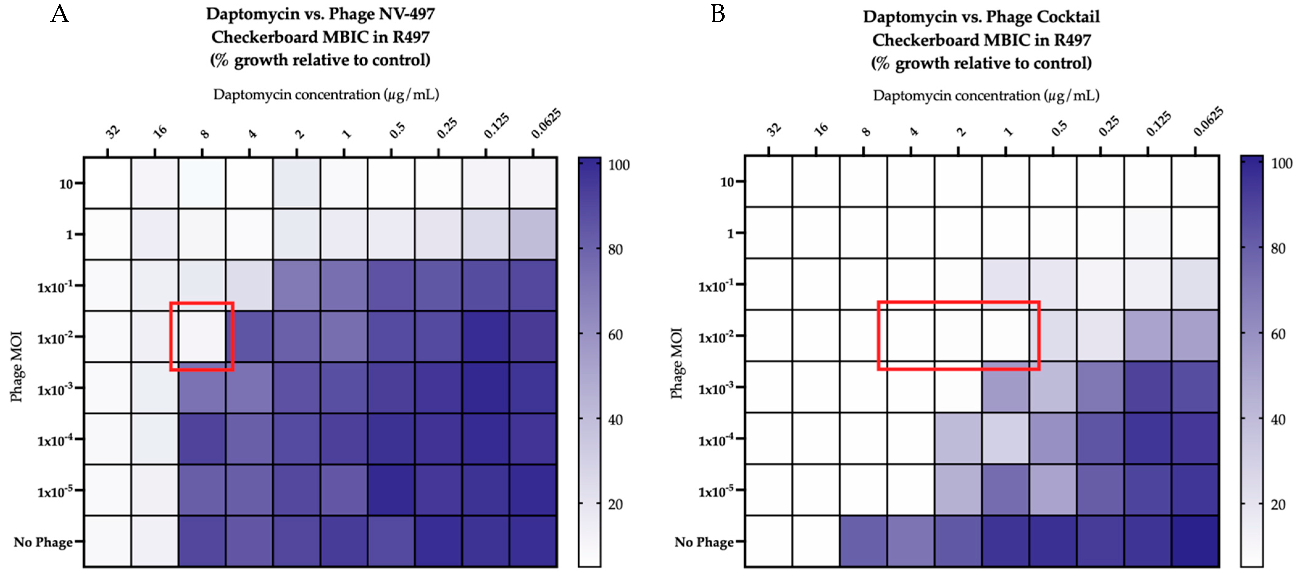
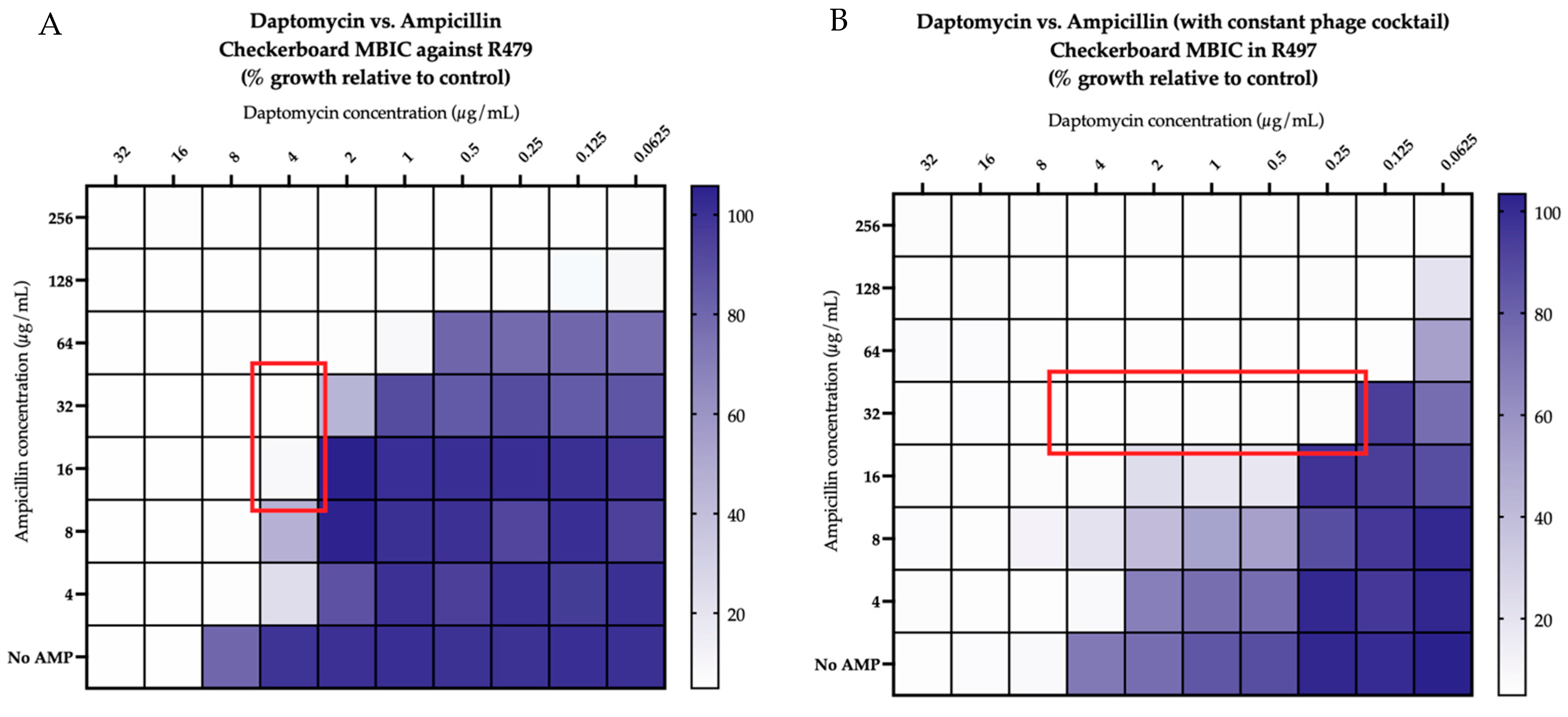
2.2. Checkerboard Analyses

Against both R497 and HOU503, DAP in combination with phage NV-497 at a multiplicity of infection (MOI) of 0.01 was additive, with an FIC index of 1 (Figure 1A), while DAP in the presence of a 4-phage cocktail (ATCC 113, NV-497, NV-503-01, NV-503-2; each at an MOI of 0.01), was synergistic, with an FIC index of 0.5 (Figure 1B). The addition of AMP to DAP in the presence of phage NV-497 (MOI 0.01) against DAP-resistant R497 in biofilm was synergistic, with an FIC index of 0.5 (Figure 2A) with additional bacterial killing identified with DAP + AMP in the presence of each 3- and 4-phage cocktail (each at an MOI of 0.01) (Figure 2B not all data shown).

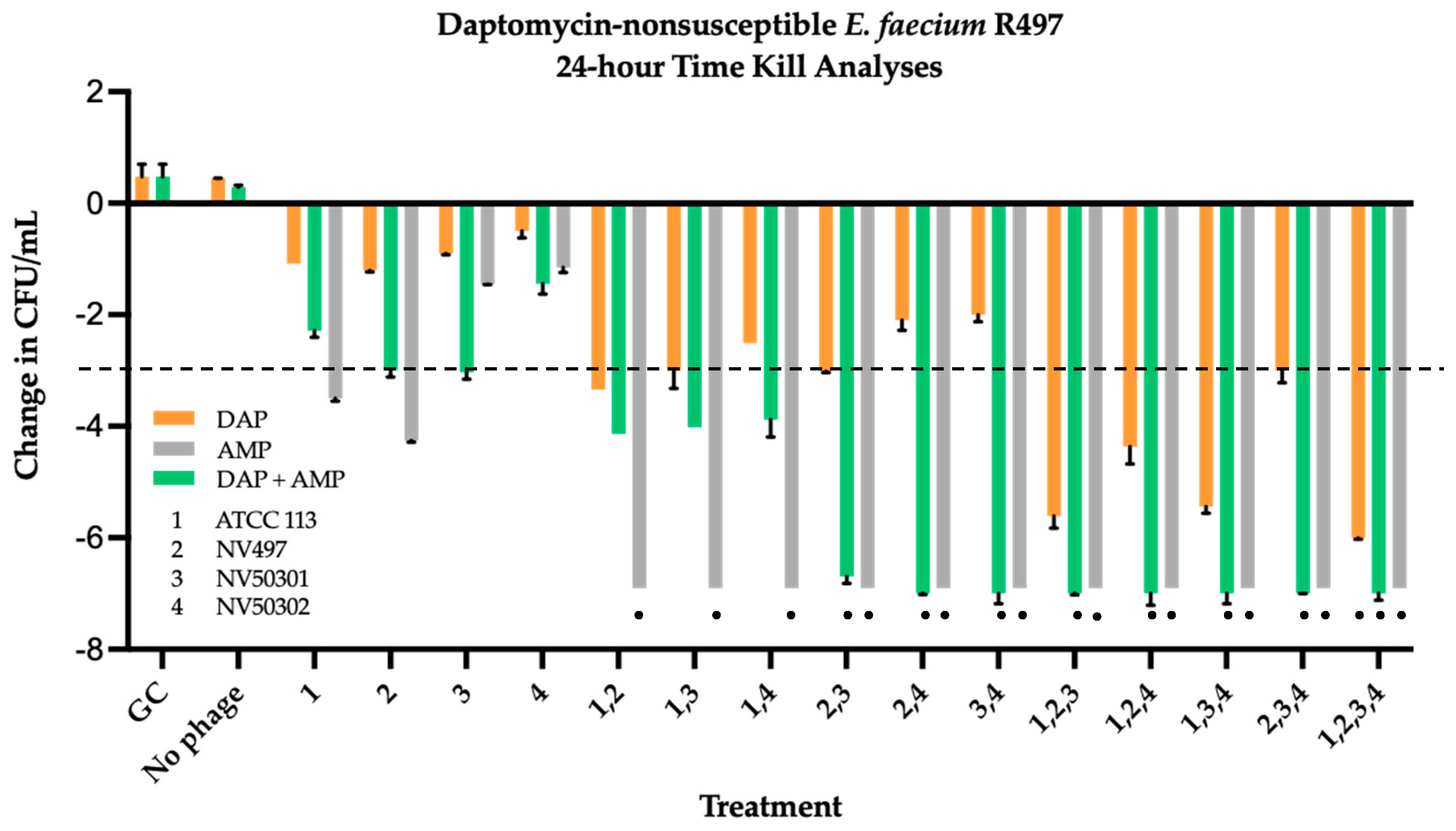
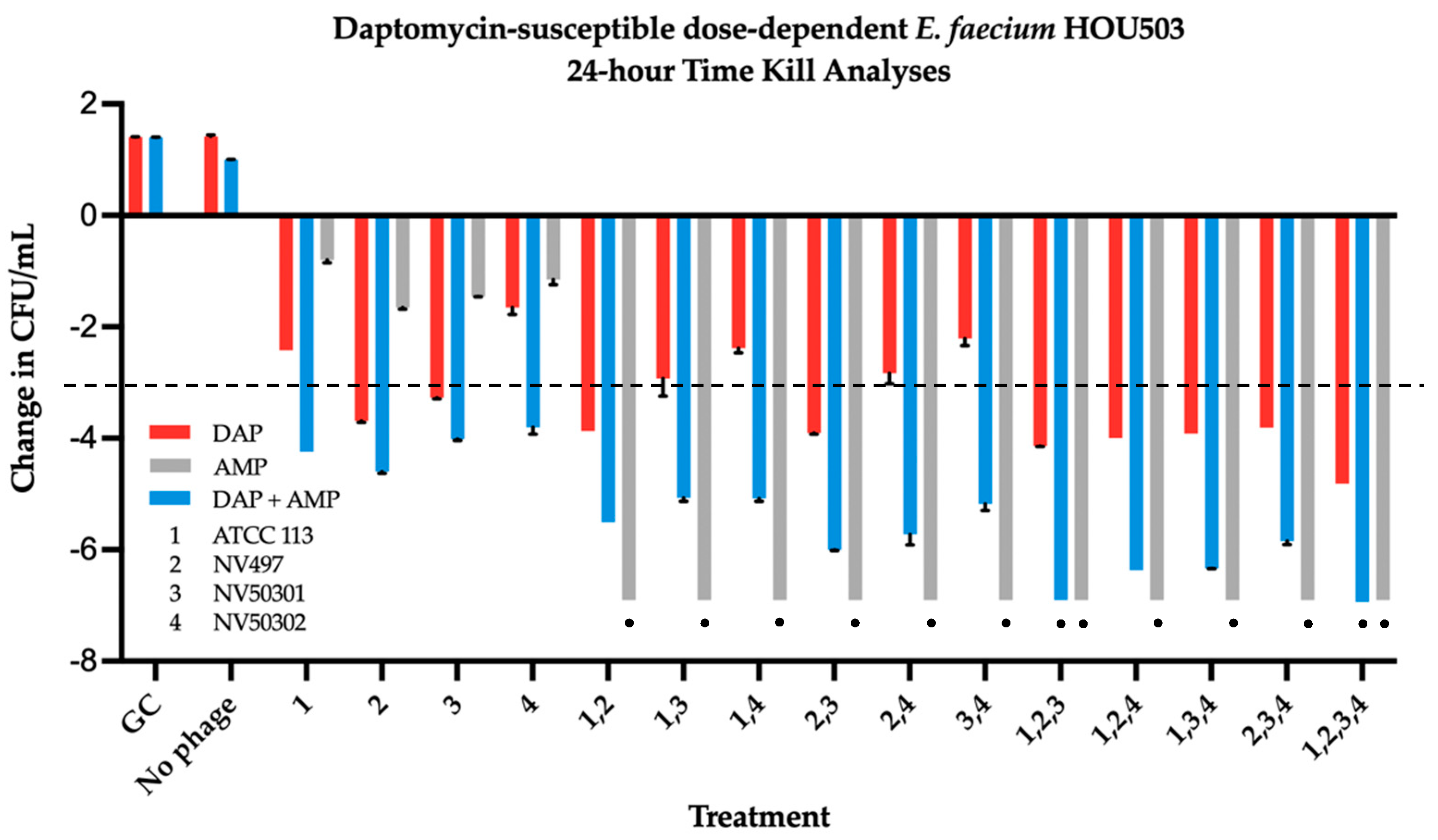
2.3. Time Kill Analyses

DAP-resistant R497 and DAP-SDD HOU503 were evaluated in 24 h biofilm TKA against DAP (0.5× MBIC), AMP (free peak = 72 µg/mL), combination DAP + AMP, single phage (MOI 0.01), phage cocktails (each at an MOI of 0.01), and multiple PAC (Figure 3 and Figure 4). Results from checkerboard analyses were used to determine phage MOI to be used in TKA. Against R497, AMP combined with any 2-, 3-, or 4-phage cocktail demonstrated significant bactericidal and synergistic activity compared to the most effective double combination regimen (ANOVA range of mean differences 2.998 to 3.102 log10 colony forming units (CFU)/mL; p = 0.011) (Figure 3). This was similar to DAP + AMP PAC, other than those with 2-phage cocktails that contained ATCC 113 Phage (ANOVA range of mean differences 2.548 to 2.868 log10 colony forming units (CFU)/mL; p = 0.023). PAC with DAP monotherapy required the addition of the 4-phage cocktail to demonstrate bactericidal and synergistic killing (ANOVA range of mean differences 2.006 to 2.329 log10 colony forming units (CFU)/mL; p = 0.039).
Against DAP-SDD HOU503, AMP combined with each 2-, 3-, or 4-phage cocktail was once again demonstrated significant bactericidal and synergistic activity compared to the most effective double combination, while DAP + AMP required addition of the 3-phage cocktail containing ATCC 113 Phage, NV-497, and NV-503-01, or the 4-phage cocktail for significant bactericidal and synergistic killing (ANOVA range of mean differences 2.251 to 2.466 log10 colony forming units (CFU)/mL; p = 0.044 and 2.119 to 2.350 log10 colony forming units (CFU)/mL; p = 0.028, respectively) (Figure 4). DAP monotherapy in PAC, while bactericidal in combination with multiple single and phage cocktail combinations, did not demonstrate synergistic killing against HOU503.

2.4. Bacteriophage and Antibiotic Resistance Testing

R497 and HOU503 that survived 24 h TKA involving phage were assessed for phage resistance. Bacteriophage resistance was observed in 24 h R497 TKA samples for each of the four phages in all phage +/− antibiotic regimens except those containing DAP + AMP and NV-497 in 2-, 3-, or 4-phage cocktails (Table 2). In HOU503, phage resistance in TKA samples at 24 h occurred in all combinations of phage plus DAP or AMP, while phage resistance in DAP + AMP combinations was only identified in DAP + AMP combinations that included single phage. Notably, R497 and HOU503 exposure to DAP, AMP, or DAP + AMP in the presence of single phage or phage cocktail resulted in antibiotic resistance stabilization (i.e., no antibiotic MBIC elevation compared to baseline) without identified antibiotic MBIC reversion (i.e., lowering of antibiotic MBIC compared to baseline in DAP-resistant and DAP-SDD isolates) at the end of 24 h TKA.

3. Discussion

Recognizing the critical knowledge gap in PAC against VRE, this study aimed to evaluate whether the addition of phage cocktails to SOC antibiotics in PAC demonstrated fundamental PAS components in biofilm state including (i) bacterial eradication; (ii) circumvention of phage:bacterial resistance; and (iii) reversion of resistance (‘resensitization’). Specifically, four different phages (ATCC 113, NV-497, NV-503-01, NV-503-02) were evaluated alone and in combination with DAP, AMP, or DAP + AMP against two clinical vancomycin-resistant E. faecium strains isolated from patients with bacteremia, DAP-resistant R497 and DAP-SDD HOU503. Prior to 24 h TKA, modified checkerboard MBICs were used as a preliminary screening method to efficiently evaluate multiple PAC for PAS. Compared to the use of modified checkerboard analyses in planktonic state, which provide information pertaining to the suppression of bacterial growth, modified checkerboards in biofilm state provides additional information related to eradication of biofilm-embedded VRE at 24 h, similar to TKA. The use of modified checkerboard MBIC in this study facilitated accurate and higher throughput screening of effective PAC at various antibiotic concentrations and phage MOI compared to more resource intensive TKA. Results of the modified checkerboard MBIC against R497 and HOU503 were reflective of TKA results with increased bacterial killing identified with DAP combined with a 3-phage cocktail compared to DAP plus single phage. Additional bacterial killing that was then visualized with the addition of AMP to DAP in the presence of a single phage or phage cocktail. These data align with TKA results in which DAP in combination with 3- and 4-phage cocktails demonstrated synergistic activity, however, the addition of AMP achieved killing to detection limit. These data provide validation for the use of a modified checkerboard MBIC to screen for killing effect of standard of care antibiotics and phage cocktails against biofilm-embedded VRE and synergy assessment prior to TKA.
In 24 h biofilm TKA, multiple DAP, AMP, and DAP + AMP combinations that included ≥2 phages in cocktail demonstrated detection level killing at 24 h. However, the prevention of treatment-emergent phage resistance at the end of 24 h TKA required DAP + AMP in combination with 2-, 3- or 4-phage combinations. Similar to previous data in planktonic and biofilm state demonstrating that the VRE strain with high phage susceptibility (R497) showed less emergence of bacteriophage resistance at the end of 24 h TKA compared to the strain with medium phage susceptibility (HOU503), these data demonstrate that while PAC containing DAP, AMP, or DAP + AMP prevented phage resistance at the end of 24 h TKA against R497, against HOU503, DAP + AMP was required in PAC to prevent phage resistance [31]. Regarding antibiotic resistance stabilization, no difference in DAP or AMP MBIC was identified in post-TKA analyses compared to baseline. The observation that phage resistance can be a fitness trade-off under antibiotic pressure in enterococci has been studied to a limited extent previously. In one study by Canfield et al., antimicrobial susceptibility of cell wall and membrane-acting antibiotics, including ampicillin and daptomycin, was tested for phage-resistant E. faecium and E. faecalis strains harboring mutations in sagA and epa genes, respectively [40]. They identified that mutations in sagA and epa genes, but not phage capsule mutations, manifested as enhanced antibiotic susceptibility compared to phage-sensitive strains. Notably, the epa gene is associated with teichoic acid biosynthesis so altered teichoic acids at the cell surface of E. faecalis may enable enhanced or at least stabilized daptomycin and beta-lactam susceptibility in phage-resistant strains. Further studies are warranted including those with whole genome sequencing and comparative genomics to further evaluate genes important for phage infection of E. faecium and antibiotic susceptibility enhancement or stabilization.
Limitations of this study include the assessment of only two MDR E. faecium clinical isolates in modified checkerboard MBIC and 24 h TKA. In the context of the current study, the primary goal was to identify which phage cocktails, when combined with standard of care antibiotics, demonstrated bactericidal killing, phage-antibiotic synergy, possible reversion of baseline antibiotic nonsusceptibility, and protection against treatment-emergent phage and antibiotic resistance in 24 h TKA. Additional evaluations of the phage cocktails use in this study against an array of other clinical VRE strains would be highly beneficial in clinical context to identify a phage cocktail with the widest host range. Additionally, based on the dataset generated by this work, there is not a clear formula for the expected outcome when combining these specific phages, and although phage cocktails have been designed and their efficacy reported in the literature previously, guidelines for the design and development of optimized phage cocktails do not exist. Synergistic effects of phage-antibiotic combinations evaluated in this study may be due to specific phage properties not assessed here including (1) adsorption, (2) rate of infection, and (3) progeny production, to name a few. Answering these questions may help determine effective future cocktail design. Furthermore, genotypic analysis of included phages prior to and following their use in 24 h TKA may decipher differences in the emergence of phage resistance at the end of 24 h TKA.
In summary, this study demonstrates that in instances of biofilm-embedded MDR E. faecium, the addition of select bacteriophage cocktails to DAP + AMP may be a promising option to eradicate biofilm-mediated infections while preserving baseline antibiotic MBIC and phage susceptibility. Additional complementary studies are warranted to assess phage host range against a larger panel of DAP-resistant and DAP-SDD isolates and evaluation of PAS components including bacterial eradication, circumvention of phage:antibiotic resistance, and reversion of antibiotic and phage resistance over longer time periods in simulated biofilm pharmacokinetic/pharmacodynamic (PK/PD) models to validate the findings of this study. Furthermore, genetic analysis of additional E. faecium strains and bacteriophages would provide further insight as to the trends we report here.

4. Materials and Methods

4.1. Bacterial Isolates

E. faecium strains R497 and HOU503, both with LiaFSR mutations and isolated from patients with bacteremia, were selected from a panel of 8 vancomycin-resistant E.faecium isolates located in the Anti-Infective Research Laboratory library and evaluated in further experiments [33,34,35,41,42].

4.2. Antimicrobial Agents and Media

Antibiotics used in this study (DAP and AMP) were purchased from Sigma Chemical Company (St. Louis, MO, USA). Prior to each biofilm assay, 1% glucose supplemented tryptic soy broth (TSB) (GSTSB) was incubated for 24 h. Brain heart infusion (BHI) broth (Difco, Detroit, MI, USA) was used for biofilm susceptibility, checkerboard, and time kill. In each assay, the BHI was supplemented with 50 mg/L calcium and 12.5 mg/L magnesium. E. faecium was plated for colony counts on BHI agar (Difco). BHI agar was prepared for use at 0.5 and 1.5%, dependent on assay (Oxoid, Lenexa, KS, USA). Broth used in assays containing DAP was supplemented with an additional 25 mg/L of calcium [42].

4.3. Bacteriophage Source and Propagation

Phages NV-497, NV-503-01, and NV-503-02 as well as phages 9181, 9183, and 9184, were isolated from wastewater treatment facilities in Maryland and Colorado and provided by the Department of the NAVY and the Duerkop laboratory, respectively [43]. ATCC 113 Phage (ATCC 19950-B1) and propagating organism E. faecium (ATCC 19950) were purchased from ATCC (Manassas, VA, USA). Phages were propagated in liquid culture to yield high titer stocks (≥109 PFU/mL) with lysates filtered with a 0.2 µm filter to remove remaining bacteria and cell debris [31,37,44]. Filtered phage was then stored, protected from light at 2–8 °C.

4.4. Biofilm Quantification Assay

Biofilm production for each E. faecium strain was evaluated, as previously described [38,39]. In brief, E. faecium-inoculated BHI broth was added to each well of a microtiter plate (96-well) and placed in a 37 °C shaker incubator for 24 h for biofilm formation around the bottom ring of each well. Next, planktonic cells are washed from the wells with sterile water and stained with 0.2% crystal violet for 30 min. The plate was then washed, and biofilm dissolved with 33% glacial acetic acid. Biofilm quantification was measured at OD560 (OD) before and after the addition of glacial acetic acid. Samples were analyzed for production compared to wells containing media alone, which was used as a negative control (ODc). Classification of adherence capabilities for each strain was categorized into one of four categories: none (OD ≤ ODc), low (ODc < OD ≤ 2xODc), medium (2xODc < OD ≤ 4xODc), or high (4xODc < OD) [38,39,45].

4.5. Phage Sensitivity Assay

Phage activity for eight E. faecium strains was tested using the small drop overlay method or “spot testing” on BHI plates, as previously described [44,46]. First, 100 µL of E. faecium planktonic 18 h overnight culture was mixed with 5 mL of 50 °C 0.5% BHI overlay. Next, the mixture was poured uniformly onto the BHI plate. Once the overlay was set (approximately 10 min after pouring), phages were spotted in 5 µL increments onto the overlay in 10-fold serial dilutions, incubated overnight at 37 °C, then counted [46]. Phage was noted to be active if clearing of the bacterial lawn was identified where phage was spotted, and discrete plaques were observed in the diluted phage spots. Plaques at specific dilutions were counted to determine the phage titer [46]. Phage susceptibility was classified based on phage titer, which was measured with plaque-forming units (PFUs). High susceptibility was indicated with >107 PFU (green), medium susceptibility was indicated with 103 to 107 PFU (yellow), and low susceptibility with <103 PFU (orange). If no PFU were identified then the phage was considered to be nonsusceptible (NS, red).

4.6. Antibiotic Susceptibility Testing

The pin-lid method using the Calgary Biofilm Device (CBD) was conducted in duplicate to determine minimum biofilm inhibitory concentration (MBIC) values for each E. faecium strain, as described previously [47,48,49]. E. faecium-inoculated GSTSB was added to the 96-well microtiter plate with the pin-lid placed on top of the microtiter plate then incubated at 37 °C for 24 h. The following day, the lid was removed from the microtiter plate, rinsed with phosphate buffer solution, then placed on a separate microtiter plate containing serial antibiotic dilutions according to the Clinical and Laboratory Standards Institute (CLSI) broth microdilution (BMD) method and inoculated at 37 °C for 24 h [50,51,52]. The pin-lid was removed to record the MBIC, which was defined per CLSI as the column with highest antibiotic dilution demonstrating no bacterial growth.

4.7. Modified Checkerboard for Antibiotic and Bacteriophage Synergy Screening

A modified checkerboard MBIC assay was used to assess PAS against E.faecium isolates R497 and HOU503, as previously described. In brief, 200 µL of 1% GSTSB inoculated with 5 × 10⁵ starting inoculum of test organism was distributed in 96-well round-bottom microtiter plates covered with a 96-pin lid and statically incubated for 24 h at 37 °C, allowing biofilm formation on the pins. The next day, in a separate 96-well plate, 100 µL of BHI was placed in each well, then 100 µL of a single antibiotic (DAP or AMP) at 4xMBIC was added to each well in column 1, then serially diluted 2-fold through the tray through column 9. Next, 100 µL of the other antibiotic was added to columns 1 to 10 of row one and serially diluted 2-fold from row 1 through row 7. In checkerboards where phage was added to single antibiotic (DAP or AMP), 20 µL of phage at an MOI of 10 was added to each well in columns 1 to 10 of row 1. Phage was then serially diluted 10-fold from row 1 through row 7. To assess PAS in checkerboards containing both DAP and AMP, phage was added at a constant subinhibitory MOI (based on single PAS checkerboard results) following completion of DAP and AMP dilutions. In each checkerboard, columns 11 and 12 were designated as growth control and media control, respectively. Once dilutions were complete, then pin-lid from the original tray was removed from the first 96-well plate and placed on the 96-well plate containing the checkerboard dilutions. The plate was then at 37 °C for 24 h, and then read with a spectrophotometer at OD570. The fractional inhibitory concentration (FIC) for each checkerboard was calculated with FIC index of ≤0.5, 1–4, and >4 indicating synergy, additivity, and antagonism, respectively [53,54].

4.8. Time Kill Analyses

Evaluation of bacterial growth suppression was performed with TKA over a 24 h time course to evaluate suppression of bacterial growth and PAS in microwell plates, as previously described [55,56]. First, four sterile 3-mm polyurethane beads were placed in each well followed by 2 mL total volume of E. faecium at 6 log10 CFU/mL and 1% GSTSB in 1:9 ratio. Plates were incubated for 24 h at 37 °C to yield biofilm formation on the beads. The 2 mL of inoculated broth was then aspirated from each well, without disturbing the biofilm-covered beads, and replaced with BHI broth supplemented with 250 µL of calcium chloride per 50 mL of broth. The starting inoculum on each bead for each TKA was 106.5–7 CFU/mL. DAP and AMP were added to their designated wells at 0.5xMBIC and free physiological peak concentration (free peak = 72 µg/mL), respectively. Phage dosing was optimized to a subinhibitory MOI (ratio of phage to target organism) of 0.01 for each phage based on modified checkerboard MBIC results. Antibiotic was added to each well first directly followed by the addition of phage. TKA growth curves were constructed from sterilely removed beads at 0 (prior to the addition of phage and/or antibiotic), 4, 8, and 24 h. Each bead was placed in an Eppendorf tubed containing 0.9 mL of 0.9% saline and stored at 2–8 °C for 24 h to inactivate the phage. Each sample was then thawed and processed to remove biofilm with 1-min vortex and sonication (20 Hz, Bransonic 12 Branson Ultrasonic Corporation) intervals for a total of 6 min. Antibiotic carryover was eliminated from each sample with dilutions in 0.9% saline, as appropriate [57]. Diluted samples were plated on BHI agar (easySpiral, Interscience for Microbiology, Saint Nom la Breteche, France, detection limit of 102 CFU/mL), and incubated at 37 °C for 24 h followed by counting of bacterial colonies (Scan 1200, Interscience for Microbiology, Saint Nom la Breteche, France). Synergy and bactericidal activity were defined as a ≥2 log10 CFU/mLkill compared to the most effective agent (or double-combination regimen) and a ≥3 log10 CFU/mL reduction from baseline at 24 h. Single drug/phage exposures in biofilm TKA included DAP, AMP and each of the four phages. Additionally, combination evaluations were performed with DAP, AMP, and DAP plus AMP with combinations of two, three, and four phages. Statistical analysis was carried out using SPSS version 21.0 (IBM Corp., Armonk, NY, USA) software. Significant differences between phage-antibiotic regimens in terms of bacterial killing metrics (i.e., extent of TKA reductions in log10 CFU/mL counts at time 0 vs. 24 h) was assessed by analysis of varianace (ANOVA) with Tukey’s post hoc test (p < 0.05).

4.9. Resistance Testing

Following the infection of E. faecium strains R497 and HOU503 in 24 h TKA with selected phages, modified bacteriophage insensitive mutants (BIM) testing was conducted as previously described to evaluate frequency of resistance (FOR) [41,46,58]. First, for each TKA sample, a mixture of 100 µL high titer phage and 10 µL TKA sample was incubated at 37 °C for 10 min. The mixture was then added to 5 mL of 0.5% BHI overlay and quickly poured onto square BHI plates. Colonies arising on the plate were counted following incubation at 37 °C for 24 h and again after an additional 24 h at room temperature. FOR was calculated by taking the colony count from 24 h TKA samples and dividing it by the number of colonies identified on the 24 h and 48 h plates.
The double drop method was then used to evaluate phage sensitivity in colonies surviving 24 h TKA in the following manner: first, 10 µL aliquots of high titer phage were spotted on BHI plates then 5 µL of overnight E. faecium culture from each BIM was spotted on top of the first 10 µL aliquot. Plates were incubated at 37 °C for 24 h with aliquot spots then compared to bacterial control spots without phage. If there was no difference between the phage-bacteria spot and the control spot the phage was considered resistant, if phage activity was identified in the bacteria spot then the phage was intermediate, and if <10 colonies were identified in the spot then the phage was sensitive [41].

Author Contributions

A.J.K.C., K.S., R.K., D.J.H., A.E.G., T.M., B.B., G.S.C., B.A.D., C.A.A. and M.J.R. were involved in the conceptualization, methodology, validation, formal analysis, investigation, writing—Original draft preparation, writing—Review and editing of this manuscript. M.W. and M.V.D. contributed to isolation, amplification, and purification of the phages. All authors have read and agreed to the published version of the manuscript.

Funding

This research received no external funding. M.J.R. is supported by NIH grants R21 AI163726. C.A.A. is supported by NIH grants K24AI121296, R01AI134637, R01AI48342, and P01AI152999.

Institutional Review Board Statement

Not applicable.

Informed Consent Statement

Not applicable.

Data Availability Statement

Not applicable.

Conflicts of Interest

A.J.K.C., K.S., R.K., D.J.H., A.E.G., T.M., B.B., M.W., M.V.D., G.S.C., B.A.D., C.A.A., M.J.R. have nothing to declare. M.J.R. has received grant support, has consulted or spoken on behalf of Allergan, Melinta, Merck, Paratek, Shionogi, Spero and Tetraphase. C.A.A. has received grant support from Merck, MeMed Diagnostics, is a co-founder and Entasis Therapeutics shareholder of Ancilia Biosciences, C.

Author Disclaimer

The views expressed in this article reflect the results of research conducted by the authors and do not necessarily reflect the official policy or position of the Department of the Navy, Department of Defense, nor the United States Government.

Support Statement

This work was supported by work unit number A1417.

Copyright Statement

Dr. Biswas is a federal employee of the United States government. This work was prepared as part of his official duties. Title 17 U.S.C. 105 provides that ‘copyright protection under this title is not available for any work of the United States Government’. Title 17 U.S.C. 101 defines a U.S. Government work as work prepared by a military service member or employee of the U.S. Government as part of that person’s official duties.

References

  1. Arias, C.A.; Murray, B.E. The rise of the Enterococcus: Beyond vancomycin resistance. Nat. Rev. Microbiol. 2012, 10, 266–278. [Google Scholar] [CrossRef]
  2. Nigo, M.; Munita, J.M.; Arias, C.A.; Murray, B.E. What’s New in the Treatment of Enterococcal Endocarditis? Curr. Infect. Dis. Rep. 2014, 16, 431. [Google Scholar] [CrossRef]
  3. Chuang, Y.-C.; Lin, H.-Y.; Chen, P.-Y.; Lin, C.-Y.; Wang, J.-T.; Chen, Y.-C.; Chang, S.-C. Effect of Daptomycin Dose on the Outcome of Vancomycin-Resistant, Daptomycin-Susceptible Enterococcus faecium Bacteremia. Clin. Infect. Dis. 2017, 64, 1026–1034. [Google Scholar] [CrossRef]
  4. Zasowski, E.J.; Claeys, K.C.; Lagnf, A.M.; Davis, S.L.; Rybak, M.J. Time Is of the Essence: The Impact of Delayed Antibiotic Therapy on Patient Outcomes in Hospital-Onset Enterococcal Bloodstream Infections. Clin. Infect. Dis. 2016, 62, 1242–1250. [Google Scholar] [CrossRef]
  5. Cole, K.A.; Kenney, R.M.; Perri, M.B.; Dumkow, L.E.; Samuel, L.P.; Zervos, M.J.; Davis, S.L. Outcomes of Aminopenicillin Therapy for Vancomycin-Resistant Enterococcal Urinary Tract Infections. Antimicrob. Agents Chemother. 2015, 59, 7362–7366. [Google Scholar] [CrossRef]
  6. Lee, R.A.; Vo, D.T.; Zurko, J.C.; Griffin, R.L.; Rodriguez, J.M.; Camins, B.C. Infectious Diseases Consultation Is Associated with Decreased Mortality in Enterococcal Bloodstream Infections. Open Forum Infect. Dis. 2020, 7, ofaa064. [Google Scholar] [CrossRef]
  7. Higuita, N.I.; Huycke, M.M. Enterococcal Disease, Epidemiology, and Implications for Treatment. In Enterococci: From Commensals to Leading Causes of Drug Resistant Infection [Internet]; Gilmore, M.S., Clewell, D.B., Ike, Y., Shankar, N., Eds.; Massachusetts Eye and Ear Infirmary: Boston, MA, USA, 2014. [Google Scholar]
  8. Hidron, A.I.; Edwards, J.R.; Patel, J.; Horan, T.C.; Sievert, D.M.; Pollock, D.A.; Fridkin, S.K. Antimicrobial-Resistant Pathogens Associated with Healthcare-Associated Infections: Annual Summary of Data Reported to the National Healthcare Safety Network at the Centers for Disease Control and Prevention, 2006–2007. Infect. Control Hosp. Epidemiol. 2008, 29, 996–1011. [Google Scholar] [CrossRef]
  9. Weiner, L.M.; Webb, A.K.; Limbago, B.; Dudeck, M.A.; Patel, J.; Kallen, A.J.; Edwards, J.R.; Sievert, D.M. Antimicrobial-Resistant Pathogens Associated with Healthcare-Associated Infections: Summary of Data Reported to the National Healthcare Safety Network at the Centers for Disease Control and Prevention, 2011–2014. Infect. Control Hosp. Epidemiol. 2016, 37, 1288–1301. [Google Scholar] [CrossRef]
  10. Crank, C.; O’Driscoll, T. Vancomycin-resistant enterococcal infections: Epidemiology, clinical manifestations, and optimal management. Infect. Drug Resist. 2015, 8, 217. [Google Scholar] [CrossRef]
  11. Jahanbakhsh, S.; Singh, N.B.; Yim, J.; Kebriaei, R.; Smith, J.R.; Lev, K.; Tran, T.T.; Rose, W.E.; Arias, C.A.; Rybak, M.J. Impact of Daptomycin Dose Exposure Alone or in Combination with β-Lactams or Rifampin against Vancomycin-Resistant Enterococci in an In Vitro Biofilm Model. Antimicrob. Agents Chemother. 2020, 64, e02074-19. [Google Scholar] [CrossRef]
  12. Benamu, E.; Deresinski, S. Vancomycin-resistant enterococcus infection in the hematopoietic stem cell transplant recipient: An overview of epidemiology, management, and prevention. F1000Research 2018, 7, 3. [Google Scholar] [CrossRef]
  13. Stewart, P.S.; Davison, W.M.; Steenbergen, J.N. Daptomycin Rapidly Penetrates a Staphylococcus epidermidis Biofilm. Antimicrob. Agents Chemother. 2009, 53, 3505–3507. [Google Scholar] [CrossRef]
  14. Lellek, H.; Franke, G.C.; Ruckert, C.; Wolters, M.; Wolschke, C.; Christner, M.; Büttner, H.; Alawi, M.; Kröger, N.; Rohde, H. Emergence of daptomycin non-susceptibility in colonizing vancomycin-resistant Enterococcus faecium isolates during daptomycin therapy. Int. J. Med. Microbiol. 2015, 305, 902–909. [Google Scholar] [CrossRef]
  15. Kamboj, M.; Cohen, N.; Gilhuley, K.; Babady, N.E.; Seo, S.K.; Sepkowitz, K.A. Emergence of Daptomycin-Resistant VRE: Experience of a Single Institution. Infect. Control Hosp. Epidemiol. 2011, 32, 391–394. [Google Scholar] [CrossRef]
  16. Munita, J.M.; Murray, B.E.; Arias, C.A. Daptomycin for the treatment of bacteraemia due to vancomycin-resistant enterococci. Int. J. Antimicrob. Agents 2014, 44, 387–395. [Google Scholar] [CrossRef]
  17. El Haddad, L.; Hanson, B.M.; Arias, C.A.; Ghantoji, S.S.; Harb, C.P.; Stibich, M.; Chemaly, R.F. Emergence and Transmission of Daptomycin and Vancomycin-Resistant Enterococci Between Patients and Hospital Rooms. Clin. Infect. Dis. 2021, 73, 2306–2313. [Google Scholar] [CrossRef]
  18. Wudhikarn, K.; Gingrich, R.D.; de Magalhaes Silverman, M. Daptomycin nonsusceptible enterococci in hematologic malignancy and hematopoietic stem cell transplant patients: An emerging threat. Ann. Hematol. 2013, 92, 129–131. [Google Scholar] [CrossRef]
  19. Kebriaei, R.; Rice, S.A.; Singh, K.V.; Stamper, K.C.; Dinh, A.Q.; Rios, R.; Diaz, L.; Murray, B.E.; Munita, J.M.; Tran, T.T.; et al. Influence of Inoculum Effect on the Efficacy of Daptomycin Monotherapy and in Combination with β-Lactams against Daptomycin-Susceptible Enterococcus faecium Harboring LiaSR Substitutions. Antimicrob. Agents Chemother. 2018, 62, e00315-18. [Google Scholar] [CrossRef]
  20. Hall, A.D.; Steed, M.E.; Arias, C.A.; Murray, B.E.; Rybak, M.J. Evaluation of Standard- and High-Dose Daptomycin versus Linezolid against Vancomycin-Resistant Enterococcus Isolates in an In Vitro Pharmacokinetic/Pharmacodynamic Model with Simulated Endocardial Vegetations. Antimicrob. Agents Chemother. 2012, 56, 3174–3180. [Google Scholar] [CrossRef]
  21. Kebriaei, R.; Stamper, K.C.; Singh, K.V.; Khan, A.; Rice, S.A.; Dinh, A.Q.; Tran, T.T.; Murray, B.E.; Arias, C.A.; Rybak, M.J. Mechanistic Insights into the Differential Efficacy of Daptomycin Plus β-Lactam Combinations Against Daptomycin-Resistant Enterococcus faecium. J. Infect. Dis. 2020, 222, 1531–1539. [Google Scholar] [CrossRef]
  22. Gordillo Altamirano, F.L.; Barr, J.J. Phage Therapy in the Postantibiotic Era. Clin. Microbiol. Rev. 2019, 32, e00066-18. [Google Scholar] [CrossRef]
  23. Morrisette, T.; Kebriaei, R.; Lev, K.L.; Morales, S.; Rybak, M.J. Bacteriophage Therapeutics: A Primer for Clinicians on Phage-Antibiotic Combinations. Pharmacotherapy 2020, 40, 153–168. [Google Scholar] [CrossRef]
  24. Tkhilaishvili, T.; Lombardi, L.; Klatt, A.-B.; Trampuz, A.; Luca, M.D. Bacteriophage Sb-1 enhances antibiotic activity against biofilm, degrades exopolysaccharide matrix and targets persisters of Staphylococcus aureus. Int. J. Antimicrob. Agents 2018, 52, 842–853. [Google Scholar] [CrossRef]
  25. Harper, D.R.; Parracho, H.M.R.T.; Walker, J.; Sharp, R.J.; Hughes, G.; Werthén, M.; Lehman, S.M.; Morales, S. Bacteriophages and Biofilms. Antibiotics 2014, 3, 270–284. [Google Scholar] [CrossRef]
  26. Parasion, S.; Kwiatek, M.; Gryko, R.; Mizak, L.; Malm, A. Bacteriophages as an alternative strategy for fighting biofilm development. Pol. J. Microbiol. 2014, 63, 137–145. [Google Scholar] [CrossRef]
  27. Kumaran, D.; Taha, M.; Yi, Q.; Ramirez-Arcos, S.; Diallo, J.-S.; Carli, A.V.; Abdelbary, H. Does Treatment Order Matter? Investigating the Ability of Bacteriophage to Augment Antibiotic Activity against Staphylococcus aureus Biofilms. Front. Microbiol. 2018, 9, 127. [Google Scholar] [CrossRef]
  28. Morrisette, T.; Lev, K.L.; Kebriaei, R.; Abdul-Mutakabbir, J.C.; Stamper, K.C.; Morales, S.; Lehman, S.M.; Canfield, G.S.; Duerkop, B.A.; Arias, C.A.; et al. Bacteriophage-Antibiotic Combinations for Enterococcus faecium with Varying Bacteriophage and Daptomycin Susceptibilities. Antimicrob. Agents Chemother. 2020, 64, e00993-20. [Google Scholar] [CrossRef]
  29. Torres-Barceló, C. Phage Therapy Faces Evolutionary Challenges. Viruses 2018, 10, 323. [Google Scholar] [CrossRef]
  30. Oechslin, F. Resistance Development to Bacteriophages Occurring during Bacteriophage Therapy. Viruses 2018, 10, 351. [Google Scholar] [CrossRef]
  31. Lev, K.; Kunz Coyne, A.J.; Kebriaei, R.; Morrisette, T.; Stamper, K.; Holger, D.J.; Canfield, G.S.; Duerkop, B.A.; Arias, C.A.; Rybak, M.J. Evaluation of Bacteriophage-Antibiotic Combination Therapy for Biofilm-Embedded MDR Enterococcus faecium. Antibiotics 2022, 11, 392. [Google Scholar] [CrossRef]
  32. Kebriaei, R.; Lev, K.; Morrisette, T.; Stamper, K.C.; Abdul-Mutakabbir, J.C.; Lehman, S.M.; Morales, S.; Rybak, M.J. Bacteriophage-Antibiotic Combination Strategy: An Alternative against Methicillin-Resistant Phenotypes of Staphylococcus aureus. Antimicrob. Agents Chemother. 2020, 64, e00461-20. [Google Scholar] [CrossRef]
  33. Tran, T.T.; Panesso, D.; Gao, H.; Roh, J.H.; Munita, J.M.; Reyes, J.; Diaz, L.; Lobos, E.A.; Shamoo, Y.; Mishra, N.N.; et al. Whole-Genome Analysis of a Daptomycin-Susceptible Enterococcus faecium Strain and Its Daptomycin-Resistant Variant Arising during Therapy. Antimicrob. Agents Chemother. 2012, 57, 261–268. [Google Scholar] [CrossRef]
  34. Diaz, L.; Tran, T.T.; Munita, J.M.; Miller, W.R.; Rincon, S.; Carvajal, L.P.; Wollam, A.; Reyes, J.; Panesso, D.; Rojas, N.L.; et al. Whole-Genome Analyses of Enterococcus faecium Isolates with Diverse Daptomycin MICs. Antimicrob. Agents Chemother. 2014, 58, 4527–4534. [Google Scholar] [CrossRef]
  35. Reyes, J.; Panesso, D.; Tran, T.T.; Mishra, N.N.; Cruz, M.R.; Munita, J.M.; Singh, K.V.; Yeaman, M.R.; Murray, B.E.; Shamoo, Y.; et al. A liaR Deletion Restores Susceptibility to Daptomycin and Antimicrobial Peptides in Multidrug-Resistant Enterococcus faecalis. J. Infect. Dis. 2015, 211, 1317–1325. [Google Scholar] [CrossRef]
  36. Munita, J.M.; Tran, T.T.; Diaz, L.; Panesso, D.; Reyes, J.; Murray, B.E.; Arias, C.A. A liaF Codon Deletion Abolishes Daptomycin Bactericidal Activity against Vancomycin-Resistant Enterococcus faecalis. Antimicrob. Agents Chemother. 2013, 57, 2831–2833. [Google Scholar] [CrossRef]
  37. Bonilla, N.; Rojas, M.I.; Netto Flores Cruz, G.; Hung, S.-H.; Rohwer, F.; Barr, J.J. Phage on tap–a quick and efficient protocol for the preparation of bacteriophage laboratory stocks. PeerJ 2016, 4, e2261. [Google Scholar] [CrossRef]
  38. Stepanović, S.; Vuković, D.; Dakić, I.; Savić, B.; Švabić-Vlahović, M. A modified microtiter-plate test for quantification of staphylococcal biofilm formation. J. Microbiol. Methods 2000, 40, 175–179. [Google Scholar] [CrossRef]
  39. Christensen, G.D.; Simpson, W.A.; Younger, J.J.; Baddour, L.M.; Barrett, F.F.; Melton, D.M.; Beachey, E.H. Adherence of coagulase-negative staphylococci to plastic tissue culture plates: A quantitative model for the adherence of staphylococci to medical devices. J. Clin. Microbiol. 1985, 22, 996–1006. [Google Scholar] [CrossRef]
  40. Canfield, G.S.; Chatterjee, A.; Espinosa, J.; Mangalea, M.R.; Sheriff, E.K.; Keidan, M.; McBride, S.W.; McCollister, B.D.; Hang, H.C.; Duerkop, B.A. Lytic Bacteriophages Facilitate Antibiotic Sensitization of Enterococcus faecium. Antimicrob. Agents Chemother. 2021, 65, e00143-21. [Google Scholar] [CrossRef]
  41. Panesso, D.; Reyes, J.; Gaston, E.P.; Deal, M.; Londoño, A.; Nigo, M.; Munita, J.M.; Miller, W.R.; Shamoo, Y.; Tran, T.T.; et al. Deletion of liaR Reverses Daptomycin Resistance in Enterococcus faecium Independent of the Genetic Background. Antimicrob. Agents Chemother. 2015, 59, 7327–7334. [Google Scholar] [CrossRef] [Green Version]
  42. Grein, F.; Müller, A.; Scherer, K.M.; Liu, X.; Ludwig, K.C.; Klöckner, A.; Strach, M.; Sahl, H.-G.; Kubitscheck, U.; Schneider, T. Ca2+-Daptomycin targets cell wall biosynthesis by forming a tripartite complex with undecaprenyl-coupled intermediates and membrane lipids. Nat. Commun. 2020, 11, 1455. [Google Scholar] [CrossRef] [PubMed]
  43. Morrisette, T.; Lev, K.L.; Canfield, G.S.; Duerkop, B.A.; Kebriaei, R.; Stamper, K.C.; Holger, D.; Lehman, S.M.; Willcox, S.; Arias, C.A.; et al. Evaluation of Bacteriophage Cocktails Alone and in Combination with Daptomycin against Daptomycin-Nonsusceptible Enterococcus faecium. Antimicrob. Agents Chemother. 2022, 66, e0162321. [Google Scholar] [CrossRef] [PubMed]
  44. Lehman, S.M.; Mearns, G.; Rankin, D.; Cole, R.A.; Smrekar, F.; Branston, S.D.; Morales, S. Design and Preclinical Development of a Phage Product for the Treatment of Antibiotic-Resistant Staphylococcus aureus Infections. Viruses 2019, 11, 88. [Google Scholar] [CrossRef] [PubMed]
  45. Bose, J.L.; Lehman, M.K.; Fey, P.D.; Bayles, K.W. Contribution of the Staphylococcus aureus Atl AM and GL Murein Hydrolase Activities in Cell Division, Autolysis, and Biofilm Formation. PLoS ONE 2012, 7, e42244. [Google Scholar] [CrossRef]
  46. Mazzocco, A.; Waddell, T.E.; Lingohr, E.; Johnson, R.P. Enumeration of Bacteriophages Using the Small Drop Plaque Assay System. In Bacteriophages; Clokie, M.R.J., Kropinski, A.M., Eds.; Humana Press: Totowa, NJ, USA, 2009; Volume 501, pp. 81–85. [Google Scholar]
  47. Ceri, H.; Olson, M.; Morck, D.; Storey, D.; Read, R.; Buret, A.; Olson, B. The MBEC assay system: Multiple equivalent biofilms for antibiotic and biocide susceptibility testing. In Methods in Enzymology; Elsevier: Amsterdam, The Netherlands, 2001; Volume 337, pp. 377–385. [Google Scholar]
  48. Ceri, H.; Olson, M.E.; Stremick, C.; Read, R.R.; Morck, D.; Buret, A. The Calgary Biofilm Device: New technology for rapid determination of antibiotic susceptibilities of bacterial biofilms. J. Clin. Microbiol. 1999, 37, 1771–1776. [Google Scholar] [CrossRef]
  49. Ali, L.; Khambaty, F.; Diachenko, G. Investigating the suitability of the Calgary Biofilm Device for assessing the antimicrobial efficacy of new agents. Bioresour. Technol. 2006, 97, 1887–1893. [Google Scholar] [CrossRef]
  50. Clinical and Laboratory Standards Institute. Methods for dilution antimicrobial susceptibility tests for bacteria that grow aerobically: M07-A10. In Approved Standard, 10th ed.; Committee for Clinical Laboratory Standards: Wayne, PA, USA, 2015. [Google Scholar]
  51. Humphries, R.M.; Ambler, J.; Mitchell, S.L.; Castanheira, M.; Dingle, T.; Hindler, J.A.; Koeth, L.; Sei, K.; on behalf of the CLSI Methods Development and Standardization Working Group of the Subcommittee on Antimicrobial Susceptibility Testing. CLSI Methods Development and Standardization Working Group Best Practices for Evaluation of Antimicrobial Susceptibility Tests. J. Clin. Microbiol. 2018, 56, e01934-17. [Google Scholar] [CrossRef] [PubMed]
  52. Rice, L.B.; Carias, L.L.; Rudin, S.; Hutton, R.; Marshall, S.; Hassan, M.; Josseaume, N.; Dubost, L.; Marie, A.; Arthur, M. Role of Class A Penicillin-Binding Proteins in the Expression of β-Lactam Resistance in Enterococcus faecium. J. Bacteriol. 2009, 191, 3649–3656. [Google Scholar] [CrossRef]
  53. Rodriguez-Gonzalez, R.A.; Leung, C.Y.; Chan, B.K.; Turner, P.E.; Weitz, J.S. Quantitative Models of Phage-Antibiotic Combination Therapy. mSystems 2020, 5, e00756-19. [Google Scholar] [CrossRef]
  54. Zhvania, P.; Hoyle, N.S.; Nadareishvili, L.; Nizharadze, D.; Kutateladze, M. Phage Therapy in a 16-Year-Old Boy with Netherton Syndrome. Front. Med. 2017, 4, 94. [Google Scholar] [CrossRef] [Green Version]
  55. Barber, K.E.; Werth, B.J.; McRoberts, J.P.; Rybak, M.J. A Novel Approach Utilizing Biofilm Time-Kill Curves To Assess the Bactericidal Activity of Ceftaroline Combinations against Biofilm-Producing Methicillin-Resistant Staphylococcus aureus. Antimicrob. Agents Chemother. 2014, 58, 2989–2992. [Google Scholar] [CrossRef] [PubMed]
  56. Barber, K.E.; Shammout, Z.; Smith, J.R.; Kebriaei, R.; Morrisette, T.; Rybak, M.J. Biofilm Time-Kill Curves to Assess the Bactericidal Activity of Daptomycin Combinations against Biofilm-Producing Vancomycin-Resistant Enterococcus faecium and faecalis. Antibiotics 2021, 10, 897. [Google Scholar] [CrossRef] [PubMed]
  57. Rybak, M.J.; Hershberger, E.; Moldovan, T.; Grucz, R.G. In Vitro Activities of Daptomycin, Vancomycin, Linezolid, and Quinupristin-Dalfopristin against Staphylococci and Enterococci, Including Vancomycin- Intermediate and -Resistant Strains. Antimicrob. Agents Chemother. 2000, 44, 1062–1066. [Google Scholar] [CrossRef] [PubMed]
  58. O’Flynn, G.; Ross, R.P.; Fitzgerald, G.F.; Coffey, A. Evaluation of a Cocktail of Three Bacteriophages for Biocontrol of Escherichia coli O157:H7. Appl. Environ. Microbiol. 2004, 70, 3417–3424. [Google Scholar] [CrossRef] [PubMed] [Green Version]
Figure 1. (A,B). Modified checkerboard minimum biofilm inhibitory concentration (MBIC) analyses of daptomycin combined with phage NV-497 (A) and a 4-phage cocktail of ATCC 113 + NV-497 + NV-503-01 + NV-503-02 (B) (each phage at MOI 0.01) against DAP-resistant R497. Additivity, defined as an FIC index >0.5 but <4, is indicated by the red outline in (A). Synergy, defined as an FIC index ≤0.5, is indicated by the red outline in (B). Comparisons are versus growth control and depicted by the purple color gradient as percent of growth.
Antibiotics 11 01175 g001
Figure 2. (A,B). Modified checkerboard minimum biofilm inhibitory concentration (MBIC) analyses of combination daptomycin and ampicillin without (A) and a phage cocktail of ATCC 113 + NV-497 + NV-503-01 (B) (each at MOI 0.01) against DAP-resistant R497. Synergy, defined as an FIC index ≤0.5, is indicated by the red outline in (A,B). Comparisons are versus growth control and depicted by the purple color gradient as percent of growth.
Antibiotics 11 01175 g002
Figure 3. Time kill analyses of DAP at 0.5x MBIC, AMP at free peak = 72 µg/mL, and DAP + AMP, alone and in combination with four bacteriophages, ATCC 113, NV-497, NV-503-01, NV-503-02, each at an MOI of 0.01 against DAP-resistant R497. Change in CFU/mL was in comparison to initial inoculum. Synergy was defined as a ≥2-log10-CFU/mL kill compared to the most effective agent (or double-combination regimen) at 24 h (synergistic regimens indicated by black circle). Bactericidal activity was defined as a ≥3-log10-CFU/mL reduction from baseline (bactericidal regimens indicated by the bars at or extending below the dashed black line).
Antibiotics 11 01175 g003
Figure 4. Time kill analyses of DAP at 0.5x MBIC, AMP at free peak = 72 µg/mL, and DAP + AMP, alone and in combination with four bacteriophages, ATCC 113, NV-497, NV-503-01, NV-503-02, each at an MOI of 0.01 against DAP-susceptible dose-dependent HOU503. Change in CFU/mL was in comparison to initial inoculum. Synergy was defined as a ≥2-log10-CFU/mL kill compared to the most effective agent (or double-combination regimen) at 24 h (synergistic regimens indicated by black circle). Bactericidal activity was defined as a ≥3-log10-CFU/mL reduction from baseline (bactericidal regimens indicated by the bars at or extending below the dashed black line).
Antibiotics 11 01175 g004
Table 1. Biofilm quantification (optical density (OD), compared to control) and phage susceptibility against E. faecium clinical isolates.
Enterococcus Faecium Clinical IsolatesDAP VAN Biofilm Quantification Phage Susceptibility
ATCC Phage 113NV-497NV-503-01NV-503-02NV-S447-01NV-S447-02918191839184
R497RRMedium
HOU503SDDRHigh
5938RRLow
S447SDDRLow
S80849SDDRLow
SF11499SDDRLow
SF12047SDDRLow
12311SDDRLow
Phage susceptibility was classified as high, medium, or low based on plaque-forming unit (PFU) counts where >107 PFU/mL was defined as high (green), 103 to 107 PFU/mL was defined as medium, and <103 PFU/mL was defined as low. Phage was classified as non-susceptible (NS) if no PFU were identified (red). Abbreviations: DAP, daptomycin; VAN, vancomycin; R, resistant; SDD, susceptible dose-dependent.
Table 2. Evaluation of phage resistance in R497 and HOU503 at the end of 24 h time kill analyses. Phages are represented in the table as follows: 1, ATCC 113; 2, NV-497; 3, NV-503-01; 4, NV-503-02.
Single Phage2-Phage Cocktails3-Phage Cocktails4-Phage Cocktail
12341,21,31,42,32,43,41,2,31,2,41,3,42,3,41,2,3,4
R497
DAP
AMP
DAP + AMP
HOU503
DAP
AMP
DAP + AMP
Antibiotics 11 01175 i001 = resistant Antibiotics 11 01175 i002 = susceptible
Publisher’s Note: MDPI stays neutral with regard to jurisdictional claims in published maps and institutional affiliations.

