Article

Nisin Mutant Prevention Concentration and the Role of Subinhibitory Concentrations on Resistance Development by Diabetic Foot Staphylococci

1
CIISA—Centro de Investigação Interdisciplinar em Sanidade Animal, Faculdade de Medicina Veterinária, Universidade de Lisboa, Av. da Universidade Técnica de Lisboa, 1300-477 Lisboa, Portugal
2
Laboratório Associado para Ciência Animal e Veterinária (AL4AnimalS), 1300-477 Lisbon, Portugal
3
Biosystems and Integrative Sciences Institute, Faculdade de Ciências, Universidade de Lisboa, Campo Grande, 1749-016 Lisboa, Portugal
*
Author to whom correspondence should be addressed.
These authors contributed equally to this study.
Antibiotics 2022, 11(7), 972; https://doi.org/10.3390/antibiotics11070972
Submission received: 9 June 2022 / Revised: 14 July 2022 / Accepted: 15 July 2022 / Published: 19 July 2022
(This article belongs to the Special Issue Antimicrobial Resistance and Virulence – 3rd Volume)

Abstract

:
The most prevalent microorganism in diabetic foot infections (DFI) is Staphylococcus aureus, an important multidrug-resistant pathogen. The antimicrobial peptide nisin is a promising compound for DFI treatment, being effective against S. aureus. However, to avoid the selection of resistant mutants, correct drug therapeutic doses must be established, being also important to understand if nisin subinhibitory concentrations (subMIC) can potentiate resistant genes transfer between clinical isolates or mutations in genes associated with nisin resistance. The mutant selection window (MSW) of nisin was determined for 23 DFI S. aureus isolates; a protocol aiming to prompt vanA horizontal transfer between enterococci to clinical S. aureus was performed; and nisin subMIC effect on resistance evolution was assessed through whole-genome sequencing (WGS) applied to isolates subjected to a MEGA-plate assay. MSW ranged from 5–360 μg/mL for two isolates, from 5–540 μg/mL for three isolates, and from 5–720 μg/mL for one isolate. In the presence of nisin subMIC values, no transconjugants were obtained, indicating that nisin does not seem to promote vanA transfer. Finally, WGS analysis showed that incubation in the presence of nisin subMIC did not promote the occurrence of significant mutations in genes related to nisin resistance, supporting nisin application to DFI treatment.

1. Introduction

Diabetic foot infections (DFI) are one of the major complications of Diabetes mellitus [1,2]. DFI are characterized by their polymicrobial feature, being Staphylococcus aureus the most frequently isolated species [3,4]
One of the biggest concerns about the treatment of S. aureus infections is the resistance ability of this bacterial species to antimicrobials action. Since Methicillin Resistant Staphylococcus aureus (MRSA) strains are also usually resistant to other classes of β-lactam antibiotics, vancomycin became one of the alternatives available for the treatment of infections caused by these strains [5,6]. However, S. aureus soon became resistant to vancomycin, through conjugation with enterococci followed by transfer of vanA, which can be located in the chromosome or in a plasmid. Researchers believe that vancomycin-resistant S. aureus (VRSA) develop due to single and independent acquisitions of Enterococcus Tn1546 transposon, which carries vanA, by MRSA from clonal complex 5 (CC5). Almost all VRSA isolated so far were obtained from patients with DFI [7,8,9]. In 2013, Zhu et al. associated the transfer of the transposon Tn1546, which contains the vanA operon, from Enterococcus to S. aureus, with the pSK41-like plasmid, a class of conjugative staphylococci plasmids that can integrate multiple mobile genetic elements [10,11].
The development of new DFI treatments is mandatory, representing a challenge to the scientific community [12]. Antimicrobial peptides (AMP) represent a promising strategy, being oligopeptides naturally produced by prokaryotes and eukaryotes as part of their innate immune response against several microorganisms [13,14,15].
Nisin, produced by Lactococcus lactis subsp. lactis, is one of the better described AMP [16,17,18]. Nisin is an amphiphilic peptide with five lanthionine rings and a positive overall charge [16,17]. It acts by two independent mechanisms, producing pores on the bacterial membrane and blocking cell wall synthesis [17,19,20]. Inhibition of cell wall synthesis and pore formation is promoted through the bonding of nisin with lipid II, a peptidoglycan subunit, although nisin may disturb the membrane independently of the lipid II presence [19,21].
Nisin has been used in the food industry for 90 years, being a promising product for biomedical applications [22]. It was also demonstrated to be effective against a wide range of Gram-positive bacteria, including antimicrobial resistant strains, such as MRSA, vancomycin-resistant enterococci (VRE), and VRSA [19,20,21]. Bacterial biofilms are also susceptible to this AMP, pointing out its potential use against biofilm-related infections, such as DFI [23,24].
As previously referred, antimicrobial resistance has been a growing problem in the last decades as demonstrated by the increment in reported resistances. To avoid the administration of doses that could promote a selective mutant environment, Zhao and Drlica proposed the mutant selection window (MSW) concept [25], referring to an antibiotic concentration range that has as a lower limit the minimum inhibitory concentration (MIC) and as the higher limit the mutant prevention concentration (MPC) [26]. The MIC is the lowest concentration of an antimicrobial that inhibits the growth of the majority of the susceptible cells, while the MPC is the concentration that inhibits the growth of the least susceptible mutant [27]. These are usually single-step mutants, since for a cell to multiply in the presence of antibiotic concentrations above MPC values would require the simultaneous occurrence of two or more mutations, which is a rare event [28].
The use of AMP for human infections’ treatment can trigger different responses by different bacteria [29]. In the presence of high concentrations of AMP, cell membranes can be damaged and break down, but at lower concentrations, peptides can translocate to the cytoplasm and have interactions with the ribosome and with the DNA at an electrostatic level [30,31]. In fact, when used in non-lethal subMIC concentrations, AMP can have an impact on the expression of the bacterial genome and affect bacteria virulence and resistance [29].
As such, the main goals of this work were to determine the MSW of nisin regarding a collection of S. aureus clinical isolates obtained from patients with DFI; to evaluate if subinhibitory concentrations of nisin and vancomycin could prompt the horizontal transfer of vanA from E. faecium to the S. aureus DFI isolates; and to assess the impact of nisin subMIC on DFI isolates’ genome, more specifically on genes related to nisin resistance.

2. Results and Discussion

2.1. Mutant Selection Window

Nowadays, antibiotic concentrations established in the therapeutic protocols for in vivo administration have the MIC determination as a reference. However, the clinical application of antimicrobial doses based on MIC values could exert a selective pressure on bacteria, allowing the selection of resistant mutants [28]. Although there are few reports of bacterial resistance to nisin, the appearance of some cases reveals the importance of determining the MSW of this AMP. To our knowledge, the determination of the MPC of nisin regarding S. aureus DFI isolates has not been previously performed.
In this study, MSW and MPC determination were performed for all the 23 S. aureus isolates and for the reference strain S. aureus ATCC 29213, using 1010 CFU/mL bacterial suspensions. Determinations were performed twice, as previously reported [32,33,34]. This concentration was selected based on the fact that the usual bacterial concentration found in infections, around 105 CFU/mL per gram of tissue, is five-fold lower than the concentration used in this protocol, which guarantees that the antimicrobial concentration obtained will be able to eliminate all the bacteria present in in vivo infections [8]. The mean MPC values obtained ranged from 360 µg/mL to more than 720 µg/mL (Table 1). Nisin MPC mean values were 360 µg/mL for 8.33% of the tested isolates (n = 2), 540 µg/mL for 12.5% of the isolates (n = 3) and 720 µg/mL for 4.17% (n = 1) of the isolates. MPC value could not be determined regarding 18 isolates (75%), since they were able to grow in the presence of the highest concentration of nisin tested (720 µg/mL). Considering the minimum inhibitory concentration (MIC) values previously described for these isolates [35], the MSW ranged from 5 to 360 μg/mL for two isolates, from 5 to 540 μg/mL for three isolates, and from 5 to 720 μg/mL for one isolate. The MPC/MIC ratios varied from 29 to 144 (Table 1).
The MPC values and MPC/MIC ratio obtained in this study are in accordance with a previously reported study which determined that the vancomycin MPC80 value for 855 S. aureus clinical isolates was 64 times higher than the MIC80 value [36]. Both compounds, vancomycin and nisin, act at the same target, the lipid II, although with a different mode of action [37]. Vancomycin inhibits the cell wall synthesis by binding to the sequence of the C-terminal D-ala-D-ala of the lipid II, while on the other hand, the lanthionine rings of nisin bind to the pyrophosphate of lipid II, using it as a docking molecule to form pores on the target membranes [37,38]. In fact, other authors already observed a high MPC variation between bacteria and drugs, and suggested that, considering the high variability of MPC values for a given bacterial strain–antimicrobial combination, MPC values should be understood as a range with confidence intervals, rather than an absolute value [39].
Despite these results regarding the MSW determination, the potential of nisin as a biomedical repurposed agent remains a reality. In a study performed in 2008, nisin was applied to the nipple and mammary areola of four women with clinical signs of mastitis by S. aureus, at concentrations selected based on the 2006 European Food Safety Authority (EFSA) report on nisin toxicity after oral administration, which referred that the acceptable daily intake (ADI) of nisin was 0.13 mg/kg body weight [40]. Since the nipples presented infected fissures (infected wound) and no signs of toxicity were observed after nisin application, the more recent nisin ADI established by EFSA was also considered in our study for comparison purposes. The updated nisin ADI value is 1 mg/kg body weight [41], which means that a person with medium weight (65 kg) can ingest a maximum of 65 mg of nisin per day. Considering that in this study nisin MPC values regarding most isolates were higher than 720 µg/mL, if 2 mL of a biogel supplemented with nisin at this concentration was applied to a DFI 3 times a day, this would correspond to the application of 4 mg of nisin to the wound, which is 16 times below the ADI for a medium weight individual.

2.2. Horizontal Gene Transfer

Horizontal gene transfer (HGT) is related to the emergence and dissemination of highly resistant strains, such as S. aureus and enterococci, being these microorganisms classified by the WHO as high-priority pathogens due to their resistance ability [42]. One example is the emergence of VRSA since vancomycin is often a last resort antibiotic applied in the treatment of several infections promoted by resistant bacteria, including DFI [43,44,45].
In our study, the HGT of vanA to S. aureus from our DFI collection was evaluated, in the absence of environmental pressure and in the presence of subinhibitory concentrations of nisin and vancomycin (Table 2). Initially, a multiplex PCR confirmed the absence of the vanA gene in all the S. aureus isolates under study, allowing to use the 23 clinical isolates as recipients to evaluate the occurrence of HGT.
Since the pSK41 plasmid has been described as required for the transfer of the vanA gene from enterococci to staphylococci, a PCR analysis was performed regarding all the clinical S. aureus isolates to evaluate the presence of this plasmid. Results showed that none of the isolates under study presented the pSK41 plasmid, in spite of it being already detected in several staphylococci strains, including in isolates responsible for community-acquired MRSA (CA-MRSA) (e.g., CC8) and hospital-acquired MRSA infections (HA-MRSA) (e.g., CC5). Therefore, the HGT protocol was performed using the 23 S. aureus clinical isolates and the reference strain as potential recipients, and the E. faecium CCUG 36804 as the vanA gene donor. After the mating experiments, PCR analysis was performed regarding all the possible transconjugants recovered. A band matching the vanA positive control was obtained from one transconjugant (4.17%, n = 1/24) resulting from the mating between the recipient S. aureus Z5.2 and the donor E. faecium CCUG 36804 (Figure 1 and Table 2) in the absence of environmental pressure. Then, the obtained PCR product was evaluated by DNA sequencing, allowing to confirm the presence of the vanA gene in the transconjugant.
The receptor DFI isolate S. aureus Z5.2 is a methicillin-susceptible S. aureus (MSSA) and belongs to clonal complex 5, as the majority of the clinical isolates under study (69.5%) (Table 1) [46]. Clones belonging to the CC5 are the predominant cause of HA-MRSA, being also present in the community. Additionally, the majority of the VRSA strains reported so far belong to CC5 [47,48]. Another interesting fact is the methicillin-susceptible profile of the transconjugant S. aureus Z5.2, since almost all the VRSA reported are also MRSA [49,50]. The association between the emergence of VRSA and MRSA is probably associated with vancomycin treatment being only recommended when semi-synthetic penicillin fail, which indicates the presence of methicillin-resistant mutants at the site of infection when the new vancomycin-based antibiotherapy is started. Results from this study seem to indicate that the MSSA strains also have the ability to acquire other resistant determinants besides mecA.
The fact that CC5 strains are repeatedly acquiring resistance to vancomycin is probably related to some predisposition of these strains to HGT [8]. In our study, the acquisition of vanA was accomplished by a CC5 MSSA not presenting pSK41, which supports the hypothesis that other plasmids could be related to the transfer of the vanA gene.
In addition, two matting experiments were performed using the same isolates, in the environmental pressure conditions caused by the presence of nisin and vancomycin, both at subMIC concentrations. None of these experiments allowed the recovery of transconjugants. Similar results were obtained by Cunha et al. [39], who used subMIC concentrations of nisin, as environmental pressure, and no vanA transfer was observed. These were interesting results, since it has been described that the presence of subMIC values of antibiotics promotes HGT and the emergence of resistant bacteria [51]. However, vanA HGT is a complex process, not entirely clarified, that may also be facilitated by some molecules, such as pheromone-inducible surface proteins and specific plasmids [39]. Further studies on this issue will help to understand this process.

2.3. MEGA-Plate Assays and Variant Call Analysis

To evaluate the influence of nisin subinhibitory concentrations on the S. aureus genome, short-read genome sequencing of the isolates collected during the MEGA-plate assays was performed at BioISI Genomics for variant call analysis (Supplementary file). A total of 21 isolates were obtained in the MEGA-plate assays, from which 7 isolates were collected in the first assay, 8 isolates in the second assay, and 6 isolates in the third assay (Table 3).
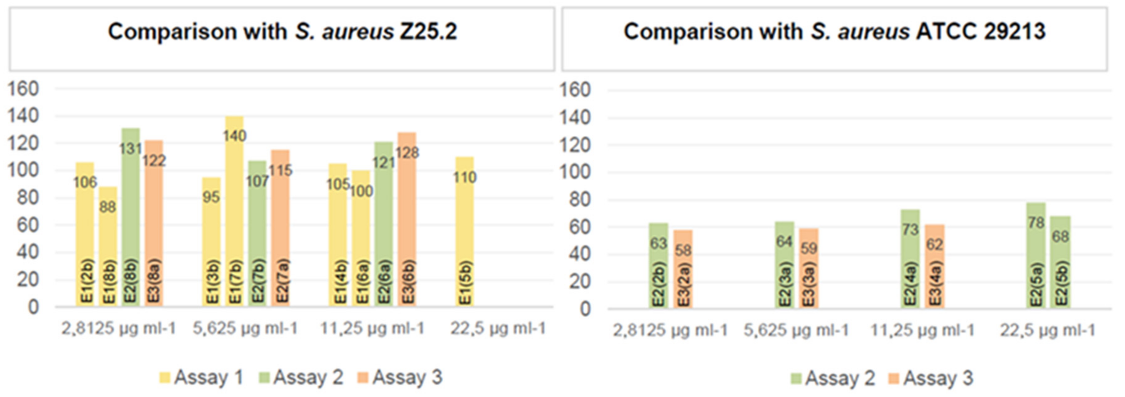
Comparing the sequences of the original isolates and the ones obtained throughout the MEGA-plate assays, it was possible to assess the number of genes that presented mutations (Figure 2).
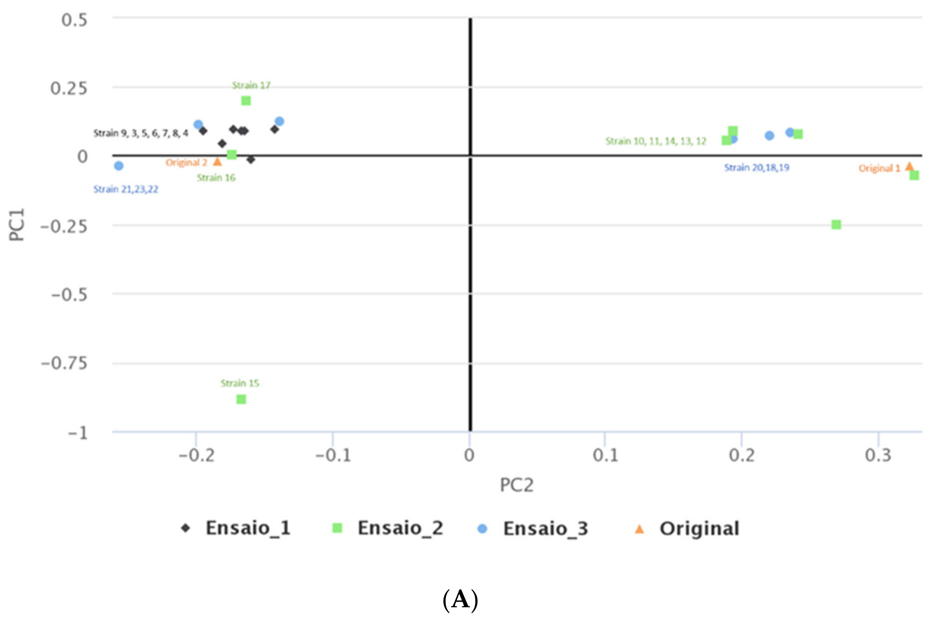
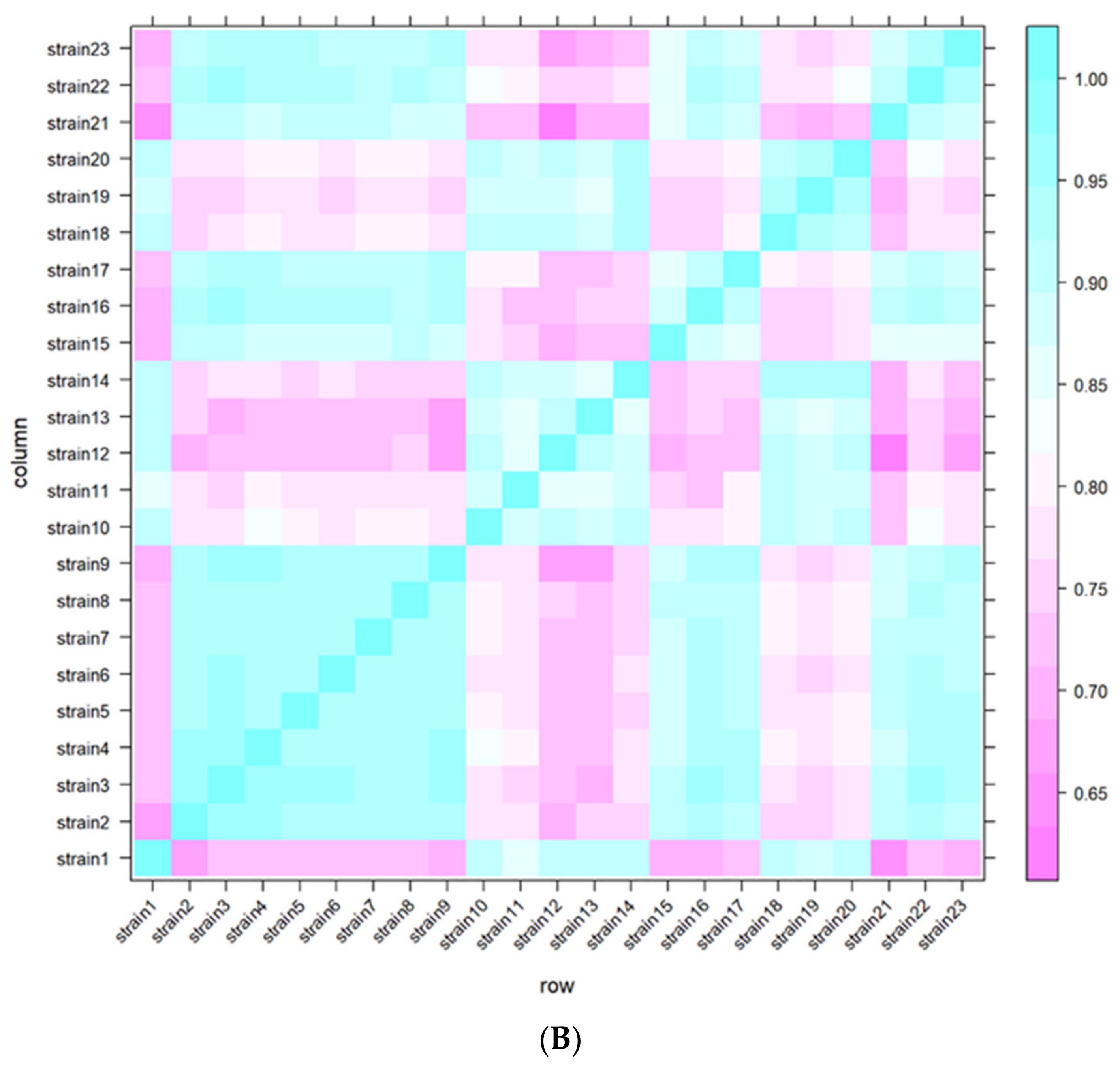
Figure 3 shows the number of genes that presented mutations detected after comparison with the original S. aureus Z25.2 (original 2, left graphic) and S. aureus ATCC 29213 (original 1, right graphic) strains, grouped according to the MEGA-plate division from which they were collected.
It was possible to observe that isolates derived from the original S. aureus ATCC 29213 presented far less genetic mutations (with the number of mutated genes ranging between 58 and 78), than the ones derived from the original S. aureus Z25.2 DFI isolate (with the number of mutated genes ranging from 88 to 140). It was not possible to detect a pattern regarding the number of mutated genes and the different assays or the nisin concentration to which they were exposed. However, in the isolates derived from S. aureus ATCC 29213, the number of genes that presented mutations tended to increase in isolates collected from divisions closer to the middle MEGA-plate division, where nisin’s concentration was the highest used in this study (MIC).
The analysis performed in this study focused on the evaluation of specific mutations in genes involved in S. aureus resistance to nisin. Currently, the glycopeptide resistance-associated two-component system (GraSR) has been identified as a very important two-component system in S. aureus known for being involved in nisin resistance [52,53]. GraSR is mainly responsible for controlling the vraFG operon, which is positioned right downstream of the graXRS genes, ultimately encoding for an ABC transporter that plays a huge role regarding resistance to cationic AMP [53]. One of the genes of this system encodes for the GraX protein, an essential component of this system. This two-component system was first discovered when investigators saw that its overexpression led to a higher vancomycin resistance; but to date, it has already been associated with the expression of the mprF gene and other operons [52]. Additionally, this system was shown to have an impact on biofilm formation [54]. In this study, only one isolate presented a mutation in a gene of this complex, graR, namely an isolate derived from the S. aureus Z25.2 DFI isolate, which was collected in the first assay from a division where nisin’s concentration was at its lowest (2.8125 μg mL−1). Moreover, El Shazely et al. [55], who studied the resistance evolution in S. aureus, observed mutations in the vraF gene in strains submitted to subMIC concentrations of the AMP pexiganan, and also in other genes associated with detoxification by efflux, which may be associated with nisin resistance.
Another two-component system of great importance regarding bacitracin and nisin resistance is NsaRS, also known as BraRS [53,56,57]. Nisin resistance often comes associated with mutations in the nsaS gene, which encodes for a sensor kinase. A study conducted by Arii et al. showed that the exposure of S. aureus to sub-inhibitory concentrations of nisin led to the occurrence of spontaneous mutations in genes associated with this system, which resulted in higher resistance to nisin [58], through mechanisms yet unknown [56]. In this study, no mutations were detected in any genes related to this system, allowing to conclude that isolates’ exposure to nisin subMIC values did not promote genomic alteration that conferred resistance to nisin.
In conclusion, results showed that, to avoid the development of resistant mutants, nisin therapeutic doses should be established based on the MSW, that nisin does not seem to promote vanA transfer, and that nisin subMIC values do not prompt significant mutations in genes related to nisin resistance. As such, nisin seems to be a good candidate to be applied to DFI treatment in the future.

3. Materials and Methods

3.1. Bacterial Isolates

In this study, a collection of 23 S. aureus DFI isolates was used. These isolates were previously collected from patients with DFI [7] and further selected and characterized in terms of clonality, antimicrobial resistance, and virulence profiles [46]. Additionally, the reference strain S. aureus ATCC 29213 was also included as a control. Each isolate was maintained at −80 °C in buffered peptone water with 20% of glycerol throughout the duration of this study.

3.2. Nisin Solution

A nisin (Sigma-Aldrich, St. Louis, MO, USA) with a purity of 2.5% (1000 IU/mg) was used. To obtain a 1000 µg/mL stock solution, 1 g was dissolved in 25 mL of 0.02M HCl (Merck, Darmstadt, Germany) [35]. Afterwards, nisin was sterilized by filtration using a 0.22 μm filter (Frilabo, Maia, Portugal) and stored at 4 °C.

3.3. Determination of the Mutant Prevention Concentration

A modified version of the protocol described by Sinel et al. [59] was used to determine the MPC of nisin regarding the 23 S. aureus DFI isolates under study.
Each isolate was inoculated in brain heart infusion (BHI) agar (VWR Chemicals, Leuven, Belgium), and, after a 24 h incubation at 37 °C, a suspension of 0.5 MacFarland (1 × 10⁸ CFU/mL) was performed and used to inoculate two BHI agar plates. After a 24 h incubation at 37 °C, the bacterial lawn was collected from the two BHI plates and resuspended in 1mL of BHI broth to achieve a bacterial suspension with a concentration of 1010 CFU/mL. To confirm the concentration values of the bacterial suspensions, these were serially diluted for viable cell count. Afterwards, 50 µL of the original 1010 CFU/mL suspensions was inoculated in Mueller–Hinton agar (MHA) (Oxoid, Hampshire, UK) supplemented with the following concentrations of nisin: 5.63, 11.25, 22.5, 45, 90, 180, 360, and 720 µg/mL. These concentrations were selected considering a two-fold increase in the mean MIC value (11.25 µg/mL) that was previously determined [35]. A subMIC value was also included (5.63 µg/mL). Finally, plates were incubated for 72 h at 37 °C for MPC determination.
The MPC corresponded to the minimum concentration of nisin that prevented the growth of resistant mutants after the incubation period. For each isolate, the mutants grown in the presence of nisin concentrations below the MPC were isolated and stored at −80 °C in a solution of buffered peptone water with 20% glycerol (VWR Chemicals, Leuven, Belgium). The MPC values of nisin were determined in two independent rounds.

3.4. Horizontal Gene Transfer

3.4.1. DNA Extraction

DNA extraction was performed according to the protocol described by Mottola et al. [46]. All isolates were inoculated in BHI agar and incubated for 24 h at 37 °C. Then, four to five bacterial colonies were collected using a sterile loop and resuspended in 100 μL of TBE buffer (0.9 M Tris-Borate, 0.01 M EDTA, pH 8.3, Omega, Norcross, GA, USA) supplemented with 0.1% Tween 20 (Merck, Darmstadt, Germany) solution. After homogenization, the suspension was incubated for seven minutes at 97 °C, centrifuged at 15,000 rpm for 5 min, and the supernatant was collected for PCR screening.

3.4.2. Multiplex PCR for vanA Detection

Before the horizontal gene transfer protocol, it was necessary to confirm the absence of vanA gene in the collection of 23 S. aureus DFI isolates under study, using multiplex PCR [60]. Two pairs of primers, synthesized by STABVIDA® (Lisbon, Portugal), targeting vanA (5′ GGG AAA ACG ACA ATT GC 3′) and mecA (5′ TCCAGAT-TACAACTTCACCAGG 3′) were used [46,60].
The PCR mixture had a final volume of 28.5 μL, including 10 μL of the Supreme NZYTaq 2x Green Master Mix (NZYTech, Lisbon, Portugal), 0.29 μL of the vanA primer (0.5 µM), 0.23 μL of the mecA primer (0.4 µM), 12.46 μL of PCR-grade water and 5 μL DNA template (170 ng/μL).
PCR amplification was completed in a MyCycler Thermal Cycler (BioRad, Lisbon, Portugal) using the following conditions: initial denaturation at 94 °C for 4 min; 10 cycles including denaturation at 94 °C for 30 s, annealing at 64 °C for 30 s, and elongation at 72 °C for 45 s; 25 cycles comprising denaturation at 94 °C for 30 s, annealing at 50 °C for 45 s and elongation at 72 °C for 2 min, and a final extension step at 72 °C for 10 min.
An electrophoresis gel was performed to perceive the amplified products, using a 1.5% agarose gel (NZYTech, Lisbon, Portugal) and a buffer stained with GreenSafe (NZYTech, Lisbon, Portugal) at 90 V for 45 min. A molecular weight marker, NZYDNA ladder VI (NZYTech, Lisbon, Portugal), was also included. Results were visualized by transillumination (ChemiDoc XRS+, BioRad, Lisbon, Portugal).
Two positive control strains, Staphylococcus aureus 01-00694 (mecA positive) and Enterococcus faecium CCUG 36804 (vanA positive), were included in each PCR amplification protocol, as well as a negative control, with no DNA.

3.4.3. Horizontal Gene Transfer protocol

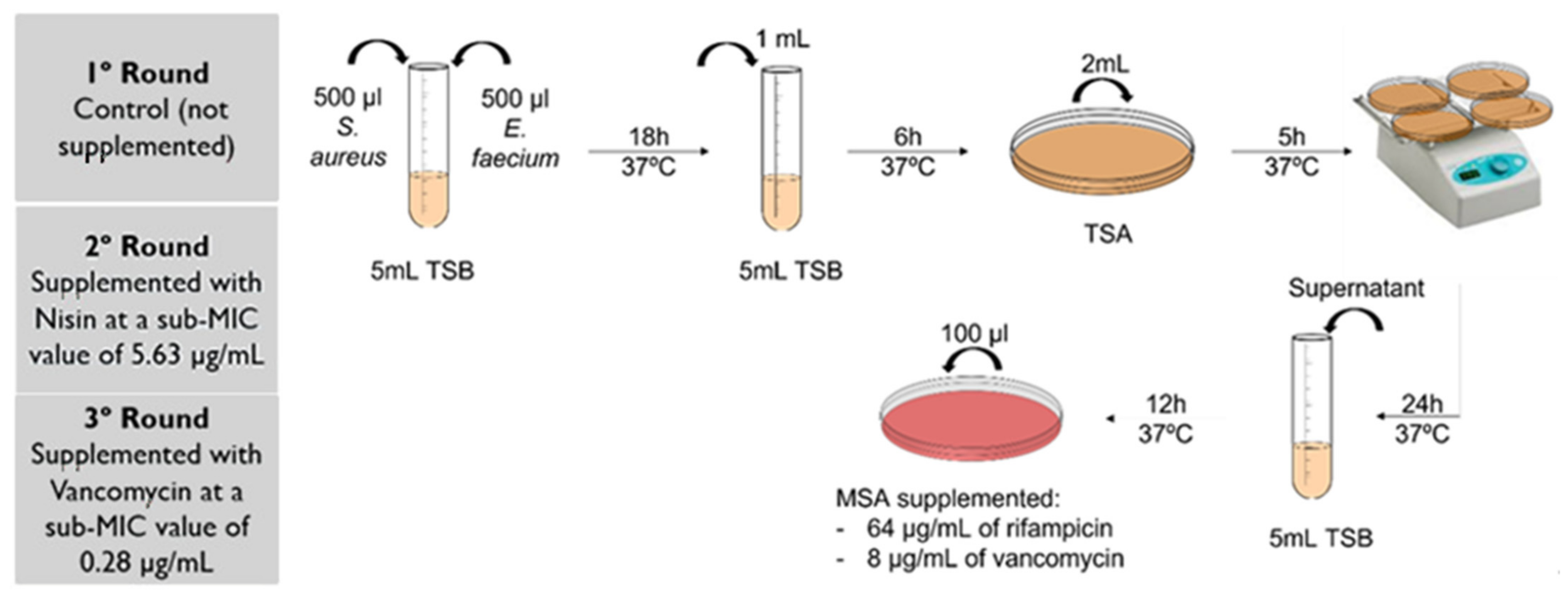
To test if nisin selective pressure induced horizontal gene transfer, a protocol adapted from Niederhäusern et al. [61] was performed (Figure 4). Mating experiments were performed in three rounds, using the VRE rifampicin-susceptible (VanR RifS) E. faecium CCUG 36804 strain as a donor of the vanA gene [39], and as recipients of all the 23 S. aureus DFI isolates of our collection. Initially, resistance to rifampicin was induced in the recipients, as described elsewhere [61], allowing to obtain strains resistant to rifampicin and susceptible to vancomycin (VanS RifR)
After performing a 0.5 MacFarland suspension for each isolate, 500 μL of the donor and 500 μL of each one of the recipients were added to 5 mL of non-supplemented TSB (tryptic soy broth, VWR Chemicals, Leuven, Belgium) and incubated at 35 °C for 18 h. After incubation, 1 mL of the bacterial suspension was added to 5 mL of TSB and further incubated for 6 h at 37 °C. Afterwards, 2 mL of each suspension was inoculated in TSA (tryptic soy agar, VWR Chemicals, Leuven, Belgium) and incubated for 5 h at 37 °C on a shaker, to promote mating. Then, the plates were incubated at 37 °C for 24 h, after which the bacterial suspension that remained at the surface of the agar plates was removed and inoculated in 5 mL of TSB. After an incubation period of 12 h at 37 °C, 100 μL of the suspension was inoculated in mannitol salt agar (MSA, PanReac AppliChem, Barcelona, Spain) supplemented with 64 μg/mL of rifampicin and 8 μg/mL of vancomycin (vancomycin hydrochloride, Abcam, Cambridge, UK) to allow VanRRifR transconjugants selection. Transconjugants were stored at −80°C in buffered peptone water with 20% glycerol and a PCR analysis was performed to confirm the presence of the vanA gene in these isolates. Moreover, strains with a positive vanA PCR were submitted for DNA Sanger sequencing by STABVIDA® (Lisbon, Portugal), to confirm vanA gene transference.
This first mating round was performed with non-supplemented TSB, as control. Then, a second mating round was performed in the presence of nisin, with all media used being supplemented with nisin at subMIC (5.63 μg/mL). A third mating round was also performed as a control, in the presence of a subMIC value of vancomycin (0.28 μg/mL), selected based on the previous MIC determination [46].

3.4.4. PCR for pSK41-like Plasmid Detection

To evaluate the presence of the pSK41-like plasmid in the 23 clinical isolates under study, a PCR protocol was performed, using a pair of primers targeting traE (5′ ACA AAT GCG TAC TAC AGA CCC TAA ACG A 3′), synthesized by STABVIDA® (Lisbon, Portugal) [11,62].
The PCR mixture had a final volume of 50 μL, including 10 μL of the Supreme NZYTaq 2x Green Master Mix (NZYTech, Lisbon, Portugal), 0.4 μL of traE primer (0.4 uM), 34.2 μL of PCR-grade water, and 5 μL DNA template (170 ng/μL). PCR amplification was completed in a MyCycler Thermal Cycler (BioRad, Lisbon, Portugal) using the following conditions: initial denaturation at 94 °C for 2 min; 30 cycles comprising denaturation at 95 °C for 15 s, annealing at 53 °C for 90 s and elongation at 72 °C for 90 s, and a final extension step at 72 °C for 7 min. An electrophoresis gel was performed to perceive the amplified products, using a 1.5% agarose gel (NZYTech, Lisbon, Portugal) and a buffer stained with GreenSafe (NZYTech, Lisbon, Portugal) at 90 V for 45 min. A molecular weight marker, NZYDNA ladder VII (NZYTech, Lisbon, Portugal), was also included. Results were visualized by transillumination (ChemiDoc XRS+, BioRad, Lisbon, Portugal).
A positive control strain, Staphylococcus aureus RN4220 (pGO1 positive), gently provided by Dr. Alex O’Neill from the University of Leeds, was included in the PCR amplification protocol, as well as a negative control, with no DNA [63].

3.5. MEGA-Plate Assays and Variant Call Analysis

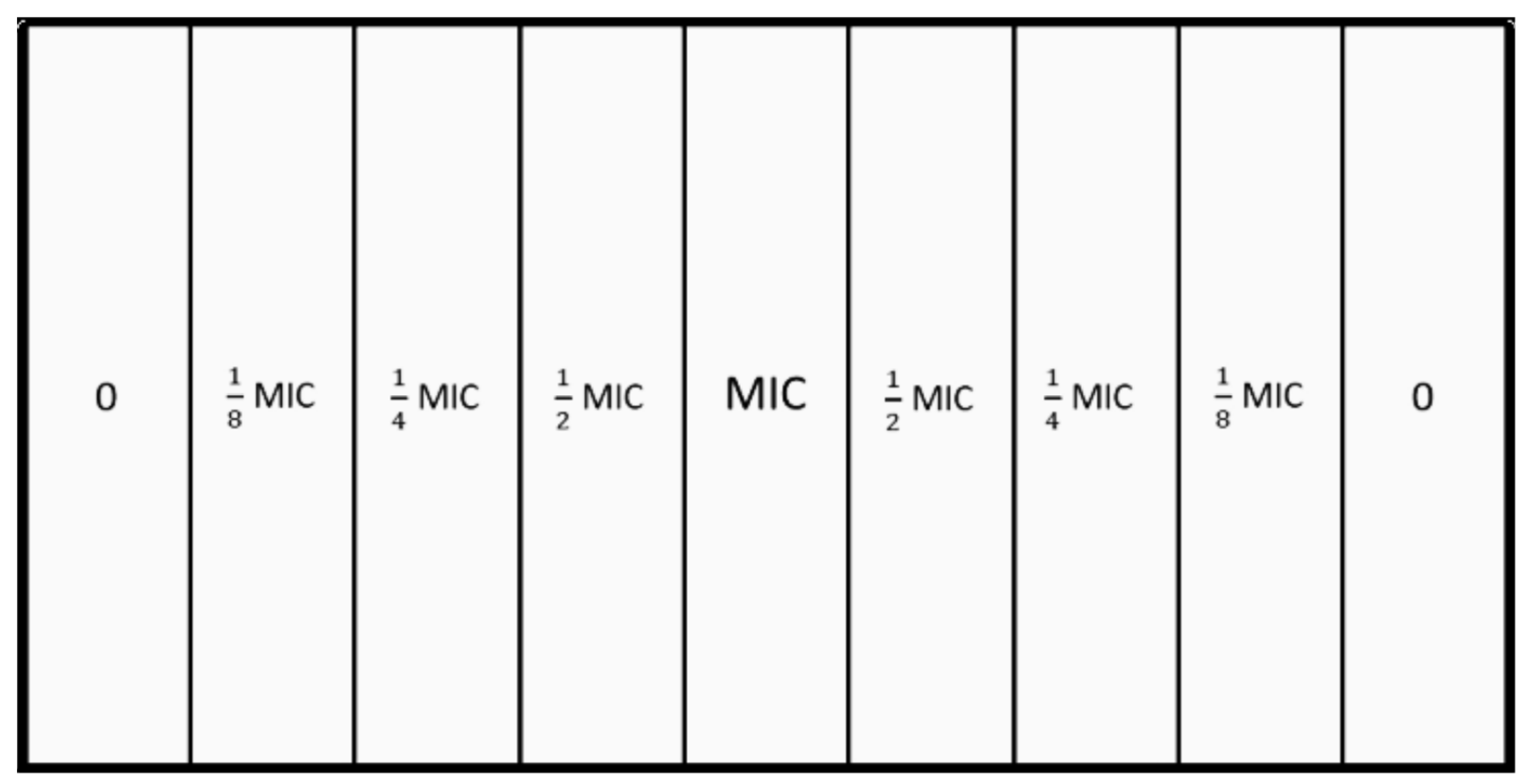
Three assays were performed using the microbial evolution and growth arena (MEGA)-plate described by Baym et al. [64], using a glass plate with 90 × 45 × 5 cm and 9 equal internal divisions of 45 × 10 × 3 cm and a glass cover (Figure 5). Prior to its use, the MEGA-plate was sterilized with ethanol (96%) and exposed to UV light for 2 h in a laminar flow chamber. The divisions were later filled with nonselective brain heart infusion (BHI) medium (VWR® Chemicals, Belgium), supplemented with bacteriological agar (VWR® Chemicals, Belgium) at 1.5 % (15 g/L) and with different nisin concentrations (2.8125 µg/mL, 5.625 µg/mL, 11.25 µg/mL, and 22.5 µg/mL, corresponding to nisin 1/8 MIC, ¼ MIC, ½ MIC, and MIC values (Figure 5). Additionally, semi-solid BHI media (BHI broth with bacteriological agar at 8 g/L), supplemented with amphotericin B (2 µg/mL), was prepared and poured on top of the previous medium.
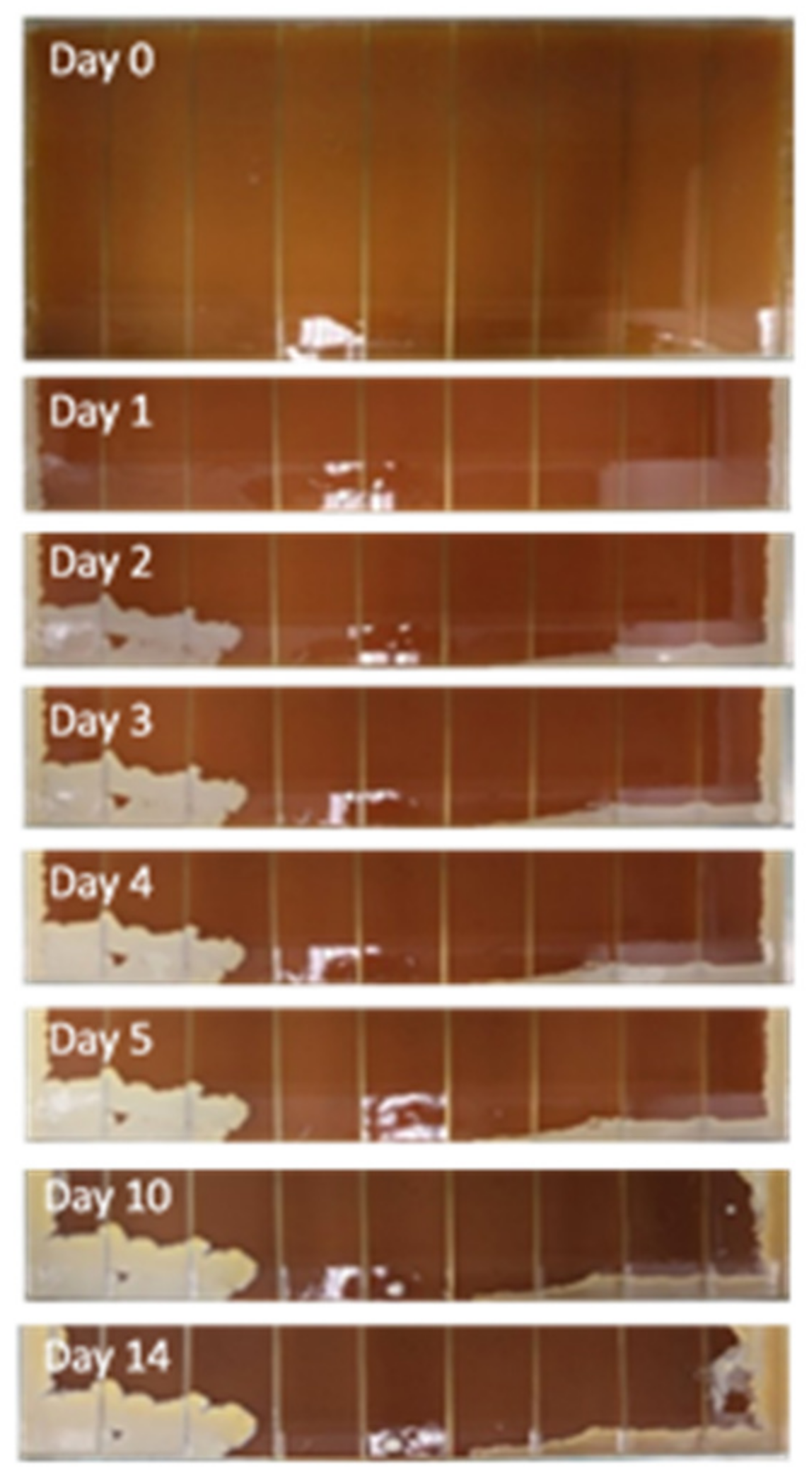
Then, a suspension of the reference strain S. aureus ATCC 29213 (with 108 CFU/mL) was inoculated on the left extremity of the MEGA-plate, and a suspension of a selected DFI isolate, S. aureus Z.25.2, was inoculated on the right extremity, in an area of 1 cm of thickness. The plates were incubated at 37 °C for a period of 15 days and, each day, the growth of the staphylococci was accessed and registered via time-lapse photography twice a day using a Canon® M50 camera, in order to evaluate the bacterial multiplication progress through time (Figure 6).
After incubation, isolates were collected from each division and inoculated in BHI agar medium plates. After a 24 h incubation at 37 °C, isolates’ identification was confirmed through Gram staining, catalase and coagulase tests, and inoculation on MSA. This protocol was performed in three independent assays.
All Isolates obtained were analyzed by whole-genome sequencing using Illumina MiSeq paired-end short read sequencing at Biosystems and Integrative Sciences Institute (BioISI) Genomics. It was obtained between 0.6 and 1.3 million reads, corresponding to a sequencing depth between 34 and 105 times the length of the S. aureus genome, respectively. The reads length ranged between 157 bps and 227 bps, and the quality of the sequenced reads ranged between a Phred score of 30.63 and 38.06. A customized analytical pipeline developed by BioISIGenomics® for short-read genomic sequencing was used. During the pre-processing stage, the quality of the reads was assessed using FASTQC v0.11.9 [65]. Trimmomatic v0.38 was used to trim and filter the reads according to their length and quality score [66]. The alignment against the ENSEMBL reference sequences: Staphylococcus aureus Newman strain (genome assembly: ASM1046v1); and S. aureus ATCC 29213 (genome assembly: ASM126771v2), was performed using Bowtie2 v.2.4.1 [67]. The mapping was performed against the S. aureus Newman strain because, contrary to the Z25.2 strain, the Newman strain is fully sequenced and annotated. Variant calling was performed using Mpileup (Samtools v 1.10) [68]. The variant call events were filtered according to the sequencing depth (DP > 100) and to an estimate of the probability of observing a call plainly by chance (%QUAL ≥ 20). The original strains inoculated on the MEGA-plate (S. aureus Z.25.2 and S. aureus ATCC 29213) and the isolates collected in the assays were compared using VCFTools v0.1.16 [69]. ENSEMBL Variant Effect Predictor tool (release/100.2) was used to annotate the VCF files obtained in the previous step [70], within QuickGO release-1.0.1 to retrieve the GO terms annotation [71]. Finally, SNiPlay v1.0_TTS toolkit and PLINK v2.3 were used to generate heatmaps (IBS matrix) and multi-dimensional scaling (MDS) plots [72,73].

Supplementary Materials

The following supporting information can be downloaded at: https://www.mdpi.com/article/10.3390/antibiotics11070972/s1, Supplementary file.

Author Contributions

Conceptualization, M.O.; methodology, M.C., C.M., E.C. and M.O.; formal analysis, M.C., C.M., M.P. and R.D.; investigation, M.C. and C.M.; resources, L.T., M.O.; writing—original draft preparation, M.C., E.C. and D.G.; writing—review and editing, E.C., R.D., L.T. and M.O.; supervision, M.O.; project administration, M.O.; funding acquisition L.T. and M.O. All authors have read and agreed to the published version of the manuscript.

Funding

This research was funded by Foundation for Science and Technology (FCT), Project PTDC/SAU-INF/28466/2017—Polyphasic validation of antimicrobial peptides as alternative treatment for diabetic foot.

Informed Consent Statement

Not applicable.

Data Availability Statement

The datasets used and/or analyzed during the current study are available from the corresponding author on reasonable request.

Acknowledgments

Authors would like to acknowledge the Foundation for Science and Technology for financial support (project PTDC/SAU-INF/28466/2017—Polyphasic validation of antimicrobial peptides as alternative treatment for diabetic foot infections), to CIISA–Centro de Investigação Interdisciplinar em Sanidade Animal, Faculdade de Medicina Veterinária, Universidade de Lisboa, Project UIDB/00276/2020 (Funded by FCT); and to the Laboratório Associado para Ciência Animal e Veterinária (LA/P/0059/2020—AL4AnimalS).

Conflicts of Interest

The authors declare no conflict of interest.

References

  1. Singh, N. Preventing Foot Ulcers in Patients with Diabetes. JAMA 2005, 293, 217–228. [Google Scholar] [CrossRef] [PubMed]
  2. Skrepnek, G.H.; Mills, J.L.; Lavery, L.A.; Armstrong, D.G. Health Care Service and Outcomes Among an Estimated 6.7 Million Ambulatory Care Diabetic Foot Cases in the U.S. Diabetes Care 2017, 40, 936–942. [Google Scholar] [CrossRef] [PubMed] [Green Version]
  3. Citron, D.M.; Goldstein, E.J.C.; Merriam, C.V.; Lipsky, B.A.; Abramson, M.A. Bacteriology of Moderate-to-Severe Diabetic Foot Infections and In Vitro Activity of Antimicrobial Agents. J. Clin. Microbiol. 2007, 45, 2819–2828. [Google Scholar] [CrossRef] [PubMed] [Green Version]
  4. Hobizal, K.B.; Wukich, D.K. Diabetic Foot Infections: Current Concept Review. Diabet. Foot Ankle 2012, 3, 18409. [Google Scholar] [CrossRef] [PubMed]
  5. Weigel, L.M.; Clewell, D.B.; Gill, S.R.; Clark, N.C.; McDougal, L.K.; Flannagan, S.E.; Kolonay, J.F.; Shetty, J.; Killgore, G.E.; Tenover, F.C. Genetic Analysis of a High-Level Vancomycin-Resistant Isolate of Staphylococcus aureus. Science 2003, 302, 1569–1571. [Google Scholar] [CrossRef]
  6. Sujatha, S.; Praharaj, I. Glycopeptide Resistance in Gram-Positive Cocci: A Review. Interdiscip. Perspect. Infect. Dis. 2012, 2012, 781679. [Google Scholar] [CrossRef] [Green Version]
  7. Mendes, J.J.; Marques-Costa, A.; Vilela, C.; Neves, J.; Candeias, N.; Cavaco-Silva, P.; Melo-Cristino, J. Clinical and Bacteriological Survey of Diabetic Foot Infections in Lisbon. Diabetes Res. Clin. Pract. 2012, 95, 153–161. [Google Scholar] [CrossRef]
  8. Kos, V.N.; Desjardins, C.A.; Griggs, A.; Cerqueira, G.; Van Tonder, A.; Holden, M.T.G.; Godfrey, P.; Palmer, K.L.; Bodi, K.; Mongodin, E.F.; et al. Comparative Genomics of Vancomycin-Resistant Staphylococcus aureus Strains and Their Positions within the Clade Most Commonly Associated with Methicillin-Resistant S. Aureus Hospital-Acquired Infection in the United States. mBio 2012, 3, e00112-12. [Google Scholar] [CrossRef] [Green Version]
  9. Gardete, S.; Tomasz, A. Mechanisms of Vancomycin Resistance in Staphylococcus aureus. J. Clin. Investig. 2014, 124, 2836–2840. [Google Scholar] [CrossRef]
  10. McDougal, L.K.; Fosheim, G.E.; Nicholson, A.; Bulens, S.N.; Limbago, B.M.; Shearer, J.E.S.; Summers, A.O.; Patel, J.B. Emergence of Resistance among USA300 Methicillin-Resistant Staphylococcus aureus Isolates Causing Invasive Disease in the United States. Antimicrob. Agents Chemother. 2010, 54, 3804–3811. [Google Scholar] [CrossRef] [Green Version]
  11. Zhu, W.; Clark, N.; Patel, J.B. PSK41-like Plasmid Is Necessary for Inc18-like VanA Plasmid Transfer from Enterococcus faecalis to Staphylococcus aureus in Vitro. Antimicrob. Agents Chemother. 2013, 57, 212–219. [Google Scholar] [CrossRef] [PubMed] [Green Version]
  12. Gottrup, F.; Apelqvist, J. Present and New Techniques and Devices in the Treatment of DFU: A Critical Review of Evidence. Diabetes Metab. Res. Rev. 2012, 28, 64–71. [Google Scholar] [CrossRef]
  13. Bahar, A.A.; Ren, D. Antimicrobial Peptides. Pharm. Basel Switz. 2013, 6, 1543–1575. [Google Scholar] [CrossRef] [PubMed] [Green Version]
  14. Mahlapuu, M.; Håkansson, J.; Ringstad, L.; Björn, C. Antimicrobial Peptides: An Emerging Category of Therapeutic Agents. Front. Cell. Infect. Microbiol. 2016, 6, 194. [Google Scholar] [CrossRef] [PubMed] [Green Version]
  15. Zhang, L.; Gallo, R.L. Antimicrobial Peptides. Curr. Biol. 2016, 26, R14–R19. [Google Scholar] [CrossRef] [PubMed]
  16. Abee, T.; Delves-Broughton, J. Bacteriocins—Nisin. In Food Preservatives; Russell, N.J., Gould, G.W., Eds.; Springer US: Boston, MA, USA, 2003; pp. 146–178. [Google Scholar] [CrossRef]
  17. Hassan, M.; Kjos, M.; Nes, I.F.; Diep, D.B.; Lotfipour, F. Natural Antimicrobial Peptides from Bacteria: Characteristics and Potential Applications to Fight against Antibiotic Resistance. J. Appl. Microbiol. 2012, 113, 723–736. [Google Scholar] [CrossRef] [PubMed]
  18. Mitchell, S.A.; Truscott, F.; Dickman, R.; Ward, J.; Tabor, A.B. Simplified Lipid II-Binding Antimicrobial Peptides: Design, Synthesis and Antimicrobial Activity of Bioconjugates of Nisin Rings A and B with Pore-Forming Peptides. Bioorg. Med. Chem. 2018, 26, 5691–5700. [Google Scholar] [CrossRef]
  19. Field, D.; Cotter, P.D.; Hill, C.; Ross, R.P. Bioengineering Lantibiotics for Therapeutic Success. Front. Microbiol. 2015, 6, 1363. [Google Scholar] [CrossRef] [Green Version]
  20. Lagedroste, M.; Reiners, J.; Smits, S.H.J.; Schmitt, L. Systematic Characterization of Position One Variants within the Lantibiotic Nisin. Sci. Rep. 2019, 9, 935. [Google Scholar] [CrossRef]
  21. Zhou, H.; Fang, J.; Tian, Y.; Lu, X.Y. Mechanisms of Nisin Resistance in Gram-Positive Bacteria. Ann. Microbiol. 2014, 64, 413–420. [Google Scholar] [CrossRef]
  22. Shin, J.M.; Gwak, J.W.; Kamarajan, P.; Fenno, J.C.; Rickard, A.H.; Kapila, Y.L. Biomedical Applications of Nisin. J. Appl. Microbiol. 2016, 120, 1449–1465. [Google Scholar] [CrossRef] [PubMed] [Green Version]
  23. Santos, R.; Veiga, A.S.; Tavares, L.; Castanho, M.; Oliveira, M. Bacterial Biofilms in Diabetic Foot Ulcers: Potential Alternative Therapeutics. In Microbial Biofilms—Importance and Applications; IntechOpen: London, UK, 2016. [Google Scholar] [CrossRef] [Green Version]
  24. Cunha, E.; Trovão, T.; Pinheiro, A.; Nunes, T.; Santos, R.; Moreira da Silva, J.; São Braz, B.; Tavares, L.; Veiga, A.S.; Oliveira, M. Potential of Two Delivery Systems for Nisin Topical Application to Dental Plaque Biofilms in Dogs. BMC Vet. Res. 2018, 14, 375. [Google Scholar] [CrossRef] [PubMed] [Green Version]
  25. Xilin, Z.; Drlica, K. Restricting the Selection of Antibiotic-Resistant Mutant Bacteria: Measurement and Potential Use of the Mutant Selection Window. J. Infect. Dis. 2002, 185, 561–565. [Google Scholar] [CrossRef]
  26. Cairns, B.J.; Payne, R.J.H. Bacteriophage Therapy and the Mutant Selection Window. Antimicrob. Agents Chemother. 2008, 52, 4344–4350. [Google Scholar] [CrossRef] [PubMed] [Green Version]
  27. Drlica, K. The Mutant Selection Window and Antimicrobial Resistance. J. Antimicrob. Chemother. 2003, 52, 11–17. [Google Scholar] [CrossRef] [PubMed] [Green Version]
  28. Drlica, K.; Zhao, X. Mutant Selection Window Hypothesis Updated. Clin. Infect. Dis. 2007, 44, 681–688. [Google Scholar] [CrossRef] [PubMed]
  29. Vasilchenko, A.S.; Rogozhin, E.A. Sub-inhibitory effects of antimicrobial peptides. Front Microbiol. 2019, 10, 1160. [Google Scholar] [CrossRef] [Green Version]
  30. Gottschalk, S.; Gottlieb, C.T.; Vestergaard, M.; Hansen, R.; Gram, L.; Ingmer, H.; Thomsen, L.E. Amphibian antimicrobial peptide fallaxin analogue FL9 affects virulence gene expression and DNA replication in Staphylococcus aureus. J. Med. Microbiol. 2015, 64, 1504–1513. [Google Scholar] [CrossRef] [Green Version]
  31. Polikanov, Y.S.; Aleksashin, N.A.; Beckert, B.; Wilson, D.N. The mechanisms of action of ribosome-targeting peptide antibiotics. Front. Mol. Biosci. 2018, 5, 48. [Google Scholar] [CrossRef] [Green Version]
  32. Shimizu, T.; Harada, K.; Kataoka, Y. Mutant Prevention Concentration of Orbifloxacin: Comparison between Escherichia coli, Pseudomonas aeruginosa, and Staphylococcus pseudintermedius of Canine Origin. Acta Vet. Scand. 2013, 55, 37. [Google Scholar] [CrossRef] [Green Version]
  33. López, Y.; Tato, M.; Gargallo-Viola, D.; Cantón, R.; Vila, J.; Zsolt, I. Mutant Prevention Concentration of Ozenoxacin for Quinolone-Susceptible or -Resistant Staphylococcus aureus and Staphylococcus epidermidis. PLoS ONE 2019, 14, e0223326. [Google Scholar] [CrossRef]
  34. Raj Dwivedi, G.; Khwaja, S.; Singh Negi, A.; Panda, S.S.; Swaroop Sanket, A.; Pati, S.; Chand Gupta, A.; Bawankule, D.U.; Chanda, D.; Kant, R.; et al. Design, Synthesis and Drug Resistance Reversal Potential of Novel Curcumin Mimics Van D: Synergy Potential of Curcumin Mimics. Bioorganic Chem. 2021, 106, 104454. [Google Scholar] [CrossRef] [PubMed]
  35. Santos, R.; Gomes, D.; Macedo, H.; Barros, D.; Tibério, C.; Veiga, A.S.; Tavares, L.; Castanho, M.; Oliveira, M. Guar gum as a new antimicrobial peptide delivery system against diabetic foot ulcers Staphylococcus aureus isolates. J. Med. Microbiol. 2016, 65, 1092–1099. [Google Scholar] [CrossRef] [PubMed]
  36. Fujimura, S.; Nakano, Y.; Watanabe, A. A Correlation between Reduced Susceptibilities to Vancomycin and Daptomycin among the MRSA Isolates Selected in Mutant Selection Window of Both Vancomycin and Daptomycin. J. Infect. Chemother. 2014, 20, 752–756. [Google Scholar] [CrossRef] [PubMed]
  37. Hasper, H.E.; Kramer, N.E.; Smith, J.L.; Hillman, J.D.; Zachariah, C.; Kuipers, O.P.; de Kruijff, B.; Breukink, E. An Alternative Bactericidal Mechanism of Action for Lantibiotic Peptides That Target Lipid II. Science 2006, 313, 1636–1637. [Google Scholar] [CrossRef] [PubMed] [Green Version]
  38. Breukink, E.; de Kruijff, B. Lipid II as a Target for Antibiotics. Nat. Rev. Drug Discov. 2006, 5, 321–332. [Google Scholar] [CrossRef]
  39. Cunha, E.; Janela, R.; Costa, M.; Tavares, L.; Veiga, A.S.; Oliveira, M. Nisin Influence on the Antimicrobial Resistance Ability of Canine Oral Enterococci. Antibiotics 2020, 9, 890. [Google Scholar] [CrossRef]
  40. Fernández, L.; Delgado, S.; Herrero, H.; Maldonado, A.; Rodríguez, J.M. The Bacteriocin Nisin, an Effective Agent for the Treatment of Staphylococcal Mastitis During Lactation. J. Hum. Lact. 2008, 24, 311–316. [Google Scholar] [CrossRef]
  41. Younes, M.; Aggett, P.; Aguilar, F.; Crebelli, R.; Dusemund, B.; Filipič, M.; Frutos, M.J.; Galtier, P.; Gundert-Remy, U.; Kuhnle, G.G.; et al. Safety of nisin (E 234) as a food additive in the light of new toxicological data and the proposed extension of use. EFSA J. 2017, 15, e05063. [Google Scholar] [CrossRef]
  42. WHO. Global Priority List of Antibiotic-Resistant Bacteria to Guide Research, Discovery, and Development of New Antibiotics. Available online: http://www.who.int/medicines/publications/global-priority-list-antibiotic-resistant-bacteria/en/ (accessed on 11 February 2021).
  43. Hsu, S.-T.D.; Breukink, E.; Tischenko, E.; Lutters, M.A.G.; de Kruijff, B.; Kaptein, R.; Bonvin, A.M.J.J.; van Nuland, N.A.J. The Nisin–Lipid II Complex Reveals a Pyrophosphate Cage That Provides a Blueprint for Novel Antibiotics. Nat. Struct. Mol. Biol. 2004, 11, 963–967. [Google Scholar] [CrossRef] [Green Version]
  44. Bader, M.S. Diabetic Foot Infection. Am. Fam. Physician 2008, 78, 71–79. [Google Scholar]
  45. Butler, M.S.; Hansford, K.A.; Blaskovich, M.A.T.; Halai, R.; Cooper, M.A. Glycopeptide Antibiotics: Back to the Future. J. Antibiot. 2014, 67, 631–644. [Google Scholar] [CrossRef] [PubMed]
  46. Mottola, C.; Semedo-Lemsaddek, T.; Mendes, J.J.; Melo-Cristino, J.; Tavares, L.; Cavaco-Silva, P.; Oliveira, M. Molecular Typing, Virulence Traits and Antimicrobial Resistance of Diabetic Foot Staphylococci. J. Biomed. Sci. 2016, 23, 33. [Google Scholar] [CrossRef] [PubMed] [Green Version]
  47. Rossi, F.; Diaz, L.; Wollam, A.; Panesso, D.; Zhou, Y.; Rincon, S.; Narechania, A.; Xing, G.; Di Gioia, T.S.R.; Doi, A.; et al. Transferable Vancomycin Resistance in a Community-Associated MRSA Lineage. N. Engl. J. Med. 2014, 370, 1524–1531. [Google Scholar] [CrossRef] [PubMed] [Green Version]
  48. King, J.M.; Kulhankova, K.; Stach, C.S.; Vu, B.G.; Salgado-Pabón, W. Phenotypes and Virulence among Staphylococcus aureus USA100, USA200, USA300, USA400, and USA600 Clonal Lineages. mSphere 2016, 1, e00071-16. [Google Scholar] [CrossRef] [Green Version]
  49. Friães, A.; Resina, C.; Manuel, V.; Lito, L.; Ramirez, M.; Melo-Cristino, J. Epidemiological Survey of the First Case of Vancomycin-Resistant Staphylococcus aureus Infection in Europe. Epidemiol. Infect. 2015, 143, 745–748. [Google Scholar] [CrossRef]
  50. Kohler, V.; Vaishampayan, A.; Grohmann, E. Broad-Host-Range Inc18 Plasmids: Occurrence, Spread and Transfer Mechanisms. Plasmid 2018, 99, 11–21. [Google Scholar] [CrossRef]
  51. Wistrand-Yuen, E.; Knopp, M.; Hjort, K.; Koskiniemi, S.; Berg, O.G.; Andersson, D.I. Evolution of High-Level Resistance during Low-Level Antibiotic Exposure. Nat. Commun. 2018, 9, 1599. [Google Scholar] [CrossRef] [Green Version]
  52. Falord, M.; Mäder, U.; Hiron, A.; Dbarbouillé, M.; Msadek, T. Investigation of the Staphylococcus aureus GraSR regulon reveals novel links to virulence, stress response and cell wall signal transduction pathways. PLoS ONE 2011, 6, e21323. [Google Scholar] [CrossRef]
  53. Falord, M.; Karimova, G.; Hiron, A.; Msadeka, T. GraXSR proteins interact with the VraFG ABC transporter to form a five-component system required for cationic antimicrobial peptide sensing and resistance in Staphylococcus aureus. Antimicrob. Agents Chemother. 2012, 56, 1047–1058. [Google Scholar] [CrossRef] [Green Version]
  54. Boles, B.R.; Thoende, M.; Roth, A.J.; Horswill, A.R. Identification of genes involved in polysaccharide-independent Staphylococcus aureus biofilm formation. PLoS ONE. 2010, 5, e10146. [Google Scholar] [CrossRef] [Green Version]
  55. El Shazely, B.; Yu, G.; Johnston, P.R.; Rolff, J. Resistance Evolution Against Antimicrobial Peptides in Staphylococcus aureus Alters Pharmacodynamics Beyond the MIC. Front. Microbiol. 2020, 11, 103. [Google Scholar] [CrossRef]
  56. Hiron, A.; Falord, M.; Valle, J.; Débarbouillé, M.; Msadek, T. Bacitracin and nisin resistance in Staphylococcus aureus: A novel pathway involving the BraS/BraR two-component system (SA2417/SA2418) and both the BraD/BraE and VraD/VraE ABC transporters. Mol. Microbiol. 2011, 81, 602–622. [Google Scholar] [CrossRef] [PubMed]
  57. Randall, C.P.; Gupta, A.; Utley-Drew, B.; Lee, S.Y.; Morrison-Williams, G.; O’Neill, A.J. Acquired nisin resistance in Staphylococcus aureus involves constitutive activation of an intrinsic peptide antibiotic detoxification module. MSphere 2018, 3, e00633-18. [Google Scholar] [CrossRef] [PubMed] [Green Version]
  58. Arii, K.; Kawada-Matsuo, M.; Oogai, Y.; Noguchi, K.; Komatsuzawa, H. Single mutations in BraRS confer high resistance against nisin A in Staphylococcus aureus. Microbiol. Open 2019, 8, e791. [Google Scholar] [CrossRef] [Green Version]
  59. Sinel, C.; Jaussaud, C.; Auzou, M.; Giard, J.C.; Cattoir, V. Mutant prevention concentrations of daptomycin for Enterococcus faecium clinical isolates. Int. J. Antimicrob. Agents 2016, 48, 449–452. [Google Scholar] [CrossRef] [PubMed]
  60. Ramos-Trujillo, E.; Pérez-Roth, E.; Méndez-Alvarez, S.; Claverie-Martín, F. Multiplex PCR for simultaneous detection of enterococcal genes VanA and VanB and staphylococcal genes MecA, IleS-2 and FemB. Int. Microbiol. Off. J. Span. Soc. Microbiol. 2003, 6, 113–115. [Google Scholar] [CrossRef]
  61. Niederhäusern, S.; Bondi, M.; Messi, P.; Iseppi, R.; Sabia, C.; Manicardi, G.; Anacarso, I. Vancomycin-resistance transferability from VanA enterococci to Staphylococcus aureus. Curr. Microbiol. 2011, 62, 1363–1367. [Google Scholar] [CrossRef]
  62. Albrecht, V.S.; Zervos, M.J.; Kaye, K.S.; Tosh, P.K.; Arshad, S.; Hayakawa, K.; Kallen, A.J.; McDougal, L.K.; Limbago, B.M.; Guh, A.Y. Prevalence of and risk factors for vancomycin-resistant Staphylococcus aureus precursor organisms in southeastern Michigan. Infect. Control Hosp. Epidemiol. 2014, 35, 1531–1534. [Google Scholar] [CrossRef]
  63. Caryl, J.A.; O’Neill, A.J. Complete Nucleotide Sequence of PGO1, the prototype conjugative plasmid from the staphylococci. Plasmid 2009, 62, 35–38. [Google Scholar] [CrossRef]
  64. Baym, M.; Lieberman, T.M.; Kelsic, E.D.; Chait, R.; Gross, R.; Yelin, I.; Kishony, R. Spatiotemporal microbial evolution on antibiotic landscapes. Science 2016, 353, 1147–1151. [Google Scholar] [CrossRef] [PubMed] [Green Version]
  65. Ghosh, A.; Som, A. Decoding molecular markers and transcriptional circuitry of naive and primed states of human pluripotency. Stem Cell Res. 2021, 53, 102334. [Google Scholar] [CrossRef] [PubMed]
  66. Bolger, A.M.; Lohse, M.; Usadel, B. Trimmomatic: A flexible trimmer for Illumina sequence data. Bioinformatics 2014, 30, 2114–2120. [Google Scholar] [CrossRef] [Green Version]
  67. Langmead, B.; Trapnell, C.; Pop, M.; Salzberg, S.L. Ultrafast and memory-efficient alignment of short DNA sequences to the human genome. Genome Biol. 2009, 10, R25. [Google Scholar] [CrossRef] [PubMed] [Green Version]
  68. Li, H.; Handsaker, B.; Wysoker, A.; Fennell, T.; Ruan, J.; Homer, N.; Marth, G.; Abecasis, G.; Durbin, R. The Sequence Alignment/Map format and SAMtools. Bioinformatics 2009, 25, 2078–2079. [Google Scholar] [CrossRef] [Green Version]
  69. Danecek, P.; Auton, A.; Abecasis, G.; Albers, C.A.; Banks, E.; DePristo, M.A.; Handsaker, R.E.; Lunter, G.; Marth, G.T.; Sherry, S.T.; et al. The variant call format and VCFtools. Bioinformatics 2011, 27, 2156–2158. [Google Scholar] [CrossRef]
  70. McLaren, W.; Gil, L.; Hunt, S.E.; Riat, H.S.; Ritchie, G.R.S.; Thormann, A.; Flicek, P.; Cunningham, F. The Ensembl Variant Effect Predictor. Genome Biol. 2016, 17, 122. [Google Scholar] [CrossRef] [Green Version]
  71. Dimmer, E.C.; Huntley, R.P.; Barrell, D.G.; Binns, D.; Draghici, S.; Camon, E.B.; Hubank, M.; Talmud, F.J.; Apweiler, R.; Lovering, R.C. The gene ontology—Providing a functional role in proteomic studies. Proteomics 2008, 8. [Google Scholar] [CrossRef]
  72. Dereeper, A.; Homa, F.; Andres, G.; Sempere, G.; Sarah, G.; Hueber, Y.; Dufayard, J.F.; Ruiz, M. SNiPlay3: A web-based application for exploration and large-scale analyses of genomic variations. Nucleic Acids Res. 2015, 43, W295–W300. [Google Scholar] [CrossRef] [Green Version]
  73. Purcell, S.; Neale, B.; Todd-Brown, K.; Thomas, L.; Ferreira, M.A.R.; Bender, D.; Maller, J.; Sklar, P.; de Bakker, P.I.; Daly, M.J.; et al. PLINK: A tool set for whole-genome association and population-based linkage analyses. Am. J. Hum. Genet. 2007, 81, 559–575. [Google Scholar] [CrossRef] [Green Version]
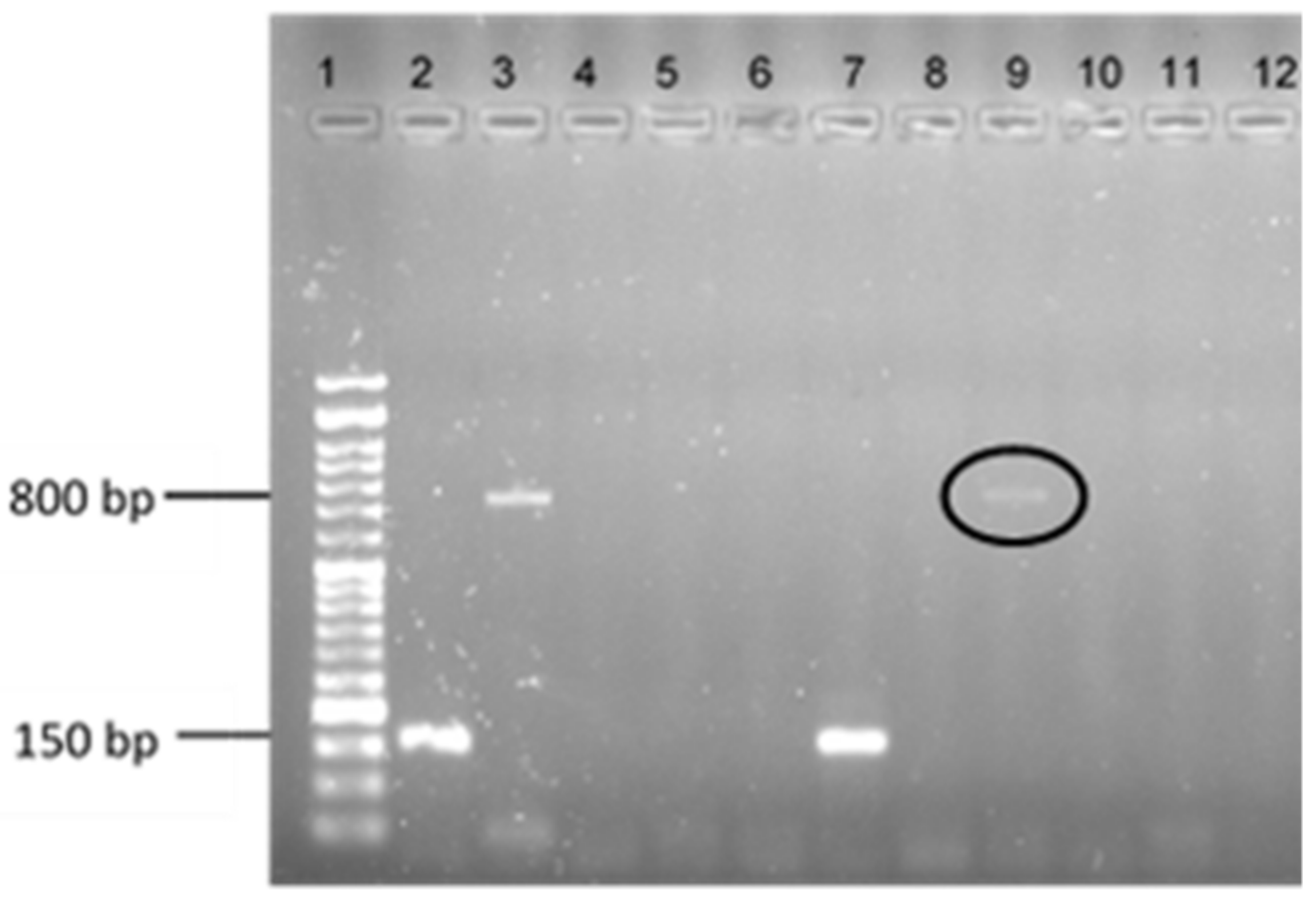
Figure 1. Electrophoresis results of a multiplex PCR reaction to determine the presence of the vanA gene in the transconjugants obtained after MPC protocol. Lane 1: ladder VI (Nzytech®, Lisbon, Portugal). Lane 2: mecA positive control S. aureus 01-00694; Lane 3: vanA positive control Enterococcus faecium CCUG 36804; Lane 4: negative control; Lane 5: S. aureus ATCC 29213; Lanes 6 to 12: the S. aureus DFI clinical isolates under study in the following order: A6.3, B14.2, Z2.2, Z5.2, Z17.2, Z.27.2, Z32.2.
Antibiotics 11 00972 g001
Figure 2. Graphical representation of the number of genes from each sequenced isolate that presented mutations, per assay and per MEGA-plate division from which they were collected, when compared to the original strain from which they derived. On the left, it is possible to compare each isolate with the original S. aureus Z25.2 and, on the right, with the original S. aureus ATCC 29213. E1—assay 1; E2—assay 2; E3—assay 3.
Antibiotics 11 00972 g002
Figure 3. Different plots representing the differentiation and/or similarity between the S. aureus isolates collected and sequenced in this study, including the original strains (S. aureus ATCC 29213 and S. aureus Z25.2). (A)—multidimensional scaling (MDS) plot of the S. aureus isolates sequenced, including of the original strains; (B)—heatmap representing the similarity of the different isolates. The intensity of the blue color represents the level of similarity between isolates, while the intensity of the pink color represents the level of dissimilarities between isolates.
Antibiotics 11 00972 g003aAntibiotics 11 00972 g003b
Figure 4. Schematic representation of the adapted horizontal gene transfer protocol. TSB—tryptic soy broth; TSA—tryptic soy agar; MSA—mannitol salt agar.
Antibiotics 11 00972 g004
Figure 5. Schematic representation of the different nisin concentrations tested in each internal division of the MEGA plate. The MIC value of 22.5 μg/mL was previously assessed. The ½ MIC division corresponds to a nisin concentration of 11.25 μg/mL, the ¼ MIC division corresponds to a nisin concentration of 5.625 μg/mL, and the 1/8 MIC division corresponds to a nisin concentration of 2.8125 μg/mL. Suspensions were inoculated along the plate extremity, at division 0.
Antibiotics 11 00972 g005
Figure 6. Photographic record of one of the MEGA-plate’s assays, in which it is possible to assess the multiplication through time and space of S. aureus ATCC 29213 on the left side of the plate and of S. aureus Z25.2 on the right side of the plate.
Antibiotics 11 00972 g006
Table 1. Nisin mutant prevention concentration (MPC) values and MPC/MIC ratio for the 24 S. aureus isolates under study.
Isolate IdentificationmecA GeneClonal ComplexMPC ValueMPC/MIC Ratio
S. aureus A1.1+5>720-
S. aureus A5.2-8>720-
S. aureus A6.3-7>720-
S. aureus B3.2-5>720-
S. aureus B3.3-5>720-
S. aureus B7.3+5>720-
S. aureus B13.1+5720144
S. aureus B14.2+22>720-
S. aureus Z1.1+22>720-
S. aureus Z2.2-5>720-
S. aureus Z3.1-5>720-
S. aureus Z5.2-5>720-
S. aureus Z12.2-554043
S. aureus Z14.1-5>720-
S. aureus Z16.1+5>720-
S. aureus Z17.2-3036072
S. aureus Z21.1+5>720-
S. aureus Z21.3+5>720-
S. aureus Z23.2-45540108
S. aureus Z25.2-18254043
S. aureus Z27.2-5>720-
S. aureus Z27.3-5>720-
S. aureus Z32.2-536029
S. aureus ATCC 29213-?>720-
Table 2. Results obtained after the protocol of horizontal gene transfer in each matting round performed.
Isolate IdentificationControl
(Not Supplemented)
2 Round
Supplemented with Nisin
(Sub-MIC Value of 5.63 µg/mL)
3 Round
Supplemented with Vancomycin
(Sub-MIC Value of 0.28 µg/mL)
S. aureus A1.1---
S. aureus A5.2---
S. aureus A6.3---
S. aureus B3.2---
S. aureus B3.3---
S. aureus B7.3---
S. aureus B13.1---
S. aureus B14.2---
S. aureus Z1.1---
S. aureus Z2.2---
S. aureus Z3.1---
S. aureus Z5.2+ (vanA detected)--
S. aureus Z12.2---
S. aureus Z14.1---
S. aureus Z16.1---
S. aureus Z17.2---
S. aureus Z21.1---
S. aureus Z21.3---
S. aureus Z23.2---
S. aureus Z25.2---
S. aureus Z27.2---
S. aureus Z27.3---
S. aureus Z32.2---
S. aureus ATCC 29213---
Legend: - vanA gene not detected; + vanA gene detected.
Table 3. Origin of the S. aureus isolates collected in the MEGA-plates assays.
Isolate CodeAssayOriginal S. aureus StrainMEGA-Plate Division Concentration
E1(2b)1S. aureus Z25.21/8 MIC-2.8125 µg mL−1
E1(3b)1S. aureus Z25.21/4 MIC-5.625 µg mL−1
E1(4b)1S. aureus Z25.21/2 MIC-11.25 µg mL−1
E1(5b)1S. aureus Z25.2MIC-22.5 µg mL−1
E1(6a)1S. aureus Z25.21/2 MIC-11.25 µg mL−1
E1(7b)1S. aureus Z25.21/4 MIC-5.625 µg mL−1
E1(8b)1S. aureus Z25.21/8 MIC-2.8125 µg mL−1
E2(2b)2S. aureus ATCC 292131/8 MIC-2.8125 µg mL−1
E2(3a)2S. aureus ATCC 292131/4 MIC-5.625 µg mL−1
E2(4a)2S. aureus ATCC 292131/2 MIC-11.25 µg mL−1
E2(5a) 2S. aureus ATCC 29213MIC-22.5 µg mL−1
E2(5b) 2S. aureus ATCC 29213MIC-22.5 µg mL−1
E2(6a)2S. aureus Z25.21/2 MIC-11.25 µg mL−1
E2(7b)2S. aureus Z25.21/4 MIC-5.625 µg mL−1
E2(8b)2S. aureus Z25.21/8 MIC-2.8125 µg mL−1
E3(2a)3S. aureus ATCC 292131/8 MIC-2.8125 µg mL−1
E3(3a)3S. aureus ATCC 292131/4 MIC-5.625 µg mL−1
E3(4a)3S. aureus ATCC 292131/2 MIC-11.25 µg mL−1
E3(6b)3S. aureus Z25.21/2 MIC-11.25 µg mL−1
E3(7b)3S. aureus Z25.21/4 MIC-5.625 µg mL−1
E3(8b)3S. aureus Z25.21/8 MIC-2.8125 µg mL−1
Publisher’s Note: MDPI stays neutral with regard to jurisdictional claims in published maps and institutional affiliations.