Article

Temporal Variation of Meropenem Resistance in E. coli Isolated from Sewage Water in Islamabad, Pakistan

1
Department of Microbiology, Quaid-i-Azam University, Islamabad 45320, Pakistan
2
National Leading Research Laboratory of Drug Resistance Proteomics, Department of Biological Sciences, Myongji University, Yongin 17058, Korea
*
Authors to whom correspondence should be addressed.
Antibiotics 2022, 11(5), 635; https://doi.org/10.3390/antibiotics11050635
Submission received: 5 April 2022 / Revised: 24 April 2022 / Accepted: 5 May 2022 / Published: 9 May 2022

Abstract

:
The WHO has classified carbapenem-resistant Enterobacteriaceae in most critical priority pathogens that pose a threat to human health. The present study investigated the prevalence of meropenem-resistant Escherichia coli (E. coli) in relation to its temporal variation in different seasons along with its resistance markers in sewage water. E. coli was selected on MacConkey agar containing meropenem (3 µg/mL). There were 27% of sites/sewage samples carrying meropenem-resistant E. coli. All E. coli were confirmed through the amplification of the uidA gene. All isolated E. coli were multidrug-resistant (MDR), and among them, 51% were extensively drug-resistant (XDR). An antibiogram determined against 15 antibiotics showed the highest resistance to ampicillin and cefotaxime (98% each) and lowest resistance to fosfomycin (2%). Phylogenetic groups and resistance gene analysis through PCR showed a significant co-occurrence of carbapenemases with extended spectrum beta lactamases (ESBLs), plasmid encoded quinolone, and colistin resistance genes. The higher number of resistance genes in E. coli isolates in community sewage indirectly indicate that these isolates circulate abundantly in the community.

1. Introduction

Bacterial infectious diseases are a leading cause of morbidity and mortality worldwide. The carbapenem-resistant Enterobacteriaceae has been listed among priority 1 pathogens and is a critical threat to human health as declared by the World Health Organization (WHO) [1]. Carbapenems are considered last resort antibiotics, when quinolones and cephalosporins are not effective to treat the infections. The presence of carbapenem resistance in E. coli emphasizes the need for their effective surveillance and interventions to prevent their spread in the community [2]. Carbapenem-resistant E. coli are considered a critical threat because strains expressing carbapenemases often harbor resistance mechanisms against several other antibiotics, giving them a broad resistance spectrum [3]. Multiple genes contribute towards carbapenem resistance, including those encoding extended spectrum beta lactamases (ESBLs) and carbapenemases, e.g., blaCTX-M, blaOXA-like, blaSHV, blaTEM, blaVIM, blaIMP, blaNDM, and blaKPC [4], which can be on a mobile genetic element and able to horizontally transfer to other species in the environment [5].
Surveillance is an important tool used to manage antibiotic resistance. Up to date information on local resistance patterns is crucial for indicating the usefulness and efficacy of antibiotics [6]. For surveillance, sewage water is considered to be a reliable indicator of community carriage as it contains fecal bacteria from individuals in the community. It opens the possibilities to generate antibiotic resistance data at the community level without the need to sample individuals. It also avoids ethical challenges associated with individual screenings, such as stigmatization of carriers of resistant bacteria [7]. Antibiotic-resistant bacteria are abundant in environmental water systems because the water cycle continually picks and disseminates the microbes through the ecosystem. Sewage water contains antibiotics, biocides, heavy metals, and other chemicals naturally present in it to provide a selection pressure for the resistant bacteria. Wastewater can also act as a mixing vessel and reservoir for transferring antimicrobial resistance genes (ARGs) to co-residing micro-organisms, such as E. coli [8]. Numerous environmental factors including biotic (e.g., microbial communities and bacteriophages) and abiotic factors (temperature, water, nutrient availability, pH, and solar radiation) contribute to the survival and spread of resistant bacteria in the environment [9]. Factors involved in the spread of resistant bacteria are likely to change with seasonal changes, rainfall variability, and human activities which may contaminate surface and drinking waters [10]. Considering the potential of horizontal ARG transfer to reservoirs of human pathogens, the identification/surveillance of ARGs in bacteria isolated from environmental waters is gaining importance [11]. Monitoring of antimicrobial resistant bacteria in wastewater could be used to predict clinical antibiotic resistance levels that in turn could provide precise directions for antibiotic use and antimicrobial resistance (AMR) management [12].
Keeping in view the clinical importance of carbapenem-resistant E. coli, and the role of sewage water in the prevalence of antibiotic-resistant bacteria, the present study was designed to monitor sewage water for the presence of antibiotic-resistant E. coli, to understand the phenotypic and genotypic resistance profiles of E. coli isolates, and to examine their phylogenetic groups with seasonal variations. E. coli, being a fecal indicator due to its extensive presence in wastewater as excreted from the gut of humans and animals, have been used for microbial source tracking; since antibiotics are conventionally used to treat E. coli infections in people and animals, this can provide an understanding of AMR status in the environment [13,14]. Thus, by looking into antibiotic-resistant E. coli in the environment, we can obtain insights into community spread of antibiotic-resistant bacteria and potential hazards for the community.

2. Results

A total of 240 (60 samples/season) sewage samples were collected over four seasons from October 2016 to September 2017. Of these, 27% (n = 64) were positive for meropenem-resistant E. coli. From 64 sewage samples, there was a total of 124 confirmed meropenem-resistant E. coli isolates. The prevalence of meropenem-resistant E. coli across the same 60 sites varied in different seasons. The highest number of sites showing meropenem-resistant E. coli was in summer at 50% (n = 30, E. coli isolates = 50) and lowest in winter at 17% (n = 10, E. coli isolates = 20). Meanwhile, prevalence in fall was 18% (n = 11, E. coli isolates = 30) and in spring was 22% (n = 13, E. coli isolates = 24), as shown in Figure 1. Average temperatures during sampling were as follows: fall, 16–21 °C; winter, 8–12 °C; spring, 16–23 °C; and summer, 25–31 °C. There was not a single site that continually carried meropenem-resistant E. coli in all four seasons. Details of each site along with number of meropenem-resistant E. coli isolates are shown in Table 1.

2.1. Antibiotic Susceptibility Profiles

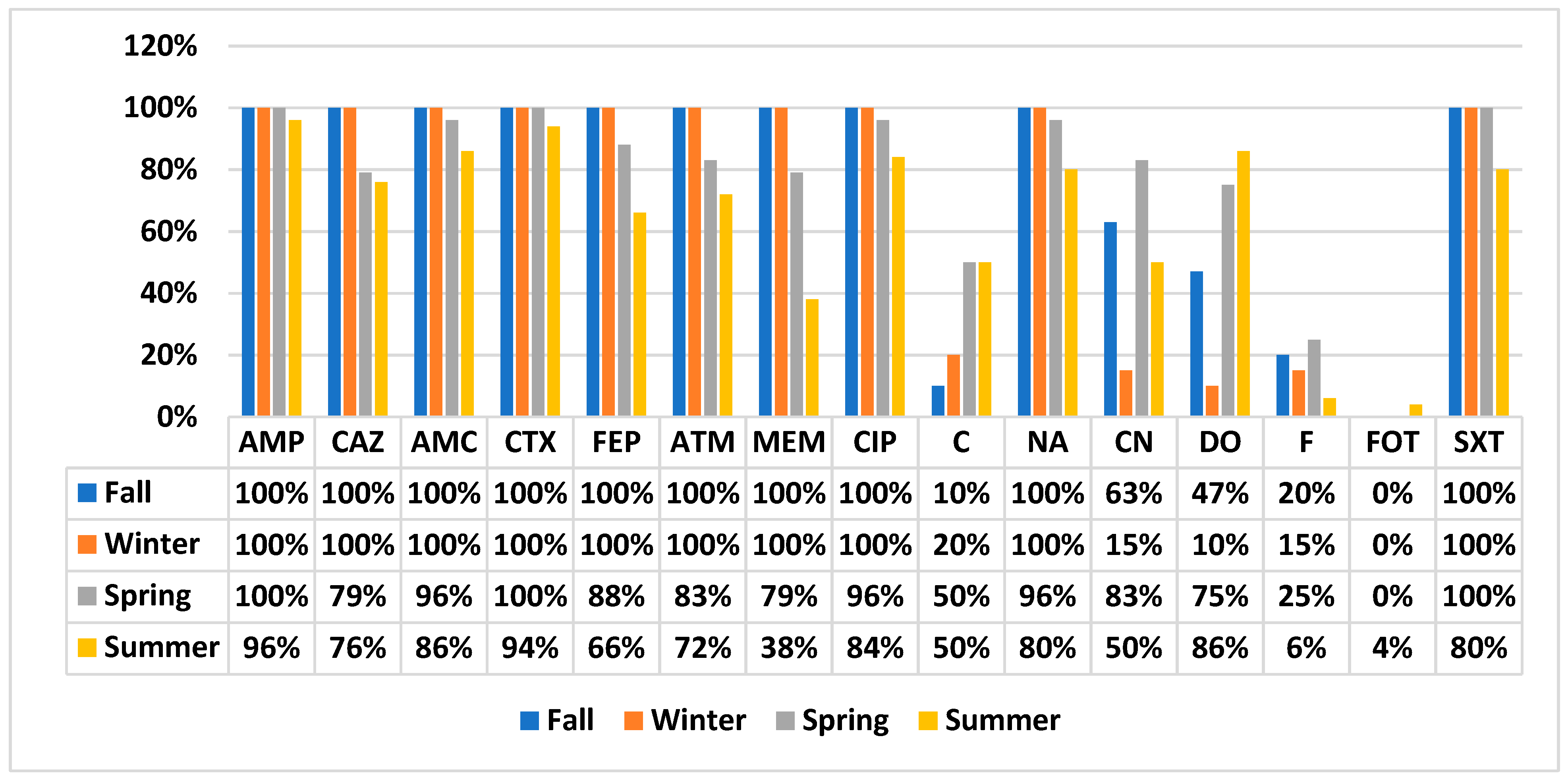
All study isolates were subjected to a disc diffusion test to determine their susceptibility against 15 different antibiotics belonging to 13 different antibiotic classes. All isolates were found to be MDR (resistant to ≥3 tested antibiotic classes). Among all MDR isolates, 51% (n = 63) were XDR (susceptible to ≤3 tested antibiotic classes), while there was no PDR isolate (resistant to all 15 tested antibiotics). Comparatively higher percentages of XDR were found in spring (79%) and summer (56%) compared to winter (20%) and fall (40%). The antibiotic resistance profile of study isolates against all tested antibiotics is as follows: ampicillin, 98%; ceftazidime, 86%; ampicillin–clavulanic acid, 94%; cefotaxime, 98%; cefepime, 84%; aztreonam, 85%; meropenem, 71%; ciprofloxacin, 93%; chloramphenicol, 35%; nalidixic acid, 91%; gentamicin, 54%; doxycycline, 62%; nitrofurantoin, 15%; fosfomycin, 2%; and sulfamethoxazole-trimethoprim, 92%. We observed intermediate resistance to doxycycline (11%), meropenem (29%), and fosfomycin (29%).
The temporal variation of antibiotic susceptibility profile of study isolates is shown in Figure 2. E. coli isolated in summer showed a slightly different pattern in comparison to other season’s E. coli isolates. Only summer E. coli showed resistance to fosfomycin (4%, n = 2). E. coli isolates of fall and winter were 100% resistant to meropenem as well. However, in spring and summer, 79% and 38% isolates were resistant and 21% and 62% were intermediately resistant, respectively.

2.2. Phylogenetic Groups of E. coli

Among the typeable isolates, the following phylogenetics groups were found: B1 (2%), B2 (6%), Clad I/II (38%), E/cladI/II (2%), A/C (7%), and F (~1%). E. coli isolated in the summer season showed a higher diversity of phylogenetic groups, and F phylogroup was only observed in summer. On the other hand, B1 isolates were only found in fall. There were 44% isolates un-typeable by quadruplex PCR and remain unknown. Seasonal prevalence comparison of E. coli phylogenetic groups is shown in Table 2.

2.3. Molecular Screening of Antimicrobial Resistance Markers

All E. coli were screened to obtain an insight into their resistance markers encoding carbapenemases, ESBLs, plasmid-mediated quinolones resistance (PMQRs), and plasmid-mediated colistin-resistance genes. Among carbapenemases, the most prevalent β-lactamases were blaOXA-48 (65%) and blaIMP (34%), followed by blaNDM (25%), blaKPC (~1%), and blaBIC (~1%). Among ESBLs, the most prevalent was blaTEM (55%), followed by blaCTX-M (46%), and blaSHV (15%). Among PMQR markers, the most prevalent was qnrS (32%), followed by qnrB (17%) and qnrA (3%). Among the mcr group of genes (from mcr-1 to mcr-5), only mcr-1 (35%) was found among study isolates.
The comparative prevalence of resistance markers in four seasons is shown in Table 2. Summer showed co-occurrence of 12 resistance markers, and blaKPC and blaBIC were only found in summer. Among the 16 screened ARGs, the lowest co-occurrence was found among winter isolates. None of the isolates in winter were positive for qnrA, qnrB, blaKPC, or blaBIC, but the highest prevalence of mcr-1 was in winter.
There were 5% (n = 6) isolates that carried all four classes of resistance genes (ARGs), with at least one of the carbapenemases, ESBL, PMQRs, and mcr group of genes. Moreover, there were 5% (n = 6) isolates which did not show the presence of any of the screened ARGs by PCR. All isolates were MDR phenotypically, and there were 44% (n = 55) E. coli carrying a combination of three classes of ARGs. The highest number of study isolates were carrying a combination of carbapenemases, ESBLs, and PMQRs. Isolates categorized based on the combination of carriage of antibiotic resistance genes are shown in Figure 3.

3. Discussion

The present study was carried out in order to obtain insights into the prevalence and temporal variation of meropenem-resistant E. coli in sewage water, along with their carriage of resistance genes. The study showed that 27% of the sites/sewage samples were positive for meropenem-resistant E. coli, and a comparatively higher prevalence was observed in summer. In contrast, a previous study from Islamabad of E. coli isolated from wastewater reported 100% carbapenem susceptibility of E. coli isolated from sewage water [15]. Another study from the neighboring country India reported only 3% carbapenem-resistant E. coli from sewage water [16]. The difference between studies suggests an increasing resistant E. coli burden in the environment over time. An increasing AMR trend in the environment can be correlated with increasing antibiotic usage over the time period. A global study published in 2018 reported comprehensive data of antibiotic usage in 76 countries over a period of 16 years (2000–2015). Overall, there was a 65% increase in antibiotic consumption. Among low- and middle-income countries, it was found that Pakistan (65% growth) was the 3rd largest among those with an increasing trend of antibiotic usage after India (103% growth) and China (79% growth). Of particular concern was the increase in usage of last-resort antibiotic classes, which was same in low-, middle-, and high-income countries, including carbapenems, polymyxins, glycylcyclins, and oxazolidinones. In particular, usage of carbapenems and polymyxins (largely colistin) increased during the study period in low- and middle-income countries. [17]. Another possible reason could be due to antibiotic-based selection of E. coli in the present study, which better enabled us to sort and isolate meropenem-resistant E. coli.
The present study reports 51% XDR. The most effective drugs were fosfomycin, nitrofurantoin, and chloramphenicol. Resistance to fluoroquinolones, quinolones, and cephalosporins was above 80%. One study from India found a maximum of 60% E. coli isolates to be resistant to fluoroquinolones, with less than 60% to quinolones and cephalosporins, and the most effective drug was carbapenem [16]. The difference of phenotypic antibiotic resistance in the present study in comparison to previous reports could be because the present study is focused on meropenem-resistant isolates, which are known for the co-occurrence of resistance markers against other antibiotics [5]. The comparatively lower resistance to chloramphenicol (35%) could be due to its lower level of use/prescription in comparison to other antibiotics. The present study reports higher susceptibility to fosfomycin. This could be because fosfomycin is among the less often prescribed antibiotics and is reserved as the last treatment option for infections caused by carbapenemase-producing Enterobacteriaceae [18].
In phylogenetic analysis, 44% of E. coli were un-typeable, and the typeable isolates mostly belonged to clad I/II, which suggests that most of the E. coli were environmental/commensals. A study [16] from India reported that most of the antibiotic-resistant E. coli belonged to the B2 phylogenetic group (67%), which is considered a virulent/pathogenic group. In contrast, the present study reported only 6% of MDR E. coli belonging to the B2 group. This contradiction shows that resistant isolates do not necessarily belong to the B2 group, but can be commensals. This difference could be because the previous study was only based on hospital wastewater, which can carry more pathogenic E. coli than other sites. These results also draw attention towards the level of horizontal gene transfer of antibiotic resistance genes from pathogenic to commensals in the environment, making them MDR and XDR.
Results of ARGs showed the co-prevalence of multiple resistance genes with carbapenemases. blaCTX-M-type ESBLs have been reported worldwide as the primary cause of resistance to third-generation cephalosporins in E. coli isolated from clinical samples and commensals [19]. The human colonization of ESBL-producing E. coli is estimated to be around 14% globally, with rates as high as 22% in Asia and Sub-Saharan Africa [20]. The present study reported 15–55% ESBLs from environmental E. coli, which showed a comparatively higher ARG burden in the environment than previous studies. A recent E. coli study from wastewater reported 84% blaTEM, followed by 52% blaCTX-M and no blaSHV. In the present study, a similar pattern was found, where the most prevalent ESBL gene was blaTEM (55%), followed by blaCTX-M (46%). Among carbapenemases, the other study found 16% each for blaNDM-1 and blaKPC-2 [21]. In the present study, carbapenemase prevalence is quite different, as the highest level of carbapenemase found was blaOXA-48 (65%), followed by blaIMP (34%), blaNDM (25%), and ~1% blaKPC. The difference in carbapenemase pattern among E. coli in different geographical regions could be due to different prevalent sequence types (STs) with different resistance mechanisms.
Previous studies from Pakistan reported mcr-1 in E. coli isolated from wild migratory birds [22], humans [23], and broiler chickens [24] over the last 5 years. Recently, a study form Pakistan reported less than 1% mcr-1 co-carriage with ESBLs in clinical isolates [25], while we found 9% of E. coli co-harboring mcr-1 with carbapenemases, ESBLs, and PMQRs. Theearlier preliminary reports from Pakistan were during 2016–2018, while the present study conducted on sewage water samples, collected during 2016–2017, found 35% mcr-1 in E. coli, indicating that there was much more colistin resistance burden in the environment than that found in clinical or other settings, which is a point of deep concern.
E. coli isolated in summer showed maximum diversity of phylogenetic groups and carriage of antimicrobial resistance genes. The comparatively greater prevalence of meropenem-resistant E. coli in summer could be due to favorable and survival-supporting environmental factors, including temperature; in summer, the mean environmental temperature is above 30 °C in plain areas of Pakistan, which is optimum for bacterial growth. Another possible reason could be the higher infection rate of meropenem-resistant E. coli in the community in summer, which ultimately disseminates in sewage water through fecal contamination. A study from India investigating pathogen inhabitant rate in water in different seasons found the presence of total and thermotolerant coliforms in different microenvironments of water, which were highest in the monsoon, followed by summer, and least in winter [26]. One retrospective study from Pakistan reported more bacterial pathogens in stool samples collected from patients in summer. They reported Vibrio cholera 01 Ogawa (32.8%), Campylobacter jejuni (17.3%), Enteropathogenic E. coli (9.9%), Salmonella paratyphi b (6.6%), and Shigella flexneri (6.2%). The study suggested seasonal variation in organism detection, demonstrating an inclination towards summer [27]. Diverse types of phylogenetic groups and ARGs in summer could be because of the higher number of E. coli isolated in summer, increasing the probability of variation.
The findings of the present study, including detection of high phenotypic antibiotic resistance patterns and ARGs in environmental commensal E. coli, are thought provoking. These commensals are expected to be carried in the gut of humans and animals and disseminate into the environment through fecal contamination, where these MDR commensal E. coli can easily transfer plasmid-mediated ARGs to other co-residing bacteria in environment. Through sewage water, antibiotic resistance can disseminate and reach all living beings (human, animals, and birds) and non-living things (soil, agriculture, and the rest of the water system) which can also ultimately become disseminators. It can become part of an ecosystem and disseminate more extensively than the control measures can counter. Thus, continuous checks of the water to gain an insight into AMR and taking prompt actions for its specified treatment to break the chain of dissemination are most necessary.

4. Materials and Methods

4.1. Sampling

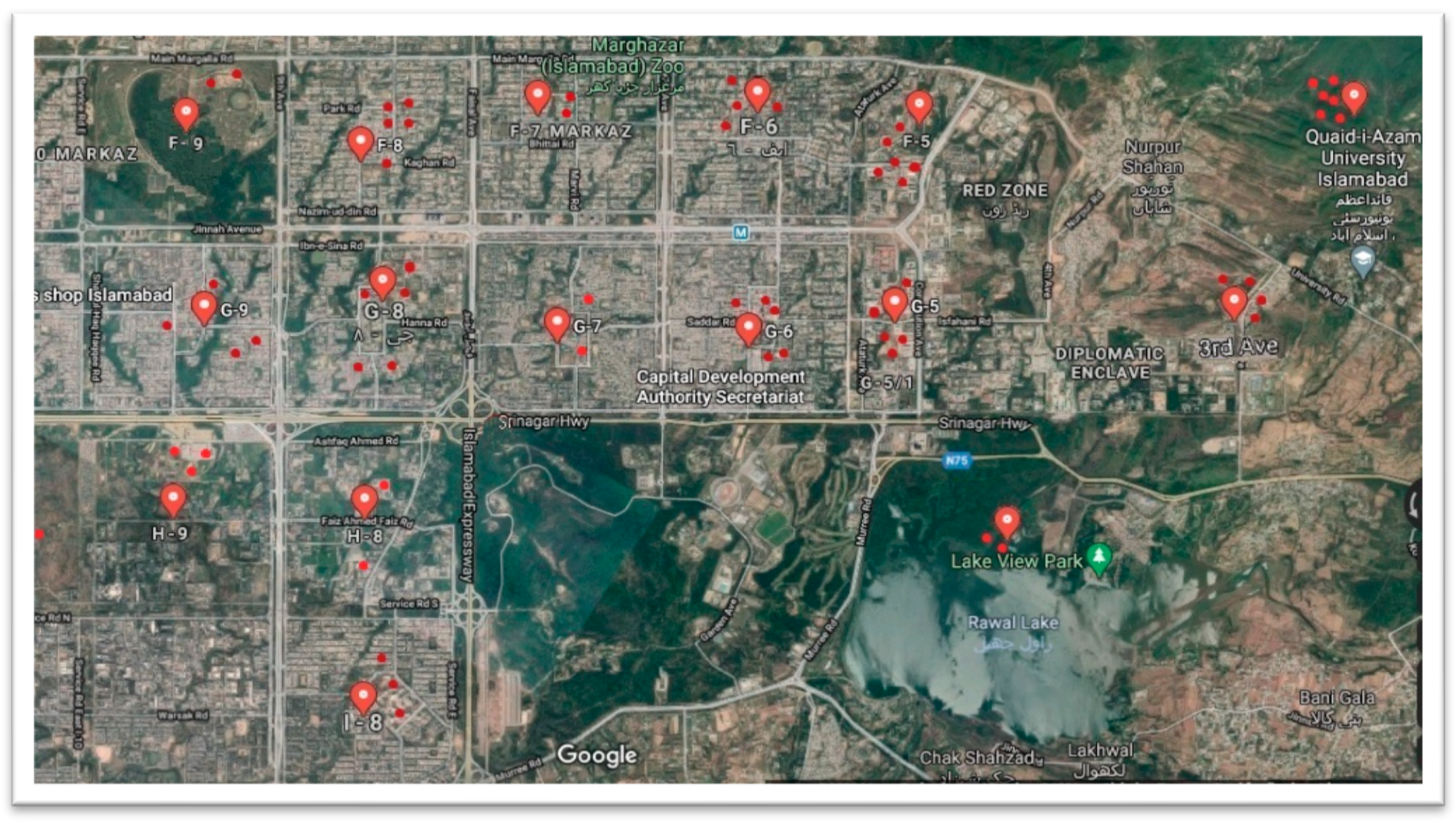
A total of 60 different sites were targeted across the capital city of Islamabad, Pakistan, as shown in the map (Figure 4). The sites included educational institutes, hospitals, commercial markets, office buildings, residential areas, and downstream waterfalls (Nullahs). From each site highlighted, 30–45 mL of sewage water was collected during October 2016–September 2017, covering all four seasons (fall, winter, spring, and summer).

4.2. Isolation and Identification of Meropenem-Resistant E. coli

Sewage samples were spun down at 1500 rpm for 3–5 min to remove visible garbage. From each sample, 100 µL water was added to screening agar (MacConkey agar supplemented with 3 µg/mL meropenem) by the spread plate method and incubated at 37 °C for 24 hrs. Suspected E. coli colonies were processed for confirmation by standard biochemical tests and through amplification of uidA gene by using primers as mentioned in Table 1. All confirmed E. coli isolates were preserved in 20% glycerol at −80 °C for further study.

4.3. Antibiotic Susceptibility Test

All study isolates’ susceptibility profiles were analyzed against 15 different antibiotics using the Kirby–Bauer disk diffusion method [28]. Zone diameters were interpreted according to CLSI 2019 guidelines. Antibiotic discs and their concentrations were as follows: Ampicillin (AMP 10 µg), ceftazidime (CAZ 30 µg), amoxicillin–clavulanic acid (AMC 10/20 µg), cefotaxime (CTX 30 µg), cefepime (FEP 30 µg), aztreonam (ATM 30 µg), meropenem (MEM 10 µg), ciprofloxacin (CIP 5 µg), Chloramphenicol (C 30 µg), Nalidixic acid (NA 30 µg), Gentamicin (CN 120 µg), Doxycycline (DO 30 µg), Nitrofurantoin (F 300 µg), Fosfomycin (FOT 200 µg), and trimethoprim-sulfamethoxazole (SXT 1.25/25 µg).

4.4. DNA Extraction

Whole genomic extraction of E. coli was performed using the boil colony method. The overnight 3–5 colonies of E. coli were suspended in 200 μL sterile distilled water. The suspension was thoroughly mixed by vortex, boiled at 95 °C for 15 min, and subsequently centrifuged at 14,000 rpm for 8 min. The supernatant was collected and stored at −20 °C for further processing [29].

4.5. Phylogenetic Analysis of E. coli

All study isolates were subjected to quadruplex PCR for phylogenetic analysis using the Clermont method [30].

4.6. Molecular Screening for Antibiotic Resistance Genes

All isolates were screened for ARGs using conventional singleplex and multiplex PCRs. Primers used along with their annealing temperature are given in Table 3. The PCR cycle conditions were as follows.
The reaction mixture contained 12.5 μL of 2X mastermix (Thermo scientific Dream TaqTm green PCR master mix), 0.2 μL each of forward and reverse primers (100 pmoles/µL), 2 μL of DNA template, and PCR-grade water up to the final volume of 25 μL. The cycling conditions for the PCR reaction were as follows: an initial denaturation for 10 min at 94 °C, followed by 35 cycles containing denaturation at 94 °C for 40 sec, annealing for 40 sec, initial extension at 72 °C for 1 min, and a final extension for 5 min at 72 °C. Agarose gel electrophoresis was performed at 90 V for 55 min in 1.5% agarose gel made in 10X Tris boric acid-EDTA (TBE) buffer.

5. Conclusions

The present study was focused on surveillance of meropenem-resistant E. coli along with other resistance mechanisms in wastewater. From our findings, we suggest conducting integrating research to elucidate their genetic details, sequence types, and study of environmental factors contributing to their successful survival in environmental water. Wastewater carriage of MDR E. coli along with combinations of multiple resistance genes (including carbapenemases and plasmid-mediated colistin resistance) is an early warning sign of potential hazard for the community, so the present study concerns public health, calling for a comprehensive monitoring and control system for clinically important antibiotic-resistant bacteria in the environment. Through control measures, antibiotic-resistant bacteria circulating in the environment should be minimized to prevent community-acquired infections, occupational hazards, and a possible public health hazard.

Author Contributions

Conceptualization, resources, supervision, project administration, funding acquisition, writing–original draft and review and approval of final draft were done by R.Z.; resources, funding acquisition, and reviewing and approval of the final draft were done by S.-H.L.; experimental work, data analysis, and writing of the original draft were done by S.Y.; data analysis and refining and reviewing of the final draft was done by A.-M.K. All authors have read and agreed to the published version of the manuscript.

Funding

This work was supported by research grants from the Bio and Medical Technology Development Program of the National Research Foundation of Korea (NRF), funded by the Ministry of Science, Information, and Communications Technology (MSIT; grant number NRF-2017M3A9E4078014); and the NRF, funded by the MSIT (grant numbers NRF-2021R1A2C3004826 and NRF-2019R1C1C1008615) to S.-H.L. and the International Development Research Centre (IDRC; grant 109304-002, EMBARK under the JPI AMR framework) to R.Z.

Institutional Review Board Statement

Not applicable.

Informed Consent Statement

Not applicable.

Data Availability Statement

Not applicable.

Conflicts of Interest

The authors declare no conflict of interest.

References

  1. Willyard, C. The drug-resistant bacteria that pose the greatest health threats. Nature 2017, 543, 15. [Google Scholar] [CrossRef] [PubMed] [Green Version]
  2. Hawkey, P.M.; Livermore, D.M. Carbapenem antibiotics for serious infections. BMJ 2012, 344, e3236. [Google Scholar] [CrossRef] [PubMed]
  3. Tzouvelekis, L.; Markogiannakis, A.; Psichogiou, M.; Tassios, P.T.; Daikos, G.L. Carbapenemases in Klebsiella pneumoniae and other Enterobacteriaceae: An evolving crisis of global dimensions. Clin. Microbiol. Rev. 2012, 25, 682–707. [Google Scholar] [CrossRef] [PubMed] [Green Version]
  4. Van Duin, D.; Doi, Y. The global epidemiology of carbapenemase-producing Enterobacteriaceae. Virulence 2017, 8, 460–469. [Google Scholar] [CrossRef]
  5. Paterson, D.L.; Bonomo, R.A. Extended-spectrum β-lactamases: A clinical update. Clin. Microbiol. Rev. 2005, 18, 657–686. [Google Scholar] [CrossRef] [PubMed] [Green Version]
  6. World Health Organization. Antimicrobial Resistance Global Report on Surveillance: 2014 Summary; WHO: Geneva, Switzerland, 2014. [Google Scholar]
  7. Rump, B.; Timen, A.; Hulscher, M.; Verweij, M. Ethics of infection control measures for carriers of antimicrobial drug–resistant organisms. Emerg. Infect. Dis. 2018, 24, 1609. [Google Scholar] [CrossRef] [Green Version]
  8. Berendonk, T.U.; Manaia, C.M.; Merlin, C.; Fatta-Kassinos, D.; Cytryn, E.; Walsh, F.; Bürgmann, H.; Sørum, H.; Norström, M.; Pons, M.-N.; et al. Tackling antibiotic resistance: The environmental framework. Nat. Rev. Microbiol. 2015, 13, 310–317. [Google Scholar] [CrossRef]
  9. Rochelle-Newall, E.; Nguyen, T.M.H.; Le, T.P.Q.; Sengtaheuanghoung, O.; Ribolzi, O. A short review of fecal indicator bacteria in tropical aquatic ecosystems: Knowledge gaps and future directions. Front. Microbiol. 2015, 6, 308. [Google Scholar] [CrossRef]
  10. Ercumen, A.; Pickering, A.J.; Kwong, L.H.; Arnold, B.F.; Parvez, S.M.; Alam, M.; Sen, D.; Islam, S.; Kullmann, C.; Chase, C.; et al. Animal feces contribute to domestic fecal contamination: Evidence from E. coli measured in water, hands, food, flies, and soil in Bangladesh. Environ. Sci. Technol. 2017, 51, 8725–8734. [Google Scholar] [CrossRef] [Green Version]
  11. Haberecht, H.B.; Nealon, N.J.; Gilliland, J.R.; Holder, A.V.; Runyan, C.; Oppel, R.C.; Ibrahim, H.M.; Mueller, L.; Schrupp, F.; Vilchez, S.; et al. Antimicrobial-resistant Escherichia coli from environmental waters in northern Colorado. J. Environ. Public Health 2019, 2019, 3862949. [Google Scholar] [CrossRef] [Green Version]
  12. Huijbers, P.M.; Larsson, D.J.; Flach, C.-F. Surveillance of antibiotic resistant Escherichia coli in human populations through urban wastewater in ten European countries. Environ. Pollut. 2020, 261, 114200. [Google Scholar] [CrossRef] [PubMed]
  13. Khan, F.M.; Gupta, R. Escherichia coli (E. coli) as an Indicator of Fecal Contamination in Water: A Review. In Proceedings of the International Conference on Sustainable Development of Water and Environment, Incheon, Korea, 13–14 January 2020. [Google Scholar]
  14. World Health Organization; World Health Organisation Staff. Guidelines for Drinking-Water Quality; WHO: Geneva, Switzerland, 2004; Volume 1. [Google Scholar]
  15. Zahra, R.; Javeed, S.; Malala, B.; Babenko, D.; Toleman, M.A. Analysis of Escherichia coli STs and resistance mechanisms in sewage from Islamabad, Pakistan indicates a difference in E. coli carriage types between South Asia and Europe. J. Antimicrob. Chemother. 2018, 73, 1781–1785. [Google Scholar] [CrossRef] [Green Version]
  16. Chandran, S.; Diwan, V.; Tamhankar, A.J.; Joseph, B.V.; Rosales-Klintz, S.; Mundayoor, S.; Lundborg, C.S.; Macaden, R. Detection of carbapenem resistance genes and cephalosporin, and quinolone resistance genes along with oqxAB gene in Escherichia coli in hospital wastewater: A matter of concern. J. Appl. Microbiol. 2014, 117, 984–995. [Google Scholar] [CrossRef] [PubMed]
  17. Klein, E.Y.; Van Boeckel, T.P.; Martinez, E.M.; Pant, S.; Gandra, S.; Levin, S.A.; Goossens, H.; Laxminarayan, R. Global increase and geographic convergence in antibiotic consumption between 2000 and 2015. Proc. Natl. Acad. Sci. USA 2018, 115, E3463–E3470. [Google Scholar] [CrossRef] [PubMed] [Green Version]
  18. Pontikis, K.; Karaiskos, I.; Bastani, S.; Dimopoulos, G.; Kalogirou, M.; Katsiari, M.; Oikonomou, A.; Poulakou, G.; Roilides, E.; Giamarellou, H. Outcomes of critically ill intensive care unit patients treated with fosfomycin for infections due to pandrug-resistant and extensively drug-resistant carbapenemase-producing Gram-negative bacteria. Int. J. Antimicrob. Agents 2014, 43, 52–59. [Google Scholar] [CrossRef] [PubMed]
  19. D’Andrea, M.M.; Arena, F.; Pallecchi, L.; Rossolini, G.M. CTX-M-type β-lactamases: A successful story of antibiotic resistance. Int. J. Med. Microbiol. 2013, 303, 305–317. [Google Scholar] [CrossRef]
  20. Karanika, S.; Karantanos, T.; Arvanitis, M.; Grigoras, C.; Mylonakis, E. Fecal colonization with extended-spectrum beta-lactamase–producing Enterobacteriaceae and risk factors among healthy individuals: A systematic review and metaanalysis. Rev. Infect. Dis. 2016, 63, 310–318. [Google Scholar] [CrossRef] [Green Version]
  21. Adegoke, A.A.; Madu, C.E.; Aiyegoro, O.A.; Stenstrom, T.A.; Okoh, A.I. Antibiogram and beta-lactamase genes among cefotaxime resistant E. coli from wastewater treatment plant. Antimicrob. Resist. Infect. 2020, 9, 46. [Google Scholar] [CrossRef]
  22. Mohsin, M.; Raza, S.; Roschanski, N.; Schaufler, K.; Guenther, S. First description of plasmid-mediated colistin-resistant extended-spectrum β-lactamase-producing Escherichia coli in a wild migratory bird from Asia. Int. J. Antimicrob. Agents 2016, 4, 463–464. [Google Scholar] [CrossRef]
  23. Mohsin, M.; Raza, S.; Roschanski, N.; Guenther, S.; Ali, A.; Schierack, P. Description of the first Escherichia coli clinical isolate harboring the colistin resistance gene mcr-1 from the Indian subcontinent. Antimicrob. Agents Chemother. 2017, 61, e01945-16. [Google Scholar] [CrossRef] [Green Version]
  24. Lv, J.; Mohsin, M.; Lei, S.; Srinivas, S.; Wiqar, R.T.; Lin, J.; Feng, Y. Discovery of a mcr-1-bearing plasmid in commensal colistin-resistant Escherichia coli from healthy broilers in Faisalabad, Pakistan. Virulence 2018, 9, 994–999. [Google Scholar] [CrossRef] [PubMed] [Green Version]
  25. Bilal, H.; Rehman, T.U.; Khan, M.A.; Hameed, F.; Jian, Z.G.; Han, J.; Yang, X. Molecular Epidemiology of mcr-1, blaKPC-2, and blaNDM-1 Harboring Clinically Isolated Escherichia coli from Pakistan. Infect. Drug Resist. 2021, 14, 1467. [Google Scholar] [CrossRef] [PubMed]
  26. Yadav, N.; Singh, S.; Goyal, S.K. Effect of seasonal variation on bacterial inhabitants and diversity in drinking water of an office building, Delhi. Air Soil Water Res. 2019, 12, 1178622119882335. [Google Scholar] [CrossRef] [Green Version]
  27. Alam, M.; Akhtar, Y.N.; Ali, S.S.; Ahmed, M.; Atiq, M.; Ansari, A.; Chaudhry, F.A.; Bashir, H.; Bangash, M.A.; Awais, A.; et al. Seasonal variation in bacterial pathogens isolated from stool samples in Karachi, Pakistan. JPMA 2003, 53, 125–129. [Google Scholar]
  28. Hudzicki, J. Kirby-Bauer disk diffusion susceptibility test protocol. ASM 2009, 15, 55–63. [Google Scholar]
  29. Salehi, T.Z.; Madani, S.A.; Karimi, V.; Khazaeli, F.A. Molecular genetic differentiation of avian Escherichia coli by RAPD-PCR. Braz. J. Microbiol. 2008, 39, 494–497. [Google Scholar] [CrossRef] [Green Version]
  30. Clermont, O.; Christenson, J.K.; Denamur, E.; Gordon, D.M. The Clermont Escherichia coli phylo-typing method revisited: Improvement of specificity and detection of new phylo-groups. Environ. Microbiol. Rep. 2013, 5, 58–65. [Google Scholar] [CrossRef]
  31. Bej, A.K.; Dicesare, J.L.; Haff, L.; Atlas, R.M. Detection of Escherichia coli and Shigella spp. in water by using the polymerase chain reaction and gene probes for uid. Appl. Environ. Microbiol. 1991, 57, 1013–1017. [Google Scholar] [CrossRef] [Green Version]
  32. Nordmann, P.; Poirel, L.; Carrër, A.; Toleman, M.A.; Walsh, T.R. How to detect NDM-1 producers. J. Clin. Microbiol. 2011, 49, 718–721. [Google Scholar] [CrossRef] [Green Version]
  33. Mostachio, A.K.; Heidjen, I.; Rossi, F.; Levin, A.S.; Costa, S.F. Multiplex PCR for rapid detection of genes encoding oxacillinases and metallo-β-lactamases in carbapenem-resistant Acinetobacter spp. J. Med. Microbiol. 2009, 58, 1522–1524. [Google Scholar] [CrossRef] [Green Version]
  34. Poirel, L.; Walsh, T.R.; Cuvillier, V.; Nordmann, P. Multiplex PCR for detection of acquired carbapenemase genes. Diagn. Microbiol. Infect. Dis. 2011, 70, 119–123. [Google Scholar] [CrossRef] [PubMed]
  35. Azizi, M.; Mortazavi, S.H.; Etemadimajed, M.; Gheini, S.; Vaziri, S.; Alvandi, A.; Kashef, M.; Ahmadi, K. Prevalence of extended-spectrum β-Lactamases and antibiotic resistance patterns in Acinetobacter baumannii isolated from clinical samples in Kermanshah, Iran. Jundishapur J. Microbiol. 2017, 10, e61522. [Google Scholar] [CrossRef] [Green Version]
  36. Lal, P.; Kapil, A.; Das, B.K.; Sood, S. Occurrence of TEM & SHV gene in extended spectrum b-lactamases (ESBLs) producing Klebsiella sp. isolated from a tertiary care hospital. Indian J. Med. Res. 2007, 125, 173. [Google Scholar] [PubMed]
  37. Cattoir, V.; Poirel, L.; Rotimi, V.; Soussy, C.-J.; Nordmann, P. Multiplex PCR for detection of plasmid-mediated quinolone resistance qnr genes in ESBL-producing enterobacterial isolates. J. Antimicrob. Chemother. 2007, 60, 394–397. [Google Scholar] [CrossRef] [Green Version]
  38. Rebelo, A.R.; Bortolaia, V.; Kjeldgaard, J.S.; Pedersen, S.K.; Leekitcharoenphon, P.; Hansen, I.M.; Guerra, B.; Malorny, B.; Borowiak, M.; Hammerl, J.A.; et al. Multiplex PCR for detection of plasmid-mediated colistin resistance determinants, mcr-1, mcr-2, mcr-3, mcr-4 and mcr-5 for surveillance purposes. Eurosurveillance 2018, 23, 17-00672. [Google Scholar] [CrossRef]
Figure 1. Temporal prevalence of meropenem-resistant E. coli in sewage samples.
Antibiotics 11 00635 g001
Figure 2. Temporal variation in antibiotic susceptibility profile of E. coli.
Antibiotics 11 00635 g002
Figure 3. The number of isolates carrying a combination of antibiotic resistance genes.
Antibiotics 11 00635 g003
Figure 4. Map showing sites of sewage water samples across the capital city Islamabad.
Antibiotics 11 00635 g004
Table 1. Meropenem-resistant E. coli in different seasons of each site/sample.
Sewage Sampling in Different Seasons
Site/LocationFallWinterSpringSummer
C-type colony, QAU + (n = 1)
B-type colony, QAU + (n = 2)
QAU D-type colony entry point of filtration plant+ (n = 6)
QAU D-type colony after filtration plant
Bari imam Nullah 1+ (n = 1)+ (n = 1)
Bari imam Nullah 2+ (n = 4)
Sector G-5 Nullah+ (n = 2)
Sector G-5 Fata house exit+ (n = 1)
Ministry of water and power indoor+ (n = 1)+ (n = 1)
Govt. hostel lodge 2+ (n = 2)
Federal lodge 1+ (n = 1)
MNA HOSTEL+ (n = 1)+ (n = 2)
PM office indoor+ (n = 1)
PARC head office indoor+ (n = 1)+ (n = 6)+ (n = 1)
Ambassador hotel+ (n = 1)
Ministry of climate change indoor+ (n = 1)+ (n = 2)+ (n = 2)
PHRC indoor+ (n = 2)+ (n = 3)
Sector G-5 Comsats indoor+ (n = 2)+ (n = 4)+ (n = 2)
Sector G-6 Nullah 1+ (n = 1) + (n = 1)
Sector G-6 Nullah 2
ICB Islamabad indoor+ (n = 6)+ (n = 1)+ (n = 2)
CDA indoor+ (n = 3)
Masjid road Nullah
Lal masjid indoor+ (n = 1)
G-6 girls Govt. School
G6 Residential outdoor+ (n = 1)
Sector F-6 Nullah
Sector F-5 Nullah+ (n = 1)+ (n = 1)
Baluchistan house indoor+ (n = 1)
PTV office indoor
FPSC office indoor+ (n = 1)
KPK house indoor+ (n = 1)+ (n = 1)
Sindh house indoor+ (n = 1)+ (n = 1)
Punjab house indoor+ (n = 2)+ (n = 2)
F-6/3 primary school
F-6/3 masjid
Poly clinic outdoor+ (n = 11)
Poly clinic emergency+ (n = 2)+ (n = 1)
Nullah F-7
GPO indoor+ (n = 2)+ (n = 1)
Sector H8-sector Nullah+ (n = 3)+ (n = 1)
Preston university indoor
Nullah back side of Turk school+ (n = 1)
Pak Turk school indoor+ (n = 2)
Preston university front Nullah+ (n = 2)
Shah-Abdul Latif auditorium indoor+ (n = 1)+ (n = 2)
HEC indoor
Institute of science and Tech. indoor+ (n = 6)
Post graduate hostel
Post graduate college outdoor+ (n = 1)
AIOU indoor
Red Crescent indoor+ (n = 1)
Fire brigade+ (n = 2)
QAU.GH.2+ (n = 1)
QAU.BH 8
QAU.BH.3
QAU.BH.4
QAU.BH6.7+ (n = 1)
QAU.GH.5
QAU colony+ (n = 1)
– = No isolates detected; + = sample carrying meropenem-resistant E. coli; n = number of E. coli isolates detected; MNA—Member of National Assembly; PM—Prime Minister; PARC—Pakistan Agricultural Research Council; PHRC—Pakistan Health Research Council; CDA—Capital Development Authority; PTV—Pakistan Television Corporation; FPSC—Federal Public Service Commission; KPK—Khyber Pakhtunkhwa; GPO—Pakistan Post office; HEC—Higher Education Commission; AIOU—Allama Iqbal Open University; QAU—Quaid-i-Azam University; GH–Girls Hostel; BH–Boys Hostel.
Table 2. Antibiotic resistance-coding genes present in the Escherichia coli isolates from sewage water in different seasons in Islamabad, Pakistan, and their phylogenic groups.
Sewage Water Samples in Different Seasons
Antibiotic Resistance GenesFall, n = 93Winter, n = 64Spring, n = 87Summer, n = 164p-Value
n (%)n (%)n (%)n (%)
qnrA1 (3)01 (4)2 (4)0.4
qnrB7 (23)04 (17)10 (20)0.6
qnrS5 (17)3 (15)11 (46)21 (42)0.7
blaOXA4820 (66)17 (85)10 (42)34 (68)0.67
blaTEM15 (50)13 (65)18 (75)22 (44)0.56
blaSHV2 (7)2 (10)8 (33)6 (12)0.25
BlaCTX-M4 (13)12 (60)14 (58)27 (54)0.77
blaIMP14 (46)5 (25)8 (33)15 (30)0.65
blaNDM11 (36)2 (10)8 (33)10 (20)0.48
blaBIC0 (0)0 (0)0 (0)1 (2)0.32
blaKPC0 (0)0 (0)0 (0)1 (2)0.32
mcr-114 (47)10 (50)5 (21)15 (30)0.55
mcr-20 (0)0 (0)0 (0)0 (0)-
mcr-30 (0)0 (0)0 (0)0 (0)-
mcr-40 (0)0 (0)0 (0)0 (0)-
mcr-50 (0)0 (0)0 (0)0 (0)-
Phylogenic groups
B12 (7)0 (0)0 (0)0 (0)0.32
B27 (23)0 (0)0 (0)1 (2)0.4
Clad I/II7 (23)7 (35)6 (25)27 (54)0.7
Unknown14 (47)13 (65)12 (50)15 (30)0.5
E/clad II0 (0)0 (0)2 (8)1 (2)0.34
A/C0 (0)0 (0)4 (17)5 (10)0.33
F0 (0)0 (0)0 (0)1 (2)0.3
p-values were extracted from Fisher test. n = number of E. coli isolates in water samples.
Table 3. Primer sequences and their product size.
Primer Primer Sequence 5′ to 3′Annealing TemperatureProduct SizeReference
1.uidAF:5′-AAAACGGCAAGAAAAAGCAG-3′55 °C147 bp[31]
R:5′-ACGCGTGGTTACAGTCTTGCG-3′
2.blaNDMF:5′-GGTTTGGCGATCTGGTTTTC-352 °C621 bp[32]
R:5′-CGGAATGGCTCATCACGATC-3
3.blaIMPF: 5′-GAATAGAATGGTTAACTCTC-3′52 °C188 bp[33]
R:5′-CCAAACCACTAGGTTATC-3′
4.blaOXA-48F:5′-GCGTGGTTAAGGATGAACAC-3′52 °C438 bp[34]
R:5′-CATCAAGTTCAACCCAACCG-3′
5.blaKPCF:5′-CGTCTAGTTCTGCTGTCTTG-3′52 °C798 bp[34]
R:5′-CTTGTCATCCTTGTTAGGCG-3′
6.blaBICF:5′-TATGCAGCTCCTTTAAGGGC-3′52 °C537 bp[34]
R:5′-TCATTGGCGGTGCCGTACAC-3′
7.blaCTX-MF:5′-ATGTGCAGTACCAGTAAGGT-3′52 °C594 bp[35]
R:5′-TGGGTAAAGTAGGTCACCAGA-3′
8.blaTEMF:5′-CTTCCTGTTTTTGCTCACCCA-3′52 °C717 bp[36]
R:5′-TACGATACGGGAGGGCTTAC-3′
9.blaSHVF:5′-TCAGCGAAAAACACCTTG-3′52 °C471 bp[36]
R:5′-TCCCGCAGATAAATCACC-3′
10.qnrAF:5′-AGAGGATTTCTCACGCCAGG-3′54 °C580 bp[37]
R:5′-TGCCAGGCACAGATCTTGAC-3′
11.qnrBF:5′-GGMATHGAAATTCGCCACTG-3′54 °C264 bp[37]
R:5′-TTTGCYGYYCGCCAGTCGAA-3′
12.qnrSF:5′-GCAAGTTCATTGAACAGGGT-3′54 °C428 bp[37]
R:5′-TCTAAACCGTCGAGTTCGGCG-3′
13.mcr-1F:5′-AGTCCGTTTGTTCTTGTGGC-3′55 °C320 bp[38]
R:5′-AGATCCTTGGTCTCGGCTTG-3′
14.mcr-2F:5′-CAAGTGTGTTGGTCGCAGTT-3′55 °C715 bp[38]
R:5′-TCTAGCCCGACAAGCATACC-3′
15.mcr-3F:5′-AAATAAAAATTGTTCCCCGCTTATG-3′55 °C929 bp[38]
R:5′-AATGGAGATCCCCGTTTTT-3′
16.mcr-4F:5′-TCACTTTCATCACTGCGTTG-3′55 °C1116 bp[38]
R:5′-TTGGTCCATGACTACCAATG-3′
17.mcr-5F:5′-ATGCGGTTGTCTGCATTTATC-3′55 °C1644 bp[38]
R:5′-CATTGTGGTTGTCCTTTTCTG-3′
Publisher’s Note: MDPI stays neutral with regard to jurisdictional claims in published maps and institutional affiliations.